mx6 autofocus options revb - molecular devices
TRANSCRIPT
MetaXpress® 6 GuideAutofocus Options
April 2021 revB
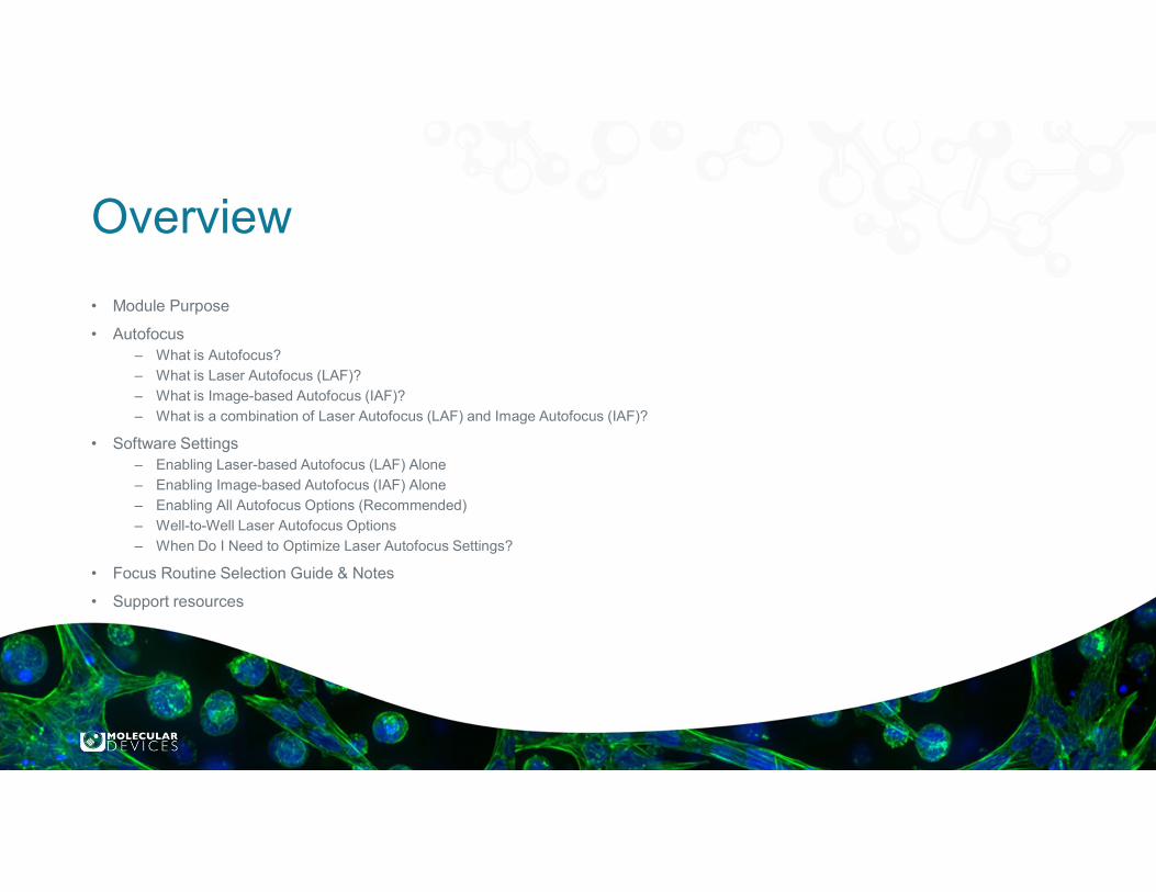
Overview
• Module Purpose
• Autofocus– What is Autofocus?
– What is Laser Autofocus (LAF)?
– What is Image-based Autofocus (IAF)?
– What is a combination of Laser Autofocus (LAF) and Image Autofocus (IAF)?
• Software Settings– Enabling Laser-based Autofocus (LAF) Alone
– Enabling Image-based Autofocus (IAF) Alone
– Enabling All Autofocus Options (Recommended)
– Well-to-Well Laser Autofocus Options
– When Do I Need to Optimize Laser Autofocus Settings?
• Focus Routine Selection Guide & Notes
• Support resources
Module Purpose
The purpose of this module is to familiarize the user with the different focus options including the Laser Autofocus
(LAF) and Image-based Autofocus (IAF) on the ImageXpress instruments with MetaXpress software
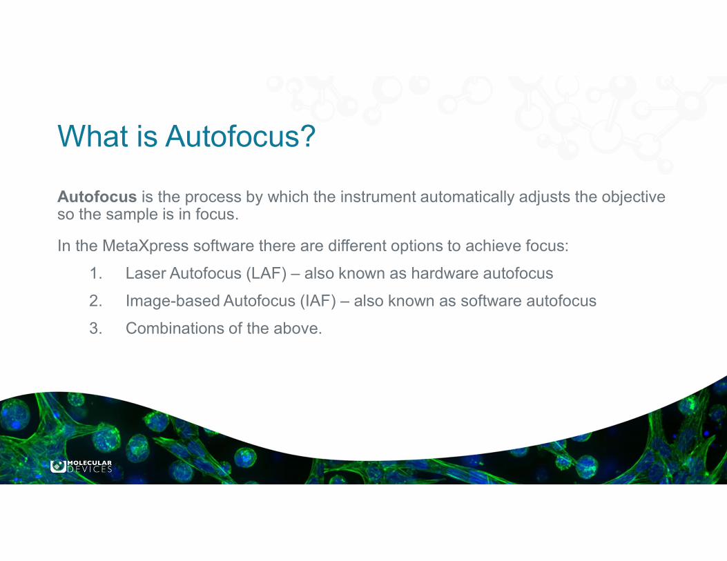
What is Autofocus?
Autofocus is the process by which the instrument automatically adjusts the objective so the sample is in focus.
In the MetaXpress software there are different options to achieve focus:
1. Laser Autofocus (LAF) – also known as hardware autofocus
2. Image-based Autofocus (IAF) – also known as software autofocus
3. Combinations of the above.
For research use only. Not for use in diagnostic procedures.© 2019 Molecular Devices, LLC. Trademarks are the property of Molecular Devices, LLC or their respective owners. | p5
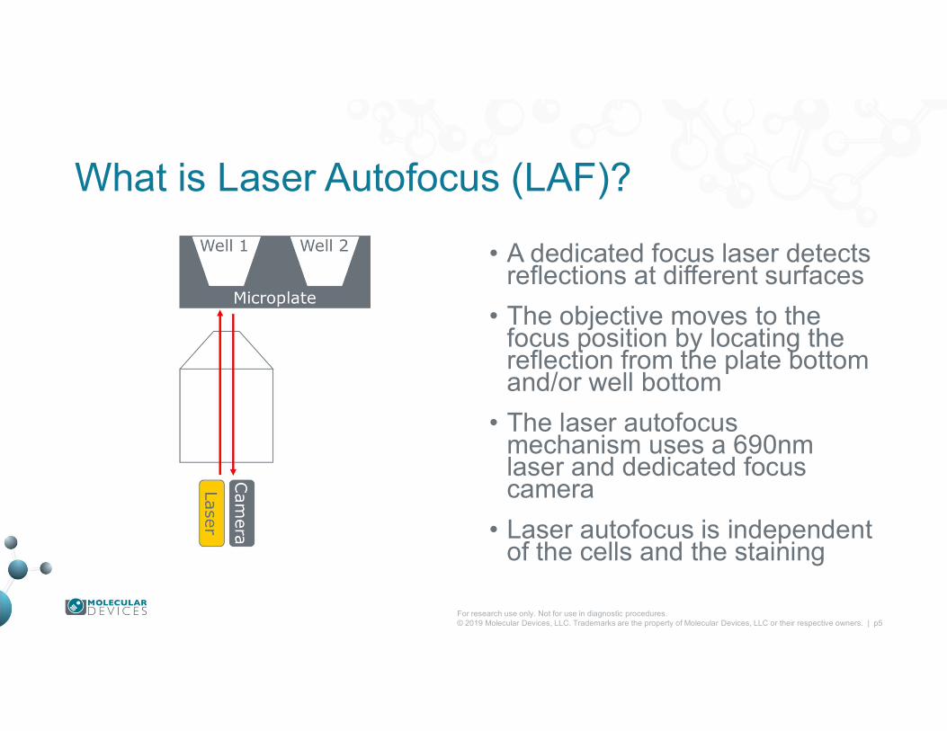
What is Laser Autofocus (LAF)?
• A dedicated focus laser detects reflections at different surfaces
• The objective moves to the focus position by locating the reflection from the plate bottom and/or well bottom
• The laser autofocus mechanism uses a 690nm laser and dedicated focus camera
• Laser autofocus is independent of the cells and the staining
For research use only. Not for use in diagnostic procedures.© 2019 Molecular Devices, LLC. Trademarks are the property of Molecular Devices, LLC or their respective owners. | p6
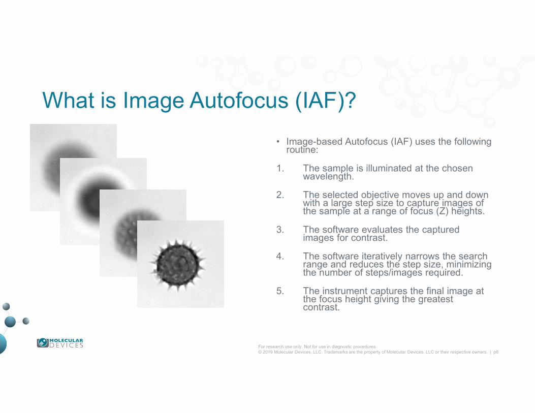
What is Image Autofocus (IAF)?
• Image-based Autofocus (IAF) uses the following routine:
1. The sample is illuminated at the chosen wavelength.
2. The selected objective moves up and down with a large step size to capture images of the sample at a range of focus (Z) heights.
3. The software evaluates the captured images for contrast.
4. The software iteratively narrows the search range and reduces the step size, minimizing the number of steps/images required.
5. The instrument captures the final image at the focus height giving the greatest contrast.
For research use only. Not for use in diagnostic procedures.© 2019 Molecular Devices, LLC. Trademarks are the property of Molecular Devices, LLC or their respective owners. | p7
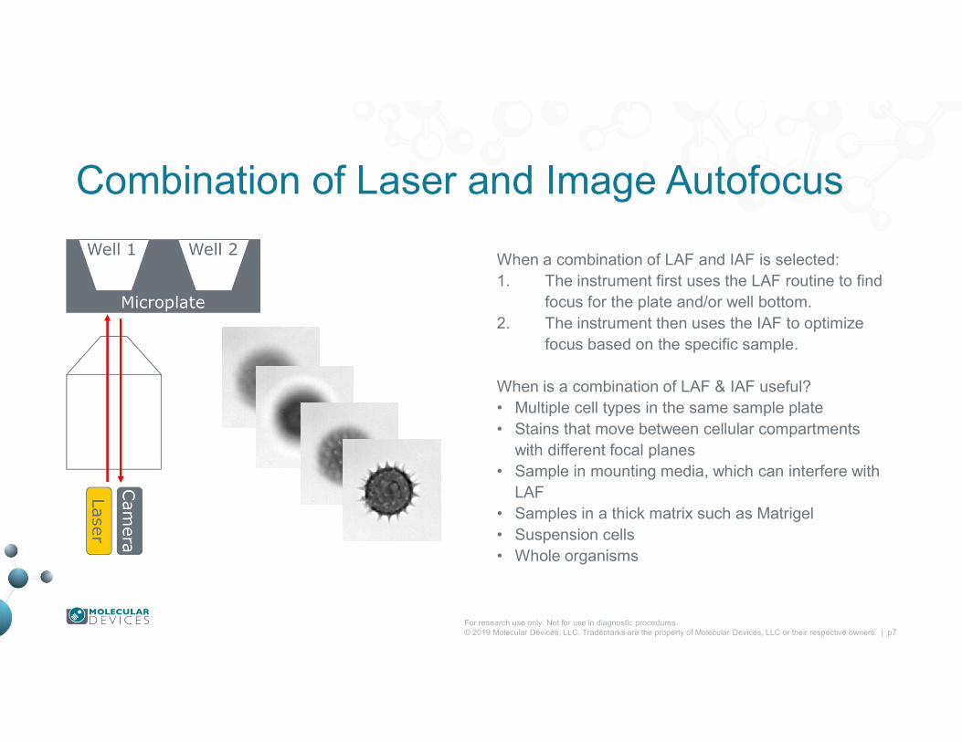
Combination of Laser and Image Autofocus
When a combination of LAF and IAF is selected:1. The instrument first uses the LAF routine to find
focus for the plate and/or well bottom. 2. The instrument then uses the IAF to optimize
focus based on the specific sample.
When is a combination of LAF & IAF useful?• Multiple cell types in the same sample plate• Stains that move between cellular compartments
with different focal planes• Sample in mounting media, which can interfere with
LAF• Samples in a thick matrix such as Matrigel• Suspension cells • Whole organisms

Autofocus Settings in MetaXpress 6• Enabling Laser-based Autofocus (LAF) alone
• Enabling Image-based Autofocus (IAF) alone
• Enabling All Autofocus options (Recommended)
• Well-to-Well Laser Autofocus options
• Find Sample options
• Site Autofocus options
• Timelapse Autofocus options
• Autofocus Routine Selection Guide
• When Do I Need to Optimize Laser Autofocus Settings?

Enabling Laser-based Autofocus (LAF) Alone• Plate Acquisition Setup > Acquisition tab
– Select Enable laser-based focusing
– Deselect Enable image-based focusing
• W1 autofocus options:– Laser with z-offset
• W2 – W8 autofocus options:– Z-offset from W1
• LAF alone is the default setting:– Fastest performance
– Minimizes photobleaching
– Independent of sample/stain quality
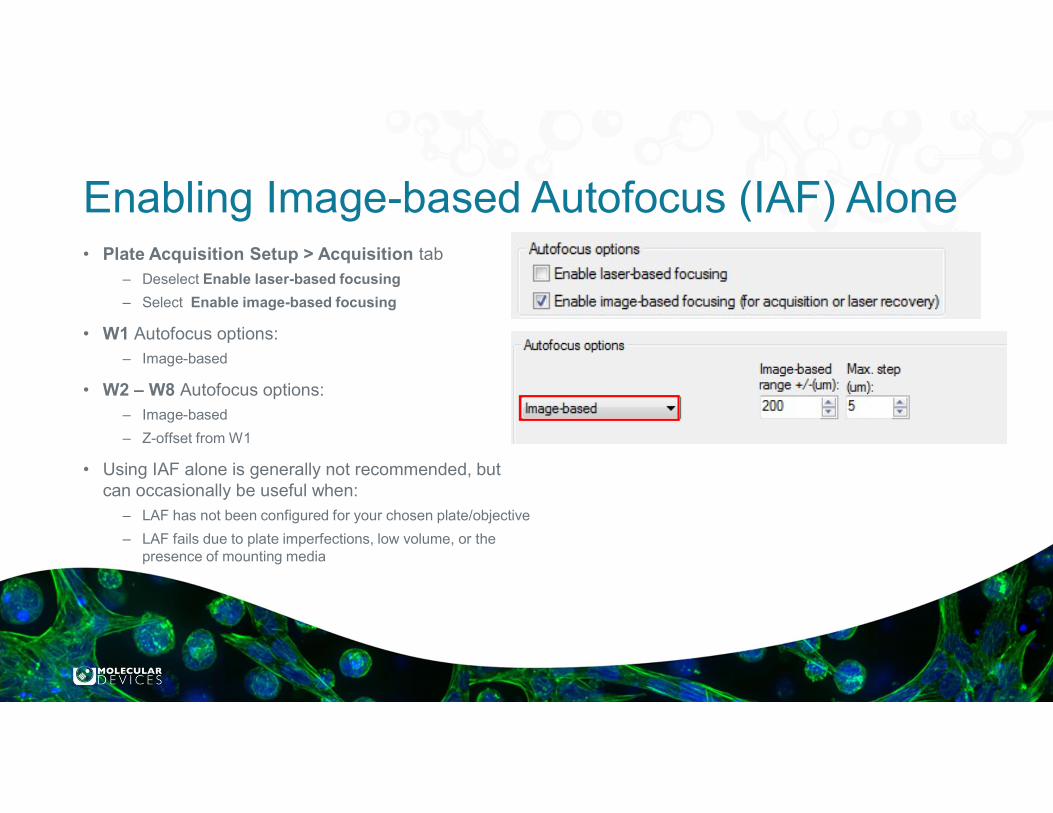
Enabling Image-based Autofocus (IAF) Alone• Plate Acquisition Setup > Acquisition tab
– Deselect Enable laser-based focusing
– Select Enable image-based focusing
• W1 Autofocus options:– Image-based
• W2 – W8 Autofocus options:– Image-based
– Z-offset from W1
• Using IAF alone is generally not recommended, but can occasionally be useful when:
– LAF has not been configured for your chosen plate/objective
– LAF fails due to plate imperfections, low volume, or the presence of mounting media
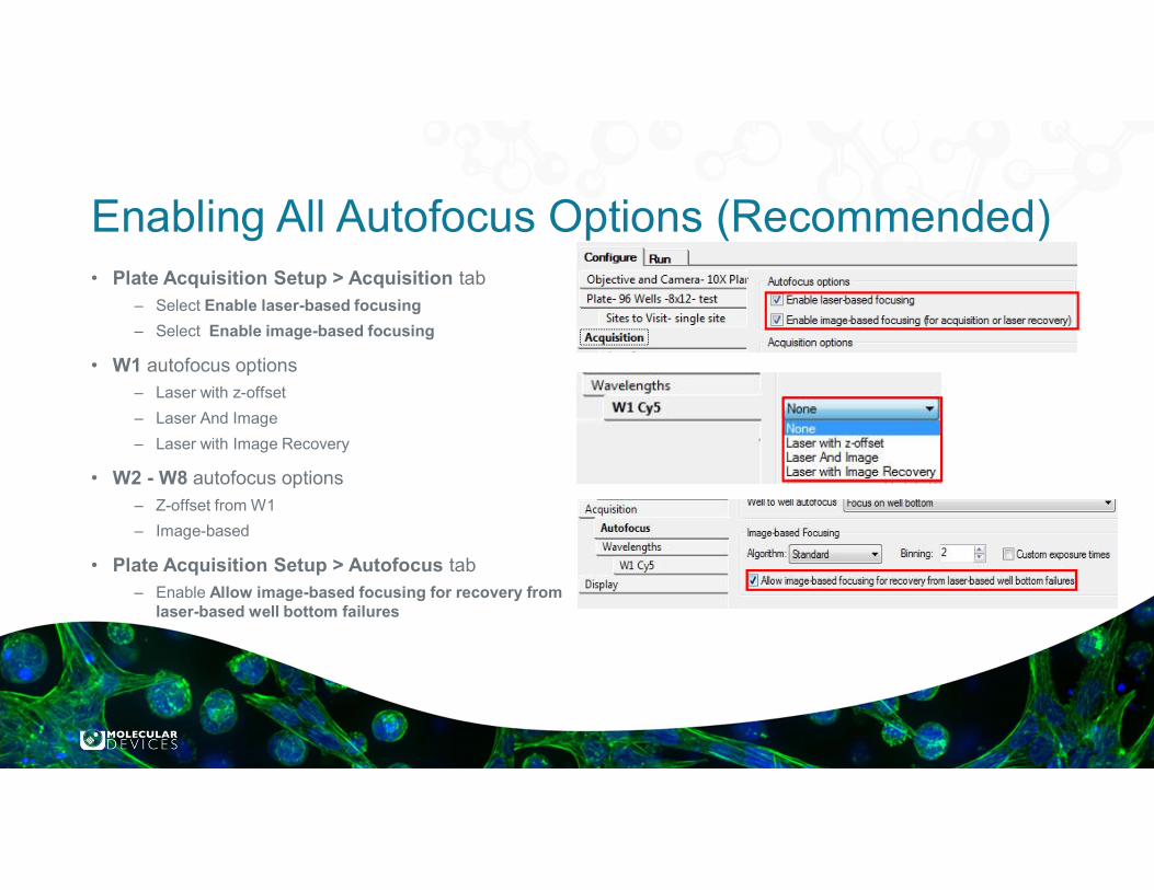
Enabling All Autofocus Options (Recommended)• Plate Acquisition Setup > Acquisition tab
– Select Enable laser-based focusing
– Select Enable image-based focusing
• W1 autofocus options– Laser with z-offset
– Laser And Image
– Laser with Image Recovery
• W2 - W8 autofocus options– Z-offset from W1
– Image-based
• Plate Acquisition Setup > Autofocus tab– Enable Allow image-based focusing for recovery from
laser-based well bottom failures

For research use only. Not for use in diagnostic procedures.© 2019 Molecular Devices, LLC. Trademarks are the property of Molecular Devices, LLC or their respective owners. | p12
Well to well Laser Autofocus OptionsWell-to-well option When to use
Focus on well bottom • Thicker plates with 10x and above• Very flat thin plates with 10x and above• For fastest performance
Focus on plate bottom, then offset by bottom thickness
• Most plates with 4x and 2x objectives• Slide/coverslips with all objectives• Together with IAF for all objectives with:
• Samples in thick matrices (Matrigel)• Ultra-thin plates
Focus on plate and well bottom • Most thin plates with 10x and above• When Focus on well bottom gives
inconsistent results• Thicker plates: Physical bottom thickness ≥ 0.35 mm
Optical thickness ≥ 220 um• Thin plates: 0.15 mm < Physical bottom thickness < 0.35 mm
100 um < Optical thickness < 220 um• Ultra-thin plates: Physical bottom thickness ≤ 0.15 mm
Optical thickness ≤ 100 um
10x
10x
10x
For research use only. Not for use in diagnostic procedures.© 2019 Molecular Devices, LLC. Trademarks are the property of Molecular Devices, LLC or their respective owners. | p13
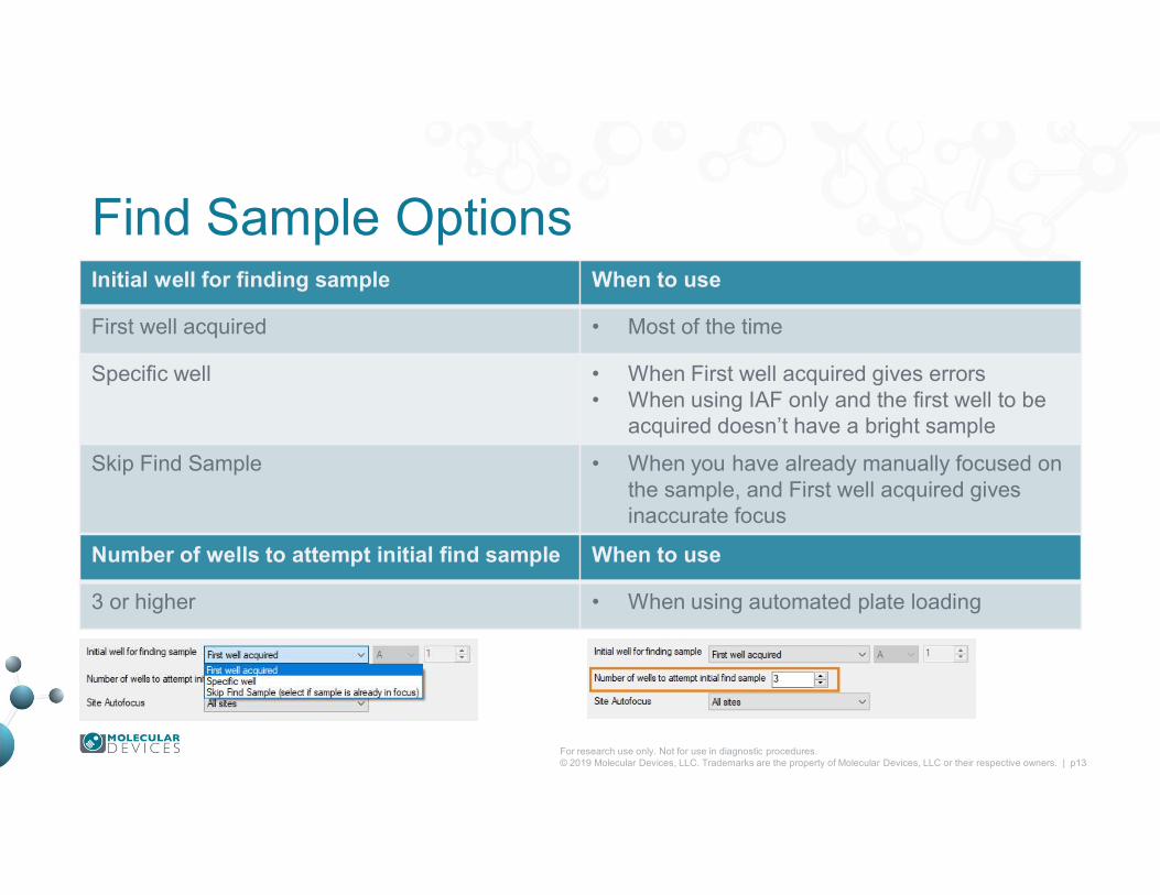
Find Sample OptionsInitial well for finding sample When to use
First well acquired • Most of the time
Specific well • When First well acquired gives errors• When using IAF only and the first well to be
acquired doesn’t have a bright sample
Skip Find Sample • When you have already manually focused on the sample, and First well acquired gives inaccurate focus
Number of wells to attempt initial find sample When to use
3 or higher • When using automated plate loading
For research use only. Not for use in diagnostic procedures.© 2019 Molecular Devices, LLC. Trademarks are the property of Molecular Devices, LLC or their respective owners. | p14
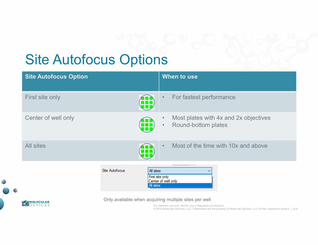
Site Autofocus OptionsSite Autofocus Option When to use
First site only • For fastest performance
Center of well only • Most plates with 4x and 2x objectives• Round-bottom plates
All sites • Most of the time with 10x and above
Only available when acquiring multiple sites per well
For research use only. Not for use in diagnostic procedures.© 2019 Molecular Devices, LLC. Trademarks are the property of Molecular Devices, LLC or their respective owners. | p15
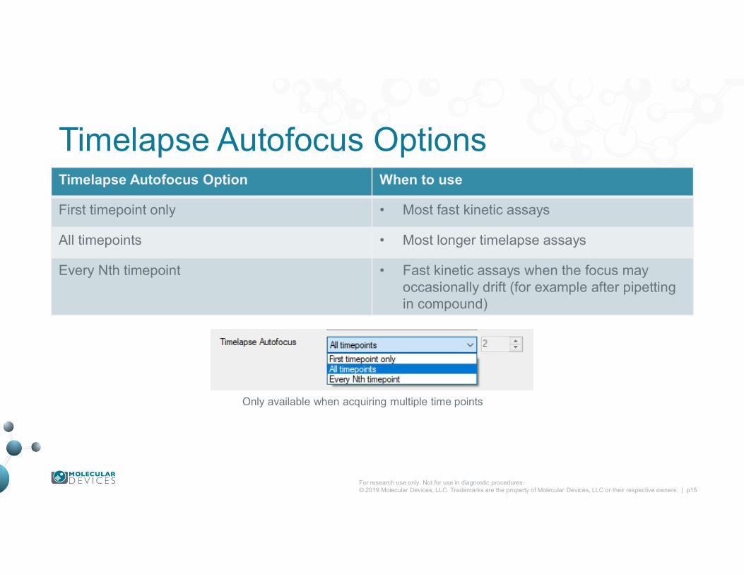
Timelapse Autofocus OptionsTimelapse Autofocus Option When to use
First timepoint only • Most fast kinetic assays
All timepoints • Most longer timelapse assays
Every Nth timepoint • Fast kinetic assays when the focus may occasionally drift (for example after pipetting in compound)
Only available when acquiring multiple time points
For research use only. Not for use in diagnostic procedures.© 2019 Molecular Devices, LLC. Trademarks are the property of Molecular Devices, LLC or their respective owners. | p16
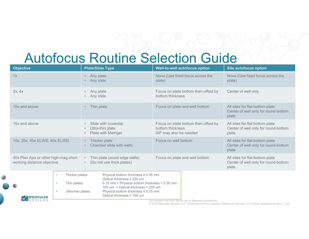
Autofocus Routine Selection Guide
• Thicker plates: Physical bottom thickness ≥ 0.35 mm Optical thickness ≥ 220 um
• Thin plates: 0.15 mm < Physical bottom thickness < 0.35 mm100 um < Optical thickness < 220 um
• Ultra-thin plates: Physical bottom thickness ≤ 0.15 mmOptical thickness ≤ 100 um
Objective Plate/Slide Type Well-to-well autofocus option Site autofocus option
1x • Any plate • Any slide
None (Use fixed focus across the plate)
None (Use fixed focus across the plate)
2x, 4x • Any plate • Any slide
Focus on plate bottom then offset by bottom thickness
Center of well only
10x and above • Thin plate Focus on plate and well bottom All sites for flat-bottom plateCenter of well only for round-bottom plate
10x and above • Slide with coverslip• Ultra-thin plate• Plate with Matrigel
Focus on plate bottom then offset by bottom thicknessIAF may also be needed
All sites for flat-bottom plateCenter of well only for round-bottom plate
10x, 20x, 40x ELWD, 60x ELWD • Thicker plate• Chamber slide with wells
Focus on well bottom All sites for flat-bottom plateCenter of well only for round-bottom plate
40x Plan Apo or other high-mag short working distance objective
• Thin plate (avoid edge wells)• (Do not use thick plates)
Focus on plate and well bottom All sites for flat-bottom plateCenter of well only for round-bottom plate

Additional notes on Autofocus Routines1. 2x and 4x objectives: For these low-magnification objectives, the plate and well bottom laser reflections cannot reliably be
distinguished because of the wide depth of field.
2. Thin-bottom plates: For thin-bottom plates with high variation, the large search range for the well bottom may accidentally detect the plate bottom. The Focus on plate and well bottom option forces the system to search for both surfaces.
3. Short working distance objectives: These objectives may bump the skirt of the plate when focusing on edge wells, pushing the plate up.
4. Matrigel-based samples: The LAF cannot detect the Matrigel or the cells. The LAF locates the plate bottom and a smaller image-based autofocus is used to find the cells.
5. Slides with coverslips: There is no equivalent well bottom to produce a reflection at the interface between the coverslip, mounting media, and slide. Generally if the sample is located on the bottom surface (e.g. cells on the coverslip/coverslip down or tissue on the slide/coverslip up) then LAF alone is sufficient. If the sample is on the top surface (e.g. tissue on the slide / coverslip down) then you may need LAF + IAF.
• Thicker plates: Physical bottom thickness ≥ 0.35 mm Optical thickness ≥ 220 um
• Thin plates: 0.15 mm < Physical bottom thickness < 0.35 mm100 um < Optical thickness < 220 um
• Ultra-thin plates: Physical bottom thickness ≤ 0.15 mmOptical thickness ≤ 100 um
For research use only. Not for use in diagnostic procedures.© 2019 Molecular Devices, LLC. Trademarks are the property of Molecular Devices, LLC or their respective owners. | p18
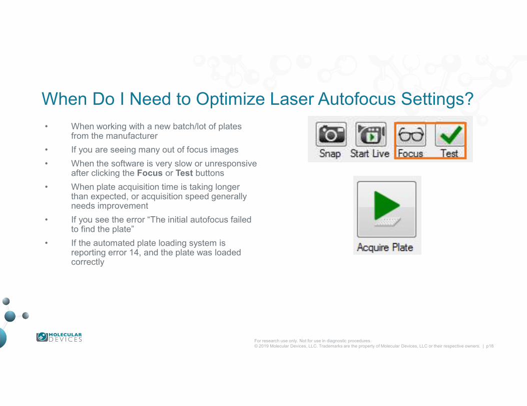
When Do I Need to Optimize Laser Autofocus Settings?
• When working with a new batch/lot of plates from the manufacturer
• If you are seeing many out of focus images
• When the software is very slow or unresponsive after clicking the Focus or Test buttons
• When plate acquisition time is taking longer than expected, or acquisition speed generally needs improvement
• If you see the error “The initial autofocus failed to find the plate”
• If the automated plate loading system isreporting error 14, and the plate was loaded correctly

For research use only. Not for use in diagnostic procedures.© 2019 Molecular Devices, LLC. Trademarks are the property of Molecular Devices, LLC or their respective owners. | p19
Support Resources• F1 / HELP within MetaXpress® Software
• Support and Knowledge Base: https://support.moleculardevices.com/
• Contact us: https://www.moleculardevices.com/contact