mutually exclusive cytoplasmic dynein regulation by nude-lis1
TRANSCRIPT

Mutually Exclusive Cytoplasmic Dynein Regulation byNudE-Lis1 and Dynactin*□S
Received for publication, August 3, 2011, and in revised form, September 2, 2011 Published, JBC Papers in Press, September 12, 2011, DOI 10.1074/jbc.M111.289017
Richard J. McKenney1, Sarah J. Weil, Julian Scherer, and Richard B. Vallee2
From the Department of Pathology and Cell Biology, Columbia University, New York, New York 10032
Background: Cytoplasmic dynein performs a great variety of cellular functions using a diversity of regulators.Results: NudE and dynactin compete for a common site within the dynein complex.Conclusion: This mechanism prevents dual regulation by dynactin and LIS1 and suggests a major new mode of regulatorycontrol.Significance: This is the first insight into coordination of cytoplasmic dynein regulators.
Cytoplasmic dynein is responsible for a wide range of cellularroles. How this single motor protein performs so many func-tions has remained a major outstanding question for manyyears. Part of the answer is thought to lie in the diversity ofdynein regulators, but how the effects of these factors are coor-dinated in vivo remains unexplored.We previously foundNudEto bind dynein through its light chain 8 (LC8) and intermediatechain (IC) subunits (1), the latter of which also mediates thedynein-dynactin interaction (2). We report here that NudE anddynactin bind to a common region within the IC, and competefor this site. We find LC8 to bind to a novel sequence withinNudE, without detectably affecting the dynein-NudE interac-tion. We further find that commonly used dynein inhibitoryreagents have broad effects on the interaction of dynein with itsregulatory factors. Together these results reveal an unantici-pated mechanism for preventing dual regulation of individualdynein molecules, and identify the IC as a nexus for regulatoryinteractions within the dynein complex.
Cytoplasmic dynein is a 1.2 MDa protein complex that func-tions as the predominant microtubule minus end-directedmolecular motor in most cell types. It is involved in a very widerange of cellular roles, but the underlying basis for its greatfunctional diversity is poorly understood. A variety of regula-tory factors appear to tailor the motor protein for specific cel-lular roles. These factors are responsible for recruitment ofdynein to appropriate sites within the cell, proper temporalactivation of motor activity, andmodulation of mechanochem-ical behavior to accommodate different cellular tasks.Themostwell-studied dynein regulatory factors are dynactin
(3–7) and the LIS1-NudE/L complex (8–15). Each of these sys-tems is involved both in dynein cargo recruitment and mecha-
nochemical regulation, though via different mechanisms. Dyn-actin is itself a �1 MDa multi-subunit complex. It was initiallyfound to be required for dynein vesicular transport in vitro (4,16), and to recruit themotor tomitotic kinetochores, and vesic-ular organelles (5, 17). Dynactin has also been found to increasedynein processivity by up to 2-fold in single molecule in vitroassays (7, 18, 19). The mechanism responsible for this effect isincompletely understood. Processivity of mammalian dynein isstimulated in both the plus- and minus-end directions alongmicrotubules (20, 21), though yeast dynein with or withoutdynactin is primarily unidirectional (19). Although the micro-tubule binding CAP-Gly domain of the dynactin p150Glued sub-unit had been assumed to contribute to the enhancement ofdynein processivity, recent studies showed no effect after itsremoval. Nonetheless, it was still required for complete dynac-tin function in vivo (6, 19, 22, 23).LIS1 and its binding partners NudE and NudEL form a tri-
partite complexwith dynein (15). LIS1 andNudE/L play criticalroles in a subset of dynein functions, many of which appear toinvolve high-load dynein mediated transport. LIS1 is requiredfor nuclear migration in neural progenitors and post mitoticneurons in vertebrates, and for nucleokinesis in several organ-isms (24–27). LIS1 and its interactors have also been implicatedin translocation or reorientation of the entiremicrotubule cyto-skeleton during mitosis and cell migration, as well as in centro-some and kinetochore dynamics, (1, 25, 28–32). The range ofcellular functions involving LIS1 and NudE/L and their extentof overlap with dynactin-requiring functions remains incom-pletely resolved. Aspects of vesicular transport that involvedynactin were found not to require LIS1 (32, 33), though gen-eral (34–38) or conditional (39) roles for LIS1, NudE, andNudEL have been reported in other studies.NudE and NudEL have been implicated in recruiting cyto-
plasmic dynein to cargo (1, 30, 40–42) as well as in recruitingLIS1 to dynein (15). We recently identified effects of LIS1 andNudE/L on dynein motor activity, and found them to be com-plex and distinct from those reported for dynactin (15). LIS1stabilized the dynein-MT interaction during the transitionstate of the cross-bridge cycle, resulting in persistent force pro-duction under load. NudE alone inhibited the dynein-MTinteraction. Strikingly, the tripartite complex of LIS1, NudE,and dynein transformed the motor to a persistent force-pro-
* This work was supported by National Institutes of Health Grants GM47434and HD40182 (to R. B. V.).
□S The on-line version of this article (available at http://www.jbc.org) containssupplemental Figs. S1 and S2.
1 Present address: Department of Cellular and Molecular Pharmacology, Uni-versity of California, San Francisco, CA 94158.
2 To whom correspondence should be addressed: Department of Pathologyand Cell Biology, Columbia University Physicians and Surgeons Building,Room 15-409 630 West 168th St., New York, NY 10032. Tel.: 212-342-0546;E-mail: [email protected].
THE JOURNAL OF BIOLOGICAL CHEMISTRY VOL. 286, NO. 45, pp. 39615–39622, November 11, 2011© 2011 by The American Society for Biochemistry and Molecular Biology, Inc. Printed in the U.S.A.
NOVEMBER 11, 2011 • VOLUME 286 • NUMBER 45 JOURNAL OF BIOLOGICAL CHEMISTRY 39615
by guest on February 3, 2018http://w
ww
.jbc.org/D
ownloaded from

ducing state and enhanced multiple motor transport underload (15). This behavior is likely to be important in cellularscenarios requiring dynein to produce force against largeopposing loads, such as nuclear migration (25).Dynactin, NudE, and NudEL each interact with the tail
region of the dynein complex. Dynactin binds via the centralregion of its p150Glued subunit to the N terminus of the dyneinintermediate chain (IC)3 (2, 43, 44). NudE and NudEL havebeen found to bind to both the dynein IC and LC8 subunits (1,15).NudE andNudELwere initially reported to contain aC-ter-minal dynein-interaction site (12), but a separate N-terminalsite has also recently been reported as well (45, 46).The current study was initiated to define the nature of the
NudE-dynein interaction in greater detail. We find the primarybinding site for NudE to lie within the dynein IC N terminus,the same region implicated in dynactin binding (2, 43). Weobserve clear competition between NudE and dynactin fordynein, identifying a novel mechanism for coordinating dyneinregulators. The common interaction site is also a target for fre-quently used inhibitory probes, and our results, therefore, haveimportant implications for phenotypic analysis of dynein func-tion in vivo.
EXPERIMENTAL PROCEDURES
DNA Cloning and Protein Purification—Full-length mouseNudE was cloned into pGEX6P-1 (GE Biosciences) with N-ter-minal HA- and C-terminal His tags. NudE fragments were alsocloned into this vector. p150Glued fragments were cloned from afull-length rat construct into pGEX6P-1 with an N-terminalFLAG-tag and human LC8 (accession number NM_003746)was also cloned into this vector. Dynein IC fragments from ratwere also cloned into pGEX6P-1 with aMyc tag at the C termi-nus, or into pCDNA 3.1 (IC2C 1–260 and 123–280) or pEGFP(IC2C 1–100) formammalian cell expression. For expression inbacteria, constructs were transformed into BL21-CodonPlusRIPL competent cells (Agilent Technologies) and expressed inLuria broth or Terrific broth. Protein production was inducedby addition of 0.1–0.5 mM IPTG, and the culture was moved to18 °C overnight. Bacteria were broken by sonication, and pro-teins were purified by batch incubation of a high speed super-natant with glutathione resin (GE Biosciences) in lysis buffer(PBS, 1mMDTT, protease inhibitormixture (Sigma), 1%TritonX-100) for 1–2 h at 4 °C. The beads were collected and washedextensively with lysis buffer in a column. The beads were thenwashed into PMEG buffer (100 mM PIPES, 5 mM EGTA, 4 mM
MgCl, 0.1 mM EDTA, 0.9 M glycerol, 1 mM DTT, pH 7.0) forfreezing, or washed into PreScission protease cleavage buffer(50 mM Tris-HCl pH 7.0, 150 mM NaCl, 1 mM EDTA), andincubated with PreScission protease (GE Biosciences) over-night to cleave off the GST moiety according to the manufac-turer’s instructions. The cleaved proteins were collected, con-centrated using Amicon concentrators (Amicon) and flashfrozen in liquid nitrogen for storage. For full-length NudE, theprotein was then incubated with Talon resin for 1 h at 4 °C, thebeads were washed extensively and eluted in PMEG containing
350 mM imidazole. Protein containing fractions were pooled,concentrated, and flash frozen in liquid nitrogen. Cytoplasmicdynein was purified from rat brain tissue as described (47) andfrozen in liquid nitrogen. This dynein preparation is essentiallyfree of dynactin (15). Baculovirus expressed LIS1 was purifiedas described (15).Protein Biochemistry—Protein interaction experiments were
performed in buffer A: 50mMHEPES (pH 7.4), 150mMNaCl, 2mM DTT, 0.2 mg/ml BSA, 0.1% Nonidet P-40. For GST pull-downs, proteins were bound to glutathione-agarose and incu-bated with interactors in 350 �l total volume for 1 h at 4 °C. ForHA or FLAG pull-downs, tagged proteins were first incubatedwith an equal molar amount of monoclonal anti-HA or anti-FLAG antibodies for 1 h on ice. The protein-antibody com-plexes were then bound to protein A beads (Invitrogen) for 1 hat 4 °C, washed twice to remove unbound protein, and thenused for pull-downs as above. Beads were washed four timeswith 350 �l of buffer A before being processed for SDS-PAGEanalysis. For pull-downs from brain lysate, a high speed super-natant (47) of rat brain was used. In competition experiments,beads coated with dynein interactors were incubated with �4nM purified brain dynein for 1 h at 4 °C, followed by three bufferwashes of 350 �l each. The beads were then resuspended tovolume, and the indicated amount of competitor protein wasadded for an additional hour, followed by four 350 �l washes.Beads were resuspended in 50 �l and processed for gel analysis.Western blots were processed on an Odyssey IR scanner (LI-COR Biosciences). Densitometry was performed using ImageJsoftware (NIH).Antibodies—Antibodies used in this study were: monoclonal
anti-dynein intermediate chain clone 74.1 (a gift fromDr. KevinPfister), or clone 70.1 (Sigma).Monoclonal and polyclonal anti-FLAG, anti-HA, anti-Myc, anti-GFP, and anti-LC8 (Abcam)and monoclonal anti-LIS1 (Sigma). Polyclonal anti-p150Glued(D’art) (48) and polyclonal anti-NudE/NudEL (1).Pepscan—Amembrane array spottedwith overlapping dode-
capeptides was generated based on mouse NudE (GenBankTMaccessionnumberQ9CZA6)C-terminal residues 192–344 (JPTPeptide Techonologies, Berlin). Each spot contains �5 nmol ofa 12-amino acid long peptide that is covalently linked to a cel-lulose-�-alanine membrane. The sequence of peptides in adja-cent spots are shifted C-terminally by two residues such thattwo neighboring spots overlap by ten residues (Fig. 2B). Beforeuse, the membrane was reconstituted at room temperature inmethanol for 5min, followed by three 10-minwasheswithTBS.Blockingwas performed for 1 hwith 5%milk inTBS-T followedby a 1-h incubation simultaneously with primary monoclonalLC8 antibody at a dilution of 1:2500 and secondary anti-rabbitAlexa Fluor 680 (Invitrogen, A10043) at a dilution of 1:10,000 in5% milk in TBS-T at room temperature. The membrane wasthen scanned using Odyssey Imaging System (LI-COR) toassess nonspecific interactions of the antibodies. 200 nM ofrecombinant purified human LC8 in 5% milk in TBS-T wasincubated with the membrane overnight at 4 °C and for 1 h atroom temperature the following day, followed by three 10-minwashes with TBS-T, sequential probing with primary and sec-ondary antibodies, and scanning as before. Scans of the mem-brane before and after incubation with LC8 were compared to
3 The abbreviations used are: IC, intermediate chain; HC, heavy chain; LC, lightchain.
Competition between Dynein Regulatory Factors
39616 JOURNAL OF BIOLOGICAL CHEMISTRY VOLUME 286 • NUMBER 45 • NOVEMBER 11, 2011
by guest on February 3, 2018http://w
ww
.jbc.org/D
ownloaded from

identify the residues in the C terminus of NudE that areinvolved in binding LC8.Inhibition of CC1/NudE-Dynein Interaction with Antibodies—
Monoclonal anti-dynein intermediate chain antibodies 74.1and 70.1 were incubated with rat brain purified cytoplasmicdynein at �10-fold molar excess for 60 min at 4 °C with gentlerotating. Antibody-dynein complexes or dynein alonewere sub-sequently incubatedwith bacterially expressedGST-CC1orGST-NudE on glutathione beads or beads alone for 90min at 4 °C withgentle rotating in buffer A. In a similar experiment, GST-CC1 orGST-NudE either alone or after preincubation with a polyclonalanti-NudE/L antibody for 60 min at 4 °C were incubated with ratbrain purified cytoplasmic dynein for 90 min at 4 °C with gentlerotating inbufferA.After the incubationperiods, unbounddyneinwas separated from the beads by centrifugation. The beads werewashed three times with buffer (15-fold bead volume) and resus-pended in protein sample buffer. Coomassie-stained gels werescanned using the Odyssey IR system.
RESULTS
NudE Binds to the Dynein Intermediate Chain N Terminus—In a previous studywe screened an array of dynein and dynactinsubunits for NudE binding, and identified interactions with thedynein IC and LC8 subunits (1, 15). To gain further insight intothe nature of these interactions we first determined whereNudE bound within the dynein IC. This subunit consists of a
shortN-terminal�-helical coiled-coil, followed by binding sitesfor dynein’s three LC classes, a dimerization domain, and finallyby a WD40 domain responsible for dynein heavy chain (HC)binding (49, 50) (Fig. 1B). Expression of theWD40 domain wastoxic in HeLa cells, as previously reported (43), but the N-ter-minal fragments expressed well. Pull-downs with GST-NudElocalized IC binding activity to the first 100 a.a. of rat IC2C (Fig.1A). GST-NudE also interacted with the endogenous dyneincomplex as evidenced by the presence of the full-length dyneinIC in the pulldowns (1). To test for a direct NudE-IC interac-tion, we expressed a series of GST-tagged N-terminal IC con-structs in Escherichia coli (Fig. 1B). All constructs that con-tained the N-terminal coiled-coil domain were capable ofinteracting with native dynactin complex in rat brain lysates(Fig. 1C) indicating the proteins were correctly folded. The ICconstructs were screened for interactions with purified HA-NudE, which mapped NudE binding to IC amino acids 1–70(Fig. 1, B–D). NudE bound to each of six alternatively splicedvariants of rat IC1 and IC2 (supplemental Fig. S1B), which allcontain a common N-terminal predicted coiled-coil sequence,but diverge immediately downstream (2, 49) (supplemental Fig.S1A), arguing that IC isoform composition is unlikely to affectthe IC-NudE interaction.Dynein LC8BindsDirectly toNudE—Although LC8 interacts
with both NudE and dynein, the specific role of the LC in the
FIGURE 1. Binding site for NudE on the dynein IC. A, GST NudE pull-downs of rat dynein IC2C fragments expressed in HeLa cells, immunoblotted withantibodies to GFP and Myc tags, and anti-IC antibody (74.1) to indicate endogenous dynein, labeled endog-IC. S, supernatant; P, pellet. B, diagram of the dyneinIC2C polypeptide and GST-IC fragments used in this study, and a Coomassie Brilliant Blue (CBB)-stained electrophoretic gel of the purified bacterially expressedprotein fragments. C, GST-IC pull-downs of dynactin from rat brain cytosol. Retention of native dynactin complex was assayed using antibodies against bothp150Glued and p50/dynamitin subunits. D, GST-IC pull-downs of purified bacterially expressed HA-NudE.
Competition between Dynein Regulatory Factors
NOVEMBER 11, 2011 • VOLUME 286 • NUMBER 45 JOURNAL OF BIOLOGICAL CHEMISTRY 39617
by guest on February 3, 2018http://w
ww
.jbc.org/D
ownloaded from
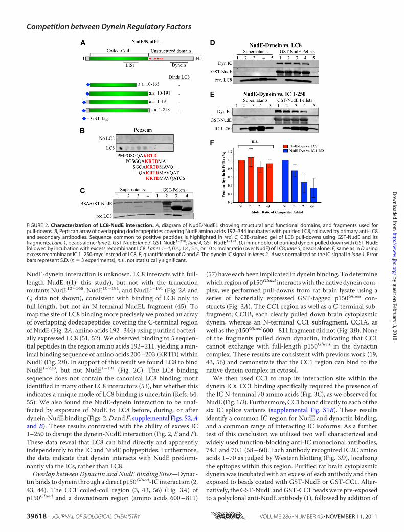
NudE-dynein interaction is unknown. LC8 interacts with full-length NudE ((1); this study), but not with the truncationmutants NudE10–165, NudE10–191, and NudE1–191 (Fig. 2A andC; data not shown), consistent with binding of LC8 only tofull-length, but not an N-terminal NudEL fragment (45). Tomap the site of LC8 binding more precisely we probed an arrayof overlapping dodecapeptides covering the C-terminal regionof NudE (Fig. 2A, amino acids 192–344) using purified bacteri-ally expressed LC8 (51, 52). We observed binding to 5 sequen-tial peptides in the region amino acids 192–211, yielding amin-imal binding sequence of amino acids 200–203 (KRTD) withinNudE (Fig. 2B). In support of this result we found LC8 to bindNudE1–218, but not NudE1–191 (Fig. 2C). The LC8 bindingsequence does not contain the canonical LC8 binding motifidentified in many other LC8 interactors (53), but whether thisindicates a unique mode of LC8 binding is uncertain (Refs. 54,55). We also found the NudE-dynein interaction to be unaf-fected by exposure of NudE to LC8 before, during, or afterdynein-NudEbinding (Figs. 2,D and F, supplemental Figs. S2,Aand B). These results contrasted with the ability of excess IC1–250 to disrupt the dynein-NudE interaction (Fig. 2, E and F).These data reveal that LC8 can bind directly and apparentlyindependently to the IC and NudE polypeptides. Furthermore,the data indicate that dynein interacts with NudE predomi-nantly via the ICs, rather than LC8.Overlap between Dynactin and NudE Binding Sites—Dynac-
tin binds to dynein through a direct p150Glued-IC interaction (2,43, 44). The CC1 coiled-coil region (3, 43, 56) (Fig. 3A) ofp150Glued and a downstream region (amino acids 600–811)
(57) have each been implicated in dynein binding. Todeterminewhich region of p150Glued interactswith the native dynein com-plex, we performed pull-downs from rat brain lysate using aseries of bacterially expressed GST-tagged p150Glued con-structs (Fig. 3A). The CC1 region as well as a C-terminal sub-fragment, CC1B, each clearly pulled down brain cytoplasmicdynein, whereas an N-terminal CC1 subfragment, CC1A, aswell as the p150Glued 600–811 fragment did not (Fig. 3B). Noneof the fragments pulled down dynactin, indicating that CC1cannot exchange with full-length p150Glued in the dynactincomplex. These results are consistent with previous work (19,43, 56) and demonstrate that the CC1 region can bind to thenative dynein complex in cytosol.We then used CC1 to map its interaction site within the
dynein ICs. CC1 binding specifically required the presence ofthe IC N-terminal 70 amino acids (Fig. 3C), as we observed forNudE (Fig. 1D). Furthermore, CC1bounddirectly to each of thesix IC splice variants (supplemental Fig. S1B). These resultsidentify a common IC region for NudE and dynactin binding,and a common range of interacting IC isoforms. As a furthertest of this conclusion we utilized two well characterized andwidely used function-blocking anti-IC monoclonal antibodies,74.1 and 70.1 (58–60). Each antibody recognized IC2C aminoacids 1–70 as judged by Western blotting (Fig. 3D), localizingthe epitopes within this region. Purified rat brain cytoplasmicdynein was incubated with an excess of each antibody and thenexposed to beads coated with GST-NudE or GST-CC1. Alter-natively, theGST-NudE andGST-CC1beadswere pre-exposedto a polyclonal anti-NudE antibody (1), followed by addition of
FIGURE 2. Characterization of LC8-NudE interaction. A, diagram of NudE/NudEL showing structural and functional domains, and fragments used forpull-downs. B, Pepscan array of overlapping dodecapeptides covering NudE amino acids 192–344 incubated with purified LC8, followed by primary anti-LC8and secondary antibodies. Sequence common to positive peptides is highlighted in red. C, CBB-stained gel of LC8 pull-downs using GST-NudE and itsfragments. Lane 1, beads alone; lane 2, GST-NudE; lane 3, GST-NudE1–218; lane 4, GST-NudE1–191. D, immunoblot of purified dynein pulled down with GST-NudEfollowed by incubation with excess recombinant LC8. Lanes 1– 4, 0�, 1�, 5�, or 10� molar ratio (over NudE) of LC8; lane 5, beads alone. E, same as in D usingexcess recombinant IC 1–250-myc instead of LC8. F, quantification of D and E. The dynein IC signal in lanes 2– 4 was normalized to the IC signal in lane 1. Errorbars represent S.D. (n � 3 experiments), n.s., not statistically significant.
Competition between Dynein Regulatory Factors
39618 JOURNAL OF BIOLOGICAL CHEMISTRY VOLUME 286 • NUMBER 45 • NOVEMBER 11, 2011
by guest on February 3, 2018http://w
ww
.jbc.org/D
ownloaded from
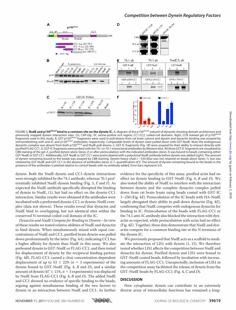
dynein. Both the NudE-dynein and CC1-dynein interactionswere strongly inhibited by the 74.1 antibody, whereas 70.1 pref-erentially inhibited NudE-dynein binding (Fig. 3, E and F). Asexpected the NudE antibody specifically disrupted the bindingof dynein to NudE, (1), but had no effect on the dynein-CC1interaction. Similar results were obtained if the antibodies wereincubatedwith a preformed dynein-CC1 or dynein-NudE com-plex (data not shown). These results reveal that dynactin andNudE bind to overlapping, but not identical sites within theconserved N-terminal coiled-coil domain of the IC.Dynactin and NudE Compete for Binding to Dynein—In view
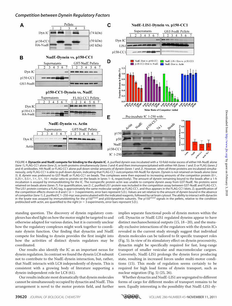
of these results we tested relative abilities of NudE and dynactinto bind dynein. When simultaneously mixed with equal con-centrations of NudE and CC1, purified brain dynein was pulleddown predominantly by the latter (Fig. 4A), indicating CC1 hasa higher affinity for dynein than NudE in this assay. We alsoprebound dynein to GST-NudE or FLAG-CC1, and then testedfor displacement of dynein by the reciprocal binding partner(Fig. 4B). FLAG-CC1 caused a clear concentration-dependentdisplacement of up to 53 � 22% (n � 3 experiments) of thedynein bound to GST-NudE (Fig. 4, B and D), and a similaramount of dynein (47� 11%, n� 3 experiments) was displacedby NudE from FLAG-CC1 (Fig. 4, B and D). The added NudEand CC1 showed no evidence of specific binding to the beads,arguing against simultaneous binding of the two factors todynein or an interaction between NudE and CC1. As further
evidence for the specificity of this assay, purified actin had noeffect on dynein binding to GST-NudE (Fig. 4, B and D). Wealso tested the ability of NudE to interfere with the interactionbetween dynein and the complete dynactin complex pulleddown from rat brain lysate using beads coated with GST-IC1–250 (Fig. 4E). Preincubation of the IC beads with HA-NudElargely abrogated their ability to pull-down dynactin (Fig. 4E),confirming that NudE competes with endogenous dynactin forbinding to IC. Preincubation of the beads with FLAG-CC1 orthe 74.1 anti-IC antibody also blocked the interactionwith dyn-actin as expected, while preincubation with actin had no effect(Fig. 4E). Together, these data demonstrate thatNudE and dyn-actin compete for a common binding site at the N terminus ofthe dynein IC.Wepreviously proposed thatNudE acts as a scaffold tomedi-
ate the interaction of LIS1 with dynein (1, 15). We thereforetested whether LIS1 affects the competition betweenNudE anddynactin for dynein. Purified dynein and LIS1 were bound toGST-NudE coated beads, followed by incubation with increas-ing amounts of FLAG-CC1. Unexpectedly, inclusion of LIS1 inthe competition assay facilitated the release of dynein from theGST-NudE beads by FLAG-CC1 (Fig. 4, C and D).
DISCUSSION
How cytoplasmic dynein can contribute to an extremelydiverse array of intracellular functions has remained a long-
FIGURE 3. NudE and p150Glued bind to a common site on the dynein IC. A, diagram of the p150Glued subunit of dynactin showing domain architecture andpreviously mapped dynein interaction sites. CG, CAP-Gly; SP, serine-proline rich region; CC1-CC2, coiled-coil domains. Right, CCB stained gel of p150Glued
fragments used in this study. B, GST-p150Glued fragments were used in pull-downs from rat brain cytosol and dynein and dynactin binding was assayed byimmunoblotting with anti-IC and p150Glued antibodies, respectively. Comparable levels of dynein were pulled down with GST-NudE. Note the endogenousdynactin complex was absent from both p150Glued and NudE pull-downs. C, GST-IC fragments (Fig. 1B) were assayed for their ability to interact directly withpurified FLAG-CC1. D, GST-IC fragments were probed with the 74.1 or 70.1 monoclonal antibodies by Western blot. All three GST-IC fragments are visualized byCBB staining of the gel. E, purified dynein alone (lanes 2) or after preincubation with the indicated antibodies (lanes 3) was bound to beads containing eitherGST-NudE or GST-CC1. Additionally, GST-NudE or GST-CC1 were preincubated with a polyclonal NudE antibody before dynein was added (right). The amountof dynein remaining bound to the beads was assayed by CBB staining. Dynein heavy chain (�530 kDa) was not retained on beads alone (lanes 1), but wasretained by GST-NudE and GST-CC1 in the absence of antibodies (lanes 2). F, quantification of E. The amount of dynein remaining bound to the beads in thepresence of the antibodies is plotted relative to control beads with no antibody added. Error bars represent S.D.
Competition between Dynein Regulatory Factors
NOVEMBER 11, 2011 • VOLUME 286 • NUMBER 45 JOURNAL OF BIOLOGICAL CHEMISTRY 39619
by guest on February 3, 2018http://w
ww
.jbc.org/D
ownloaded from
standing question. The discovery of dynein regulatory com-plexes has shed light on how themotormight be targeted to andotherwise adapted for various duties, but it is currently unclearhow the regulatory complexes might work together to coordi-nate dynein function. Our finding that dynactin and NudEcompete for binding to dynein provides the first insight intohow the activities of distinct dynein regulators may becoordinated.Our results also identify the IC as an important nexus for
dynein regulation. In contrast we found the dynein LC8 subunitnot to contribute to the NudE-dynein interaction, but, rather,that NudE interacts with LC8 independently of dynein. This isconsistent with a growing body of literature supporting adynein-independent role for LC8 (61).Our results indicatemost dramatically that dyneinmolecules
cannot be simultaneously occupied by dynactin andNudE.Thisarrangement is novel to the motor protein field, and further
implies separate functional pools of dynein motors within thecell. Dynactin or NudE-LIS1 regulated dyneins appear to havedistinct mechanochemical outputs (15, 18–20), and the mutu-ally exclusive interactions of the regulators with the dynein ICsrevealed in the current study strongly suggest that individualdynein molecules can be tailored to fit specific transport roles(Fig. 5). In view of its stimulatory effect on dynein processivity,dynactin might be specifically required for fast, long-rangetransport of smaller vesicular and macromolecular cargoes.Conversely, NudE-LIS1 prolongs the dynein force producingstate, resulting in increased forces under multi-motor condi-tions (15). This mode of regulation seems certainly to berequired for high load forms of dynein transport, such asnuclear migration (Fig. 5) (25, 26).Whether dynactin andNudE-LIS1 are segregated to different
forms of cargo for different modes of transport remains to beseen. Equally interesting is the possibility that NudE-LIS1-dy-
FIGURE 4. Dynactin and NudE compete for binding to the dynein IC. A, purified dynein was incubated with a 10-fold molar excess of either HA-NudE alone(lane 1), FLAG-CC1 alone (lane 2), or both proteins simultaneously (lanes 3 and 4) and then immunoprecipitated with either HA (lanes 1 and 3) or FLAG (lanes 2and 4) antibodies. HA-NudE or FLAG-CC1 alone pull down similar amounts of dynein (lanes 1 and 2). However, when all three proteins are incubated simulta-neously, only FLAG-CC1 is able to pull down dynein, indicating that FLAG-CC1 outcompetes HA-NudE for dynein. Dynein is not retained on beads alone (lane5). B, dynein was prebound to GST-NudE or FLAG-CC1 on beads. The complexes were then exposed to increasing amounts of the competitor protein (0�,0.25�, 0.5�, 1�, 5�, 10� molar ratio to protein on the beads in lanes 1– 6, respectively). The amount of dynein remaining bound to the beads after a 1-hincubation is assayed by immunoblotting for the IC. The nonspecific protein actin was unable to compete dynein away from GST-NudE. No proteins wereretained on beads alone (lanes 7). For quantification, see D. C, purified LIS1 protein was included in the competition assay between GST-NudE and FLAG-CC1.The LIS1 protein contains a FLAG tag, is approximately the same molecular weight as FLAG-CC1, and thus appears in the FLAG-CC1 blots. D, quantification ofthe competition effects shown in B and C (n � 3 experiments, error bars represent S.D.). Values are set relative to the amount of dynein bound in the absenceof competitor (lane 1). E, purified IC 1–250-myc was preincubated with the indicated reagents, followed by rat brain cytosol. The ability to interact with dynactinin the lysate was assayed by immunoblotting for the p150Glued and p50/dynamitin subunits. The p150Glued signals in the pellets, relative to the conditionpreblocked with actin, are quantified to the right (n � 3 experiments, error bars represent S.D.).
Competition between Dynein Regulatory Factors
39620 JOURNAL OF BIOLOGICAL CHEMISTRY VOLUME 286 • NUMBER 45 • NOVEMBER 11, 2011
by guest on February 3, 2018http://w
ww
.jbc.org/D
ownloaded from

nein and dynactin-dynein coexist on common cellular cargoes,adapting transport to distinct subcellular environments (Fig. 5).Indeed, recent evidence suggests that teams ofmultiple dyneinsmove membranous cargoes in vivo (62–64). Finally, it is alsopossible that the balance of a dynein regulatory factors may besubject to regulation in vivo, an issue of considerable furtherinterest. Indeed, phosphorylation of the dynein IC has beenreported to affect the affinity of this subunit for dynactin (65),andmight conceivably contribute to switching between dyneinregulatory factors. Further experiments are needed to test thishypothesis.Such a shift between regulatorymodesmay require new tools
to assay properly. Dynactin and NudE/NudEL-LIS1 each con-trol aspects of dynein recruitment to subcellular cargo, as wellas dyneinmechanochemical activity. This dual role will make itnecessary to quantify relative effects on the number of dyneinmolecules associated with cargo versus the nature of dyneinregulation. Changes in the affinity of dynein for dynactin rela-tive to NudE and NudEL could alter the number of cargo-asso-
ciated dyneins or shuttle dyneins between high-force and longtravel distance regulators, or both.Surprisingly, the addition of LIS1 caused dynein to be
released from NudE more easily in the presence of FLAG-CC1(Fig. 4, C and D). This result is unexpected given NudE estab-lished role in recruiting LIS1 to dynein (1, 15, 45, 46). These arethe first results suggesting that LIS1 may affect the NudE-dy-nein interaction and suggest further complexity in the interac-tion between dynein and its regulators. Additional informationon the structural nature of the various dynein complexes will beneeded to clarify this issue.Our study also reveals broader effects for commonly used
dynein inhibitory probes than has been assumed. The dynactinCC1 fragment, as well as the 74.1 and 70.1monoclonal antibod-ies, have been favored reagents for cytoplasmic dynein inhibi-tion in vivo. Our data indicate that the first two of these shouldinterfere with both dynactin and NudE-LIS1 binding, while thethird interferes preferentially withNudE.However, some of themore readily assayed dynein functions require both types ofregulatory factor. For this reason, physiological assays for thespecificity of the dynein and dynactin inhibitory agents mayrequire more quantitative in vivo assays for dynein behaviorthan are currently available (39). Although the effects of eachreagent provide insight into dynein function, their implicationsfor understanding dynein regulation now appear less clear. Fur-ther development of probes specific for cytoplasmic dynein andfor its individual regulatory factors will be needed to addressthese issues.
Acknowledgments—We thank Drs. Kevin Vaughan and John Wil-liams for contribution of reagents, sharing of unpublished results, andhelpful discussion.
REFERENCES1. Stehman, S. A., Chen, Y., McKenney, R. J., and Vallee, R. B. (2007) J. Cell
Biol. 178, 583–5942. Vaughan, K. T., and Vallee, R. B. (1995) J. Cell Biol. 131, 1507–15163. Schroer, T. A. (2004) Annu. Rev. Cell Dev. Biol. 20, 759–7794. Gill, S. R., Schroer, T. A., Szilak, I., Steuer, E. R., Sheetz, M. P., and Cleve-
land, D. W. (1991) J. Cell Biol. 115, 1639–16505. Echeverri, C. J., Paschal, B. M., Vaughan, K. T., and Vallee, R. B. (1996)
J. Cell Biol. 132, 617–6336. Kim, H., Ling, S. C., Rogers, G. C., Kural, C., Selvin, P. R., Rogers, S. L., and
Gelfand, V. I. (2007) J. Cell Biol. 176, 641–6517. Culver-Hanlon, T. L., Lex, S. A., Stephens, A. D., Quintyne, N. J., and King,
S. J. (2006) Nat. Cell Biol. 8, 264–2708. Reiner, O., Carrozzo, R., Shen, Y., Wehnert, M., Faustinella, F., Dobyns,
W. B., Caskey, C. T., and Ledbetter, D. H. (1993) Nature 364, 717–7219. Hirotsune, S., Fleck, M. W., Gambello, M. J., Bix, G. J., Chen, A., Clark,
G. D., Ledbetter, D. H., McBain, C. J., andWynshaw-Boris, A. (1998)Nat.Genetics 19, 333–339
10. Feng, Y., Olson, E. C., Stukenberg, P. T., Flanagan, L. A., Kirschner,M.W.,and Walsh, C. A. (2000) Neuron 28, 665–679
11. Niethammer, M., Smith, D. S., Ayala, R., Peng, J., Ko, J., Lee, M. S., Mora-bito, M., and Tsai, L. H. (2000) Neuron 28, 697–711
12. Sasaki, S., Shionoya, A., Ishida,M., Gambello,M. J., Yingling, J.,Wynshaw-Boris, A., and Hirotsune, S. (2000) Neuron 28, 681–696
13. Mesngon, M. T., Tarricone, C., Hebbar, S., Guillotte, A. M., Schmitt,E. W., Lanier, L., Musacchio, A., King, S. J., and Smith, D. S. (2006) J. Neu-rosci. 26, 2132–2139
14. Hebbar, S., Mesngon, M. T., Guillotte, A. M., Desai, B., Ayala, R., and
FIGURE 5. Model for mutually exclusive binding of dynactin and NudE todynein. Dynactin and NudE are shown at top to interact with the base of thedynein complex within and the coiled-coil N-terminal region of the ICs(expanded view). The 74.1 and 70.1 monoclonal antibodies (red) bind withinthe same region. Dynactin has been proposed to regulate dynein processivityfor fast, low-load transport, whearas NudE-LIS1 promote multi-motor trans-port of high-loads. The competition for dynein is proposed here to preventdual regulation of individual dynein complexes and, potentially, to provide amechanism to allow dyneins to shift between high-load and long travel dis-tance modes of transport.
Competition between Dynein Regulatory Factors
NOVEMBER 11, 2011 • VOLUME 286 • NUMBER 45 JOURNAL OF BIOLOGICAL CHEMISTRY 39621
by guest on February 3, 2018http://w
ww
.jbc.org/D
ownloaded from

Smith, D. S. (2008) J. Cell Biol. 182, 1063–107115. McKenney, R. J., Vershinin, M., Kunwar, A., Vallee, R. B., and Gross, S. P.
(2010) Cell 141, 304–31616. Schroer, T. A., and Sheetz, M. P. (1991) J. Cell Biol. 115, 1309–131817. Roghi, C., and Allan, V. J. (1999) J. Cell Sci. 112, 4673–468518. King, S. J., and Schroer, T. A. (2000) Nat. Cell Biol. 2, 20–2419. Kardon, J. R., Reck-Peterson, S. L., and Vale, R. D. (2009) Proc. Natl. Acad.
Sci. U.S.A. 106, 5669–567420. Ross, J. L., Wallace, K., Shuman, H., Goldman, Y. E., and Holzbaur, E. L.
(2006) Nat. Cell Biol. 8, 562–57021. Dixit, R., Ross, J. L., Goldman, Y. E., andHolzbaur, E. L. (2008) Science 319,
1086–108922. Vaughan, P. S., Miura, P., Henderson, M., Byrne, B., and Vaughan, K. T.
(2002) J. Cell Biol. 158, 305–31923. Moore, J. K., Sept, D., and Cooper, J. A. (2009) Proc. Natl. Acad. Sci. U.S.A.
106, 5147–515224. Lee,W. L., Oberle, J. R., and Cooper, J. A. (2003) J. Cell Biol. 160, 355–36425. Tsai, J. W., Bremner, K. H., and Vallee, R. B. (2007) Nat. Neurosci 10,
970–97926. Tsai, J. W., Chen, Y., Kriegstein, A. R., and Vallee, R. B. (2005) J. Cell Biol.
170, 935–94527. Xiang, X., Osmani, A. H., Osmani, S. A., Xin, M., andMorris, N. R. (1995)
Mol. Biol. Cell 6, 297–31028. Grabham, P. W., Seale, G. E., Bennecib, M., Goldberg, D. J., and Vallee,
R. B. (2007) J. Neurosci. 27, 5823–583429. Liang, Y., Yu, W., Li, Y., Yu, L., Zhang, Q., Wang, F., Yang, Z., Du, J.,
Huang, Q., Yao, X., and Zhu, X. (2007)Mol. Biol. Cell 18, 2656–266630. Vergnolle, M. A., and Taylor, S. S. (2007) Curr. Biol. 17, 1173–117931. Siller, K. H., and Doe, C. Q. (2008) Dev. Biol. 319, 1–932. Faulkner,N. E., Dujardin, D. L., Tai, C. Y., Vaughan, K. T.,O’Connell, C. B.,
Wang, Y., and Vallee, R. B. (2000) Nat. Cell Biol. 2, 784–79133. Tai, C. Y., Dujardin, D. L., Faulkner, N. E., and Vallee, R. B. (2002) J. Cell
Biol. 156, 959–96834. Smith, D. S., Niethammer, M., Ayala, R., Zhou, Y., Gambello, M. J., Wyn-
shaw-Boris, A., and Tsai, L. H. (2000) Nat. Cell Biol. 2, 767–77535. Lam, C., Vergnolle, M. A., Thorpe, L., Woodman, P. G., and Allan, V. J.
(2010) J. Cell Sci. 123, 202–21236. Zhang, J., Li, S., Fischer, R., and Xiang, X. (2003) Mol. Biol. Cell 14,
1479–148837. Zhang, J., Zhuang, L., Lee, Y., Abenza, J. F., Peñalva, M. A., and Xiang, X.
(2010) J. Cell Sci. 123, 3596–360438. Lenz, J. H., Schuchardt, I., Straube, A., and Steinberg, G. (2006)The EMBO
J. 25, 2275–228639. Yi, J., Ori-McKenney, K. M., McKenney, R. J., Vershinin, M., Gross, S. P.,
and Vallee, R. B. (2011) J. Cell Biol., in press40. Guo, J., Yang, Z., Song, W., Chen, Q., Wang, F., Zhang, Q., and Zhu, X.
(2006)Mol. Biol. Cell 17, 680–68941. Liang, Y., Yu, W., Li, Y., Yang, Z., Yan, X., Huang, Q., and Zhu, X. (2004)
J. Cell Biol. 164, 557–56642. Bolhy, S., Bouhlel, I., Dultz, E., Nayak, T., Zuccolo, M., Gatti, X., Vallee, R.,
Ellenberg, J., and Doye, V. (2011) J. Cell Biol. 192, 855–87143. King, S. J., Brown, C. L., Maier, K. C., Quintyne, N. J., and Schroer, T. A.
(2003)Mol. Biol. Cell 14, 5089–509744. Karki, S., and Holzbaur, E. L. (1995) J. Biol. Chem. 270, 28806–2881145. Wang, S., and Zheng, Y. (2011) J. Biol. Chem. 286, 587–59346. Zylkiewicz, E., Kijańska,M., Choi,W.C., Derewenda,U., Derewenda, Z. S.,
and Stukenberg, P. T. (2011) J. Cell Biol. 192, 433–44547. Paschal, B. M., Shpetner, H. S., and Vallee, R. B. (1991)Methods Enzymol.
196, 192–20148. Vaughan, K. T., Tynan, S. H., Faulkner, N. E., Echeverri, C. J., and Vallee,
R. B. (1999) J. Cell Sci. 112, 1437–144749. Myers, K. R., Lo, K. W., Lye, R. J., Kogoy, J. M., Soura, V., Hafezparast, M.,
and Pfister, K. K. (2007) J. Neurosci Res. 85, 2640–264750. Ma, S., Trivinos-Lagos, L., Collins, C. A., and Chisholm, R. L. (1995)Mol.
Biol. Cell 6, 155a51. Rodríguez-Crespo, I., Yélamos, B., Roncal, F., Albar, J. P., Ortiz de Mon-
tellano, P. R., and Gavilanes, F. (2001) FEBS Lett. 503, 135–14152. Navarro-Lérida, I., Martínez Moreno, M., Roncal, F., Gavilanes, F., Albar,
J. P., and Rodríguez-Crespo, I. (2004) Proteomics 4, 339–34653. Benison,G., Karplus, P. A., andBarbar, E. (2007) J.Mol. Biol. 371, 457–46854. Lightcap, C.M., Sun, S., Lear, J. D., Rodeck, U., Polenova, T., andWilliams,
J. C. (2008) J. Biol. Chem. 283, 27314–2732455. Lightcap, C.M., Kari, G., Arias-Romero, L. E., Chernoff, J., Rodeck, U., and
Williams, J. C. (2009) PLoS ONE 4, e602556. Quintyne, N. J., Gill, S. R., Eckley, D.M., Crego, C. L., Compton, D. A., and
Schroer, T. A. (1999) J. Cell Biol. 147, 321–33457. Deacon, S. W., Serpinskaya, A. S., Vaughan, P. S., Lopez Fanarraga, M.,
Vernos, I., Vaughan, K. T., and Gelfand, V. I. (2003) J. Cell Biol. 160,297–301
58. Steffen, W., Karki, S., Vaughan, K. T., Vallee, R. B., Holzbaur, E. L., Weiss,D. G., and Kuznetsov, S. A. (1997)Mol. Biol. Cell 8, 2077–2088
59. Steffen, W., Hodgkinson, J. L., and Wiche, G. (1996) J. Struct. Biol. 117,227–235
60. Burkhardt, J. K., Echeverri, C. J., Nilsson, T., and Vallee, R. B. (1997) J. CellBiol. 139, 469–484
61. Rapali, P., Szenes, A., Radnai, L., Bakos, A., Pal, G., and Nyitray, L. (2011)Febs J. 278, 2980–2996
62. Hendricks, A. G., Perlson, E., Ross, J. L., Schroeder, H.W., 3rd, Tokito,M.,and Holzbaur, E. L. (2010) Curr. Biol. : CB 20, 697–702
63. Soppina, V., Rai, A. K., Ramaiya, A. J., Barak, P., andMallik, R. (2009) Proc.Natl. Acad. Sci. U.S.A. 106, 19381–19386
64. Shubeita, G. T., Tran, S. L., Xu, J., Vershinin,M., Cermelli, S., Cotton, S. L.,Welte, M. A., and Gross, S. P. (2008) Cell 135, 1098–1107
65. Vaughan, P. S., Leszyk, J. D., and Vaughan, K. T. (2001) J. Biol. Chem. 276,26171–26179
Competition between Dynein Regulatory Factors
39622 JOURNAL OF BIOLOGICAL CHEMISTRY VOLUME 286 • NUMBER 45 • NOVEMBER 11, 2011
by guest on February 3, 2018http://w
ww
.jbc.org/D
ownloaded from

Richard J. McKenney, Sarah J. Weil, Julian Scherer and Richard B. ValleeMutually Exclusive Cytoplasmic Dynein Regulation by NudE-Lis1 and Dynactin
doi: 10.1074/jbc.M111.289017 originally published online September 12, 20112011, 286:39615-39622.J. Biol. Chem.
10.1074/jbc.M111.289017Access the most updated version of this article at doi:
Alerts:
When a correction for this article is posted•
When this article is cited•
to choose from all of JBC's e-mail alertsClick here
Supplemental material:
http://www.jbc.org/content/suppl/2011/09/12/M111.289017.DC1
http://www.jbc.org/content/286/45/39615.full.html#ref-list-1
This article cites 64 references, 37 of which can be accessed free at
by guest on February 3, 2018http://w
ww
.jbc.org/D
ownloaded from