mutated huntingtin causes testicular pathology in ... · pdf filepathology in transgenic...
TRANSCRIPT
Seediscussions,stats,andauthorprofilesforthispublicationat:https://www.researchgate.net/publication/297722766
MutatedHuntingtinCausesTesticularPathologyinTransgenicMinipigBoars
ArticleinNeurodegenerativeDiseases·March2016
DOI:10.1159/000443665
CITATIONS
0
READS
84
26authors,including:
MiroslavaSedlackova
MasarykUniversity
42PUBLICATIONS90CITATIONS
SEEPROFILE
IvonaValeková
AcademyofSciencesoftheCzechRepublic
9PUBLICATIONS5CITATIONS
SEEPROFILE
JiriKlempir
CharlesUniversityinPrague
32PUBLICATIONS164CITATIONS
SEEPROFILE
HanaHansikova
CharlesUniversityinPrague
167PUBLICATIONS1,458CITATIONS
SEEPROFILE
Allin-textreferencesunderlinedinbluearelinkedtopublicationsonResearchGate,
lettingyouaccessandreadthemimmediately.
Availablefrom:MonikaMačáková
Retrievedon:31August2016

E-Mail [email protected]
Original Paper
Neurodegener Dis 2016;16:245–259 DOI: 10.1159/000443665
Mutated Huntingtin Causes Testicular Pathology in Transgenic Minipig Boars
Monika Macakova a, c Bozena Bohuslavova a, c Petra Vochozkova a, c Antonin Pavlok a Miroslava Sedlackova b Daniela Vidinska a, c Klara Vochyanova a, c Irena Liskova a, d Ivona Valekova a, c Monika Baxa a, c Zdenka Ellederova a Jiri Klima a Stefan Juhas a Jana Juhasova a Jana Klouckova e Martin Haluzik e Jiri Klempir d Hana Hansikova f Jana Spacilova f Ryan Collins g Ian Blumenthal g Michael Talkowski g James F. Gusella g David S. Howland i Marian DiFiglia h Jan Motlik a
a Laboratory of Cell Regeneration and Plasticity, Institute of Animal Physiology and Genetics, Czech Academy of Science, Libechov , b Department of Histology and Embryology, Faculty of Medicine, Masaryk University in Brno, Brno , c Department of Cell Biology, Faculty of Science, and d Department of Neurology and Centre of Clinical Neuroscience, First Faculty of Medicine, Charles University in Prague, and e 3rd Department of Medicine, Department of Endocrinology and Metabolism, and f Laboratory for Study of Mitochondrial Disorders, Department of Pediatrics and Adolescent Medicine, First Faculty of Medicine, Charles University and General University Hospital in Prague, Prague , Czech Republic; g Center for Human Genetic Research, and h Department of Neurology, Massachusetts General Hospital, Boston, Mass. , and i CHDI Foundation, Princeton, N.Y. , USA
the effects of mtHtt. Results: Evidence for fertility failure of both TgHD generations was observed at the age of 13 months. Reproductive parameters declined and progres-sively worsened with age. EM revealed numerous patholog-ical features in sperm tails and in testicular epithelium from 24- and 36-month-old TgHD boars. Moreover, immunohisto-chemistry confirmed significantly lower proliferation activity of spermatogonia in transgenic testes. mtHtt was highly ex-pressed in spermatozoa and testes of TgHD boars and local-ized in all cells of seminiferous tubules. Levels of fertility-re-lated hormones did not differ in TgHD and WT siblings. Ge-nome analysis confirmed that insertion of the lentiviral construct did not interrupt any coding sequence in the pig genome. Conclusions: The sperm and testicular degenera-tion of TgHD boars is caused by gain-of-function of the high-ly expressed mtHtt. © 2016 S. Karger AG, Basel
Key Words
Huntington’s disease · Pig model · Mutant huntingtin · Spermatozoa · Testes · Degeneration
Abstract
Background: Huntington’s disease is induced by CAG ex-pansion in a single gene coding the huntingtin protein. The mutated huntingtin (mtHtt) primarily causes degeneration of neurons in the brain, but it also affects peripheral tissues, including testes. Objective: We studied sperm and testes of transgenic boars expressing the N-terminal region of human mtHtt. Methods: In this study, measures of reproductive pa-rameters and electron microscopy (EM) images of spermato-zoa and testes of transgenic (TgHD) and wild-type (WT) boars of F1 (24–48 months old) and F2 (12–36 months old) genera-tions were compared. In addition, immunofluorescence, im-munohistochemistry, Western blot, hormonal analysis and whole-genome sequencing were done in order to elucidate
Received: June 1, 2015 Accepted after revision: December 23, 2015 Published online: March 10, 2016 D i s e a s e s
Jan Motlik Laboratory of Cell Regeneration and PlasticityInstitute of Animal Physiology and Genetics Rumburska 89, CZ–27721 Libechov (Czech Republic) E-Mail motlik @ iapg.cas.cz
© 2016 S. Karger AG, Basel1660–2854/16/0164–0245$39.50/0
www.karger.com/ndd
Monika Macakova and Bozena Bohuslavova contributed equally to this work.
Dow
nloa
ded
by:
Uni
vers
ität Z
üric
h, Z
entr
albi
blio
thek
Zür
ich
13
0.60
.129
.67
- 5/
11/2
016
2:45
:57
PM
Macakova et al.
Neurodegener Dis 2016;16:245–259 DOI: 10.1159/000443665
246
Introduction
Huntington’s disease (HD) is a neurodegenerative dis-order caused by the expansion of CAG repeat in the gene encoding the huntingtin protein (Htt), which is expressed in most tissues. The onset of the disease is usually in the mid-thirties. The main target is the central nervous sys-tem, but it has an impact on the whole body. There is no available curative treatment to date. Even the pathogen-esis of the disease is not well understood. Nevertheless, it is well known that mutated Htt (mtHtt) forms cytoplas-mic and nuclear aggregates, particularly in the cerebral cortex, striatum and lateral hypothalamus [1] . Many ro-dent models of HD that express either truncated or full-length human mutant Htt display differences in the onset and severity of phenotypes. Rodent models have collec-tively provided valuable information related to target val-idation and drug therapy [2–4] . However, large animal models are expected to simulate the disease more faith-fully and moreover enable the usage of medical tech-niques and equipment applicable for human patients [5] .
Minipigs represent a desirable model for longitudinal safety studies and preclinical drug trials to fill the gap be-tween rodent models and patients [6, 7] . The advantage of minipigs is their resemblance with the human brain as well as with the whole body in terms of size, anatomy and physiology. There is a 96% homology between porcine and human huntingtin genes and proteins [8] that pro-vides further impetus to use the minipig as a model of HD. Therefore, a transgenic minipig model was gener-ated using microinjection of a lentiviral vector encoding the N-terminal (1–548 aa) of human Htt containing 145 CAG/CAA repeats under the control of the human HTT promoter [9] . The mtHtt gene with 124 glutamines was incorporated into chromosome 1 (1q24–q25), and the ex-pression of mtHtt was detected in numerous peripheral tissues. Successful germ line transmission occurred through 4 successive generations inheriting the mutation in Mendelian ratio [9] .
Even though the neurological phenotype of HD pa-tients is the most prominent, the first sign of phenotype development in TgHD boars of F1 generation was re-productive failure, starting at the age of 13 months [9] . Interestingly, among all organs, the testes display the most comparable gene expression pattern to the brain [10] . In accordance with this finding, the expression of mtHtt in R6/2 and YAC128 mouse models of HD results in atrophy of the brain and testes [11–13] . Closer ex-amination of the testes in YAC128 mice revealed disor-ganized seminiferous epithelium and a reduced number
of germ cells. YAC72 mice expressing mtHtt but lacking endogenous Htt (YAC72 –/– ) revealed an even more se-vere phenotype resulting in infertility with aspermia and massive apoptotic cell death in the testes [14] . Also, a detailed testes examination in HD patients documented testicular abnormalities as well as reduced numbers of germ cells and abnormal morphology of seminiferous tubules [13] .
The question arises whether the defect in testes is caused by the presence of mtHtt in testes or by a defect in neurons responsible for hormonal changes. In R6/2 mice, a secondary effect due to the decreased level of go-nadotropin-releasing hormone (GnRH)-immunoreac-tive neurons was suggested. Only 10% of GnRH neurons remained in R6/2 mice by 9 weeks of age, while testicular atrophy and infertility were detected at 12 weeks of age together with a decrease of testosterone levels in serum and testes [11] . Nonetheless, the direct effect of mtHtt was not considered. On top of that, a previous paper showed testicular atrophy in R6/2 mice by 4 weeks of age [15] , a week prior to the start of GnRH neuronal loss. In the YAC128 mouse model, testicular degeneration de-veloped between 9 and 12 months of age, but even at 12 months, there is no evidence for decreased testosterone levels in urinary and plasma samples or loss of GnRH neurons in the hypothalamus [13] .
In this paper, we followed reproductive parameters of TgHD and WT minipig boars from F1 and F2 generations in order to describe their sperm and testicular pathology phenotype. Furthermore, we investigated whether the phenotype is caused by the primary effect of mtHtt. We ruled out hormonal changes or interruption of any cod-ing sequence during insertion of the lentiviral construct. Here we show evidence for morphological and function-al defects in sperm and testes over two generations of TgHD minipigs that accrue before the neurology defects and hormonal changes, suggesting a direct toxic conse-quence of the expressed N-terminal mtHtt.
Materials and Methods
Animals Transgenic minipigs with the N-terminal part of human
mtHtt [9] were studied . Transgenic boars (n = 17) and their wild-type male controls (n = 13) were used in experiments. All compo-nents of this study were carried out in accordance with the Animal Care and Use Committee of the Institute of Animal Physiology and Genetics and were conducted according to current Czech regula-tions and guidelines for animal welfare and with approval by the State Veterinary Administration of the Czech Republic.
Dow
nloa
ded
by:
Uni
vers
ität Z
üric
h, Z
entr
albi
blio
thek
Zür
ich
13
0.60
.129
.67
- 5/
11/2
016
2:45
:57
PM
Testicular Pathology in TgHD Minipigs Neurodegener Dis 2016;16:245–259 DOI: 10.1159/000443665
247
For an overview of the animals used in experiments see supple-mentary material SM 1 (for all online suppl. material, see www.karger.com/doi/10.1159/000443665).
Spermatozoa Collection, Measurement of Sperm Parameters and in vitro Fertilization Test Semen was collected from boars of F1 (age 24–48 months, n =
4) and F2 (age 12–36 months, n = 8) generations. All samples were evaluated using a sperm cell analyzer (Microptic, Spain) immedi-ately after collection. The number of spermatozoa per ejaculate and the motility and progressivity of the spermatozoa were as-sessed. In vitro fertilization tests were done as previously described [9] .
Preparation of Testicular Tissue Testicular tissue was obtained from boars of F2 generation aged
24 (n = 2) and 36 months (n = 4). Animals were perfused under deep anesthesia with cold PBS. The tissue of the right testis was fixed in 4% paraformaldehyde followed by cryoprotection in 30% sucrose in 0.1 M PBS and used for immunohistochemistry and electron microscopy (EM). The tissue of the left testis was used for SDS-PAGE and Western blot.
Electron Microscopy Small blocks of testicular tissue and ejaculate samples were fixed
in 300 m M glutaraldehyde (Sigma-Aldrich) in 100 m M cacodylate buffer for 2 h at room temperature (RT), washed in the same buffer and postfixed in 40 m M osmium tetroxide (Polysciences) in 100 m M cacodylate buffer for 1 h at RT. Samples of testicular tissue were embedded in araldite resin (Durcupan ACM; Sigma-Aldrich) after rinsing in cacodylate buffer and dehydration in ethanol. Ejaculate samples were embedded in agar blocks, dehydrated in ethanol and embedded in araldite resin (Durcupan ACM; Sigma-Aldrich).
For immunohistochemical analyses, samples of ejaculate were washed in PBS and fixed in 4% paraformaldehyde with 0.1% glu-taraldehyde in PBS. The samples were embedded in agar blocks, dehydrated in ethanol and embedded in LR white resin (Sigma-Aldrich). Samples were incubated with mouse anti-polyglutamine monoclonal primary antibody (MAB1574; Millipore; 1: 50) over-night at 4 ° C. Then the sections were rinsed in PBS and incubated with anti-mouse IgG-Gold antibody (10 nm gold particles; G7652; Sigma-Aldrich; 1: 40) for 2 h at RT.
In all of the EM analyses, 60-nm-thick sections were cut using a Leica EM UC6 ultramicrotome and stained with uranyl acetate and lead citrate. Sections were examined under an FEI Morgagni 268D electron microscope (FEI Company, The Netherlands) at 70 kV.
Immunofluorescence and Immunohistochemistry Spermatozoa were spotted onto clean slides using cytospin
(800 g , 5 min). Spermatozoa were fixed and permeabilized with ice-cold absolute methanol for 5 min and then with acetone for30 s. Slides were blocked with 5% goat serum and 5% milk for 30 min at RT. Sections were incubated with mouse anti-polyQ mono-clonal antibody (3B5H10; Sigma Aldrich; 1: 500) for 2 h at 4 ° C, and then Alexa Fluor 488-conjugated goat anti-mouse antibody (A21424; Invitrogen; 1: 500) was applied for 1 h at RT. DAPI was added to the mounting medium.
Frozen testicular tissue was cut using a Leica CM1950 cryostat. Testicular sections (5 μm thick) were mounted on slides coated
with 2% (3-aminopropyl) triethoxysilane in acetone (Sigma-Al-drich). Slides were heated for 10 min at 0.7 bar overpressure in0.01 M sodium citrate buffer (pH 6.0) using a pressure cooker (Ste-ba, Germany) for antigen retrieval. Sections were blocked with 10% goat or donkey serum and stained with mouse anti-PCNA monoclonal antibody (ab29; Abcam; 1: 2,000) and rabbit anti-Ki67 monoclonal antibody (ab16667; Abcam; 1: 1,000) or rabbit anti-Htt monoclonal antibody (EPR5526; Abcam; 1: 250) overnight at 4 ° C. Sections stained with EPR5526 were further treated with Al-exa Fluor 647-conjugated goat anti-rabbit antibody (Amersham; 1: 500) for 1 h at RT and followed by mounting medium containing DAPI. Other sections were incubated with sheep anti-mouse bio-tinylated antibody or donkey anti-rabbit biotinylated antibody (Amersham; 1: 200) for 1 h at RT followed by incubation with an avidin-peroxidase complex (Vector ABC Elite) and 3,3 ′ -diamino-benzidine tetrahydrochloride (DAB; Sigma) chromogen. Sections were dehydrated in graded ethanol, cleared in xylene and then cov-erslipped using DPX. Slides were digitalized using a scanning mi-croscope (Olympus BX) and images were edited using VS-120 software. Statistical analyses were performed using GraphPad Prism 5.0 software (one-way ANOVA with Duncan’s post hoc test). PCNA-positive and Ki67-positive cells were counted in 20–30 seminiferous tubules per animal.
SDS-PAGE and Western Blot Testes were homogenized in liquid nitrogen using a mortar.
Spermatozoa and homogenized testes were lysed in RIPA buffer (150 m M NaCl, 1% NP-40, 0.5% deoxycholate, 0.1% SDS, 50 m M Tris-HCl pH 8, inhibitors of phosphatases and proteases ) , soni-cated, and centrifuged at 10,000 g for 10 min at 4 ° C. Samples (20 μg of total protein) were loaded onto 3–8% Tris-acetate gel (EA03755; LifeTech) and run at 150 V. Gel was transferred onto nitrocellulose membrane (IB301001; LifeTech) at 250 mA. Mem-branes were blocked in 5% skimmed milk, and probed overnight with anti-Htt antibody (EPR5526; Abcam; 1: 30,000 or AB1; Sigma Aldrich; 1: 000), or anti-polyQ antibody (3B5H10; Sigma Aldrich; 1: 3,000), tubulin staining was used as loading control (anti-tubu-lin; Sigma Aldrich; 1: 10,000). Secondary antibody conjugated with HRP (anti-mouse, 711-035-152; Jackson ImmunoResearch; 1: 10,000 or anti-rabbit, 711-035-152; Jackson ImmunoResearch;1: 10,000) was used. Light reaction was induced by ECL (RPN2232; GE Healthcare) and the signal was captured on CL-Xposure films (34091; Thermo Scientific).
Hormonal Assay Blood samples were collected 5 times from age-matched TgHD
(n = 15) and WT (n = 8) boars (aged 7–30 months). The samples were allowed to clot for 60 min at RT, and centrifuged twice(1,500 g , 10 min, 4 ° C). Serum levels of testosterone, luteinizing hormone (LH) and inhibin-α were determined by commercial ELISA kits (CSB-E06796p, CSB-E06791p, CSB-E12870p, CSB-EL-011718PI; CUSABIO, Wuhan, China). All measurements were performed in duplicate and according to the manufacturers’ pro-tocols. Statistical analysis was done using the Kolmogorov-Smirnov normality test followed by the unpaired t test.
Jumping Library Whole-Genome Sequencing Customized sequencing libraries were constructed based on
published protocols [16] and sequenced with paired-end 50-bp reads on an Illumina HiSeq2500. Library barcodes were demulti-
Dow
nloa
ded
by:
Uni
vers
ität Z
üric
h, Z
entr
albi
blio
thek
Zür
ich
13
0.60
.129
.67
- 5/
11/2
016
2:45
:57
PM
Macakova et al.
Neurodegener Dis 2016;16:245–259 DOI: 10.1159/000443665
248
25–36 months13–24 months25–36 months 37–48 monthsa
32
16
8
41
Tota
l num
ber o
f spe
rm ×
109
b
32
16
8
2
4
1
Tota
l num
ber o
f spe
rm ×
109
0.0015 0.00270.0007 <0.0001
F1 F2
25–36 months13–24 months25–36 months 37–48 monthsc
99
92
86
80
75
32
1
Mot
ility
(%)
d
99
86
75
32
1
Mot
ility
(%)
<0.0001 <0.00010.0435 <0.0001
F1 F2
25–36 months13–24 months25–36 months 37–48 monthse
91
64
45
32
4
Prog
ress
ivity
(%)
f
91
64
45
32
4
Prog
ress
ivity
(%)
0.0004 0.00020.0350 <0.0001
F1 F2
WTTgHD
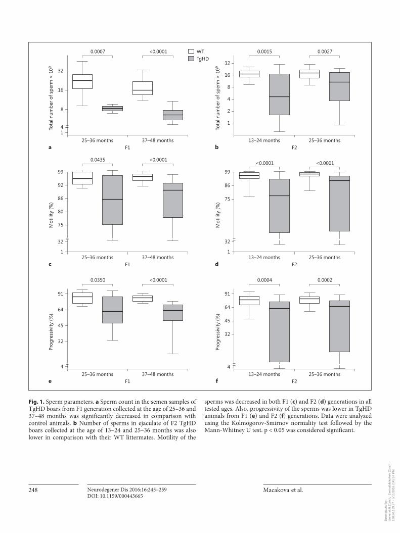
Fig. 1. Sperm parameters. a Sperm count in the semen samples of TgHD boars from F1 generation collected at the age of 25–36 and 37–48 months was significantly decreased in comparison with control animals. b Number of sperms in ejaculate of F2 TgHD boars collected at the age of 13–24 and 25–36 months was also lower in comparison with their WT littermates. Motility of the
sperms was decreased in both F1 ( c ) and F2 ( d ) generations in all tested ages. Also, progressivity of the sperms was lower in TgHD animals from F1 ( e ) and F2 ( f ) generations. Data were analyzed using the Kolmogorov-Smirnov normality test followed by the Mann-Whitney U test. p < 0.05 was considered significant.
Dow
nloa
ded
by:
Uni
vers
ität Z
üric
h, Z
entr
albi
blio
thek
Zür
ich
13
0.60
.129
.67
- 5/
11/2
016
2:45
:57
PM

Testicular Pathology in TgHD Minipigs Neurodegener Dis 2016;16:245–259 DOI: 10.1159/000443665
249
plexed with CASAVA v1.7. Read quality was assessed with FastQC v0.11.2 (http://www.bioinformatics.babraham.ac.uk). Quality and adapter trimming was performed with TrimGalore v0.3.7 (http://www.bioinformatics.babraham.ac.uk). Reads were aligned to a modified version of Sus scrofa reference genome assembly Sscro-fa10.2.74 (GCA_000003025.4; http://www.ensembl.org/Sus_scro-fa) that included the full pHIV1-HD548aa-145Q vector sequence. Reads were aligned with BWA-backtrack v0.7.10-r789 [17] . Dupli-cates were marked with Picard Tools MarkDuplicates v0.1.111 (http://picard.sourceforge.net). All alignment manipulations, in-cluding sorting and indexing, was performed with sambamba v0.4.6 [18] . Alignment quality was assessed using Picard Tools, Samtools v1.0 and BamTools v2.2.2 [19, 20] . All chimeric read pairs mapping from endogenous reference sequences to the trans-gene or vector backbone sequences were isolated and clustered us-ing our published algorithms BamStat and ReadPairCluster [21–23] . An independent algorithm, DELLY, was used to corroborate integration sites detected by principal methods [24] . Actual se-quences of the integration junctions were determined by PCR and Sanger sequencing.
Results
Sperm Pathology of TgHD Boars We showed altered reproduction parameters in 2
TgHD boars of F1 generation starting at the age of 13 months [9] as a potential HD phenotype in our porcine model. However, detailed analysis of a larger cohort of animals was needed to investigate the basis for the decline
in fertility. We provided evidence on sperm reproductive parameters of TgHD and WT boars from F1 (24–36 months old) and F2 (12–36 months old) generations. An-imals in the compared groups did not yet vary in weight, size or their motor movements.
Semen of TgHD and WT animals was collected and characterized using a sperm cell analyzer. Sperm count, motility and progressivity were evaluated. All parameters measured in semen samples of TgHD boars significantly decreased at around 13 months in both generations ( fig. 1 ) and persisted at a low level with increasing age. In vitro fertilization tests showed a continuous decreased ability of TgHD sperms to penetrate the oocytes ( fig. 2 ).
EM analysis of semen samples revealed altered mor-phology of spermatozoa between TgHD and WT boars. Structural anomalies of spermatozoa were much more numerous in TgHD samples. These abnormalities were more pronounced in the F2 generation. Nearly all the spermatozoa of TgHD animals of F2 generation had a cy-toplasmic droplet (most often proximal; fig. 3 a). Severe structural alterations in TgHD spermatozoa were local-ized mainly in the connecting piece and midpiece of the tail. Abnormalities were manifested as deformation of the mitochondrial sheath in the tail midpiece and also other tail structures. Common findings were folded or coiled tails, and sometimes a double or triple axoneme with fused mitochondrial sheaths ( fig. 3 b, d). Deformity of the
23–30 months 34–41 months 11–18 months 23–30 monthsa
100
80
60
40
20
0
90
70
50
30
10
Pene
trat
ion
abili
ty (%
)
b
100
80
WTTgHD
60
40
20
0
90
70
50
30
10
Pene
trat
ion
abili
ty (%
)
0.0072 0.0027n.s. 0.0004
F1 F2
Fig. 2. Sperm penetration ability. a The ability to penetrate into oocytes is decreased in sperms of F1 boars at the age of 23–30 months, and also at the age of 34–41 months. The most striking difference in the penetration ability between WT and TgHD F2 boars was at the age of 23–30 months. b Such alterations were ob-
served as well in animals from F2 generation who reached the age of 11–18 months (p = 0.0608). Each penetration test was repeated at least 5 times. Data were analyzed using the Kolmogorov-Smirnov normality test followed by the Mann-Whitney U test. p < 0.05 was considered significant.
Dow
nloa
ded
by:
Uni
vers
ität Z
üric
h, Z
entr
albi
blio
thek
Zür
ich
13
0.60
.129
.67
- 5/
11/2
016
2:45
:57
PM

Macakova et al.
Neurodegener Dis 2016;16:245–259 DOI: 10.1159/000443665
250
nucleus associated with incomplete chromatin condensa-tion and abnormal acrosome occurred occasionally ( fig. 3 c). Instability of acrosomes was in some cases man-ifested by a precocious acrosomal reaction. Proximal cy-toplasmic droplets were often associated with disorga-nized mitochondrial sheaths ( fig. 3 e). In the F2 genera-tion, there was a total absence of residual bodies in the ejaculate.
Testicular Pathology of TgHD Boars After having demonstrated the sperm pathology of
TgHD boars, we sacrificed one pair of 24-month-old and 2 pairs of 36-month-old animals from the F2 generation to perform the morphological analysis of testes.
At the age of 24 months, degenerative changes in sem-iniferous epithelium were more frequent in the TgHD boar in comparison with the wild-type one. Apoptosis was seen in supporting Sertoli cells as well as cells of sper-
a b
c d
e f
Fig. 3. Fresh ejaculate of TgHD boar. RB = Residual bodies; N = neck. a Spermatozoon with proximal cytoplasmic droplet (aster-isk) and residual bodies. b Spermatozoon with cytoplasmic droplet and double neck and tail structures (arrows). c Fully de-formed spermatozoon. d A tail of a sper-matozoon with triple axoneme and fused mitochondrial sheaths. e Connecting piece of tail of a spermatozoon with multiple and totally disorganized axoneme and mito-chondrial sheaths (asterisk). f Immunocy-tochemical reaction. 10-nm gold particles (arrows) indicate the presence of polyglu-tamine-containing proteins.
Dow
nloa
ded
by:
Uni
vers
ität Z
üric
h, Z
entr
albi
blio
thek
Zür
ich
13
0.60
.129
.67
- 5/
11/2
016
2:45
:57
PM

Testicular Pathology in TgHD Minipigs Neurodegener Dis 2016;16:245–259 DOI: 10.1159/000443665
251
matogenic lineage. Degeneration of Sertoli cells was char-acterized by increased density and vacuolation of cyto-plasm, dilatation of endoplasmic reticulum, structural al-terations of the nuclei and swollen mitochondria ( fig. 4 a). Degeneration of spermatogonia was manifested by cell shrinkage, increased chromatin condensation in the nu-cleus, dilatation of endoplasmic reticulum and swollen mitochondria with defects of their internal structure ( fig. 4 b). In addition, other cells of the spermatogenic lin-eage were gradually degenerated ( fig. 4 c). Strongly re-duced epithelium of the seminiferous tubules, restricted to the Sertoli cells, spermatogonia and the sparsely occur-ring spermatocytes and spermatids were occasionally ob-served. Multinucleated spermatogenic cells including spermatogonia ( fig. 4 d) were frequently recognized. The thick basal lamina was made up of several layers (2–3 lam-inae densae).
At the age of 36 months, the rate of degenerative changes in the testicular samples of 2 transgenic individ-uals was different. The changes were less pronounced in
boar K104 than in boar K63 and resembled those of a TgHD boar at the age 24 months. Morphology of semi-niferous tubules in boar K63 showed a significant reduc-tion of spermatogenesis. In some of the tubules, sper-matogenesis was preserved (but only to a limited extent; ( fig. 5 a); other tubules contained only Sertoli cells ( fig. 5 b). In the tubules with preserved spermatogenesis, the de-generative changes were detected in both Sertoli cells ( fig. 5 d) and spermatogenic elements, including the sper-matogonia. Degenerative changes exhibited characteris-tics typical for early or advanced apoptosis – the increased density of cytoplasm associated with its vacuolization, swollen mitochondria, dilated endoplasmic reticulum and clumps of heterochromatin in the nucleus. The basal lamina was thick and made up of several layers (2–3 lam-inae densae). Tubules without spermatogenic cells were lined with Sertoli cells exclusively, which, unlike the tu-bules with preserved spermatogenesis, hold signs of de-generation only in exceptional cases. Sertoli cells con-tained extreme amounts of endoplasmic reticulum, which
a b
c d
Fig. 4. Seminiferous epithelium of TgHD boar at the age of 24 months. Se = Sertoli cell undergoing apoptosis; N = nuclear structure; Sp = spermatocyte; L = lipid droplet; Sg = spermatogonia. a Sertoli cell undergoing apoptosis – increasing cyto-plasm density, vacuolation, altered nuclear structure. Note spermatocyte and large lip-id droplet. b Spermatogonia undergoing apoptosis. Note basal lamina (arrow). c De-generated spermatocytes (arrows). Note large lipid droplet. d Spermatogonia with 3 nuclei. Note basal lamina (arrow).
Dow
nloa
ded
by:
Uni
vers
ität Z
üric
h, Z
entr
albi
blio
thek
Zür
ich
13
0.60
.129
.67
- 5/
11/2
016
2:45
:57
PM
Macakova et al.
Neurodegener Dis 2016;16:245–259 DOI: 10.1159/000443665
252
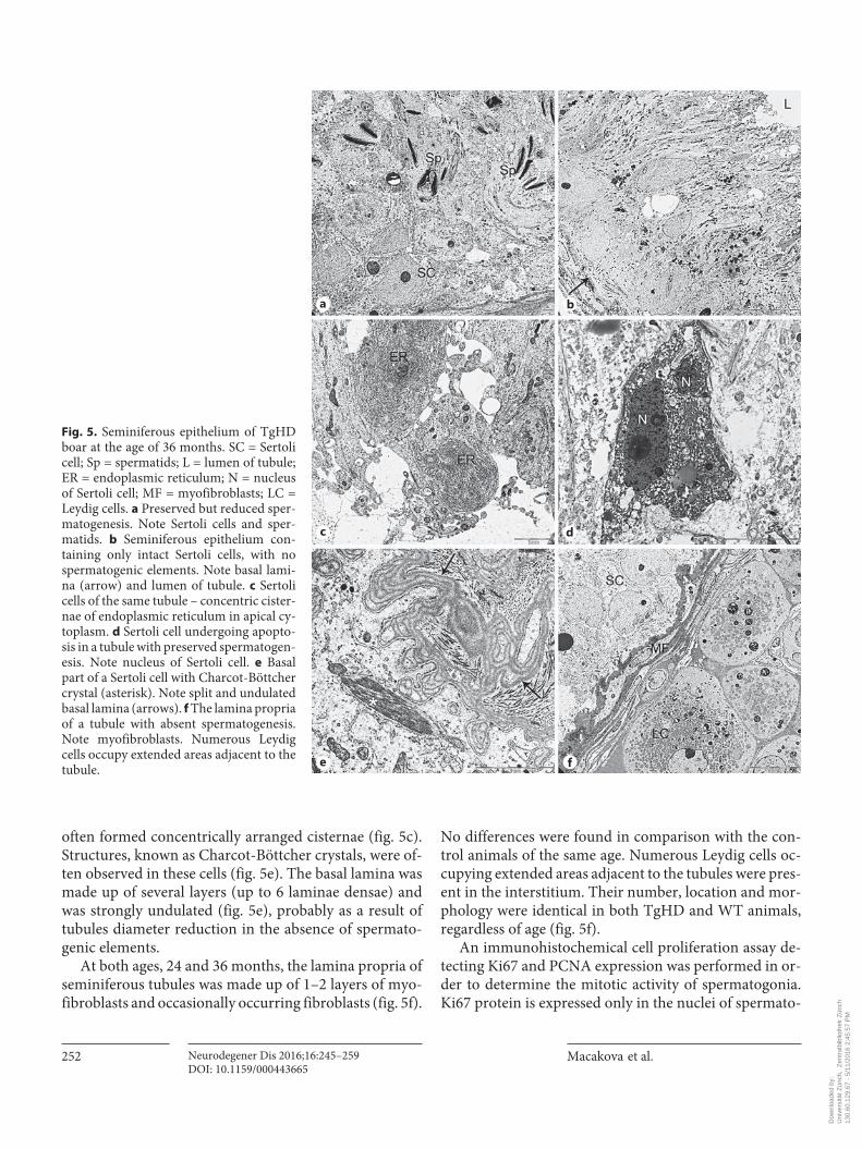
often formed concentrically arranged cisternae ( fig. 5 c). Structures, known as Charcot-Böttcher crystals, were of-ten observed in these cells ( fig. 5 e). The basal lamina was made up of several layers (up to 6 laminae densae) and was strongly undulated ( fig. 5 e), probably as a result of tubules diameter reduction in the absence of spermato-genic elements.
At both ages, 24 and 36 months, the lamina propria of seminiferous tubules was made up of 1–2 layers of myo-fibroblasts and occasionally occurring fibroblasts ( fig. 5 f).
No differences were found in comparison with the con-trol animals of the same age. Numerous Leydig cells oc-cupying extended areas adjacent to the tubules were pres-ent in the interstitium. Their number, location and mor-phology were identical in both TgHD and WT animals, regardless of age ( fig. 5 f).
An immunohistochemical cell proliferation assay de-tecting Ki67 and PCNA expression was performed in or-der to determine the mitotic activity of spermatogonia. Ki67 protein is expressed only in the nuclei of spermato-
a b
c d
e f
Fig. 5. Seminiferous epithelium of TgHD boar at the age of 36 months. SC = Sertoli cell; Sp = spermatids; L = lumen of tubule; ER = endoplasmic reticulum; N = nucleus of Sertoli cell; MF = myofibroblasts; LC = Leydig cells. a Preserved but reduced sper-matogenesis. Note Sertoli cells and sper-matids. b Seminiferous epithelium con-taining only intact Sertoli cells, with no spermatogenic elements. Note basal lami-na (arrow) and lumen of tubule. c Sertoli cells of the same tubule – concentric cister-nae of endoplasmic reticulum in apical cy-toplasm. d Sertoli cell undergoing apopto-sis in a tubule with preserved spermatogen-esis. Note nucleus of Sertoli cell. e Basal part of a Sertoli cell with Charcot-Böttcher crystal (asterisk). Note split and undulated basal lamina (arrows). f The lamina propria of a tubule with absent spermatogenesis. Note myofibroblasts. Numerous Leydig cells occupy extended areas adjacent to the tubule.
Dow
nloa
ded
by:
Uni
vers
ität Z
üric
h, Z
entr
albi
blio
thek
Zür
ich
13
0.60
.129
.67
- 5/
11/2
016
2:45
:57
PM

Testicular Pathology in TgHD Minipigs Neurodegener Dis 2016;16:245–259 DOI: 10.1159/000443665
253
WT TgHD TgHD with testis atrophy
PCN
AKi
67
a b c
d e f
WTg
250
200
150
100
50
0
PCN
A-po
sitiv
e ce
lls (n
)
TgHD (K8) TgHD (K104) TgHD (K63)
***
WTh
100
80
60
40
20
0
Ki67
-pos
itive
cel
ls (n
)
TgHD (K8) TgHD (K104) TgHD (K63)
***
*
Colo
r ver
sion
ava
ilabl
e on
line
Fig. 6. Proliferative analysis. PCNA ( a–c ) and Ki67 ( d–f ) staining revealed decreased number of spermatocytes ( c ) and spermatogo-nia ( f ) in seminiferous epithelium of the TgHD boar (K63) at the age of 36 months. g , h The number of spermatogenic cells of the TgHD boar (K63) was significantly decreased compared with the WT boar (p < 0.05). Seminiferous tubules of the 24-month-old ( b ) and 36-month-old ( e ) transgenic boars showed similar staining of
spermatogenic cells to WT ( a , d ). h The exception is testicular tis-sue from TgHD boar K104, in which spermatogonia (Ki67-posi-tive cells) were determined in a significantly reduced number of seminiferous tubules compared with WT. Scale bars = 50 μm (20 μm in insets ). Results were plotted as mean ± SD of positive cells per seminiferous tubule. p < 0.05 was considered significant. * p = 0.0151; * * * p < 0.0001.
Dow
nloa
ded
by:
Uni
vers
ität Z
üric
h, Z
entr
albi
blio
thek
Zür
ich
13
0.60
.129
.67
- 5/
11/2
016
2:45
:57
PM

Macakova et al.
Neurodegener Dis 2016;16:245–259 DOI: 10.1159/000443665
254
gonia, while PCNA occurs also in the nuclei of primary spermatocytes in normal seminiferous epithelium [25] – 90.86% (726/799) of seminiferous tubules lacked PCNA-positive cells and 90.08% (745/827) of seminiferous tu-bules contained no Ki67-positive cell in the testis of the 36-month-old TgHD boar (K63; fig. 6 a–f). Spermatogo-nia serve as stem cells in the process of differentiation to spermatozoa, and their proliferative activity secures nor-mal spermatogenesis. That means that impaired sper-matogenesis involved 90% of seminiferous tubules. These results confirmed impaired spermatogenesis observed by EM (as indicated above). The remaining 10% of seminif-erous tubules contained spermatogonia and spermato-cytes stained by anti-PCNA and anti-Ki67 antibodies, but their number was significantly reduced in comparison with the seminiferous tubules of a WT boar. A significant decrease in the quantity of spermatogonia was also ob-served in the testis of the other TgHD boar at the age of 36 months (K104) in comparison with the WT one. It suggested that the spermatogonial proliferation was re-
duced in TgHD boars at the age of 36 months (n = 2, K104 and K63). No difference was found in the testes of the 24-month-old TgHD boar (K8; fig. 6 g, h).
Sperm and Testicular Degeneration: The Direct Effect of mtHtt Supposing that sperm pathology is caused by the toxic
effect of mtHtt, experiments showing localization and ex-pression of mtHtt were done. The presence of the polyglu-tamine-containing proteins was observed in structures in the spermatozoa tail of TgHD boars using 10-nm gold particles examined under EM ( fig. 3 f). This finding was confirmed by a dotted immunohistochemical signal de-tected along the whole spermatozoa tail in all tested ages of F1- and F2-generation TgHD boars (but not in WT controls) using 3B5H10 (anti-N-terminal fragment of 171 aa of human Htt with 65Q) antibody ( fig. 7 a). Western blot analysis using EPR5526 (anti-N-terminal fragment of Htt) confirmed highly abundant mtHtt and a slightly low-er level of endogenous Htt in spermatozoa ( fig. 7 b). In ad-
117
Htt(EPR5526)
mtHtt(EPR5526)
FragmentedHtt
TgHD
TgHD
WT
WT
TgHD
TgHD
WT
WT
kDa460
71
mtHtt(polyQ-3B5H10)
FragmentedHtt
c
a b
Fig. 7. Presence of Htt in spermatozoa cells. a , b Immunofluorescence dotted sig-nal of the polyglutamine-containing pro-teins (3B5H10 antibody, yellow; color in online version only) was detected along whole spermatozoa tail of TgHD boars ( b ) but not in WT cells ( a ). Scale bar = 20 μm. c Western blot analysis detected endoge-nous (Htt) and transgenic (mtHtt) hun-tingtin by EPR5526 antibody, specific to N-terminal part of huntingtin. PolyQ an-tibody (3B5H10) as well as Htt antibody (EPR5526) detected fragments of mtHtt.
Colo
r ver
sion
ava
ilabl
e on
line
Dow
nloa
ded
by:
Uni
vers
ität Z
üric
h, Z
entr
albi
blio
thek
Zür
ich
13
0.60
.129
.67
- 5/
11/2
016
2:45
:57
PM

Testicular Pathology in TgHD Minipigs Neurodegener Dis 2016;16:245–259 DOI: 10.1159/000443665
255
dition, anti-polyQ antibody (3B5H10) as well as anti-hun-tingtin antibody (EPR5526) revealed a fragmented form of mtHtt, which was reported to cause cellular toxicity [26] . Sperm samples from all tested ages in both genera-tions (F1, F2) were analyzed, and no significant change in expression between samples was detected (only represen-tative data from two pairs of samples are shown; fig. 7 ).
Consequently, we looked at Htt localization in TgHD and WT seminiferous tubules from testes of 24- and 36-month-old boars. The signal was widely spread in spermatogenic and Sertoli cells in normal as well as in the atrophic seminiferous tubules ( fig. 8 a). Western blot analysis showed a high expression of mtHtt form com-pared with endogenous Htt. The huntingtin antibody also revealed fragmented forms of mtHtt that may con-tribute to toxicity of the transgene ( fig. 8 b).
In order to eliminate the neuronal effect on the tes-ticular phenotype, levels of fertility-related hormones were measured in the blood serum of two age groups: 7- to 17-month-old TgHD (n = 14) and WT (n = 9) boars, and 15–30-month-old TgHD (n = 11) and WT (n = 6) boars. Levels of testosterone, LH and inhibin-α were an-alyzed (see online suppl. material SM 2). No significant difference was observed between TgHD and WT ani-mals.
Mapping the HIV1-HD-548aaHTT-145Q Transgene Integration To detect any and all sites of vector integration into the
pig genome in the transgenic lineage, we performed long-insert jumping library whole-genome sequencing of transgenic animals from F0 (founder female) and F2 gen-
WT WT TgHD TgHD atrophic
EPR 5526Blanka
Htt
mtHtt
460
171
117
71
55
TgHD
TgHD
WT
WT
24months
36monthsb
Fig. 8. Expression of Htt in testes. M = Myoid cells. a Total hun-tingtin protein was visualized immunohistochemically with EPR5526 antibody in sections of WT and transgenic (TgHD) por-cine testis. EPR5526 (red; color in online version only) was de-tected in seminiferous epithelium demarcated by seminiferous tu-bule basement membrane (dashed line) and lumen (asterisk). My-oid cells were negatively stained. Cell nuclei were counterstained with DAPI (blue). Prominent Sertoli cell nuclei (arrowheads) pres-ent in atrophic seminiferous tubule (TgHD atrophic) documented the loss of germ cells. Blank, WT testis section stained only with A647-conjugated secondary antibody. Scale bar = 20 μm. b West-ern blot analysis of testis; representative sample shown. Anti-AB1 antibody detected endogenous Htt, transgenic mtHtt and frag-ments.
Colo
r ver
sion
ava
ilabl
e on
line
Dow
nloa
ded
by:
Uni
vers
ität Z
üric
h, Z
entr
albi
blio
thek
Zür
ich
13
0.60
.129
.67
- 5/
11/2
016
2:45
:57
PM
Macakova et al.
Neurodegener Dis 2016;16:245–259 DOI: 10.1159/000443665
256
erations in comparison with a negative control. This method, which involves sequencing the ends of genomic DNA fragments after circularization and size reduction, has previously been shown to be an effective platform for vector integration site discovery in transgenic sheep and mice [21] . Whole-genome sequencing of jumping librar-ies prepared from random fragments (mean size 3.6 kb) resulted in an average of 65.5× coverage of mapped in-serts across each base in the haploid genome for all 3 an-imals. The paired-end reads were examined for the signa-ture of vector integration into the genome: chimeric frag-ments consisting of pig genomic sequence on one end and a portion of the introduced vector/transgene on the other. No integrations were detected in the negative con-trol genome, while a single identical vector integration was detected in each of the F0 and F2 generations of TgHD animals. As suggested previously from FISH anal-ysis, the integration occurred into chromosome 1q [9] . The jumping library analysis revealed that 5.3 kb of the HIV1-HD-548aaHTT-145Q vector DNA integrated as expected via the HIV LTR sequences and harbored an in-tact HD-548aaHTT145Q expression cassette. Sanger se-quencing of the junctions with genomic DNA showed that the integration was in reverse orientation relative to the genomic sequence, between chr1 228,641,631 and 228,641,637, with loss of the 5 intervening bases of ge-nomic DNA. This integration does not directly disrupt any annotated gene, and no further breakpoint or inte-gration complexity or other genomic rearrangement was apparent at the resolution of the jumping library sequenc-ing.
Discussion
In this study we described sperm and testicular degen-eration, which is a result of the presence of mtHtt protein in the testes of transgenic minipig boars expressing the N-terminal part of human mtHtt. Cohorts of TgHD and their WT controls of F1 and F2 generations were directly compared.
Previous studies on HD rodent models showed male sterility that was assumed to be due to a reduction of sper-matozoa [15] . We confirmed this phenotype in a large animal model of HD, the minipig. Additionally, we re-ported both the reduction of spermatozoa and also their function measured by motility, progressivity and in vitro penetration assay. Spermatozoa of TgHD boars had se-vere problems to penetrate the minipig oocytes. The dif-ference in values of sperm parameters was evident at 13
months and worsened with age. A comparison of animals from F1 and F2 generations of the same age (25–36 months) showed slightly worse values of all observed pa-rameters in the F2 generation. There was also a wider variability of sperm parameters in the F2 generation, probably caused by a larger cohort of animals in the F2 group. Furthermore, EM analysis revealed deformation of the mitochondrial sheath in the tail midpiece of TgHD spermatozoa. Folded or coiled tails and a double or triple axoneme with fused mitochondrial sheaths were also ob-served. This can be caused by a failure of the disjunction of excess cytoplasm, which results in the presence of cy-toplasmic droplets. This phenomenon, together with spermatozoa motility dysfunction, can be related to a de-crease of mitochondrial energetic metabolism and func-tional impairment of respiratory chain complex II (un-published observations). Occasionally, nucleus deforma-tion associated with incomplete chromatin condensation and abnormal acrosome occurred in transgenic sperma-tozoa (but not in WT controls). These abnormalities were more pronounced in the F2 generation. Moreover, nearly all the spermatozoa of TgHD animals of the F2 generation contained a cytoplasmic droplet, and their ejaculate lacked residual bodies.
The testicular degeneration reported here is in agree-ment with observations in mice (R6/2 and Yac72) [14, 15] , as well as in postmortem samples from humans [13] . Multinucleated spermatogenic cells were fre-quently present in the seminiferous epithelium of 24- and 36-month-old TgHD boars. The spermatogonia were shrunk and had dilated endoplasmic reticulum, swollen mitochondria and condensed chromatin in the nucleus. Spermatocytes and spermatids were observed occasionally. Reduced numbers of developing sper-matocytes and spermatids were also observed in HD pa-tients [13] and YAC128 mice [27] . Some tubules con-tained only Sertoli cells. Sertoli cells were characterized by increased density and vacuolization of the cytoplasm, dilatation of endoplasmic reticulum, structural altera-tions of the nuclei and swollen mitochondria. These are features typical for early or advanced apoptosis. More-over, proliferative analysis of seminiferous tubules with elongated spermatozoa showed fewer cells expressing the proliferative markers PCNA and Ki67 in TgHD an-imals. The apoptotic nature of the cell death in a large number of degenerating spermatids with diffuse cyto-plasmic vacuolization, condensed nuclei and electron-dense cytoplasm which were phagocytized and degrad-ed by Sertoli cells was also observed in a mouse model lacking endogenous huntingtin YAC72 –/– [14] . Similar-
Dow
nloa
ded
by:
Uni
vers
ität Z
üric
h, Z
entr
albi
blio
thek
Zür
ich
13
0.60
.129
.67
- 5/
11/2
016
2:45
:57
PM
Testicular Pathology in TgHD Minipigs Neurodegener Dis 2016;16:245–259 DOI: 10.1159/000443665
257
ly, seminiferous tubules of YAC128 were disrupted by large vacuoles [27] . In addition, the seminiferous tubule wall was thickened in HD patients [13] . In the TgHD minipig, the thick basal lamina was made up of several layers (2–3 laminae densae) compared with the WT minipig. At the age of 36 months, spermatogenesis was more affected in comparison with the age of 24 months. Sertoli cells contained extreme amounts of endoplasmic reticulum and structures known as Charcot-Böttcher crystals. The basal lamina was made up of several layers (up to 6 laminae densae) and was strongly undulated, resulting probably from a reduction of the diameter of the tubules in the absence of spermatogenic elements. The rate of degenerative changes in the testicular sam-ples of the 2 transgenic boars at the age of 36 months was different. The changes were less pronounced in boar K104 than in boar K63 and resembled those of the TgHD boar at the age 24 months. Boar K63 also showed more change in sperm parameters, including atrophy of seminiferous epithelium and impaired spermatogene-sis. The difference in severity of pathology between age-matched boars might be a consequence of variation in the progression of the disease and genetic background of individual minipigs. The age of onset of HD depends on CAG length (around 70%), but also on other factors like polymorphism, modifier genes, etc. (30%) [28, 29] . We suggest that polymorphisms of proteins interacting with huntingtin could contribute to different degrees of testicular degeneration between transgenic boars of the same age.
After demonstrating testicular abnormalities we in-tended to clarify the reason for the pathological pheno-type. We focused on the design of the transgenic minipig model. The number of CAG repeats was chosen in order to expect an earlier phenotype. This number of repeats imitates juvenile HD in patients. It has an earlier onset and faster progress, but the disease has the same charac-teristics as the adult form. Mixed CAG/CAA repeats, in-stead of just CAG, were used to increase the stability of the insert. This has been used in several rodent models (YAC128, BACHD mice) that also showed the phenotype [30, 31] . Therefore, the design was not a problem. We also checked whether the insertion of the lentiviral construct did not interrupt any coding sequence in the pig genome. Since the result was negative, the question was whether the defect in the testes was due to the expression ofmtHtt or as a result of changed levels of fertility-related hormones.
There is evidence that the expression of mutant Htt leads to selective cellular dysfunction and degeneration
[32] . The most affected cells are neurons. However, we also provided data for spermatozoa degeneration and tes-ticular dysfunction in a minipig model of HD. We showed a high and stable expression of endogenous Htt as well as the transgenic N-truncated mutant form of human Htt in spermatozoa as well as in spermatogenic and Sertoli cells, and also in the atrophic seminiferous tubules of TgHD testes. In addition, we detected fragments of mtHtt in spermatozoa as well as in testes. It has been published that smaller fragments of mtHtt cause cellular toxicity and in-duce apoptosis [33, 34] . Furthermore, mutant N-terminal Htt fragments were also detected in tissues from HD pa-tients and mouse models in the presymptomatic stage, suggesting their role in the progression of HD [35–37] . These facts also support our statement that the N-termi-nal part of human mtHtt causes testicular pathology in transgenic minipig boars.
Although testicular degeneration in HD is well de-scribed in mouse models (R6/2 and YAC128), it is not clear whether this phenotype is independent or a conse-quence of alterations of the hypothalamic-pituitary-go-nadal axis (GnRH). A significant loss of GnRH neurons starting from 5 weeks of age followed by decreasing levels of plasma testosterone at 12 weeks of age was found in R6/2 mice [11] , while testicular atrophy without concom-itant loss of GnRH neurons was described in a YAC128 mouse model [13] . An analysis of testosterone levels in YAC128 mice did not reveal any significant difference compared with controls, even when testicular atrophy was already present [13] . Furthermore, testosterone treat-ment had no effect on the peripheral phenotype of HD, e.g. body weight loss or motor function in R6/2 mice [11] . An analysis of complete neuroendocrine status in HD pa-tients showed no significant difference in the plasma lev-els of LH, FSH and testosterone between all male HD pa-tients and controls [38] . However, Markianos et al. [39] observed significantly lower testosterone and LH levels in HD patients compared with healthy controls. These con-flicting results suggested a detailed hormonal analysis of our porcine model. We observed no significant difference in the levels of fertility-related hormones between TgHD and control boars, and no changes in libido were ob-served during regular collection of semen. Moreover, in the interstitium, the number, location and morphology of the Leydig cells were identical regardless of the age or genotype of the animal, and no differences were found in the lamina propria of seminiferous tubules of the TgHD boars in both ages compared with WT controls. Similarly, unaffected Leydig cells were observed between degener-ating tubules of stromal interstitial tissue in YAC72 –/–
Dow
nloa
ded
by:
Uni
vers
ität Z
üric
h, Z
entr
albi
blio
thek
Zür
ich
13
0.60
.129
.67
- 5/
11/2
016
2:45
:57
PM
Macakova et al.
Neurodegener Dis 2016;16:245–259 DOI: 10.1159/000443665
258
References
1 Vonsattel JP, DiFiglia M: Huntington disease. J Neuropathol Exp Neurol 1998; 57: 369–384.
2 Stanek LM, Sardi SP, Mastis B, Richards AR, Treleaven CM, Taksir T, Misra K, Cheng SH, Shihabuddin LS: Silencing mutant huntingtin by adeno-associated virus-mediated RNA in-terference ameliorates disease manifestations in the YAC128 mouse model of Huntington’s disease. Hum Gene Ther 2014; 25: 461–474.
3 Dufour BD, Smith CA, Clark RL, Walker TR, McBride JL: Intrajugular vein delivery of AAV9-RNAi prevents neuropathological changes and weight loss in Huntington’s dis-ease mice. Mol Ther 2014; 22: 797–810.
4 Squitieri F, Di Pardo A, Favellato M, Amico E, Maglione V, Frati L: Pridopidine, a dopa-mine stabilizer, improves motor performance and shows neuroprotective effects in Hun-tington disease R6/2 mouse model. J Cell Mol Med 2015; 19: 2540–2548.
5 Holm IE, Alstrup AK, Luo Y: Genetically modified pig models for neurodegenerative disorders. J Pathol 2016; 238: 267–287.
6 Dolezalova D, Hruska-Plochan M, Bjarkam CR, Sorensen JC, Cunningham M, Weingar-ten D, Ciacci JD, Juhas S, Juhasova J, Motlik J, Hefferan MP, Hazel T, Johe K, Carromeu C, Muotri A, Bui J, Strnadel J, Marsala M: Pig models of neurodegenerative disorders: utili-zation in cell replacement-based preclinical safety and efficacy studies. J Comp Neurol 2014; 522: 2784–2801.
7 Vodicka P, Smetana K Jr, Dvorankova B, Emerick T, Xu YZ, Ourednik J, Ourednik V, Motlik J: The miniature pig as an animal model in biomedical research. Ann NY Acad Sci 2005; 1049: 161–171.
8 Matsuyama N, Hadano S, Onoe K, Osuga H, Showguchi-Miyata J, Gondo Y, Ikeda JE: Identification and characterization of the miniature pig Huntington’s disease gene ho-molog: evidence for conservation and poly-
morphism in the CAG triplet repeat. Genom-ics 2000; 69: 72–85.
9 Baxa M, Hruska-Plochan M, Juhas S, Vodicka P, Pavlok A, Juhasova J, Miyanohara A, Nejime T, Klima J, Macakova M, Marsala S, Weiss A, Kubickova S, Musilova P, Vrtel R, Sontag EM, Thompson LM, Schier J, Han-sikova H, Howland DS, Cattaneo E, DiFiglia M, Marsala M, Motlik J: A transgenic minipig model of Huntington’s disease. J Huntingtons Dis 2013; 2: 47–68.
10 Guo J, Zhu P, Wu C, Yu L, Zhao S, Gu X: In silico analysis indicates a similar gene expres-sion pattern between human brain and testis. Cytogenet Genome Res 2003; 103: 58–62.
11 Papalexi E, Persson A, Bjorkqvist M, Petersen A, Woodman B, Bates GP, Sundler F, Mulder H, Brundin P, Popovic N: Reduction of GnRH and infertility in the R6/2 mouse model of Huntington’s disease. Eur J Neurosci 2005; 22: 1541–1546.
12 Van Raamsdonk JM, Pearson J, Murphy Z, Hayden MR, Leavitt BR: Wild-type hunting-tin ameliorates striatal neuronal atrophy but does not prevent other abnormalities in the YAC128 mouse model of Huntington disease. BMC Neurosci 2006; 7: 80.
13 Van Raamsdonk JM, Murphy Z, Selva DM, Hamidizadeh R, Pearson J, Petersen A, Bjorkqvist M, Muir C, Mackenzie IR, Ham-mond GL, Vogl AW, Hayden MR, Leavitt BR: Testicular degeneration in Huntington dis-ease. Neurobiol Dis 2007; 26: 512–520.
14 Leavitt BR, Guttman JA, Hodgson JG, Kimel GH, Singaraja R, Vogl AW, Hayden MR: Wild-type huntingtin reduces the cellular toxicity of mutant huntingtin in vivo. Ameri-can J Hum Genet 2001; 68: 313–324.
15 Sathasivam K, Hobbs C, Turmaine M, Man-giarini L, Mahal A, Bertaux F, Wanker EE, Doherty P, Davies SW, Bates GP: Formation of polyglutamine inclusions in non-CNS tis-
sue. Hum Mol Genet 1999; 8: 813–822. 16 Hanscom C, Talkowski M: Design of large-
insert jumping libraries for structural variant detection using Illumina sequencing. Curr Protoc Hum Genet 2014; 80: 7221–7229.
17 Li H, Durbin R: Fast and accurate long-read alignment with Burrows-Wheeler transform. Bioinformatics 2010; 26: 589–595.
18 Tarasov A, Vilella AJ, Cuppen E, Nijman IJ, Prins P: Sambamba: fast processing of NGS alignment formats. Bioinformatics 2015; 31: 2032–2034.
19 Li H, Handsaker B, Wysoker A, Fennell T, Ruan J, Homer N, Marth G, Abecasis G, Durbin R: The Sequence Alignment/Map for-mat and SAMtools. Bioinformatics 2009; 25: 2078–2079.
20 Barnett DW, Garrison EK, Quinlan AR, Strom-berg MP, Marth GT: BamTools: A c++ API and toolkit for analyzing and managing BAM files. Bioinformatics 2011; 27: 1691–1692.
21 Chiang C, Jacobsen JC, Ernst C, Hanscom C, Heilbut A, Blumenthal I, Mills RE, Kirby A, Lindgren AM, Rudiger SR, McLaughlan CJ, Bawden CS, Reid SJ, Faull RL, Snell RG, Hall IM, Shen Y, Ohsumi TK, Borowsky ML, Daly MJ, Lee C, Morton CC, MacDonald ME, Gu-sella JF, Talkowski ME: Complex reorganiza-tion and predominant non-homologous re-pair following chromosomal breakage in karyotypically balanced germline rearrange-ments and transgenic integration. Nat Genet 2012; 44: 390–397.
22 Brand H, Pillalamarri V, Collins RL, Eggert S, O’Dushlaine C, Braaten EB, Stone MR, Chambert K, Doty ND, Hanscom C, Rosen-feld JA, Ditmars H, Blais J, Mills R, Lee C, Gu-sella JF, McCarroll S, Smoller JW, Talkowski ME, Doyle AE: Cryptic and complex chromo-somal aberrations in early-onset neuropsy-chiatric disorders. Am J Hum Genet 2014; 95: 454–461.
mice [14] . Our results support the idea that testicular de-generation and fertility defects are related to mtHtt ex-pression in testes and not to peripheral hormonal changes.
In conclusion, we demonstrated a failure in sperm pa-rameters and extensive testicular pathology in a minipig model of HD. We showed that insertion of the lentiviral construct did not interrupt any coding sequence in the pig genome and suggest that the testicular defect was caused by the presence of mtHtt and its fragmented cyto-toxic form in testicular tissue, since hormonal changes were not measured between TgHD boars and their WT controls.
Acknowledgments
This work was supported by the CHDI Foundation (A-5378), the project EXAM from the European Regional Development Fund (CZ.1.05/2.1.00/03.0124), RVO 67985904, the Norwegian Financial Mechanism 2009–2014, the Ministry of Education, Youth and Sports (project contract MSMT-28477/2014) and Charles University (grant SVV 260083), and National Sustainabil-ity Programme No. LO1609 (MEYS CR).
Disclosure Statement
One of the authors, Dr. David S. Howland is employed by the CHDI Foundation, which supported this work. Dr. Howland was not involved in designing the experiments or in writing this article, and did not make the decision to publish it. He was a consultant. The authors have no other conflicts of interest to declare.
Dow
nloa
ded
by:
Uni
vers
ität Z
üric
h, Z
entr
albi
blio
thek
Zür
ich
13
0.60
.129
.67
- 5/
11/2
016
2:45
:57
PM
Testicular Pathology in TgHD Minipigs Neurodegener Dis 2016;16:245–259 DOI: 10.1159/000443665
259
23 Talkowski ME, Rosenfeld JA, Blumenthal I, Pillalamarri V, Chiang C, Heilbut A, Ernst C, Hanscom C, Rossin E, Lindgren AM, Pereira S, Ruderfer D, Kirby A, Ripke S, Harris DJ, Lee JH, Ha K, Kim HG, Solomon BD, Grop-man AL, Lucente D, Sims K, Ohsumi TK, Borowsky ML, Loranger S, Quade B, Lage K, Miles J, Wu BL, Shen Y, Neale B, Shaffer LG, Daly MJ, Morton CC, Gusella JF: Sequencing chromosomal abnormalities reveals neurode-velopmental loci that confer risk across diag-nostic boundaries. Cell 2012; 149: 525–537.
24 Rausch T, Zichner T, Schlattl A, Stutz AM, Benes V, Korbel JO: Delly: Structural variant discovery by integrated paired-end and split-read analysis. Bioinformatics 2012; 28:i333–i339.
25 Steger K, Aleithe I, Behre H, Bergmann M: The proliferation of spermatogonia in normal and pathological human seminiferous epithe-lium: an immunohistochemical study using monoclonal antibodies against Ki-67 protein and proliferating cell nuclear antigen. Mol Hum Reprod 1998; 4: 227–233.
26 Miller JP, Holcomb J, Al-Ramahi I, de Haro M, Gafni J, Zhang N, Kim E, Sanhueza M, Torcassi C, Kwak S, Botas J, Hughes RE, Eller-by LM: Matrix metalloproteinases are modi-fiers of huntingtin proteolysis and toxicity in Huntington’s disease. Neuron 2010; 67: 199–212.
27 Van Raamsdonk JM, Pearson J, Rogers DA, Bissada N, Vogl AW, Hayden MR, Leavitt BR: Loss of wild-type huntingtin influences mo-tor dysfunction and survival in the YAC128 mouse model of Huntington disease. Hum Mol Genet 2005; 14: 1379–1392.
28 Langbehn DR, Hayden MR, Paulsen JS: CAG-repeat length and the age of onset in Hunting-ton disease (HD): a review and validation study of statistical approaches. Am J Med Genet B Neuropsychiatr Genet 2010; 153B:397–408.
29 Tabrizi SJ, Reilmann R, Roos RA, Durr A, Leavitt B, Owen G, Jones R, Johnson H, Crau-furd D, Hicks SL, Kennard C, Landwehrmey-er B, Stout JC, Borowsky B, Scahill RI, Frost C, Langbehn DR: Potential endpoints for clin-ical trials in premanifest and early Hunting-ton’s disease in the TRACK-HD study: analy-sis of 24-month observational data. Lancet Neurol 2012; 11: 42–53.
30 Gray M, Shirasaki DI, Cepeda C, Andre VM, Wilburn B, Lu XH, Tao J, Yamazaki I, Li SH, Sun YE, Li XJ, Levine MS, Yang XW: Full-length human mutant huntingtin with a sta-ble polyglutamine repeat can elicit progres-sive and selective neuropathogenesis in BACHD mice. J Neurosci 2008; 28: 6182–6195.
31 Pouladi MA, Stanek LM, Xie Y, Franciosi S, Southwell AL, Deng Y, Butland S, Zhang W, Cheng SH, Shihabuddin LS, Hayden MR: Marked differences in neurochemistry and aggregates despite similar behavioural and neuropathological features of Huntington disease in the full-length BACHD and YAC128 mice. Hum Mol Genet 2012; 21: 2219–2232.
32 Bhide PG, Day M, Sapp E, Schwarz C, Sheth A, Kim J, Young AB, Penney J, Golden J, Aro-nin N, DiFiglia M: Expression of normal and mutant huntingtin in the developing brain. J Neurosci 1996; 16: 5523–5535.
33 Hackam AS, Singaraja R, Wellington CL, Metzler M, McCutcheon K, Zhang T, Kalch-man M, Hayden MR: The influence of hun-tingtin protein size on nuclear localization and cellular toxicity. J Cell Biol 1998; 141: 1097–1105.
34 Martindale D, Hackam A, Wieczorek A, Ellerby L, Wellington C, McCutcheon K, Sin-garaja R, Kazemi-Esfarjani P, Devon R, Kim SU, Bredesen DE, Tufaro F, Hayden MR: Length of huntingtin and its polyglutamine tract influences localization and frequency of intracellular aggregates. Nat Genet 1998; 18: 150–154.
35 Mende-Mueller LM, Toneff T, Hwang SR, Chesselet MF, Hook VY: Tissue-specific pro-teolysis of huntingtin (Htt) in human brain: evidence of enhanced levels of N- and C-ter-minal Htt fragments in Huntington’s disease striatum. J Neurosci 2001; 21: 1830–1837.
36 Wang CE, Tydlacka S, Orr AL, Yang SH, Gra-ham RK, Hayden MR, Li S, Chan AW, Li XJ: Accumulation of N-terminal mutant hun-tingtin in mouse and monkey models impli-cated as a pathogenic mechanism in Hunting-ton’s disease. Hum Mol Genet 2008; 17: 2738–2751.
37 Wellington CL, Singaraja R, Ellerby L, Savill J, Roy S, Leavitt B, Cattaneo E, Hackam A, Sharp A, Thornberry N, Nicholson DW, Bredesen DE, Hayden MR: Inhibiting caspase cleavage of huntingtin reduces toxicity and aggregate formation in neuronal and nonneu-ronal cells. J Biol Chem 2000; 275: 19831–19838.
38 Saleh N, Moutereau S, Durr A, Krystkowiak P, Azulay JP, Tranchant C, Broussolle E, Mo-rin F, Bachoud-Levi AC, Maison P: Neuroen-docrine disturbances in Huntington’s disease. PLoS One 2009; 4:e4962.
39 Markianos M, Panas M, Kalfakis N, Vassilo-poulos D: Plasma testosterone in male pa-tients with Huntington’s disease: relations to severity of illness and dementia. Ann Neurol 2005; 57: 520–525.
Dow
nloa
ded
by:
Uni
vers
ität Z
üric
h, Z
entr
albi
blio
thek
Zür
ich
13
0.60
.129
.67
- 5/
11/2
016
2:45
:57
PM