muscular system. functions of muscle tissue 1. body movement 2. stabilizing body positions/posture...
TRANSCRIPT

Muscular System

Functions of Muscle Tissue
1. Body Movement2. Stabilizing body positions/posture3. Storing and moving substances
within the body4. Generating Heat (rhermogenesis)

Types of Muscle●Skeletal – striated & voluntary
●Smooth – involuntary
●Cardiac - heart
The word “striated” means striped. Skeletal muscle appears striped under a microscope.
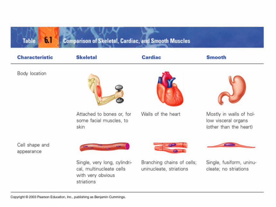
Comparison of Types of Muscle (p. 300)

Types of Muscle, cont.

Skeletal Muscle Characteristics
• Most attach to bones by tendon
• Cells are multinucleate
• Striated—have visible binding
• Voluntary• Cells surrounded &
bundled by connective tissue
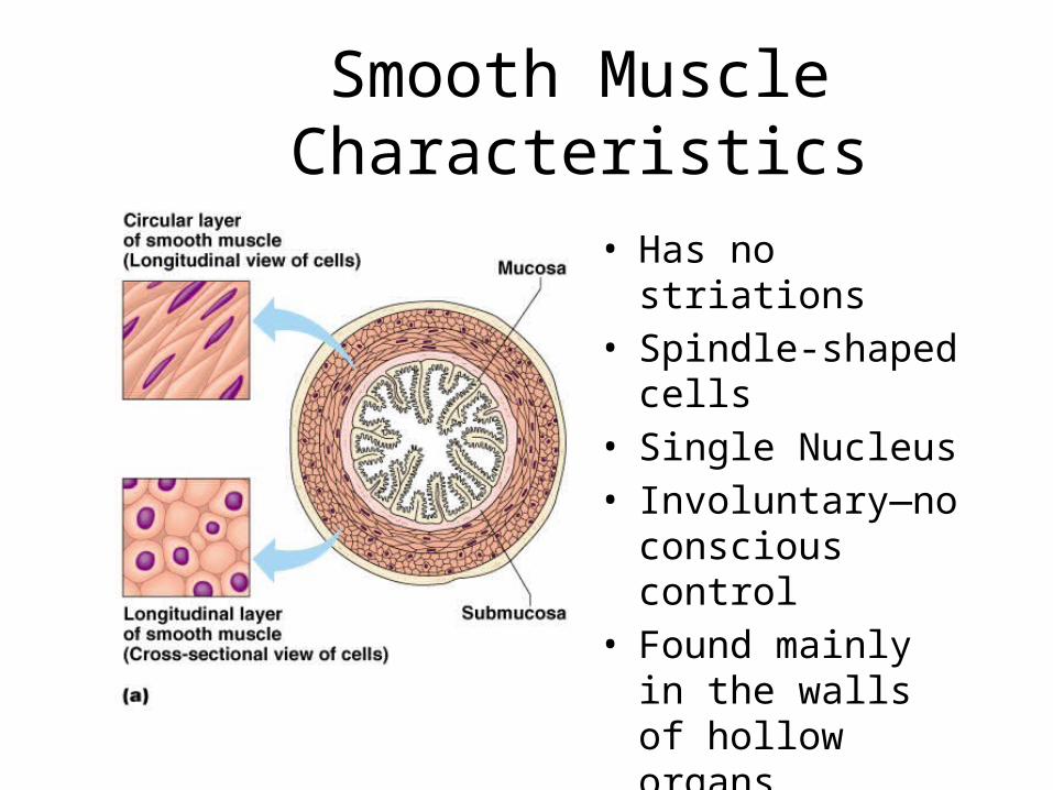
Smooth Muscle Characteristics
• Has no striations• Spindle-shaped cells• Single Nucleus• Involuntary—no
conscious control• Found mainly in the
walls of hollow organs

Characteristics of Cardiac Muscle
• Has striations• Usually has a single
nucleus• Joined to another
cardiac muscle cell• Involuntary• Found only in the
heart

Skeletal Muscle
• Functions of Skeletal Muscle– Produce Movement– Maintain posture– Stabilize joints– Generate Heat
• Sites of Muscle Attachment– Bones– Cartilage– Connective tissue
coverings
• Muscle Fibers blend into a connective tissue attachment– Tendon—cordlike structure– Aponeurosis—sheet-like structure
• Properties of Muscle– Irritability – ability to receive and respond to a stimulus– Contractibility – ability to shorten when an adequate stimulus is received– Extensibility – ability to lengthen when an adequate stimulus is received– Elasticity – ability to return to normal shape
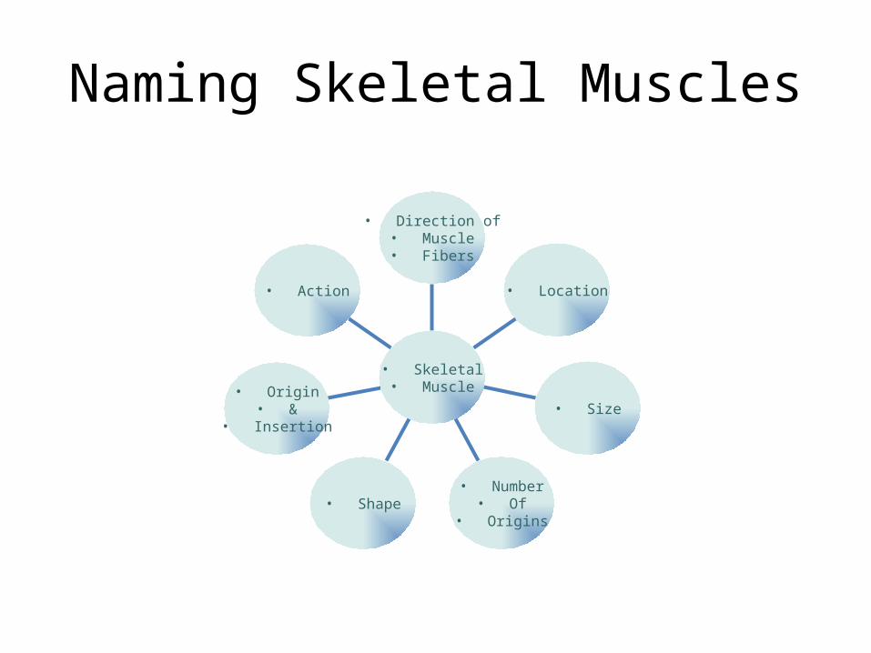
Naming Skeletal Muscles
• Action
• Origin• &
• Insertion
• Shape• Number
• Of• Origins
• Size
• Location
• Direction of• Muscle• Fibers
• Skeletal• Muscle
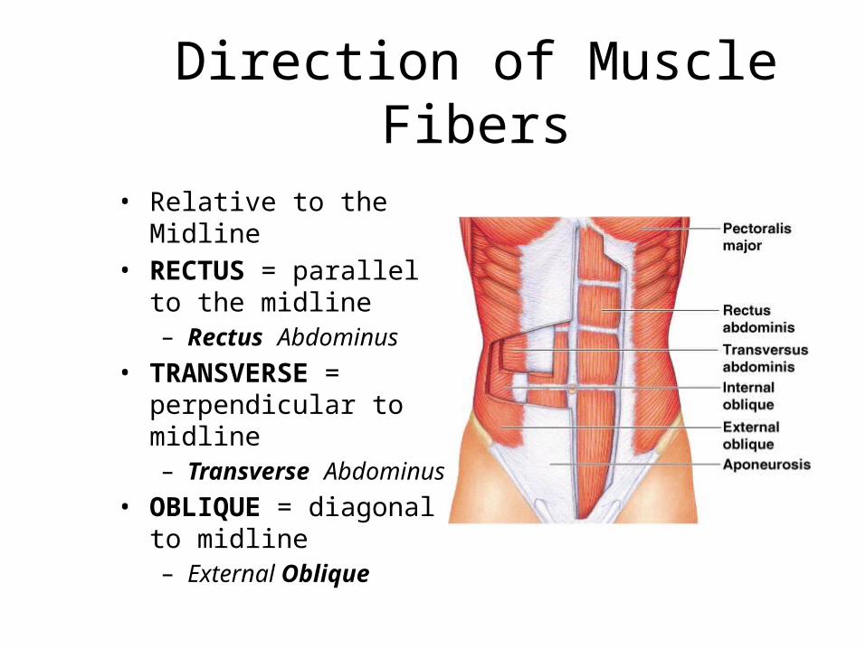
Direction of Muscle Fibers
• Relative to the Midline• RECTUS = parallel to the
midline– Rectus Abdominus
• TRANSVERSE = perpendicular to midline– Transverse Abdominus
• OBLIQUE = diagonal to midline– External Oblique
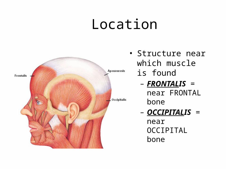
Location
• Structure near which muscle is found– FRONTALIS = near
FRONTAL bone– OCCIPITALIS = near
OCCIPITAL bone

Size
• Relative Size of Muscle• MAXIMUS = largest
– Gluteus Maximus• MEDIUS = middle
– Gluteus Medius• MINIMUS = smallest
– Gluteus Minimus• LONGUS = longest
– Fibularis Longus• BREVIS = short
– Fibularis Brevis• TERTIUS = shortest
– Fibularis Tertius
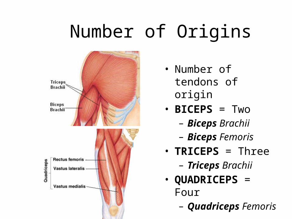
Number of Origins
• Number of tendons of origin
• BICEPS = Two– Biceps Brachii– Biceps Femoris
• TRICEPS = Three– Triceps Brachii
• QUADRICEPS = Four– Quadriceps Femoris
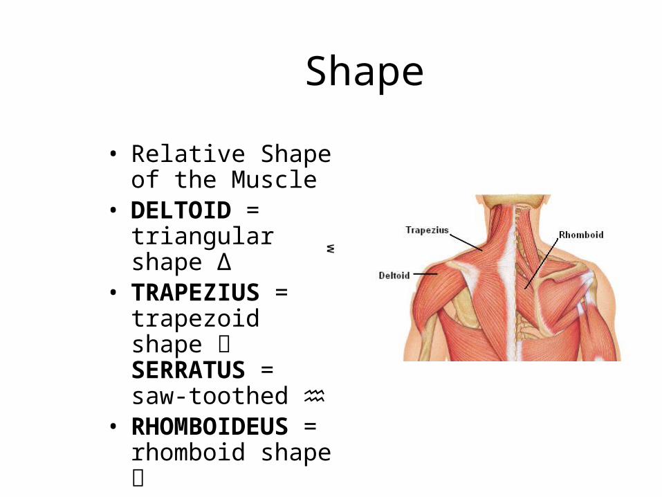
Shape
• Relative Shape of the Muscle
• DELTOID = triangular shape Δ
• TRAPEZIUS = trapezoid shape SERRATUS = saw-toothed ♒
• RHOMBOIDEUS = rhomboid shape
• TERES = round ○

Origin & Insertion
• Origin – attachment to an immoveable bone
• Insertion – attachment to a movable bone
• ILIO COSTALIS= attaches to the ilium & ribs (costal = ribs)
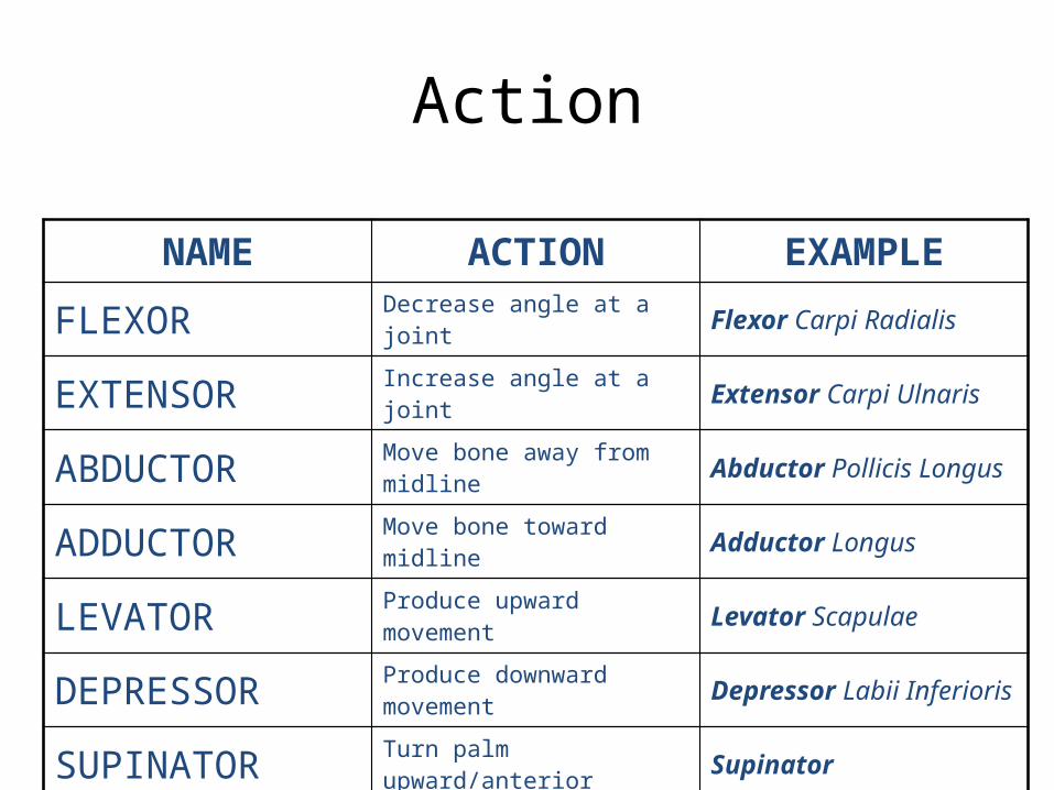
Action
NAME ACTION EXAMPLE
FLEXOR Decrease angle at a joint Flexor Carpi Radialis
EXTENSOR Increase angle at a joint Extensor Carpi Ulnaris
ABDUCTOR Move bone away from midline Abductor Pollicis Longus
ADDUCTOR Move bone toward midline Adductor Longus
LEVATOR Produce upward movement Levator Scapulae
DEPRESSOR Produce downward movement Depressor Labii Inferioris
SUPINATOR Turn palm upward/anterior Supinator
PRONATOR Turn palm downward/posterior Pronator Teres

Types of Muscle--Actions
• Prime mover (Agonist) – muscle with the major responsibility for a certain movement (When flexing forearm= Biceps Brachii)
• Antagonist – muscle that opposes or reverses a prime mover (When flexing forearm=triceps brachii)
• Synergist – muscle that aids a prime mover in a movement and helps prevent rotation

Anatomy of a Muscle Cell


Muscles and Muscle Fiber Structure
Muscles are composed of many fibers that are arranged in bundles called FASCICLES
Individual muscles are separated by FASCIA, which also forms tendons
Video on plantar fasciitis.