mus-101 modifier genes in - genetics · systematic, rnai-mediated identification of mus-101...
TRANSCRIPT
Systematic, RNAi-mediated identification of mus-101 modifier genes in C. elegans
Antonia H. Holway, Crystal Hung, and W.
The Biological Laboratories Department of Molecular and Cellular Biology
Harvard University Cambridge, MA 01238
Genetics: Published Articles Ahead of Print, published on January 16, 2005 as 10.1534/genetics.104.036137
2
Running head: RNAi modifier screen in C. elegans Keywords: Synthetic interactions RNAi TopBP1 MMS sensitivity PIAS domain SUMO ligase Corresponding author: W. Matthew Michael 2021 Biological Laboratories 16 Divinity Ave Cambridge, MA 02138 Tel. (617) 496-2940 Email: [email protected] *I do not own a FAX machine
3
ABSTRACT
The Mus101 family of chromosomal proteins, identified initially in Drosophila, is
widely conserved and has been shown to function in a variety of DNA metabolic
processes. Such functions include DNA replication, DNA damage repair, postreplication
repair, damage checkpoint activation, chromosome stability, and chromosome
condensation. Despite its conservation and widespread involvement in chromosome
biogenesis very little is known about how Mus101 is regulated and what other proteins
are required for Mus101 to exert its functions. To learn more about Mus101 we have
initiated an analysis of the protein in C. elegans. Here, we show that C. elegans mus-101
is an essential gene, that it is required for DNA replication, and that it also plays an
important role in the DNA damage response. Furthermore, we use RNA interference
(RNAi)-mediated reverse genetics to screen for genes that modify a mus-101 partial loss
of function RNAi phenotype. Using a systematic approach towards modifier gene
discovery we have found five Chromosome I genes that modify the mus-101 RNAi
phenotype, and we go on to show that one of them encodes an E3 SUMO ligase that
promotes SUMO-modification of MUS-101 in vitro. These results expand our
understanding of MUS-101 regulation, and show that genetic interactions can be
uncovered using screening strategies that rely solely on RNAi.
4
INTRODUCTION
The combination of RNAi-mediated gene knockdown methods and functional
genomics has provided an alternative to classic forward genetics for gene discovery
efforts in many organisms (Fraser et al, 2000; Gonczy et al, 2000; Boutros et al, 2004;
Berns et al, 2004; Paddison et al, 2004). RNAi is triggered when double-stranded RNA
(dsRNA) is processed in a manner that results in the destruction of mRNAs that are
homologous to the dsRNA (reviewed by Novina and Sharp, 2004). In the nematode C.
elegans dsRNA is delivered to the organism by one of three methods: soaking (Maeda et
al, 2001) injection (Fire et al, 1998), or feeding (Timmons and Fire, 1998). The
availability of a complete genome sequence in C. elegans has allowed systematic, high-
throughput RNAi screens to be routinely performed, and screens have been reported
using all three dsRNA delivery methods. While not absolute, a trend has emerged from
these studies that indicates that RNAi by feeding is less robust and less penetrant than the
other methods, suggesting that for many genes RNAi by feeding is phenotypically closer
to a hypomorphic than to the null condition.
When using RNAi to analyze the properties of genes that encode essential
functions a hypomorphic condition can be useful. Partial depletion of an essential gene
could, for example, reveal additional, nonessential functions of the gene. In addition, the
hypomorphic condition can be exploited to search for genes that modify the phenotype of
an essential gene and may therefore act in a common pathway. This last assertion is
based on considerable evidence from budding yeast where synthetic lethal screens have
been used to define and order genetic pathways. Synthetic lethal phenotypes result when
5
two mutations, neither of which effects viability on its own, cause a decrease in viability
when combined (reviewed by Hartman et al, 2001). This can occur by one of three ways:
by combining null alleles of two non-essential genes, by combining a hypomorphic allele
of an essential gene with a null allele of a non-essential gene, and by combining
hypomorphic alleles of two different essential genes. That such genetic interactions are
specific, and that they suggest inclusion of the genes in common or similar pathways, has
been addressed through Systematic Genetic Analysis (SGA) of the complete collection of
non-essential yeast deletion mutants. SGA involves making double mutants between a
mutant of interest and all 5,100 nonessential deletion mutants and then scoring for effects
on fitness (Tong et al, 2001). A recent study reported the results of 132 SGA screens
totaling roughly 4,000 interactions (Tong et al, 2004). The average number of
interactions per target was 34, demonstrating that such screens are reasonably specific in
the interactions that are revealed. Another indication of specificity was the correlation in
function between targets and hits uncovered in the screen - 27% of the interactions
involved pairs of genes that were previously known to function in the same or similar
pathways. These data reveal that systematic genetic modifier screens can be effective
tools for gene discovery.
We have been studying the DNA damage response in C. elegans and have been
particularly interested in the mus-101 locus. mus-101 (F37D6.1) encodes a 1,227 amino
acid protein that is characterized by the presence of six copies of the BRCA1 Carboxyl-
terminal (BRCT) repeat. BRCT repeats are commonly found in proteins involved in
DNA metabolism, and are thought to mediate protein-protein interactions between BRCT
6
repeat containing proteins and phosphorylated protein binding partners (Manke et al,
2003; Yu et al, 2003). The mus-101 gene is highly conserved throughout evolution, with
homologs present in humans (TopBP1)(Yamane et al, 1997), Xenopus (Xmus101)(Van
Hatten et al, 2002), Drosophila (mus101)(Boyd et al, 1976), Arabidopsis (MEI1)(Grelon
et al, 2003), and fission and budding yeast (cut5 and DPB11, respectively)(Saka and
Yanagida, 1993; Araki et al, 1995). Mus101 family members have been linked to a
variety of chromosomal pathways, including DNA replication, S phase checkpoint
activation, postreplication repair of damaged DNA, chromosome condensation,
chromosome stability, and meiosis. An important question regarding Mus101 function is
how the protein is regulated so that it can participate in multiple chromosomal pathways
without inducing inappropriate cross-talk between different pathways. To help answer
this question we have initiated an analysis of the C. elegans mus-101 gene. RNAi by
soaking resulted in robust depletion of mus-101, a failure to replicate DNA, and
embryonic lethality. RNAi using the feeding protocol resulted in less penetrant
depletion, viability, and sensitivity to the DNA damaging agent methyl methanesulfonate
(MMS). We took advantage of the hypomorphic condition caused by mus-101 RNAi by
feeding to systematically screen for genes that, when co-depleted with mus-101, modified
the mus-101 phenotype. From a library of 2,425 chromosome I genes we isolated five
modifiers. Three of the five displayed MMS sensitivity, suggesting a common function
with mus-101. Additionally, one of the identified modifiers, gei-17, encodes a
presumptive E3 SUMO ligase and was shown to stimulate SUMO modification of MUS-
101 in vitro. These results shed new light on the regulation of mus-101 and demonstrate

7
that systematic co-depletion by RNAi is a useful method for identification of genetic
modifiers.
8
MATERIALS and METHODS
C. elegans strains and culturing: The N2 Bristol strain was used in all experiments.
Worms were maintained as described (Brenner, 1974). Embryonic lethality was
determined by counting the percentage of eggs that failed to hatch 20 hours after laying.
RNA interference assays: mus-101 RNAi by soaking was performed as described
(Maeda et al., 2001). RNAi by feeding was performed as described (Timmons and Fire,
1998). For feeding RNAi all bacteria were cultured for 24 hrs at 37ºC in Terrific Broth
containing 50ug/ml ampicillin, seeded onto NGM plates containing 5mM IPTG, and
allowed to dry overnight.
Hoescht’s 33258 staining of embryos and gonads: Worms were dissected on glass
microscope slides, and permeabilized by freeze cracking. Slides were fixed for 10
minutes in methanol/formaldehyde fixative at -20°C and washed in PBST. DNA staining
was accomplished by adding 10 ul of 10ug/ul Hoechst’s 33258.
Antibody production: PCR primers were designed to amplify fragments corresponding
to the C-terminal 333 amino acids of mus-101. The fragments were subcloned into the
pET30 bacterial expression vector (Novagen) and used to produce recombinant protein.
Six histidine-tagged recombinant protein was purified on a nickel agarose column under
denaturing conditions. Purified proteins were then used as antigens to immunize rabbits.
Polyclonal antibodies were obtained, and affinity purified according to standard
procedures.
9
Propidium iodide and tubulin staining of embryos: Worms were mounted, fixed, and
washed as described for Hoescht’s staining. Slides were incubated in 200ug/ml RNAse
for 30 minutes at 37°C followed by a 5 minute incubation in 0.1mg/ml propidium iodide
solution (Sigma). Embryos were then incubated with an antibody against α-tubulin
(MAb DM1A; Sigma), diluted 1:100, for two hours at room temperature followed by
incubation with donkey anti-mouse, FITC-labeled secondary antibody. Embryos were
visualized on an Olympus BX51 microscope. Pictures were captured using a SPOT RT
monochrome camera (Diagnostic Instruments).
Embryo Culture Assays: Embryos were prepared for culture experiments based on
published protocols (Edgar and McGee, 1988) with some deviations. Briefly, dissected
adult worms were collected and incubated for two minutes in a 1:9 solution of 6%
hypochlorite (Fisher). Worms were then pelleted and resuspended in minimal egg
growth media (Edgar and McGhee, 1988) followed by another two-minute hypochlorite
treatment. The remaining eggs were pelleted, resuspended a final time in EGM, and
transferred to a gelatin-subbed slide (2%) and covered with a coverslip supported by half-
inch adhesive transfer tape. Pressure was applied to the coverslip to attach the embryos
to the slide and permeabilize the vitelline membrane. Additional EGM was added to fill
the chamber. Finally, three washes of 30 ul EGM with cytochalasin B were flushed
through the chamber and embryos were incubated for one hour. HU exposure was
accomplished by incubating permeabilized embryos in EGM containing 5mM HU and
cytochalasin B. Embryos were fixed by flushing the chamber with three 30 ul washes of
10
fixative (Edgar and McGhee, 1988) followed by PBS. DNA staining was accomplished
by flushing the chamber with three 30 ul washes of a solution containing 10ug/ul
Hoescht's 33258 followed by PBS and, finally, 30 ul of mounting medium (2% NPG
in 80% glycerol).
MMS Sensitivity assays: L4 F1 worms grown on plates containing the appropriate
bacterial expression vectors were transferred to plates containing 0.05 mg/ml MMS
(Sigma) at 25°C. Eggs laid by these worms were collected over time and scored for
survival.
Chromosome I RNAi modifier screen: The chromosome I RNAi library was purchased
from the UK HGMP Resource Centre. Bacteria were amplified as described and mixed
in a 1:1 ratio with either mus-101 RNAi bacteria or control RNAi bacteria (bacteria
expressing dsRNA against the exogenous green fluorescent protein, GFP). Additionally,
mus-101 RNAi bacteria were mixed at a 1:1 ratio with GFP RNAi bacteria to establish
baseline mus-101 RNAi lethality (which was typically less than 5%). Bacterial mixes
were then plated on media containing 5mM ITPG and allowed to dry overnight. P0
worms were seeded as L1s and cultured at 25ºC. Approximately 100 F2 embryos were
collected for each mixture and scored for survival. Genes were scored as modifiers when
a higher percentage of lethality was observed in the gene X/mus-101 RNAi combination
than the gene X/GFP RNAi or mus-101/GFP RNAi combinations after multiple rounds of
screening.
11
SUMOylation assay: A cDNA encoding full length mus-101 was transcribed and
translated in the presence of [35S]-methionine according to the manufacturer’s
instructions (Promega). The sumoylation kit was purchased from LAE Biotech
International and in vitro sumoylation reactions were performed according to the
manufacturer’s instructions with the following exceptions. Recombinant C. elegans his-
tagged UBC-9 was substituted for the human protein supplied with the kit. 20 µl
reactions contained 5 µl of the translation product, 0.36 µg C. elegans UBC-9, 0.68 µg C.
elegans GEI-17, 1 µg human SUMO-I, 1 µg human SUMO III, and 150 ng human E1.
To generate recombinant C. elegans UBC-9 and GEI-17, full-length cDNAs were
amplified from a cDNA library and cloned into the pET-28A expression vector
(Novagen). Recombinant protein was purified using standard his-tag purification
protocols. Induction of GEI-17 was performed for four hours at 16°C.
12
RESULTS
Differential depletion of mus-101 after soaking or feeding RNAi: To initiate an
analysis of mus-101 function in C. elegans we depleted the gene product using RNAi by
both soaking and feeding. To assess the relative effectiveness of feeding and soaking
RNAi on mus-101 depletion we probed whole worm lysates derived from treated animals
with an affinity purified anti-MUS-101 antibody. RNAi by soaking resulted in a robust
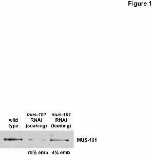
depletion of MUS-101 protein, whereas RNAi by feeding was less effective (Figure 1).
Quantification of the band intensities using NIH Image revealed that soaking RNAi
reduced MUS-101 levels to 10% of wild type while feeding RNAi reduced the level to
53% of wild type. When embryonic viability of the progeny of treated animals was
assessed we found that RNAi by soaking caused a high (78%) level of embryonic
lethality whereas RNAi by feeding resulted in only a very low level of embryonic
lethality (4%). We conclude that while mus-101 is an essential gene, a partial decrease in
mus-101 expression does not dramatically compromise viability.
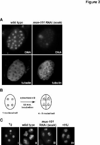
DNA replication problems in embryos depleted of mus-101 by soaking RNAi: A
conserved function of the Mus101 protein family is DNA replication (Saka and
Yanagida, 1993; Araki et al, 1995; Van Hatten et al, 2002), thus one likely explanation
for the embryonic lethality induced by mus-101 RNAi by soaking is a failure to replicate.
To investigate this further either wild type or mus-101 RNAi by soaking embryos were
treated with RNAse and then fixed and stained with the nucleic acid stain propidium
iodide to assess DNA content. As shown in Figure 2A, the propidium iodide signal
(panels “DNA”) in mus-101 RNAi by soaking embryos is far less intense than in the wild
13
type, while the signal resulting from an anti α-tubulin antibody (panels “tubulin”) is
qualitatively equivalent. The mus-101 soaking RNAi embryos therefore contain
substantially less DNA than do wild type embryos. A similar observation has been
reported for embryos depleted of the DNA replication factor CDT-1 (Zhong et al, 2003).
The data in Figure 2A suggest a role for mus-101 in DNA replication. To
examine more directly this possibility, we took advantage of previous observations that
DNA synthesis occurs normally in embryos treated with the actin poison cytochlasin
(Edgar and McGhee, 1988). Cytochlasin blocks cell division but does not prevent DNA
replication, thus cytochlasin-treated embryos will perform multiple rounds of DNA
replication and accumulate masses of DNA within individual cells (Figure 2B). This
allows a more direct assessment of the involvement of a given gene in DNA replication,
as embryos that cannot replicate DNA will simply fail to accumulate masses of DNA
over time. For this experiment, four-cell stage embryos were collected, the vitelline
membranes were permeabilized, and the samples were treated with cytochalasin B. After
a 60-minute incubation period, the embryos were fixed and the DNA was visualized by
the application of Hoescht’s 33258. As shown in Figure 2C, and consistent with previous
reports (Edgar and McGhee, 1988), wild type embryos accumulated large amounts of
DNA within individual cells after the hour-long incubation (Figure 2C, compare panel I
to panel II). By contrast, mus-101 RNAi by soaking embryos accumulated very little
DNA during the incubation period (Figure 2C, panel III). The amount of DNA
synthesized in mus-101 RNAi by soaking embryos was qualitatively similar to the
amount synthesized by wild type embryos that had been treated with the replication
14
inhibitor HU (Figure 2C, compare panel III to panel IV). From this, we conclude that
mus-101 is required for DNA replication in the early C. elegans embryo.
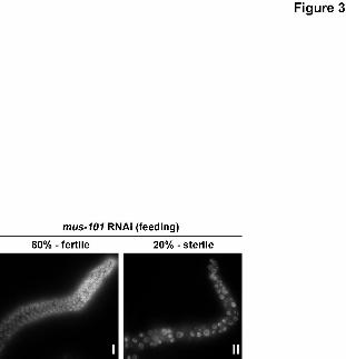
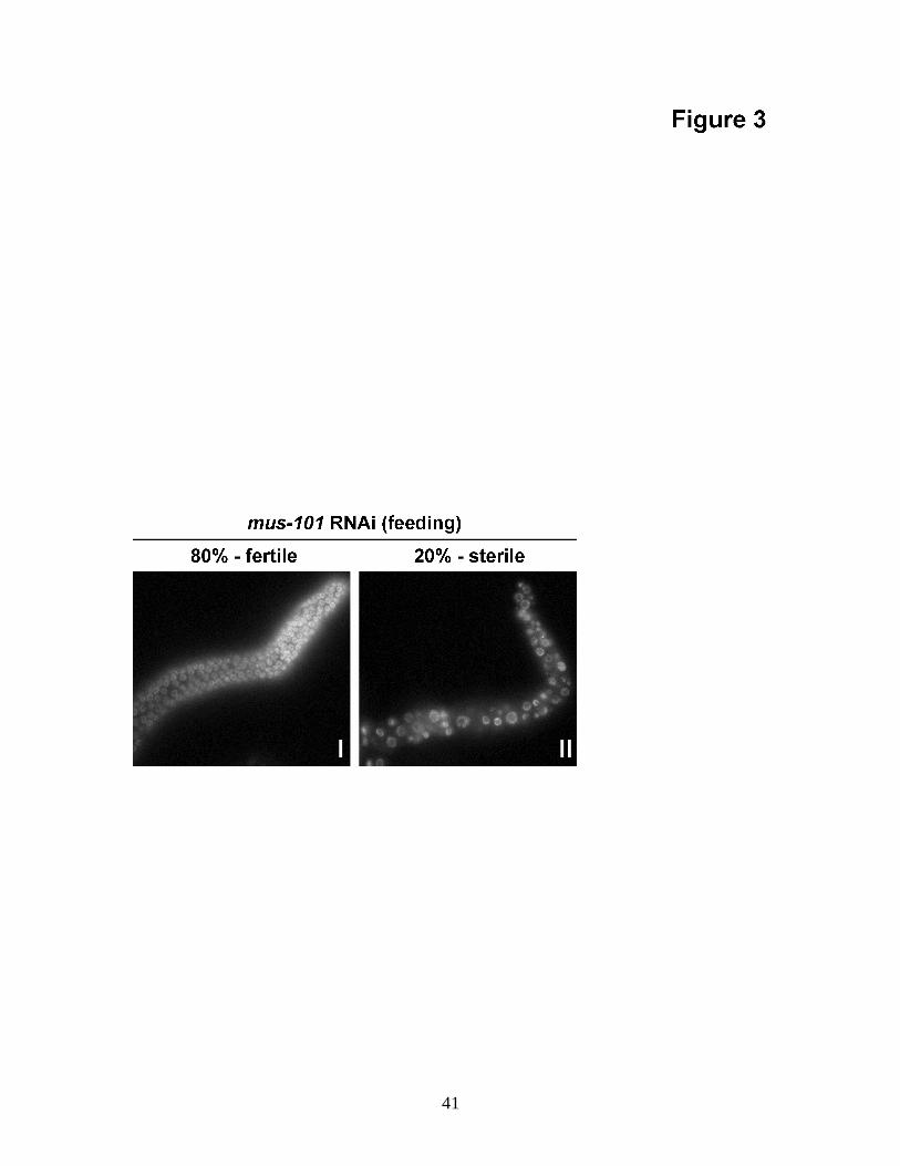
Depletion of mus-101 by feeding RNAi mimics a hypomorphic condition: By contrast
to RNAi by soaking, RNAi by feeding did not cause extensive embryonic lethality
(Figure 1). It did, however, result in 20% sterility in the F1 progeny of treated animals.
Staining of the germ lines of F1 fertile or sterile adults revealed that the sterile animals
had an abnormal gonad morphology characterized by necrotic-looking germ cell nuclei
(Figure 3). This demonstrates that, at low frequency, mus-101 feeding RNAi affects
proliferation of the germ line. Although the basis for this proliferation defect is not
known, the morphology of affected nuclei is reminiscent of wild type germ cell nuclei
that are blocked for replication with HU (MacQueen and Villeneuve, 2001) or treated
with ionizing radiation (Gartner et al, 2000). Consistent with a replication problem
causing this phenotype is the observation that RNAi by feeding depletion of the
replication initiation factor ORC-2 causes an identical sterility/abnormal nuclear
morphology defect (A. Holway, unpublished data).
In Drosophila hypomorphic alleles of mus101 exist that retain the DNA
replication function but not the DNA damage response function of the protein (Boyd et
al, 1976). In C. elegans we have shown that feeding RNAi allows embryonic viability,
and thus DNA replication, and it was therefore of interest to determine if the DNA
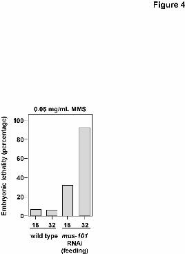
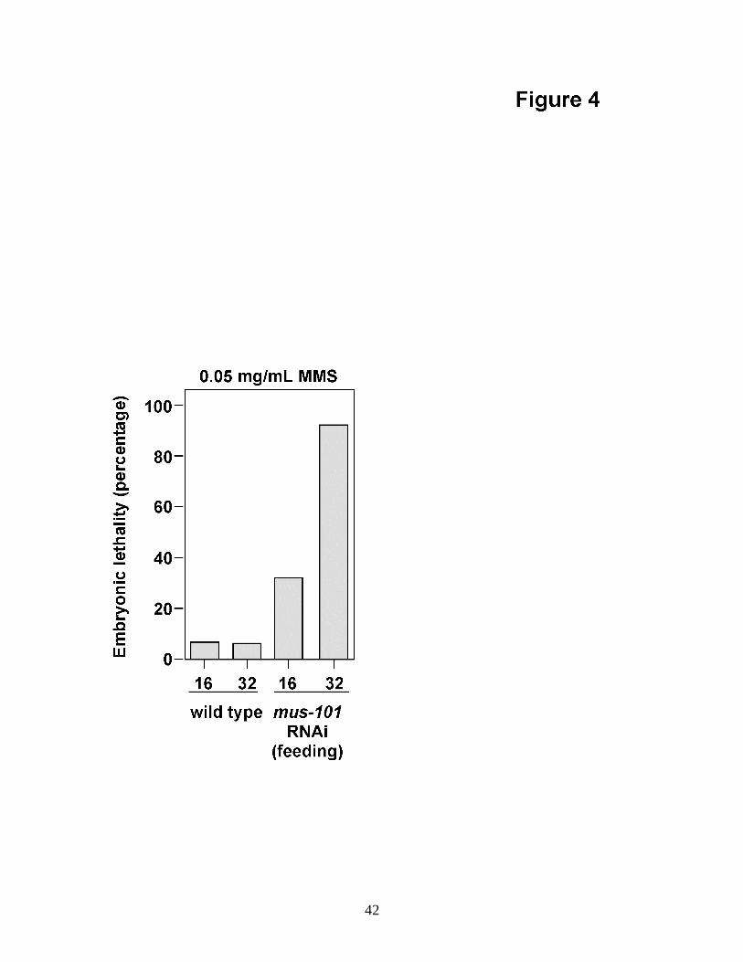
damage response function of mus-101 was also retained after feeding RNAi. To examine
this we tested for MMS sensitivity. F1 L4 stage animals were plated on media containing
15
0.05 mg/ml MMS and allowed to lay eggs for 16 hours before being transferred to a fresh
MMS plate for an additional 16 hours of egg laying. Embryonic lethality in the F2 was
then determined by counting the number of eggs that failed to hatch. As shown in Figure
4, and by contrast to wild type, mus-101 RNAi by feeding resulted in MMS sensitivity.
This result makes two important points. One, mus-101 is required for the DNA damage
response in C. elegans embryos. Two, a separation of the DNA replication from the
DNA damage response function of mus-101 can be achieved through RNAi by feeding.
Identification of mus-101 modifiers: The immunoblot in Figure 1 shows that mus-101
RNAi by feeding reduces the steady-state level of MUS-101 protein to roughly half of
what is normally present. The experiments shown in Figures 3 and 4 reveal that while
this reduction is generally tolerable there are phenotypic consequences, in both the germ
line and the embryo. RNAi by feeding therefore mimics a hypomorphic allele of mus-
101. We sought to exploit this hypomorphic condition to identify other genes that
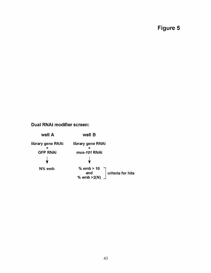
function with mus-101 by systematically searching for genes that, upon co-depletion,
modified the mus-101 feeding RNAi phenotype. In particular, we screened for genes that
when co-depleted with mus-101 caused an increase in embryonic lethality. Co-
depletions were performed by mixing, at a 1:1 ratio, bacteria expressing dsRNA against
mus-101 with bacteria expressing dsRNA against the gene to be screened. The mixture
was then seeded onto media plates prior to plating of L1 stage worms. After two
generations on the selective media embryonic viability was determined and compared to
that observed for a control co-depletion that substituted mus-101 for the irrelevant gene
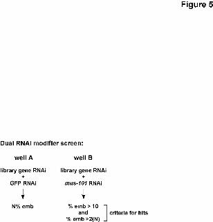
GFP. Two criteria were imposed to weed out weak and/or nonspecific interactions
16
(Figure 5). One, the percent embryonic lethality produced upon co-depletion of mus-101
and a candidate gene must be at least twice that observed when the candidate was co-
depleted with GFP. Two, the embryonic lethality produced upon co-depletion of mus-
101 and a candidate gene must be at least 10% as pilot experiments had shown that
reproducibility suffered when the cutoff was set beneath this value.
To isolate mus-101 modifiers we screened every gene present in a library
containing 2,425 chromosome I genes (representing 89% of the chromosome; see Fraser
et al, 2000 for details on the library). One concern that was raised as we were performing
this screen was that mixing two different feeding vectors in the same depletion
experiment might weaken or eliminate the ability of either feeding vector alone to
effectively silence its target gene. This did not occur. Fraser et al have reported a
chromosome I RNAi by feeding screen using a full dose of the chromosome I feeding
vectors (Fraser et al, 2000). Our screen, which used a half dose of the chromosome I
vectors, produced outcomes that were consistent with those reported by Fraser et al 95%
of the time. The 5% of the cases where the outcomes did not agree were equally divided
between examples where Fraser et al reported a phenotype for a given gene and we did
not, and examples where we could observe a phenotype and Fraser et al did not. Thus
this 5% discrepancy likely reflects the noise inherent in any large-scale functional
genomics screen. The high degree of correlation between phenotypes reported by Fraser
et al using a full complement of feeding vector and those reported here using a half
complement shows unambiguously that RNAi co-depletion by feeding allows silencing
of both target genes in the co-depletion.
17
Table 1 lists the results of this screen. Five genes were isolated that fit the criteria
outlined in Figure 5. Three of the genes, gei-17, arx-7, and let-49, have been previously
characterized, but not in significant detail. The other two, W01A8.1 and F26B1.2, have
not been characterized. W01A8.1 encodes a protein with no strong homology to
anything in the current databases, excepting its presumptive ortholog in C. briggsae.
F26B1.2 encodes a protein containing a heterogeneous nuclear ribonucleoprotein
(hnRNP) K homology (KH) domain. gei-17 encodes a protein with homology to the
mammalian protein inhibitor of activated STAT (PIAS) family of transcriptional
regulators and E3 SUMO ligases (reviewed by Schmidt and Muller, 2003). arx-7
encodes a component of the Arp2/3 complex which regulates actin dynamics, and let-49
is a transcriptional regulator that controls expression of genes required for germ line
formation and postembryonic development (Kwon et al, 2001).
The five genes identified in the screen all caused an increase in embryonic
lethality when they were co-depleted with mus-101, relative to GFP. In order to make
sure that the enhanced embryonic lethality was due to synergy with mus-101 RNAi and
was not due to an unexpected suppression of embryonic lethality by the GFP RNAi we
performed full-dose feeding RNAi experiments on all five genes. There were no
significant differences observed in the embryonic lethality after a full-dose of feeding
RNAi relative to that observed for the GFP co-depletions for four of the five genes. For
let-49 a full dose of feeding RNAi resulted in 100% sterility in the F1 thus precluding
18
analysis of the F2. We conclude that none of the genes identified in the screen were
false-positives resulting from interactions with GFP RNAi.
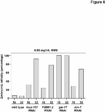
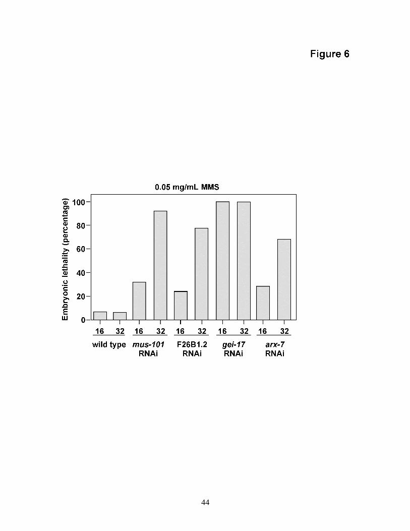
MMS sensitivity of mus-101 modifiers: Four of the screen hits were next tested for
MMS sensitivity. We did not score let-49 in this assay owing to the sterility phenotype.
Three of the four genes tested showed sensitivity (Figure 6). One of the genes, gei-17,
was exceptionally sensitive to MMS, with embryonic lethality of 100% at the first time
point. The other two, F26B1.2 and arx-7, were more modestly sensitive. For arx-7, we
did note a weak eggshell phenotype, therefore it is unclear whether the MMS sensitivity
in the arx-7 RNAi embryos was due to direct involvement in the DNA damage response
or to the weakened egg shell resulting in the embryos experiencing a higher effective
concentration of MMS during the experiment.
To understand more about why mus-101, gei-17, and F26B1.2 depleted embryos
were MMS sensitive we stained MMS-exposed embryos with the DNA stain Hoescht’s
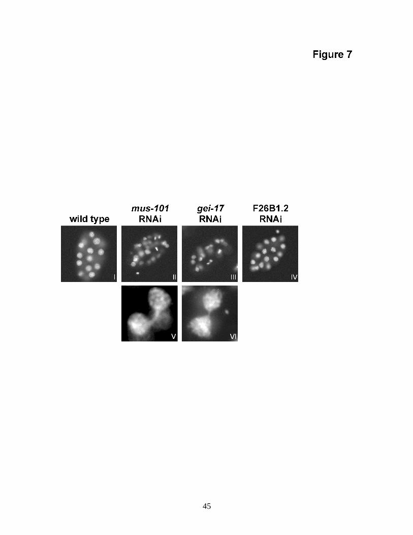
33258 to examine the morphology of the nuclear DNA. As shown in Figure 7, nuclear
morphology in F26B1.2 depleted embryos did not differ from wild type after MMS
exposure. By contrast, nuclear morphology in both mus-101 and gei-17 depleted
embryos was affected by MMS. Specifically, many nuclei were of discontinuous size
and shape, some nuclei appeared to be torn or fragmented, and numerous anaphase
bridges were also observed. Such nuclear morphology aberrations were not observed in
either mus-101 or gei-17 depleted embryos in the absence of MMS exposure. Thus, in
addition to MMS sensitivity, mus-101 and gei-17 share a similar nuclear morphology
19
defect upon MMS exposure. This suggests that mus-101 and gei-17 may be components
of a common pathway that responds to MMS-induced DNA damage in the embryo.
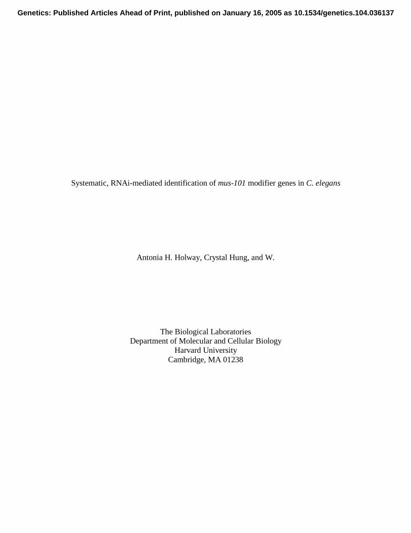
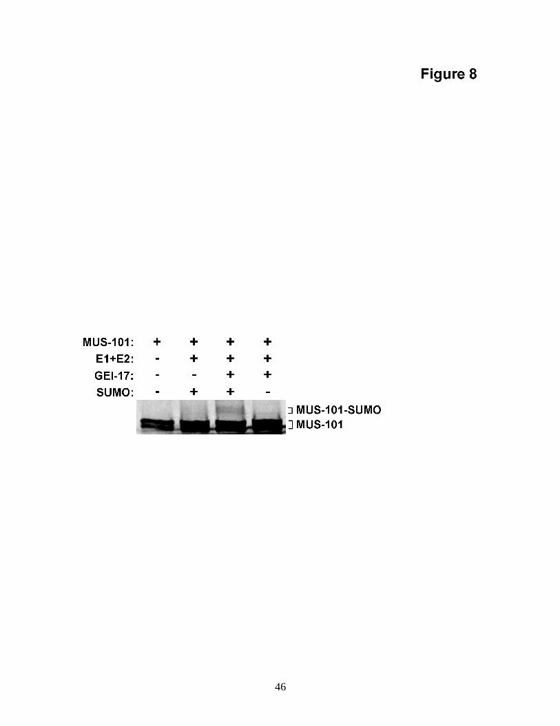
GEI-17 stimulates SUMO modification of MUS-101 in vitro: In order to strengthen
the connection between mus-101 and gei-17 we sought biochemical evidence for an
interaction between the two gene products. The PIAS domain family of proteins, of
which GEI-17 is a member, has been shown to stimulate SUMO modification of substrate
proteins (Johnson and Gupta, 2001). SUMO is a small, ubiquitin-like polypeptide that is
covalently attached to substrate proteins (reviewed by Johnson, 2004). SUMO
modification of substrates can alter their function, and chromosomal proteins in particular
are subject to regulation by the SUMO system. Attachment of SUMO to substrate is
similar to ubiquitination in that attachment is mediated through sequential transfer of
SUMO from an E1 enzyme to an E2 and then on to substrate. The last step, transfer of
SUMO from the E2 to substrate, is often facilitated by an E3 ligase such as a PIAS
domain protein. Because gei-17 modifies the mus-101 feeding RNAi phenotype we
speculated that GEI-17 may stimulate SUMO modification of MUS-101. We, therefore,
asked if GEI-17 protein could stimulate SUMO modification of MUS-101 in a
reconstituted in vitro SUMOylation assay. MUS-101 was radio-labeled through
transcription and translation in the presence of 35S-methionine to produce MUS-101 TnT.
MUS-101 TnT was mixed with the core components of the SUMOylation machinery (E1,
E2, and SUMO) as well as recombinant GEI-17, and the reaction products were resolved
on an SDS-PAGE gel. This resulted in the appearance of high molecular weight bands
that ran above MUS-101 TnT on SDS-PAGE (Figure 8). The appearance of these bands
20
was dependent upon the presence of both GEI-17 and SUMO in the reaction mixture,
demonstrating that they were derived from SUMO modification of MUS-101 and that
GEI-17 facilitates this. We conclude that GEI-17 stimulates SUMO modification of
MUS-101 in vitro, suggesting that GEI-17 may control MUS-101 function in vivo.
21
DISCUSSION
In this paper we have used RNAi to reduce expression of the mus-101 gene.
Soaking RNAi cleared 90% of MUS-101 protein, whereas feeding RNAi removed
roughly half. Accordingly, soaking RNAi caused high embryonic lethality, and this was
likely through a reduction in DNA replication. For the remainder of this work we have
focused on the mus-101 RNAi by feeding phenotype.
The mus-101 RNAi by feeding phenotype: Feeding RNAi caused a low level (20%) of
sterility in the F1, and DNA-staining of the germ lines of sterile animals revealed a defect
in germ cell proliferation. We have observed this phenotype after RNAi against other
DNA replication proteins, such as subunits of the origin recognition complex (A.
Holway, unpublished data), and thus we suspect that the phenotype is connected to a
defect in the initiation of DNA replication. It is also possible that an inability to repair
replication-induced damage may be responsible for this phenotype.
Unlike the sterility phenotype, which was modestly penetrant, the MMS
sensitivity phenotype after feeding RNAi was highly penetrant with embryonic lethality
of more than 90% as compared to less than 10% for wild type. This suggests that a 50%
reduction in MUS-101 protein is incompatible with an efficient embryonic DNA damage
response. This, in turn, suggests that the replication and damage response functions of
mus-101 can be uncoupled by feeding RNAi. Interestingly, such uncoupling has also
been observed for the Drosophila and the budding and fission yeast homologs of mus-
101. Drosophila mus101 hypomorphic alleles exist that are MMS sensitive and defective
22
in postreplication repair but are nonetheless viable (Boyd et al, 1976). In fission yeast
separation of function alleles of cut5 have been isolated that allow replication but not
activation of the DNA damage checkpoint (McFarlane et al, 1997). Finally, in budding
yeast, the dpb11-1 allele is MMS sensitive at the permissive temperature for DNA
replication (Wang et al, 2002). Thus a common theme in this gene family is the
availability of hypomorphic alleles. The data presented here shed light on this by
showing that alterations to the mus-101 coding sequences are not necessary to generate
hypomorphic alleles as limiting the expression of an otherwise wild type copy of the gene
is sufficient to do this. One interpretation of this is that the DNA damage response
function of the Mus101 family requires more protein on a per cell basis than does the
replication function.
The modifier screen: Having established a hypomorphic condition using mus-101
feeding RNAi we exploited this to screen for genes that modified the phenotype. This
resulted in the isolation of five genes that displayed enhanced embryonic lethality when
co-depleted with mus-101. Interestingly, none of the genes identified are known to
encode DNA replication factors. Indeed, when directed co-depletion experiments were
performed with mus-101 and a number of different DNA replication factors we failed to
detect any interactions (A. Holway and C. Hung, unpublished data). This is by contrast
to a synthetic lethal screen performed in budding yeast using the dpb11-1 allele
(Kamimura et al, 1998). This screen identified numerous sld genes, many of which are
DNA replication factors. One explanation for this is that in C. elegans MUS-101 is an
excess, even after feeding RNAi, for embryonic DNA replication thus making it difficult
23
to further weaken this pathway. If so, we would not expect to find many replication
genetic interactions given that embryonic lethality was the scored phenotype.
Interestingly, three of the five genes tested (gei-17, arx-7, and F26B1.2) showed
MMS sensitivity after a full-dose of feeding RNAi, implying participation in the DNA
damage response. F26B1.2 encodes a KH domain containing protein. KH proteins bind
single-stranded nucleic acids, and while roles for these proteins in the DNA damage
response are not common, we do note that the MCG10 KH protein has been shown to be
a transcriptional target of p53 and to promote cell cycle arrest and apoptosis after DNA
damage in human cells (Zhu and Chen, 2000). Our finding that F26B1.2 RNAi embryos
are MMS sensitive further strengthens the connection between KH proteins and the DNA
damage response.
The gene from the screen that showed the most robust MMS sensitivity was gei-
17. The gei-17 gene was initially identified as one of 26 genes recovered in a yeast two-
hybrid screen using the transcription factor gex-3 as bait (Tsuboi et al, 2002) and has not
been further characterized. gei-17 is highly conserved and shares homology with the
Su(var)2-10 chromosomal regulator in Drosophila (Hari et al, 2001) and the PIAS family
of mammalian SUMO ligases (reviewed in Seeler and Dejean, 2003). Three findings
reported here provide strong evidence that gei-17 and mus-101 act together in a common
pathway. One, the gei-17 gene was selected by our screen as a mus-101 modifier. Two,
like mus-101, gei-17 RNAi embryos show MMS sensitivity and an MMS-induced
nuclear morphology defect. We have observed MMS sensitivity after RNAi for a number
24
of DNA damage response genes and yet the only two genes to date that produce this
nuclear morphology defect are mus-101 and gei-17. Thus the nuclear morphology defect
is relatively rare. Three, we have demonstrated a biochemical interaction between MUS-
101 and GEI-17 by showing in an in vitro SUMOylation assay that GEI-17 stimulates
SUMO modification of MUS-101. Taken together, these results all suggest that SUMO
modification of MUS-101 by GEI-17 regulates MUS-101 function during the DNA
damage response. The connections between Mus101 and PIAS proteins are not limited to
those reported here. We note that the Drosophila gei-17 homolog, Su(var)2-10, shares a
chromosomal phenotype with fly mus101 in that both genes are required for condensation
of heterochromatic regions of the chromosome (Gatti et al, 1983; Yamamoto et al, 2000;
Hari et al, 2001). How might this phenotype be connected to the MMS sensitivity
reported here for the C. elegans orthologs? We propose that both mus-101 and gei-17 are
required to promote DNA replication under conditions where replication is difficult.
MMS-mediated damage of DNA is known to stress replication (Tercero and Diffley,
2001), and replication through heterochromatin is also thought to be difficult (reviewed
by Maison and Almouzni, 2004). Thus the hetreochromatin condensation defect may be
an indirect result of a failure to replicate heterochromatic DNA. Consistent with this
hypothesis is a recent study from budding yeast that isolated DPB11 in a screen for
chromosome integrity genes (Huang and Koshland, 2003). This work showed that
DPB11 is required during the elongation phase of replication to maintain replication fork
progression. When elongation was challenged by increasing the distance between active
origins on mini-chromosomes than hypomorphic alleles of DPB11 caused elongation
failures at higher frequencies than wild type. While much further work is required to
25
confirm this hypothesis it is intriguing to speculate that GEI-17 mediated SUMO
modification of MUS-101 allows MUS-101 to promote replication during stressful
conditions.
Dual RNAi modifier screens: This paper presents, to our knowledge, the first large-
scale screen for genetic modifiers that is based entirely on RNAi. We screened a
chromosome I library which covers 13% of the C. elegans genome and isolated five
modifiers. Through extrapolation we can estimate that a total of 38 modifier genes would
be isolated using these conditions by screening the entire genome. This is, interestingly,
in line with the average number of 34 modifiers uncovered in budding yeast SGA.
Whether or not dual RNAi modifier screens are universally useful in isolating genetic
modifiers remains to be determined. The drawbacks include that possibility of low
penetrance that can occur using feeding RNAi. Additionally, the method does appear to
isolate nonspecific interactors as it is unclear how arx-7, which encodes a cytoplasmic
component of the actin polymerization regulator Arp2/3 complex, would participate
directly with mus-101. Nonetheless, the method does offer several advantages. One,
when working with essential genes, feeding RNAi can produce the hypomorphic
conditions that are essential for screening. Thus the relative inefficiency of feeding
RNAi can be exploited to perform modifier screens that would otherwise require the
laborious and imperfect isolation of hypomorphic alleles. This allows modifier analysis
to be performed on a systematic level. Two, as is the case in all functional genomic
screens, once an interaction is uncovered then the identification of the relevant gene is
immediate. We have shown here that by combining careful analysis of a feeding RNAi

26
phenotype of an essential gene with dual RNAi modifier screening that specific and novel
genetic interactions can be discovered.
27
ACKNOWLEDGEMENTS
We thank Craig Hunter and his colleagues for generous and expert advice on working
with C. elegans. We are also grateful to Tim Schedl for thoughtful discussion and for
advice and assistance during the early stages of this work. We thank Mike Matunis for
help and advice on SUMO modification. The N2 Bristol strain and HT115(DE3) cells
used in this work were provided by the Caenorhabditis Genetics Center, which is funded
by the NIH National Center for Research Resources (NCRR). The Chromosome I RNAi
library was created by Julie Ahringer and distributed by the UK HGMP Resource Centre.
A.H.H. was supported by funds from a Genetics and Genomics training grant (National
Institute of General Medical Sciences) and a Don Wiley Award for Excellence in
Graduate Studies (funded by Merck). Additional support for this work was provided by
research grants from the National Institute of General Medical Sciences (R01GM67735),
the American Cancer Society (RSG-03-153), and a Searle Scholar Award to W.M.M.
28
LITERATURE CITED
ARAKI, H., S. H. LEEM, A. PHONGDARA and A. SUGINO, 1995 Dpb11, which interacts with
DNA polymerase II(epsilon) in Saccharomyces cerevisiae, has a dual role in S-phase
progression and at a cell cycle checkpoint. Proc Natl Acad Sci U S A 92: 11791-11795.
BERNS, K., E. M. HIJMANS, J. MULLENDERS, T. R. BRUMMELKAMP, A. VELDS et al., 2004
A large-scale RNAi screen in human cells identifies new components of the p53 pathway.
Nature 428: 431-437.
BOUTROS, M., A. A. KIGER, S. ARMKNECHT, K. KERR, M. HILD et al., 2004 Genome-wide
RNAi analysis of growth and viability in Drosophila cells. Science 303: 832-835.
BOYD, J. B., M. D. GOLINO, T. D. NGUYEN and M. M. GREEN, 1976 Isolation and
characterization of X-linked mutants of Drosophila melanogaster which are sensitive to
mutagens. Genetics 84: 485-506.
BRAUCHLE, M., K. BAUMER and P. GONCZY, 2003 Differential activation of the DNA
replication checkpoint contributes to asynchrony of cell division in C. elegans embryos.
Curr Biol 13: 819-827.
BRENNER, S., 1974 The genetics of Caenorhabditis elegans. Genetics 77: 71-94.
EDGAR, L. G., and J. D. MCGHEE, 1988 DNA synthesis and the control of embryonic
29
gene expression in C. elegans. Cell 53: 589-599.
ENCALADA, S. E., P. R. MARTIN, J. B. PHILLIPS, R. LYCZAK, D. R. HAMILL et al., 2000
DNA replication defects delay cell division and disrupt cell polarity in early
Caenorhabditis elegans embryos. Dev Biol 228: 225-238.
FIRE, A., S. XU, M. K. MONTGOMERY, S. A. KOSTAS, S. E. DRIVER et al., 1998 Potent and
specific genetic interference by double-stranded RNA in Caenorhabditis elegans. Nature
391: 806-811.
FRASER, A. G., R. S. KAMATH, P. ZIPPERLEN, M. MARTINEZ-CAMPOS, M. SOHRMANN et al.,
2000 Functional genomic analysis of C. elegans chromosome I by systematic RNA
interference. Nature 408: 325-330.
GARTNER, A., S. MILSTEIN, S. AHMED, J. HODGKIN and M. O. HENGARTNER, 2000 A
conserved checkpoint pathway mediates DNA damage--induced apoptosis and cell cycle
arrest in C. elegans. Mol Cell 5: 435-443.
GATTI, M., D. A. SMITH and B. S. BAKER, 1983 A gene controlling condensation of
heterochromatin in Drosophila melanogaster. Science 221: 83-85.
GONCZY, P., C. ECHEVERRI, K. OEGEMA, A. COULSON, S. J. JONES et al., 2000 Functional
genomic analysis of cell division in C. elegans using RNAi of genes on chromosome III.

30
Nature 408: 331-336.
GRELON, M., G. GENDROT, D. VEZON, G. PELLETIER, G. MATHILDE et al., 2003 The
Arabidopsis MEI1 gene encodes a protein with five BRCT domains that is involved in
meiosis-specific DNA repair events independent of SPO11-induced DSBs. Plant J 35:
465-475.
HARI, K. L., K. R. COOK and G. H. KARPEN, 2001 The Drosophila Su(var)2-10 locus
regulates chromosome structure and function and encodes a member of the PIAS protein
family. Genes Dev 15: 1334-1348.
HARTMAN, J. L. T., B. GARVIK and L. HARTWELL, 2001 Principles for the buffering of
genetic variation. Science 291: 1001-1004.
HUANG, D., and D. KOSHLAND, 2003 Chromosome integrity in Saccharomyces cerevisiae:
the interplay of DNA replication initiation factors, elongation factors, and origins. Genes
Dev 17: 1741-1754.
JOHNSON, E. S., 2004 Protein modification by sumo. Annu Rev Biochem 73: 355-382.
JOHNSON, E. S., and A. A. GUPTA, 2001 An E3-like factor that promotes SUMO
conjugation to the yeast septins. Cell 106: 735-744.
31
KAMIMURA, Y., H. MASUMOTO, A. SUGINO and H. ARAKI, 1998 Sld2, which interacts with
Dpb11 in Saccharomyces cerevisiae, is required for chromosomal DNA replication. Mol
Cell Biol 18: 6102-6109.
KWON, J. Y., J. KIM-HA, B. J. LEE and J. LEE, 2001 The MED-7 transcriptional mediator
encoded by let-49 is required for gonad and germ cell development in Caenorhabditis
elegans. FEBS Lett 508: 305-308.
MACQUEEN, A. J., and A. M. VILLENEUVE, 2001 Nuclear reorganization and homologous
chromosome pairing during meiotic prophase require C. elegans chk-2. Genes Dev 15:
1674-1687.
MAEDA, I., Y. KOHARA, M. YAMAMOTO and A. SUGIMOTO, 2001 Large-scale analysis of
gene function in Caenorhabditis elegans by high-throughput RNAi. Curr Biol 11: 171-
176.
MAISON, C., and G. ALMOUZNI, 2004 HP-1 and the dynamics of heterochromatin
maintenance. Nat Rev Mol Cell Biol 5: 296-304.
MANKE, I. A., D. M. LOWERY, A. NGUYEN and M. B. YAFFE, 2003 BRCT repeats as
phosphopeptide-binding modules involved in protein targeting. Science 302: 636-639.
MCFARLANE, R. J., A. M. CARR and C. PRICE, 1997 Characterisation of the
32
Schizosaccharomyces pombe rad4/cut5 mutant phenotypes: dissection of DNA
replication and G2 checkpoint control function. Mol Gen Genet 255: 332-340.
NOVINA, C. D., and P. A. SHARP, 2004 The RNAi revolution. Nature 430: 161-164.
PADDISON, P. J., J. M. SILVA, D. S. CONKLIN, M. SCHLABACH, M. LI et al., 2004 A
resource for large-scale RNA-interference-based screens in mammals. Nature 428: 427-
431.
SAKA, Y., and M. YANAGIDA, 1993 Fission yeast cut5+, required for S phase onset and M
phase restraint, is identical to the radiation-damage repair gene rad4+. Cell 74: 383-393.
SCHMIDT, D., and S. MULLER, 2003 PIAS/SUMO: new partners in transcriptional
regulation. Cell Mol Life Sci 60: 2561-2574.
TERCERO, J. A., and J. F. DIFFLEY, 2001 Regulation of DNA replication fork progression
through damaged DNA by the Mec1/Rad53 checkpoint. Nature 412: 553-557.
TIMMONS, L., and A. FIRE, 1998 Specific interference by ingested dsRNA. Nature 395:
854.
TONG, A. H., M. EVANGELISTA, A. B. PARSONS, H. XU, G. D. BADER et al., 2001
Systematic genetic analysis with ordered arrays of yeast deletion mutants. Science 294:
33
2364-2368.
TONG, A. H., G. LESAGE, G. D. BADER, H. DING, H. XU et al., 2004 Global mapping of
the yeast genetic interaction network. Science 303: 808-813.
TSUBOI, D., H. QADOTA, K. KASUYA, M. AMANO and K. KAIBUCHI, 2002 Isolation of the
interacting molecules with GEX-3 by a novel functional screening. Biochem Biophys Res
Commun 292: 697-701.
VAN HATTEN, R. A., A. V. TUTTER, A. H. HOLWAY, A. M. KHEDERIAN, J. C. WALTER et
al., 2002 The Xenopus Xmus101 protein is required for the recruitment of Cdc45 to
origins of DNA replication. J Cell Biol 159: 541-547.
WANG, H., and S. J. ELLEDGE, 2002 Genetic and physical interactions between DPB11
and DDC1 in the yeast DNA damage response pathway. Genetics 160: 1295-1304.
YAMAMOTO, R. R., J. M. AXTON, Y. YAMAMOTO, R. D. SAUNDERS, D. M. GLOVER et al.,
2000 The Drosophila mus101 gene, which links DNA repair, replication and
condensation of heterochromatin in mitosis, encodes a protein with seven BRCA1 C-
terminus domains. Genetics 156: 711-721.
YAMANE, K., M. KAWABATA and T. TSURUO, 1997 A DNA-topoisomerase-II-binding
protein with eight repeating regions similar to DNA-repair enzymes and to a cell-cycle
34
regulator. Eur J Biochem 250: 794-799.
YU, X., C. C. CHINI, M. HE, G. MER and J. CHEN, 2003 The BRCT domain is a phospho-
protein binding domain. Science 302: 639-642.
ZHONG, W., H. FENG, F. E. SANTIAGO and E. T. KIPREOS, 2003 CUL-4 ubiquitin ligase
maintains genome stability by restraining DNA-replication licensing. Nature 423: 885-
889.
ZHU, J., and X. CHEN, 2000 MCG10, a novel p53 target gene that encodes a KH domain
RNA-binding protein, is capable of inducing apoptosis and cell cycle arrest in G(2)-M.
Mol Cell Biol 20: 5602-5618.
35
TABLE 1
Genes that interact with mus-101 after co-depletion by feeding RNAi
Gene emb (GFP) emb (mus-101) presumed function
W01A8.1 3.3% (9/269) 14.7% (58/395) unknown
F26B1.2 7.7% (29/379) 14.5% (62/427) KH domain
gei-17 12.8% (20/156) 28.9% (43/149) PIAS SUMO ligase
arx-7 4.6% (11/239) 12.6% (17/135) Arp 2/3 complex
let-49 1.7 % (6/356) 14.4% (21/156) transcription factor
Emb refers to embryonic lethality and was determined by counting the number of
eggs that failed to hatch. Both the percentage and the actual counts are shown. Emb
(GFP) refers to embryonic lethality after co-depletion with GFP and emb (mus-101)
refers to embryonic lethality after co-depletion with mus-101.
36
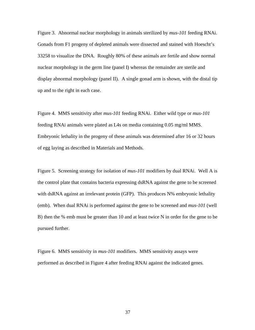
FIGURE LEGENDS
Figure 1. Immunoblot analysis of MUS-101 protein levels in wild type animals and after
soaking or feeding RNAi. Below each RNAi lane is the percentage embryonic lethality
(emb) in the F1 (soaking) or F2 (feeding) progeny of RNAi-treated animals. For soaking
RNAi N2 worms were submerged into a solution of concentrated dsRNA according to
published procedures (Maeda et al, 2001). After a recovery period whole worm lysates
were prepared for immunoblots. For feeding RNAi animals were cultured on plates
seeded with bacteria expressing the dsRNA for two generations at 25 C prior to
preparation of whole worm lysates. Each lane on the blot corresponds to 10 adult worms.
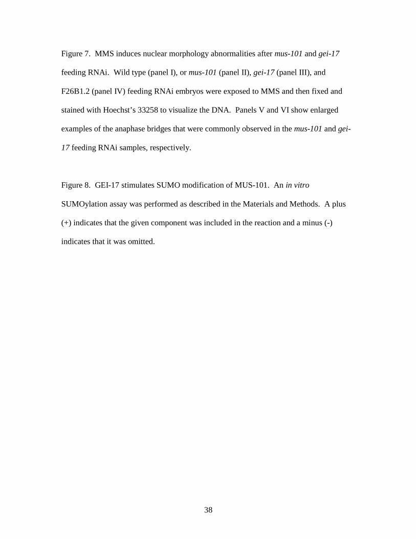
Figure 2. DNA replication is attenuated after mus-101 RNAi by soaking. (A) Wild type
or mus-101 soaking RNAi embryos were fixed, treated with RNase I, and stained with
propidium iodide to visualize the DNA (panels “DNA”) and anti α-tubulin to visualize
tubulin (panels “tubulin”). (B) Schematic depiction of the uncoupling of DNA
replication from cell division that occurs upon treatment of permeabilized embryos with
the cytokinesis inhibitor cytochlasin B. (C) Permeabilized embryos were fixed and
stained with Hoescht’s 33258 stain either immediately after permeabilization (panel I) or
after a one-hour incubation in culture media (panels II – IV). The embryos shown in
panels II and IV are wild type, while the embryo depicted in panel III was depleted of
mus-101 gene product by soaking RNAi. The embryo in panel IV was treated with HU
to block replication. The embryos depicted in panels I through IV are representative of a
larger (>25) sample set that was analyzed for each condition.
37
Figure 3. Abnormal nuclear morphology in animals sterilized by mus-101 feeding RNAi.
Gonads from F1 progeny of depleted animals were dissected and stained with Hoescht’s
33258 to visualize the DNA. Roughly 80% of these animals are fertile and show normal
nuclear morphology in the germ line (panel I) whereas the remainder are sterile and
display abnormal morphology (panel II). A single gonad arm is shown, with the distal tip
up and to the right in each case.
Figure 4. MMS sensitivity after mus-101 feeding RNAi. Either wild type or mus-101
feeding RNAi animals were plated as L4s on media containing 0.05 mg/ml MMS.
Embryonic lethality in the progeny of these animals was determined after 16 or 32 hours
of egg laying as described in Materials and Methods.
Figure 5. Screening strategy for isolation of mus-101 modifiers by dual RNAi. Well A is
the control plate that contains bacteria expressing dsRNA against the gene to be screened
with dsRNA against an irrelevant protein (GFP). This produces N% embryonic lethality
(emb). When dual RNAi is performed against the gene to be screened and mus-101 (well
B) then the % emb must be greater than 10 and at least twice N in order for the gene to be
pursued further.
Figure 6. MMS sensitivity in mus-101 modifiers. MMS sensitivity assays were
performed as described in Figure 4 after feeding RNAi against the indicated genes.
38
Figure 7. MMS induces nuclear morphology abnormalities after mus-101 and gei-17
feeding RNAi. Wild type (panel I), or mus-101 (panel II), gei-17 (panel III), and
F26B1.2 (panel IV) feeding RNAi embryos were exposed to MMS and then fixed and
stained with Hoechst’s 33258 to visualize the DNA. Panels V and VI show enlarged
examples of the anaphase bridges that were commonly observed in the mus-101 and gei-
17 feeding RNAi samples, respectively.
Figure 8. GEI-17 stimulates SUMO modification of MUS-101. An in vitro
SUMOylation assay was performed as described in the Materials and Methods. A plus
(+) indicates that the given component was included in the reaction and a minus (-)
indicates that it was omitted.
39
40
41
42
43
44
45
46