multiplex rt-pcr kit. - dna diagnosticdna-diagnostic.com/files/downloads/hemavision/26.pdf · the...
TRANSCRIPT

HemaVision®-Screen
Screening test for 28 chromosome translocations and more than 145 breakpoints associated with leukemia
del1(p32) (STIL-TAL1) t(9;12) (q34;p13) (ETV6-ABL1)
t(1;11) (p32;q23) (MLL-EPS15) t(9,22) (q34;q11) (BCR-ABL1)
t(1;11) (q21;q23) (MLL-MLLT11) t(10;11) (p12;q23) (MLL-MLLT10)
t(1;19) (q23;p13) (TCF3-PBX1) t(11;17) (q23;q21) (MLL-MLLT6)
t(3;5) (q25;q34) (NPM1-MLF1) t(11;17) (q23;q21) (ZBTB16-RARA)
t(3;21) (q26;q22) (RUNX1-MECOM) t(11;19) (q23;p13.1) (MLL-ELL)
t(4;11) (q21;q23) (MLL-AFF1) t(11;19) (q23;p13.3) (MLL-MLLT1)
t(5;12) (q33;p13) (ETV6-PDGFRB) t(12;21) (p13;q22) (ETV6-RUNX1)
t(5;17) (q35;q21) (NPM1-RARA) t(12;22) (p13;q11) (ETV6-MN1)
t(6;9) (p23;q34) (DEK-NUP214) t(15;17) (q24;q21) (PML-RARA)
t(6;11) (q27;q23) (MLL-MLLT4) inv(16) (p13;q22) (CBFB-MYH11)
t(8;21) (q22;q22) (RUNX1-RUNX1T1) t(16;21) (p11;q22) (FUS-ERG)
t(9;9) (q34;q34) (SET-NUP214) t(17;19) (q22;p13) (TCF3-HLF)
t(9;11) (p22;q23) (MLL-MLLT3) t(X;11) (q13;q23) (MLL-FOXO4)
Cat No. HV01-Screen USER MANUAL
DNA Diagnostic A/S www.dna-diagnostic.com Revision 2014.10.28
IVD

1
HemaVision®-Screen www.dna-diagnostic.com Revision 2014.10.28
HemaVision-Screen
Multiplex RT-PCR test
Screens for 28 leukemia causing translocations
User Manual
for
HemaVision-Screen, Cat. No. HV01-Screen
25 tests per kit
TABLE OF CONTENTS
1. PURPOSE OF THE TEST – SCREENING FOR 28 TRANSLOCATIONS 2
2. PRINCIPLES OF TEST 3
3. KIT COMPONENTS AND STORAGE 5
4. EQUIPMENT AND MATERIALS REQUIRED BUT NOT PROVIDED 6
5. PRECAUTIONS 7
6. PROCEDURE 8
Step 1 cDNA synthesis 8
Step 2 Master PCR-I 9
Step 3 Master PCR-II 10
Step 4 Gel electrophoresis Master PCR-II 11
Step 5 Interpretation of results from Master PCR-II 11
7. GENE ABBREVATIONS ACCORDING TO THE HGNC 14
8. REFERENCES 15
HemaVision-Screen is manufactured by DNA Diagnostic A/S Voldbjergvej 16 Tel. +45 8732 3050 DK-8240 Risskov [email protected] Denmark www.dna-diagnostic.com
2
HemaVision®-Screen www.dna-diagnostic.com Revision 2014.10.28
1. PURPURSE OF THE TEST - SCREENING FOR 28 TRANSLOCATIONS
HemaVision-Screen is a CE-marked in vitro diagnostic test for 28 leukemia causing chromosomal
translocations including more than 145 breakpoints plus associated mRNA splice variants.
Furthermore, it detects new breakpoints and mRNA splice variants for the 28 translocations. It is a fast
one day test based on the method described by Pallisgard et al. (Ref 58). The HemaVision-Screen test
has very high sensitivity (>99%) and specificity (>99%) (Ref 59, 60).
Limit of detection is 10-9 µg of fusion RNA in a sample of 1 µg total RNA when the RNA quality is good.
This test brings IVD testing deeper into a detailed description of the exon organization of fusion genes
originating from chromosome translocations. This information is important for predicting development
of the disease and selection of treatment.
HemaVision-Screen is a qualitative test using total RNA extracted from human whole blood or bone
marrow cells as starting material in the test. The test uses reverse transcription of RNA to cDNA
followed by multiplex nested polymerase chain reactions (RT-PCR), agarose gel electrophoresis, and
interpretation.
HemaVision-Screen identifies chromosomes, genes and exons at the breakpoint in fusion genes.
Furthermore, the test identifies mRNA splice variants from fusion genes.
The HemaVision-Screen kit contains primers for 25 cDNA reactions and 25 (master) nested PCR tests.
The HemaVision-Screen kit is identical to the BOX 1 of HemaVision-28N kit (Cat No HV01-28N).
Therefore, positive master reactions from HemaVision-Screen tests must be characterized further by
split-out reactions from BOX2 of the HemaVision-28N kit.
The test is for professional use only.
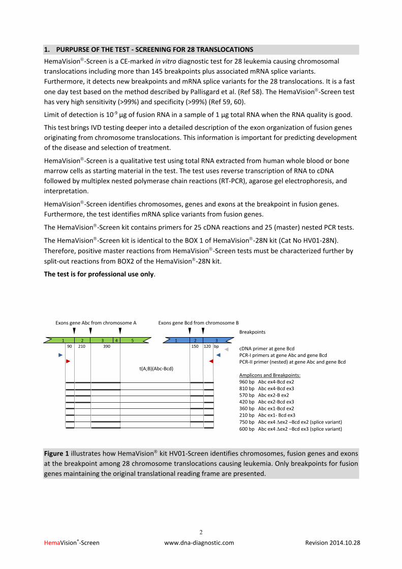
Figure 1 illustrates how HemaVision kit HV01-Screen identifies chromosomes, fusion genes and exons
at the breakpoint among 28 chromosome translocations causing leukemia. Only breakpoints for fusion
genes maintaining the original translational reading frame are presented.
Breakpoints
cDNA primer at gene Bcd PCR-I primers at gene Abc and gene Bcd PCR-II primer (nested) at gene Abc and gene Bcd Amplicons and Breakpoints: 960 bp Abc ex4-Bcd ex2 810 bp Abc ex4-Bcd ex3 570 bp Abc ex2-B ex2 420 bp Abc ex2-Bcd ex3 360 bp Abc ex1-Bcd ex2 210 bp Abc ex1- Bcd ex3
750 bp Abc ex4 ex2 –Bcd ex2 (splice variant)
600 bp Abc ex4 ex2 –Bcd ex3 (splice variant)
Exons gene Abc from chromosome A Exons gene Bcd from chromosome B 1 2 3 4 5 1 2 3 90 210 390 150 120 bp
t(A;B)(Abc-Bcd)

3
HemaVision®-Screen www.dna-diagnostic.com Revision 2014.10.28
2. PRINCIPLES OF TEST
RNA is template for synthesis of cDNA in a reaction using Reverse Transcriptase (RT) and specific cDNA
primers. The cDNA is template for PCR amplification using a hot start Taq DNA Polymerase and specific
PCR primers. Many of the fusion genes have several breakpoints. Therefore, the PCR primers are
designed to bind at positions enabling screening for all these breakpoints as illustrated in figure 1. The
workflow of the test and an example of test results are shown in figure 2.
Total RNA
cDNA Synthesis
Master PCR
Continue with Split-out PCR
reactions M6A-M6E
using HemaVision kit HV01-28N BOX 2
Figure 2. Workflow and results from a test with HemaVision Cat. No. HV01-Screen.
Result: All reactions M1-M8 are positive for the reaction control amplicon of 911 bp. M6 is also positive for an amplicon approximately 400 bp.
Conclusion: Patient is positive for a translocation. Continue testing with the split-out reactions M6A-
M6E from BOX 2 in HemaVision kit HV01-28N for identification of the translocation.
M1 M2 M3 M4 M5 M6 M7 M8 m
Reaction control amplicon 911 bp
Translocation specific amplicon M6
1000 900 800
700
600
500
400
300
200
100
4
HemaVision®-Screen www.dna-diagnostic.com Revision 2014.10.28
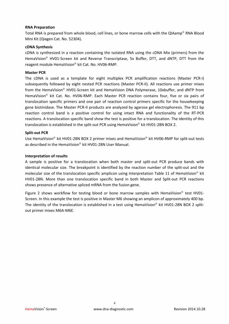
RNA Preparation
Total RNA is prepared from whole blood, cell lines, or bone marrow cells with the QIAamp RNA Blood
Mini Kit (Qiagen Cat. No. 52304).
cDNA Synthesis
cDNA is synthesized in a reaction containing the isolated RNA using the cDNA Mix (primers) from the
HemaVision HV01-Screen kit and Reverse Transcriptase, 5x Buffer, DTT, and dNTP, DTT from the
reagent module HemaVision kit Cat. No. HV06-RMP.
Master PCR
The cDNA is used as a template for eight multiplex PCR amplification reactions (Master PCR-I)
subsequently followed by eight nested PCR reactions (Master PCR-II). All reactions use primer mixes
from the HemaVision HV01-Screen kit and HemaVision DNA Polymerase, 10xbuffer, and dNTP from
HemaVision kit Cat. No. HV06-RMP. Each Master PCR reaction contains four, five or six pairs of
translocation specific primers and one pair of reaction control primers specific for the housekeeping
gene biotinidase. The Master PCR-II products are analyzed by agarose gel electrophoresis. The 911 bp
reaction control band is a positive control for using intact RNA and functionality of the RT-PCR
reactions. A translocation specific band show the test is positive for a translocation. The identity of this
translocation is established in the split-out PCR using HemaVision kit HV01-28N BOX 2.
Split-out PCR
Use HemaVision kit HV01-28N BOX 2 primer mixes and HemaVision kit HV06-RMP for split-out tests
as described in the HemaVision kit HV01-28N User Manual.
Interpretation of results
A sample is positive for a translocation when both master and split-out PCR produce bands with
identical molecular size. The breakpoint is identified by the reaction number of the split-out and the
molecular size of the translocation specific amplicon using Interpretation Table 11 of HemaVision kit
HV01-28N. More than one translocation specific band in both Master and Split-out PCR reactions
shows presence of alternative spliced mRNA from the fusion gene.
Figure 2 shows workflow for testing blood or bone marrow samples with HemaVision test HV01-
Screen. In this example the test is positive in Master M6 showing an amplicon of approximately 400 bp.
The identity of the translocation is established in a test using HemaVision kit HV01-28N BOX 2 split-
out primer mixes M6A-M6E.
5
HemaVision®-Screen www.dna-diagnostic.com Revision 2014.10.28
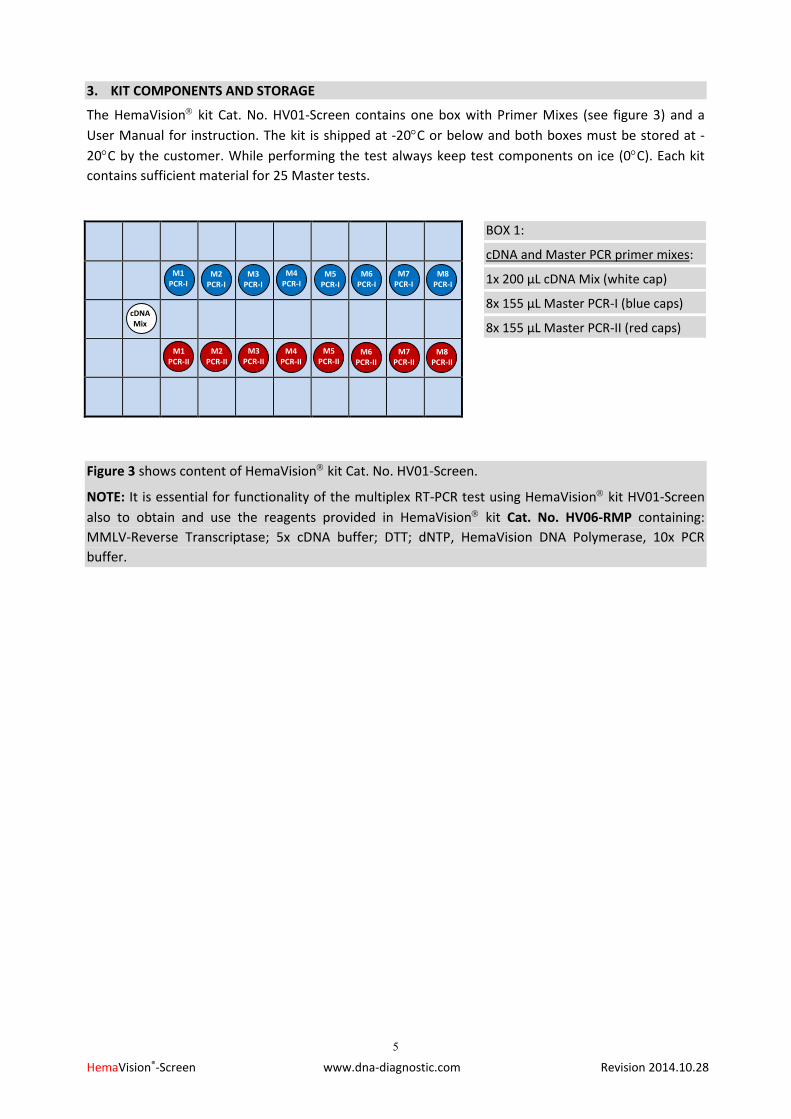
3. KIT COMPONENTS AND STORAGE
The HemaVision kit Cat. No. HV01-Screen contains one box with Primer Mixes (see figure 3) and a
User Manual for instruction. The kit is shipped at -20C or below and both boxes must be stored at -
20C by the customer. While performing the test always keep test components on ice (0C). Each kit
contains sufficient material for 25 Master tests.
Figure 3 shows content of HemaVision kit Cat. No. HV01-Screen.
NOTE: It is essential for functionality of the multiplex RT-PCR test using HemaVision kit HV01-Screen
also to obtain and use the reagents provided in HemaVision kit Cat. No. HV06-RMP containing:
MMLV-Reverse Transcriptase; 5x cDNA buffer; DTT; dNTP, HemaVision DNA Polymerase, 10x PCR
buffer.
M1 PCR-I
M2 PCR-I
M3 PCR-I
M4 PCR-I
M5 PCR-I
M6 PCR-I
M7 PCR-I
M8 PCR-I
M1 PCR-II
M2 PCR-II
M3 PCR-II
M4 PCR-II
M5 PCR-II
M6 PCR-II
M7 PCR-II
M8 PCR-II
cDNA Mix
BOX 1:
cDNA and Master PCR primer mixes:
1x 200 µL cDNA Mix (white cap)
8x 155 µL Master PCR-I (blue caps)
8x 155 µL Master PCR-II (red caps)
6
HemaVision®-Screen www.dna-diagnostic.com Revision 2014.10.28
4. EQUIPMENT AND MATERIALS REQUIRED BUT NOT PROVIDED
RNA extraction:
QIAamp RNA Blood Mini Kit from Qiagen Cat. No. 52304. Reagent Module:
HemaVision kit Cat. No. HV06-RMP containing: MMLV-RT; 5x cDNA buffer; DTT; dNTP, HemaVision DNA Polymerase, 10x PCR buffer. Use four HV06-RMP kits together with each HV01-Screen kit.
Or as an alternative use: HemaVision kit Cat. No. HV04-RM containing: MMLV-RT; 5x cDNA buffer; DTT; dNTP and from Qiagen HotStarTaq DNA Polymerase 5 Units/ µL and 10x PCR buffer. Use four HV04-RM kits and 1000 Units HotStarTaq DNA Polymerase together with each HV01-Scree kit.
Master Mix room – No templates in this room:
Micropipettes, 0.5-10 µL, 20-200 HemaVision kit Cat. No. HV06-RMP containing: MMLV-RT; 5x cDNA buffer; DTT; dNTP, HemaVision DNA Polymerase, 10x PCR buffer Aerosol barrier micropipette tips, 0.5-10 µL, and 20-200 µL Micro centrifuge Ice bath RNase free tubes Disposable gloves RNase free water -20oC freezer for storage of kits (HV01-28N and HV06-RMP) cDNA room: Micropipettes, 0.5-10 µL, 20-200 µL Aerosol barrier micropipette tips, 0.5-10 µL, and 20-200 µL Micro centrifuge Heating block/Water bath Ice bath RNase free tubes Disposable gloves RNase free water -80oC freezer for storage of RNA samples PCR room: Micropipettes, 0.5-10 µL, 20-200 µL Aerosol barrier micropipette tips, 0.5-10 µL, and 20-200 µL Micro centrifuge Thermal Cycler Ice bath PCR tubes (0.1 mL or 0.2 mL) and lids Disposable gloves Gel electrophoresis room: Micropipettes, 0.5-10 µL Aerosol barrier micropipette tips, 0.5-10 µL Micro centrifuge Equipment for agarose gel electrophoresis Disposable gloves Molecular size marker (e.g. 100 bp ladder)

7
HemaVision®-Screen www.dna-diagnostic.com Revision 2014.10.28
5. PRECAUTIONS
General precautions
1. The quality of the RNA sample greatly affects the results of this test. To minimize the risk of
degradation of RNA by ribonucleases, we strongly recommend lysing the cells in a denaturing
solution [e.g. containing guanidinium isothiocyanate (GTC)] immediately after isolation and before
freezing. Always store cell samples and aqueous RNA solutions at –80oC. Even an overnight storage
at – 20oC may result in RNA degradation. When working with RNA always use gloves to avoid
ribonuclease contamination from hands.
2. RT-PCR is a very sensitive technique. Therefore, precautions must be taken to avoid false positive
results caused by contamination with RNA, cDNA or PCR products from other samples.
Dedicate four separate rooms to the following activities:
1) Master Mix production – No templates in here
2) cDNA synthesis
3) PCR
4) Gel electrophoresis
A set of micropipettes, aerosol barrier pipette tips, disposable gloves and clean lab coats should be
kept in each of the four rooms. The work must be organized so that mixes and reaction products
only moves in the direction from Master Mix room to cDNA room to PCR room to Gel
electrophoresis room. NEVER move mixes or reaction products in the opposite direction.
3. Laboratory workbenches, pipettes, and lab coats must be cleaned on a regular basis.
4. Use of aerosol barrier pipette tips is highly recommended during the entire procedure. It is
essential to change gloves very often when handling tubes containing RNA or DNA. After PCR tubes
must be opened with extreme care to avoid spillage of high copy number DNA products.
Safety
Read and understand the procedure before starting.
Normal laboratory aseptic technique should be followed at all times.
Treat each sample as if it is infectious.
Wear eye protection and disposable gloves during all steps of the assay.
8
HemaVision®-Screen www.dna-diagnostic.com Revision 2014.10.28
6. PROCEDURE
Procedural notes
Store all test components as described in section 3: Kit Components and Storage.
Do not mix reagents from different lots.
Careful pipetting technique is essential for accurate results.
This protocol is optimized with enzymes and buffers from HemaVision kit Cat. No. HV06-RMP.
This protocol is optimized for the Perkin Elmer GeneAmp 9600/9700 thermal cycler. Use of another thermal cycler may require optimization by the user.
As a positive control for RNA quality and functionality of each RT-PCR reaction a 911 bp fragment of the housekeeping gene biotinidase must be present in all lanes except in reactions positive for a translocation specific amplicon where it may be weak or missing.
As a negative control make the cDNA reaction without RNA template.
RNA preparation
Use blood from venipuncture collected into a tube containing EDTA. Alternatively, use bone marrow collected into a tube containing EDTA. Do not freeze the blood or bone marrow sample or use samples collected in heparin tubes.
Prepared mononuclear cells from whole blood or bone marrow by the Ficoll Hypaque method.
Within 24 hours of collection, extract total RNA with QIAamp RNA Blood Mini Kit (Qiagen Cat.
No. 52304). Typically 5-10 g total RNA is extracted from 1 x10⁷ mononuclear blood cells.
Measure the RNA concentration by reading the optical density at 260 nm. An absorbance of 1
unit at 260 nm corresponds to 40 g of RNA per mL. Adjust the concentration of RNA to 0.1
g/L with RNase free H2O.
Make 20 L (0.1 g/L) RNA aliquots in RNase free tubes. Store RNA aliquots at –80oC or use RNA immediately for cDNA synthesis.
Master test
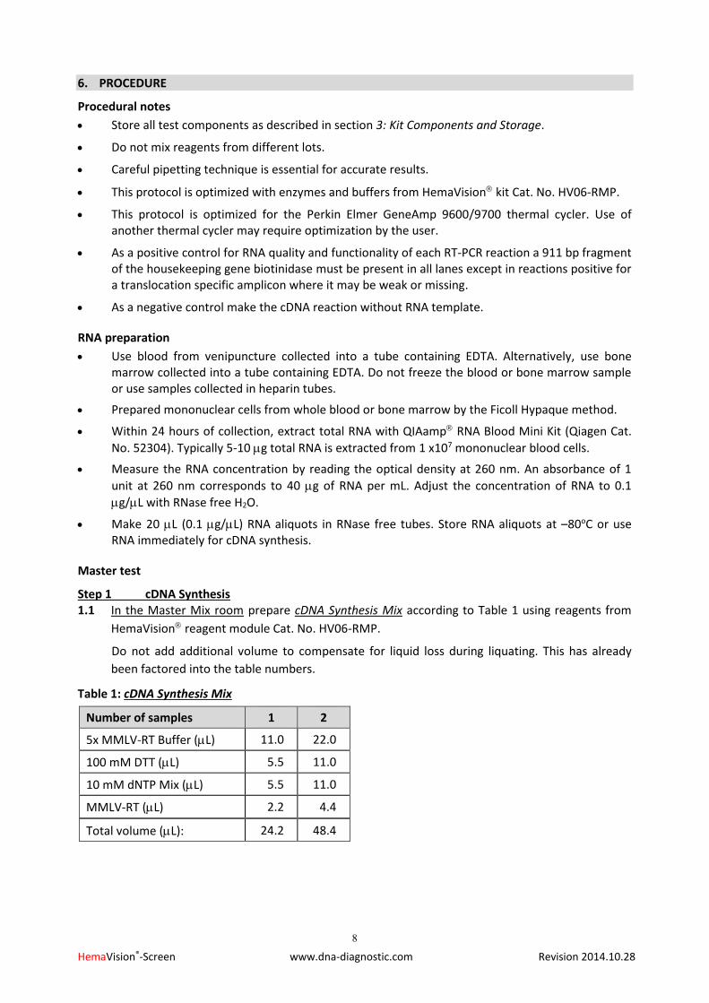
Step 1 cDNA Synthesis 1.1 In the Master Mix room prepare cDNA Synthesis Mix according to Table 1 using reagents from
HemaVision reagent module Cat. No. HV06-RMP.
Do not add additional volume to compensate for liquid loss during liquating. This has already
been factored into the table numbers.
Table 1: cDNA Synthesis Mix
Number of samples 1 2
5x MMLV-RT Buffer (L) 11.0 22.0
100 mM DTT (L) 5.5 11.0
10 mM dNTP Mix (L) 5.5 11.0
MMLV-RT (L) 2.2 4.4
Total volume (L): 24.2 48.4
9
HemaVision®-Screen www.dna-diagnostic.com Revision 2014.10.28
1.2 In the cDNA room add 8 L cDNA Mix (primers) from white capped tube in the HemaVision
HV01-28N kit to one tube containing 20 L total RNA (2 g). Mix gently and spin down for 10
seconds.
1.3 In a separate RNase free tube, add 8 µL of cDNA Mix to 20 L H2O (negative control).
1.4 Incubate the tubes in a heating block or water bath at 65C for 5 minutes. Chill and hold on ice.
1.5 Add 22 L of the cDNA Synthesis Mix to the tube with 28 L RNA+cDNA Mix and the negative
control tube from step 1.4. Mix gently and spin down for 10 seconds.
1.6 Incubate at 37C for 45 minutes.
1.7 Add 50 L H2O to each cDNA tube.
1.8 Incubate at 95C for 5 min to inactivate the MMLV-RT enzyme.
1.9 Chill and hold the cDNA tube on ice (0C, do not freeze) for a maximum of three days before use
in Master PCR and Split-out PCR.
Step 2 Master PCR-I To achieve maximal sensitivity and specificity each sample is tested with both a first and a nested
Master PCR reaction.
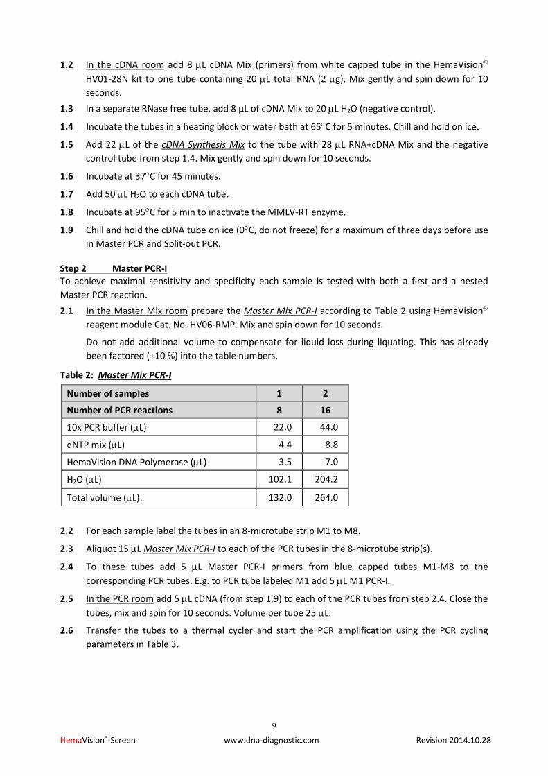
2.1 In the Master Mix room prepare the Master Mix PCR-I according to Table 2 using HemaVision
reagent module Cat. No. HV06-RMP. Mix and spin down for 10 seconds.
Do not add additional volume to compensate for liquid loss during liquating. This has already
been factored (+10 %) into the table numbers.
Table 2: Master Mix PCR-I
Number of samples 1 2
Number of PCR reactions 8 16
10x PCR buffer (L) 22.0 44.0
dNTP mix (L) 4.4 8.8
HemaVision DNA Polymerase (L) 3.5 7.0
H2O (L) 102.1 204.2
Total volume (L): 132.0 264.0
2.2 For each sample label the tubes in an 8-microtube strip M1 to M8.
2.3 Aliquot 15 L Master Mix PCR-I to each of the PCR tubes in the 8-microtube strip(s).
2.4 To these tubes add 5 L Master PCR-I primers from blue capped tubes M1-M8 to the
corresponding PCR tubes. E.g. to PCR tube labeled M1 add 5 L M1 PCR-I.
2.5 In the PCR room add 5 L cDNA (from step 1.9) to each of the PCR tubes from step 2.4. Close the
tubes, mix and spin for 10 seconds. Volume per tube 25 L.
2.6 Transfer the tubes to a thermal cycler and start the PCR amplification using the PCR cycling
parameters in Table 3.

10
HemaVision®-Screen www.dna-diagnostic.com Revision 2014.10.28
Table 3: Master PCR-I Amplification Parameters
Step Time/Temperature Cycles
1 15 minutes at 95oC 1
2
30 seconds at 95oC
30 seconds at 58oC
1 minute 30 seconds at 72oC
25
3 Hold at 4oC 1
Step 3 Master PCR-II (nested)
3.1 In the Master Mix room prepare the Master Mix PCR-II according to Table 4 using HemaVision
reagent module Cat. No. HV06-RMP. Mix and spin down for 10 seconds.
Do not add additional volume to compensate for liquid loss during liquating. This has already
been factored (+10 %) into the table numbers.
Table 4: Master Mix PCR-II
Number of samples 1 2
Number of PCR reactions 8 16
10x PCR buffer (L) 22.0 44.0
dNTP mix (L) 4.4 8.8
HemaVision DNA Polymerase (L) 3.5 7.0
H2O (L) 137.3 274.6
Total volume (L): 167.2 334.4
3.2 For each sample label the tubes in an 8-microtube strip M1 to M8.
3.3 Aliquot 19 L Master Mix PCR-II to each of the PCR tubes in the 8-microtube strip(s).
3.4 To these tubes add 5 L Master PCR-II primers from red capped tubes M1-M8 to the
corresponding PCR tubes. E.g. to PCR tube labeled M1 add 5 L M1 PCR-II.
3.5 In the PCR room carefully open without spillage the PCR tubes containing the Master PCR-I
reactions. From these tubes transfer 1 L to the corresponding Master PCR-II tubes from step
3.3. An eight channel pipette can be used for this transfer. Close the tubes, mix and spin for 10
seconds. Volume per tube 25 L.
3.6 Transfer the tubes to a thermal cycler and start the PCR amplification using the PCR cycling
parameters in Table 5.
Table 5: Master PCR-II Amplification Parameters
Step Time/Temperature Cycles
1 15 minutes at 95oC 1
2
30 seconds at 95oC
30 seconds at 58oC
1 minute 30 seconds at 72oC
20
3 10 minutes at 72oC 1
4 Hold at 4oC 1
11
HemaVision®-Screen www.dna-diagnostic.com Revision 2014.10.28
Step 4 Gel electrophoresis
4.1 Prepare a 1.5 % (w/v) agarose gel at least 10 cm long in 1X TBE buffer. Add ethidium bromide to
a final concentration of 0.5 g/mL.
4.2 In the Gel Electrophoresis room carefully open the PCR tubes with Master PCR-II without
contaminating gloves and surroundings with drops containing high copy numbers of amplicon.
Add 3 L of 10x loading buffer into each PCR tube. Load approximately 14 L per slot in the gel
(lane 1-8). Finally load a molecular size marker to lane 9.
4.3 Run the gel in 1X TBE buffer until the Bromophenol blue dye has migrated approximately 3/4 of
the gel.
4.4 Examine the gel with UV-light and document result by photography.
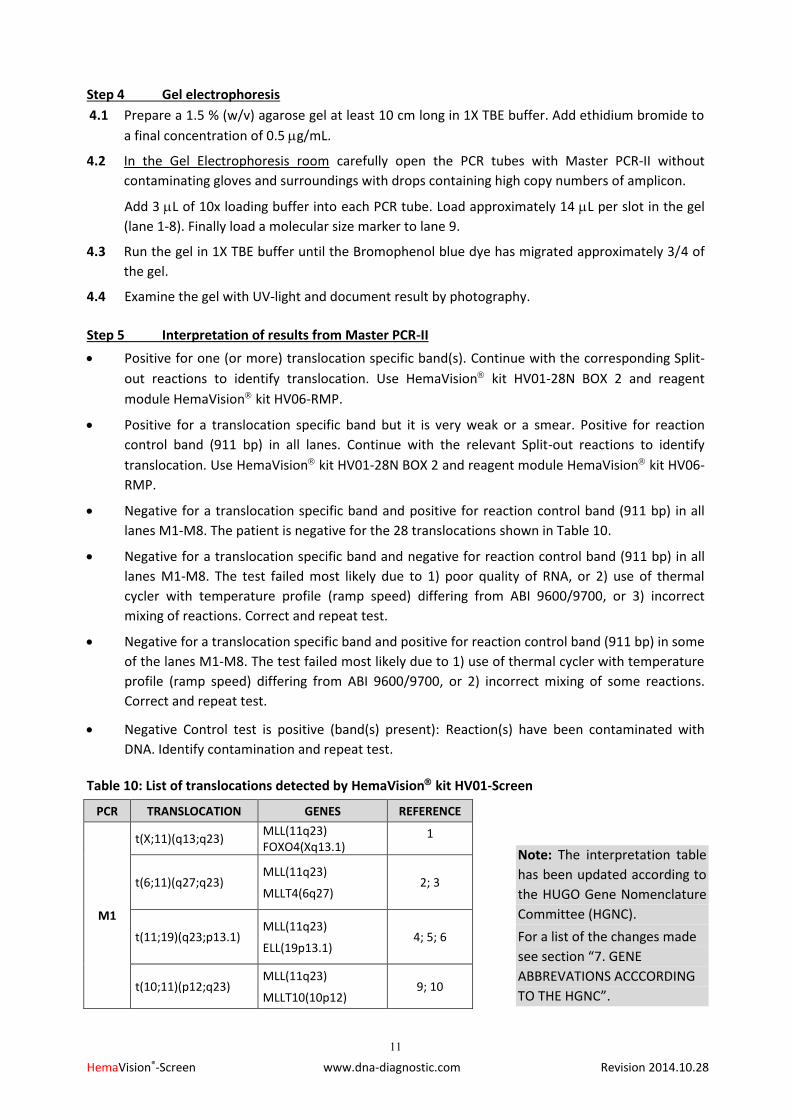
Step 5 Interpretation of results from Master PCR-II
Positive for one (or more) translocation specific band(s). Continue with the corresponding Split-
out reactions to identify translocation. Use HemaVision kit HV01-28N BOX 2 and reagent
module HemaVision kit HV06-RMP.
Positive for a translocation specific band but it is very weak or a smear. Positive for reaction
control band (911 bp) in all lanes. Continue with the relevant Split-out reactions to identify
translocation. Use HemaVision kit HV01-28N BOX 2 and reagent module HemaVision kit HV06-
RMP.
Negative for a translocation specific band and positive for reaction control band (911 bp) in all
lanes M1-M8. The patient is negative for the 28 translocations shown in Table 10.
Negative for a translocation specific band and negative for reaction control band (911 bp) in all
lanes M1-M8. The test failed most likely due to 1) poor quality of RNA, or 2) use of thermal
cycler with temperature profile (ramp speed) differing from ABI 9600/9700, or 3) incorrect
mixing of reactions. Correct and repeat test.
Negative for a translocation specific band and positive for reaction control band (911 bp) in some
of the lanes M1-M8. The test failed most likely due to 1) use of thermal cycler with temperature
profile (ramp speed) differing from ABI 9600/9700, or 2) incorrect mixing of some reactions.
Correct and repeat test.
Negative Control test is positive (band(s) present): Reaction(s) have been contaminated with
DNA. Identify contamination and repeat test.
Table 10: List of translocations detected by HemaVision kit HV01-Screen
PCR TRANSLOCATION GENES REFERENCE
M1
t(X;11)(q13;q23) MLL(11q23) FOXO4(Xq13.1)
1
t(6;11)(q27;q23) MLL(11q23)
MLLT4(6q27) 2; 3
t(11;19)(q23;p13.1) MLL(11q23)
ELL(19p13.1) 4; 5; 6
t(10;11)(p12;q23) MLL(11q23)
MLLT10(10p12) 9; 10
Note: The interpretation table
has been updated according to
the HUGO Gene Nomenclature
Committee (HGNC).
For a list of the changes made
see section “7. GENE
ABBREVATIONS ACCCORDING
TO THE HGNC”.

12
HemaVision®-Screen www.dna-diagnostic.com Revision 2014.10.28
PCR TRANSLOCATION GENES REFERENCE
M2
t(1;11)(p32;q23) MLL(11q23) EPS15 (1p32)
7
t(11;17)(q23;q12-21) MLL(11q23) MLLT6(17q21)
8
t(11;19)(q23;p13.3) MLL(11q23)
MLLT1(19p13.3) 30; 32; 34
t(10;11)(p12;q23) MLL(11q23)
MLLT10(10p12) 9; 10; 12
t(9;11)(p22;q23) MLL(11q23)
MLLT3(9p22) 3; 30; 35
PCR TRANSLOCATION GENES REFERENCE
M3
t(1;19)(q23;p13) TCF3(19p13.3) PBX1(PRL)(1q23.3)
13; 14; 15
t(17;19)(q22;p13) TCF3(19p13.3)
HLF(17q22) 16; 17; 18
t(12;21)(p13;q22) ETV6(12p13) RUNX1(21q22.3)
19; 20; 21; 55
TAL1d 40kbp deletion 1p32
STIL(1p32) TAL1(1p32)
22; 23
PCR TRANSLOCATION GENES REFERENCE
M4
t(8;21)(q22;q22) RUNX1(21q22.3)
RUNX1T1(8q22) 24; 25; 26
t(3;21)(q26;q22) RUNX1(21q22.3)
MDS1-EVI1(3q26) 25; 27; 28
t(16;21)(p11;q22) FUS(16p11.2)
ERG(21q22.3) 29; 57
t(15;17)(q24;q21) PML(15q24)
RARA(17q21)
50; 51;
52
PCR TRANSLOCATION GENES REFERENCE
M5
t(4;11)(q21;q23) MLL(11q23)
AFF1(4q21.3) 30; 31; 32; 33
t(10;11)(p12;q23) MLL(11q23)
MLLT10(10p12) 9; 10; 11
t(11;19)(q23;p13.3) MLL(11q23)
MLLT1(19p13.3) 30; 34
t(9;11)(p22;q23) MLL(11q23)
MLLT3(9p22) 3; 30; 35
t(1;11)(q21;q23) MLL(11q23)
MLLT11(1q21) 36

13
HemaVision®-Screen www.dna-diagnostic.com Revision 2014.10.28
PCR TRANSLOCATION GENES REFERENCE
M6
inv(16)(p13;q22) CBFB (16q22.1)
MYH11 (16p13.11) 47
t(9;22)(q34;q11) BCR(22q11)
ABL1(9q34.1) 37; 38; 39
t(9;12)(q34;p13) ETV6(12p13)
ABL1(9q34.1) 40; 41
t(5;12)(q33;p13) ETV6(12p13)
PDGFRB(5q33) 42
t(12;22)(p13;q11-12) ETV6(12p13)
MN1(22q12.1) 43
PCR TRANSLOCATION GENES REFERENCE
M7
t(6;9)(p23;q34) DEK(6p23)
NUP214(9q34) 44
t(9;9)(q34;q34) SET(9q34)
NUP214(9q34) 45; 46
inv(16)(p13;q22) CBFB (16q22.1)
MYH11 (16p13.11) 47
t(3;21)(q26;q22)
or
-------------------------
t(1;21)(p36;q22)
RUNX1(21q22.3)
RPL22P1(pseudogene) (3q26.2)
-------------------------
RUNX1(21q22.3)
RPL22 (1p36.3)
25; 27; 28; 56
PCR TRANSLOCATION GENES REFERENCE
M8
t(11;17)(q23;q12) ZBTB16(11q23)
RARA(17q12) 48; 49
t(3;21)(q26;q22) RUNX1(21q22.3)
EVI1(3q26) 25; 27; 28
t(15;17)(q24;q21) PML(15q24)
RARA(17q21)
50; 51;
52
t(5;17)(q35;q12) NPM1(5q35)
RARA(17q12) 53
t(3;5)(q25.1;q35) NPM1(5q35)
MLF1(3q25.1) 54
t(9;22)(q34;q11) BCR(22q11)
ABL1(9q34.1) 37; 38; 39
14
HemaVision®-Screen www.dna-diagnostic.com Revision 2014.10.28
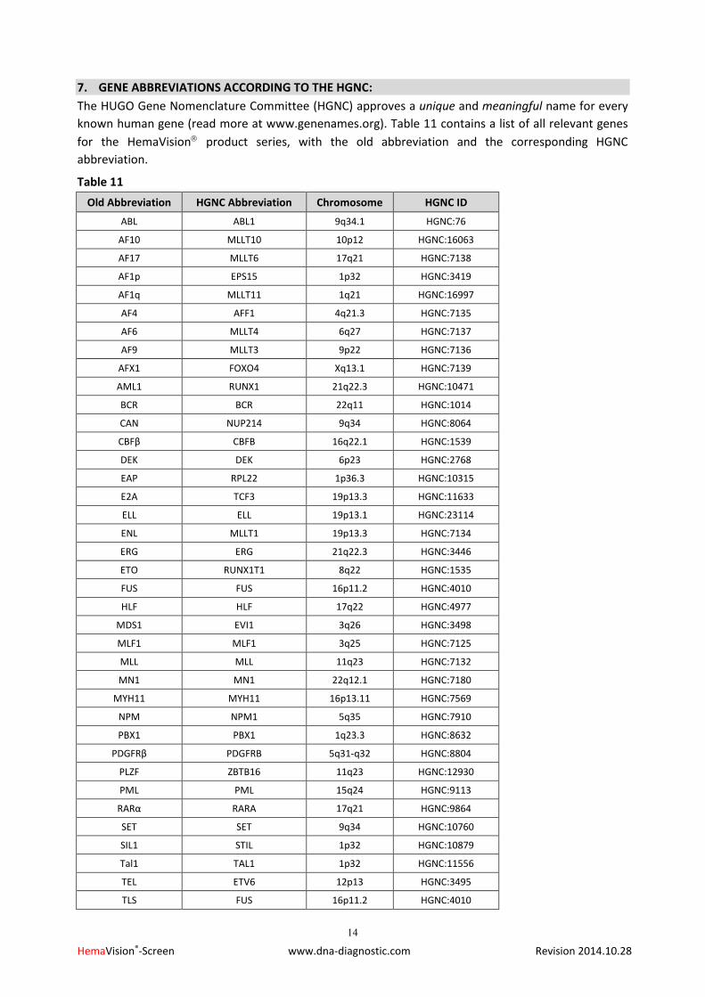
7. GENE ABBREVIATIONS ACCORDING TO THE HGNC:
The HUGO Gene Nomenclature Committee (HGNC) approves a unique and meaningful name for every
known human gene (read more at www.genenames.org). Table 11 contains a list of all relevant genes
for the HemaVision product series, with the old abbreviation and the corresponding HGNC
abbreviation.
Table 11
Old Abbreviation HGNC Abbreviation Chromosome HGNC ID
ABL ABL1 9q34.1 HGNC:76
AF10 MLLT10 10p12 HGNC:16063
AF17 MLLT6 17q21 HGNC:7138
AF1p EPS15 1p32 HGNC:3419
AF1q MLLT11 1q21 HGNC:16997
AF4 AFF1 4q21.3 HGNC:7135
AF6 MLLT4 6q27 HGNC:7137
AF9 MLLT3 9p22 HGNC:7136
AFX1 FOXO4 Xq13.1 HGNC:7139
AML1 RUNX1 21q22.3 HGNC:10471
BCR BCR 22q11 HGNC:1014
CAN NUP214 9q34 HGNC:8064
CBFβ CBFB 16q22.1 HGNC:1539
DEK DEK 6p23 HGNC:2768
EAP RPL22 1p36.3 HGNC:10315
E2A TCF3 19p13.3 HGNC:11633
ELL ELL 19p13.1 HGNC:23114
ENL MLLT1 19p13.3 HGNC:7134
ERG ERG 21q22.3 HGNC:3446
ETO RUNX1T1 8q22 HGNC:1535
FUS FUS 16p11.2 HGNC:4010
HLF HLF 17q22 HGNC:4977
MDS1 EVI1 3q26 HGNC:3498
MLF1 MLF1 3q25 HGNC:7125
MLL MLL 11q23 HGNC:7132
MN1 MN1 22q12.1 HGNC:7180
MYH11 MYH11 16p13.11 HGNC:7569
NPM NPM1 5q35 HGNC:7910
PBX1 PBX1 1q23.3 HGNC:8632
PDGFRβ PDGFRB 5q31-q32 HGNC:8804
PLZF ZBTB16 11q23 HGNC:12930
PML PML 15q24 HGNC:9113
RARα RARA 17q21 HGNC:9864
SET SET 9q34 HGNC:10760
SIL1 STIL 1p32 HGNC:10879
Tal1 TAL1 1p32 HGNC:11556
TEL ETV6 12p13 HGNC:3495
TLS FUS 16p11.2 HGNC:4010
15
HemaVision®-Screen www.dna-diagnostic.com Revision 2014.10.28
REFERENCES:
1. Parry P., Wei Y., Evans G.: Cloning and characterization of the t(X;11) breakpoint from a leukemic cell line identify a new member of the forkhead family. Genes Chromosomes Cancer. 11: 79, 1994.
2. Prasad R., Gu Y., Alderr, H., Nakamura, T., Canaani O., Saito H., Huebner K., Gale R.P.,Nowell P.C., Kuryiama K et al. Cloning of the All-1 fusion partner, The AF-6 gene, involved in acute myeloid leukemias with the t(6;11) chromosome translocation. Cancer Res. 53: 5624, 1993.
3. Poirel H., Rack K., Delabesse E., Radford Weiss I., Troussard X., Debert C., Leboeuf D., bastard C., Pichard F., Veil Buzyn A., Flandrin G., Bernard O., and Macintyre E.: Incidence and characterization of MLL gene (11q23) rearrangements in acute myeloid leukemia M1 and M5. Blood 87: 2496, 1996.
4. 4: Thirman MJ., Levitan DA., Kobayashi H., Simon MC., and Rowley JD.: Cloning of ELL, a gene that fuses to MLL in a t(11;19)(q23;p13.1) in acute myeloid leukemia. Proc. Natl. Acad. Sci., USA 91: 12110, 1994.
5. Mitani K., Kanda Y., Ogawa S., Tanaka T., Inazawa J., Yazaki Y., and Hirai H.: Cloning of several species of MLL/MEN chimeric cDNAs in myeloid leukemia with t(11;19)(q23;p13.1) translocation. Blood 85: 2017, 1995.
6. Rubnitz JE., Behm FG., Curcio Brint AM., Pinheiro RP., Carroll AJ., Raimondi SC., Shurtleff SA., and Downing JR.: Molecular analysis of t(11;19) breakpoints in childhood acute leukemias. Blood 87: 4804, 1996.
7. Bernard OA., Mauchauffe M., Mecucci C., Van den Berghe H., and Berger R.: A novel gene, AF-1p, fused to HRX in t(1;11)(p32;q23), is not related to AF-4, AF-9 nor ENL. Oncogene 9: 1039, 1994.
8. Prsasd R., Leshkowitz D., Gu Y., Alder H., Nakamura T., Saito H., Huebner K., Berger R., Croce CM., and Canaani E.: Leucine-zipper dimerization motif encoded by the AF17 gene fused to ALL-1 (MLL) in acute leukemia. . Proc. Natl. Acad. Sci., USA 91: 8107, 1994.
9. Chaplin T., Ayton P., Bernard OA., Saha V., Della Valle V., Hillion J., Gregorini A., Lillington D., Berger R., and Young BD.: A novel class of zinc finger/leucine zipper genes identified from the molecular cloning of the t(10;11) translocation in acute leukemia. Blood 85: 1435, 1995.
10. Chaplin T., Bernard O., Beverloo HB., Saha V., Hagemeijer A., Berger R., and Young BD.: The t(10;11) translocation in acute myeloid leukemia (M5) consistently fuses the leucine zipper motif of AF10 onto the HRX gene. Blood 86: 2073, 1995.
11. Hjorth-Sørensen B., Pallisgaard N., Grønholm M., Hokland P., Clausen N., and Jørgensen P.: A novel MLL-AF10 fusion mRNA variant in a patient with acute myeloid leukemia detected by a new asymmetric reverse transcription PCR method. Leukemia 11: 1588, 1997.
12. Borkhardt A., Haas OA., Strobl W., Repp R., Mann G., Gadner H., and Lampert F.: A novel type of MLL/AF10 fusion transcript in a child with acute megakaryocytic leukemia (AML-M7). Leukemia 9: 1796, 1995.
13. Kamps MP., Murre C., Sun XH., and Baltimore D.: A new homeobox gene contributes the DNA binding domain of the t(1;19) translocation protein in pre-B ALL. Cell 60: 547, 1990.
14. Nourse J., Mellentin JD., Galili N., Wilkinson J., Stanbridge E., Smith SD., and Cleary ML.: Chromosomal translocation t(1;19) results in synthesis of a homeobox fusion mRNA that codes for a potential chimeric transcription factor. Cell 60: 535, 1990.
15. Izraeli S., Kovar H., Gadner H., and Lion T.: Unexpected heterogeneity in E2A/PBX1 fusion messenger RNA detected by the polymerase chain reaction in pediatric patients with acute lymphoblastic leukemia. Blood 80: 1413, 1992.
16. 16: Hunger SP., Ohyashiki K., Toyama K., and Cleary ML.: Hlf, a novel hepatic bZIP protein, shows altered DNA-binding properties following fusion to E2A in t(17;19) acute lymphoblastic leukemia. Genes Dev. 6: 1608, 1992.
17. Inaba T., Roberts WM., Shapiro LH., Jolly KW., Raimondi SC., Smith SD., and Look AT.: Fusion of the leucine zipper gene HLF to the E2A gene in human acute B-lineage leukemia. Science 257: 531, 1992.
18. Hunger SP., Devaraj PE., Foroni L., Secker Walker LM., and Cleary ML.: Two types of genomic rearrangements create alternative E2A-HLF fusion proteins in t(17;19)-ALL. Blood 83: 2970, 1994.
19. Romana SP., Poirel H., Le Coniat M., Flexor MA., Mauchauffe M., Jonveaux P., Macintyre EA., Berger R., and Bernard OA.: High frequency of t(12;21) in childhood B-lineage acute lymphoblastic leukemia. Blood 86: 4263, 1995.
20. Romana SP., Mauchauffe M., Le Coniat M., Chumakov I., Le Paslier D., Berger R., and Bernard OA.: The t(12;21) of acute lymphoblastic leukemia results in a tel-AML1 gene fusion. Blood 85: 3662, 1995.
16
HemaVision®-Screen www.dna-diagnostic.com Revision 2014.10.28
21. Golub TR., Barker GF., Bohlander SK., Hiebert SW., Ward DC., Bray Ward P., Morgan E., Raimondi SC., Rowley JD., and Gilliland DG.: Fusion of the TEL gene on 12P13 to the AML1 gene on 21q22 in acute lymphoblastic leukemia. Proc. Natl. Acad. Sci., USA 92: 4917, 1995.
22. Bash RO., Crist WM., Shuster JJ., Link MP., Amylon M., Pullen J., Carroll AJ., Buchanan GR., Smith RG., and Baer R.: Clinical features and outcome of T-cell acute lymphoblastic leukemia in childhood with respect to alterations at the TAL1 locus: a Pediatric Oncology Group stude. Blood 81: 2110, 1993.
23. Aplan PD., Lombardi DP., Ginsberg AM., Cossman J., Bertness VL., and Kirsch IR.: Disruption of the human SCL locus by ”illegitimate” V-(D)-J recombinase activity. Science 250: 1426, 1990.
24. Miyoshi H., Kozu T., Shimizu K., Enomoto K., Maseki N., Kaneko Y., Kamada N., and Ohki M.: The t(8;21) translocation in acute myeloid leukemia results in production of an AML1-MTG8 fusion transcript. EMBO J. 12: 2715, 1993.
25. Nucifora G., and Rowley JD.: AML1 and the 8;21 and 3;21 translocations in acute and chronic myeloid leukemia. Blood 86: 1, 1995.
26. Downing JR., Head DR., Curcio Brint AM., Hulshof MG., Motroni TA., Raimondi SC., Carroll AJ., Drabkin HA., Willman C., and Theil KS.: An AML1/ETO fusion transcript is consistently detected by RNA-based polymerase chain reaction in acute myelogenous leukemia containing the (8;21)(q22;q22) translocation. Blood 81: 2860, 1993.
27. Mitani K., Ogawa S., Tanaka T., Miyoshi H., Kurokawa M., Mano H., Yazaki Y., Ohki M., and Hirai H.: Generation of the AML1-EVI-1 fusion gene in the t(3;21)(q26;q22) causes blastic crisis in chronic myelocytic leukemia. EMBO J. 13: 504, 1994.
28. Nucifora G., Begy CR., Kobayashi H., Roulston D., Claxton D., Pedersen-Bjergaard J., Parganas E., Ihle JN., and Rowley JD.: Consistent intergenic splicing and production of multiple transcripts between AML1 at 21q22 and unrelated genes at 3q26 in (3;21)(q26;q22) translocations. Proc. Natl. Acad. Sci., USA 91: 4004, 1994.
29. Ichikawa H., Shimizu K., Hayashi Y., and Ohki M.: An RNA-binding protein gene, TLS/FUS, is fused to ERG in human myeloid leukemia with t(16;21) chromosomal translocation. Cancer Res. 54: 2865, 1994.
30. Yamamoto K., Seto M., Iida S., Komatsu H., Kamada N., Kojima S., Kodera Y., Nakazawa S., Saito H., and Takahashi T.: A reverse transcriptase-polymerase chain reaction detects heterogenous chimeric mRNAs in leukemias with 11q23 abnormalities. Blood 83: 2912, 1994.
31. Biondi A., Rambaldi A., Rossi V., Elia L., Caslini C., Basso G., Battista R., Barnui T., Mandelli F., and Masera G.: Detection of ALL-1/AF4 fusion transcript by reverse transcription-polymerase chain reaction for diagnosis and monitoring of acute leukemias with the t(4;11) translocation. Blood 82: 2943, 1993.
32. Corral J., Forster A., Thompson S., Lampert F., Kaneko Y., Slater R., Kroes WG., van der Schoot CE., Ludwig WD., and Karpas A.: Acute leukemias of different lineages have similar MLL gene fusions encoding related chimeric proteins resulting from chromosomal translocation. Proc. Natl. Acad. Sci., USA 90: 8538, 1993.
33. Downing JR., Head DR., Raimondi SC., Carrol AJ., Curcio Brint AM., Motroni TA., Hulshof MG., Pullen DJ., and Domer PH.: The der(11)-encoded MLL/AF-4 fusion transcript is consistently detected in t(4;11)(q21;q23)-containing acute lymphoblastic leukemia. Blood 83: 330, 1994.
34. Yamamoto K., Seto M., Komatsu H., Iida S., Akao Y., Kojima S., Kodera Y., Nakazawa S., Ariyoshi Y., Takahashi T., and Ueda R.: Two distinct portions of LTG19/ENL at 19p13 are involved in t(11;19) leukemia. Oncogene 8: 2617, 1993.
35. Nakamura T., Alder H., Gu Y., Prasad R., Canaani O., Kamada N., Gale R.P., Lange B., Crist W.M., and Nowell P.C.: Genes on chromosome 4, 9, and 19 involved in 11q23 abnormalities in acute leukemia share sequence homology and/or common motifs. Proc. Natl. Acad. Sci., USA 90: 4631, 1993.
36. Tse W., Zhu W., Chen H.S., and Cohen A.: A novel gene, AF1q, fused to MLL in t(1;11)(q21;q23), is specifically expressed in leukemic and immature hematopoietic cells. Blood 85: 650, 1995.
37. Hermans A., Heisterkamp N., von Linden M., van Baal S., Meijer D., van der Plas D., Wiedermann L.M., Groffen J., Bootsma D., and Grosveld G.: Unique fusion of bcr and c-abl genes in Philadelphia chromosome positive acute lymphoblastic leukemia. Cell 51: 33, 1987.
38. Cross N.C., Melo J.V., Feng L., and Goldman J.M.: An optimized multiplex polymerase chain reaction (PCR) for detection of BCR-ABL fusion mRNAs in haematological disorders. Leukemia 8: 186, 1994.
39. Hochhaus, A., Reiter A., Skladny H., Melo J.V., Sick C., Berger U., Guo J.Q., Arlinghaus R.B., Hehlmann R., Goldman J.M., and Cross N.C.P.: A novel BCR-ABL fusion gene (e6a2) in a patient with Philadelphia chromosome-negative chronic myelogenous leukemia. Blood 88: 2236, 1996.
17
HemaVision®-Screen www.dna-diagnostic.com Revision 2014.10.28
40. Papadopoulos P., Ridge S.A., Boucher C.A., Stocking C., and Wiedemann L.M.: The novel activation of ABL by fusion to an ets-related gene, TEL. Cancer Res. 55: 34, 1995.
41. Andreasson P., Johansson B., Carlsson M., Jarlsfelt I., Fioretos T., Mitelman F., and Höglund M.: BCR/ABL-negative chronic myeloid leukemia with ETV6/ABL fusion. Genes, Chromosomes & Cancer 20: 299, 1997.
42. Golub T.R., Barker G.F., Lovett M., and Gilliland D.G.: Fusion of PDGF receptor beta to a novel ets-like gene, tel, in chronic myelomonocytic leukemia with t(5;12) chromosomal translocation. Cell 77: 307, 1994.
43. Buijs A., Sherr S., van Baal S., van Bezouw S., van der Plas D., Geurts van Kessel A., Riegman P., Lekanne Deprez R., Zwarthoff E., Hagemeijer A., and Grosveld G.: Translocation (12;22)(p13;q11) in myeloproliferative disorders results in fusion of the ETS-like TEL gene on 12p13 to the MN1 gene on 22q11. Oncogene 10: 1511, 1995.
44. von Lindern M., Fornerod M., van Baal S., Jaegle M., de Wit T., Buijs A., and Grosveld G.: The translocation (6;9), associated with a specific subtype of acute myeloid leukemia, results in the fusion of two genes, dec and can, and the expression of a chimeric, leukemia-specific dek-can mRNA. Mol. Cell. Biol. 12: 1687, 1992.
45. von Lindern M., van Baal S., Wiegant j., Raap A., Hagemeijer A., and Grosveld G.: Can, a putative oncogene associated with myeloid leukomogenesis, may be activated by fusion of its 3’half to different genes: characterization of the set gene. Mol. Cell. Biol. 12: 3346, 1992.
46. Nagata K., Kawase H., Handa H., Yano K., Yamasaki M., Ishimi Y., Okuda A., Kikuchi A., and Matsumoto K.: Replication factor encoded by a putative oncogene, set, associated with myeloid leukomogenesis. Proc. Natl. Acad. Sci., USA 92: 4279, 1995.
47. Liu P.P., Hajra A., Wijmenga C., and Collins F.S.: Molecular pathogenesis of the chromosome 16 inversion in the M4Eo subtype of acute myeloid leukemia. Blood 85: 2289, 1995.
48. Chen Z., Brand N.J., Chen A., Chen S.J., Tong J.H., Wang Z.Y., Waxman S., and Zelent A.: Fusion between a novel Kruppel-like zinc finger gene and the retinoic acid receptor-alpha locus due to a varient t(11;17) translocation associated with acute promyelocytic leukemia. EMBO J. 12: 1161, 1993.
49. Licht J.D., Chomienne C., Goy A., Chen A., Scott A.A., Head D.R., Michaux J.L., Wu Y., DeBlasio A., Miller W.H.Jr.: Clinical and molecular characterization of a rare syndrome of acute promyelocytic leukemia associated with translocation (11;17). Blood 85: 1083, 1995.
50. Pandolfi P.P., Alcalay M., Fagioli M., Zangrilli D., Mencarelli A., Diverio D., Biondi A., Lo Coco F., Rambaldi A., and Grignani F.: Genimic variability and alternative splicing generate multiple PML/RAR alpha transcripts that encode aberrant PML proteins and PML/RAR alpha isoforms in acute promyelocytic leukaemia. EMBO J. 11: 1397, 1992.
51. 51: Yoshida H., Naoe T., Fukutani H., Kiyoi H., Kubo K., and Ohno R.: Analysis of the joining sequences of the t(15;17) translocation in human acute promyelocytic leukemia: Sequence non-specific recombination between the PML and RARA genes within identical short stretches. Genes, Chromosomes & Cancer 12: 37, 1995.
52. Kane J.R., Head D.R., Balazs L., Hulshof M.G., Motroni T.A., Raimondi S.C. Carroll A.J., Behm F.G., Krance R.A., Shurtleff S.A., and Downing J.R.: Molecular analysis of the PML/RARalpha chimeric gene in pediatric acute promyelocytic leukemia. Leukemia 10: 1296, 1996.
53. Redner R.L., Rush E.A., Faas S., Rudert W.A. and Corey S.J.: The t(5;17) variant of acute promyelocytic leukemia expresses a nucleophosmin-retinoic acid receptor fusion. Blood 87: 882, 1996.
54. Yoneda Kato N., Look A.T., Kirstein M.N., Valentine M.B., Raimondi S.C., Cohen K.J., Carroll A.J., and Morris S.W.: The t(3;5)(q25.1;q34) of myelodysplastic syndrome and acute myeloid leukemia produces a novel fusion gene, NPM-MLF1. Oncogene 12: 265, 1996.
55. 55: Satake N., Kobayashi H., Tsunematsu Y., Kawasaki H., Horikoshi Y., Koizumi S. and Kaneko Y.: Minimal residual disease with TEL-AML1 fusion transcript in childhood acute lymphoblastic leukaemia with t(12,21). British J. of Haematology 97: 607, 1997.
56. Sacchi N., Nisson P.E., Watkins P.C., Faustinella F., Wijsman J. and Hagemeijer A.: AML1 fusion transcripts in t(3,21) positive leukemia: evidence of molecular heterogeneity and usage of splicing sites frequently involved in the generation of normal AML1 transcripts. Genes, Chromosomes & Cancer 11: 226, 1994.
57. Kong X-T., Ida K., Ichikawa H., Shimizu K., Ohki M., Maseki N., Kaneko Y., Sako M., Kobayashi Y., Tojou A., Miura I., Kakuda H., Funabiki T., Horibe K., Hamaguchi H., Akiyama Y., Bessho F., Yanagisawa M. and Hayashi Y.: Consistent detection of TLS/FUS-ERG chimeric transcripts in acute myeloid leukemia with t(16,21)(p11,q22) and identification of a novel transcript. Blood 90: 1192, 1997.
18
HemaVision®-Screen www.dna-diagnostic.com Revision 2014.10.28
58. Pallisgaard N., Hokland P., Riishøj D.C., Pedersen B. and Jørgensen P.: Multiplex reverse transcription-polymerase chain reaction for simultaneous screening of 29 translocations and chromosomal aberrations in Acute Leukemia. Blood 92: 574, 1998.
59. Kim HJ., Oh HJ., Lee JW, Jang PS., Chung NG., Kim M., Lim J., Cho B., Kim HK.: Utility of a multiplex reverse transcriptase-polymerase chain reaction assay (HemaVision) in the evaluation of genetic abnormalities in Korean children with acute leukemia: a single institution study. Korean J Pediatr. 2013 Jun;56(6):247-53.
60. Song MJ., Kim HJ., Park CH., Kim SK., Ki CS., Kim JW., Kim SH.: Diagnostic utility of a multiplex RT-PCR assay in detecting fusion transcripts from recurrent genetic abnormalities of acute leukemia by WHO 2008 classification. Diagn mol pathol. 2012 Mar;21(1):40-44.
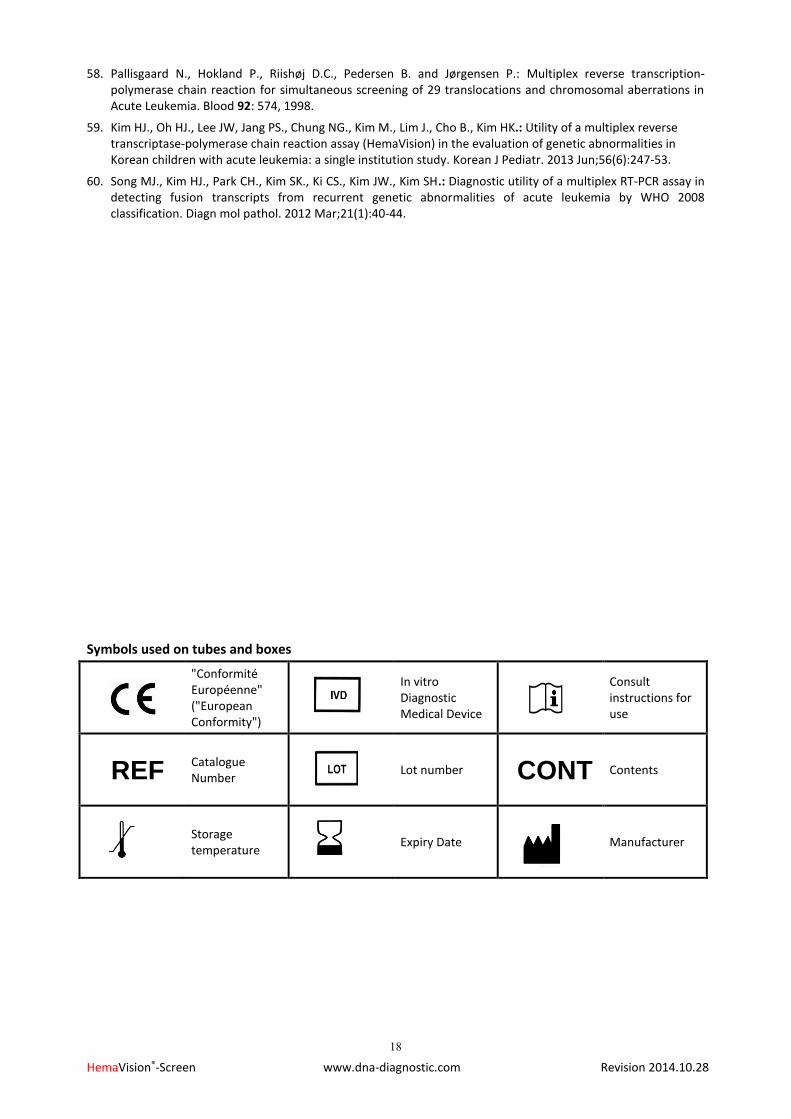
Symbols used on tubes and boxes
"Conformité Européenne" ("European Conformity")
In vitro Diagnostic Medical Device
Consult instructions for use
REF Catalogue Number
Lot number CONT Contents
Storage temperature
Expiry Date Manufacturer
Availability / questions Our team and distributors are always at hand to answer all your questions.
Contact us to find your nearest HemaVision partner. For more information, contact DNA Diagnostic A/S Voldbjergvej 16 Tel. +45 8732 3050 DK-8240 Risskov [email protected] Denmark www.dna-diagnostic.com DNA Diagnostic A/S (previously named DNA Technology A/S) was established in 1992. DNA Diagnostic A/S is an ISO 13485:2012 certified developer, manufacturer, and worldwide supplier of PCR based CE IVD marked in vitro diagnostic kits.