multiple forms of chicken a3(vi) collagen chain generated
TRANSCRIPT

Multiple Forms of Chicken a3(VI) Collagen Chain Generated by Alternative Splicing in Type A Repeated Domains Roberto Doliana, Paolo Bonaldo, and Alfonso Colomba t t i
Divisione di Oncologia Sperimentale 2 Centro Di Riferimento Oncologico, 33081 Aviano, Italy
Abstract. Type VI collagen is a structurally unique component widely distributed in connective tissues. Its molecular structure consists of monomers that have the potential to assemble intracellularly into dimers and tetramers which, once secreted, can form microfilaments by end-to-end association. Individual monomers are composed of chains of Mr = ~140,000 (cd and a2) and >300,000 (or3). Type VI collagen molecules contain a short triple helix with large globular domains at both ends. These domains are made for their greatest part of repetitive units similar to type A repeats of von Willebrand Factor. The ot 3(VI) chain, contributing most of the mass of the NH~-terminal globule, appeared heterogenous both at the mRNA and protein level. Several ot3(VI)-specific clones that lack the sequences corresponding to repeats A8 and A6 were isolated from a chicken aorta cDNA library. Northern blot hybridization of poly (A+)-enriched RNA from chicken gizzard with cDNA fragments corresponding to several individual type A repeats showed that A8- and A6-specific probes
did not hybridize to the lower Mr transcripts. Clones spanning ~o20 kb of the 5'-end of the ot 3(VI) gene were isolated from a chicken genomic library and sub- jected to analysis by restriction mapping, Southern blotting, and selective sequencing of the intron-exon boundaries. At the most 5'-end of the gene an addi- tional type A repeat (A9), previously undetected in eDNA clones, was identified. Furthermore, it was de- termined that the presumed signal peptide and repeats A9 through A6 are encoded within individual exons. Reverse transcription and polymerase chain reaction of aorta RNA suggested that a mechanism of alternative mRNA splicing by a phenomenon of exon skipping generates tx3(VI) isoform variants that contain differ- ent numbers of type A repeats. Immunohistochemistry of frozen sections of chicken embryo tissues with repeat-specific mAbs showed that an antibody directed against a conditional exon has a more restricted tissue distribution compared to an antibody against a consti- tutive exon.
T YPE VI, one of the major collagens of connective tis- sues, is a component of 100-rim-long periodic micro- filaments that are found at the surface of cells and
around or between collagen fibers (von der Mark et al., 1984; Bruns, 1984; Bruns et al., 1986; Keene et al., 1988). The widespread occurrence of these thin fibrils in embryo (Bruns et al., 1986) and adult tissues (vonder Mark et al., 1984; Keene et al., 1988) and the diversity in localization, ranging from cartilage to soft tissues (Burgeson, 1988), are characteristic features of this collagen. The molecular mech- anisms of microfilament formation are presently unknown but electron microscopic (Furthmayr et al., 1983) and bio- synthetic studies (Engvail et al., 1986; Colombatti et ai., 1987; Colombatti and Bonaldo, 1987) have provided evi- dence that the polymerization process takes place intracellu- larly soon after synthesis and leads to the formation of disulfide-bonded dimers and tetramers. Furthermore, the in- dividual chains do not seem to undergo proteolytic process- ing with removal of the large N- and C- propeptides that do not represent precursor structures. The tetramers associate
extracellularly by end-to-end to form the oligomeric microfilaments (Furthmayr et al., 1983).
Recently, we (Bonaldo et al., 1989, 1990) and others (Koller et al., 1989; Chu et al., 1989) provided evidence that a major portion of the constituent chains of chicken and hu- man type VI collagen consists of repeating units of ~ 200 residues that are closely related to the type A repeats of von Willebrand Factor (Shelton-Inloes et al., 1986). The most distinctive feature that emerged from the analysis of these se- quences was the finding that ,,085 % of the or3 (VI) chain is represented by two types of similar repeating motifs, desig- nated domains A and #/(Bonaido et ai., 1990). In a previous study Engvall et al. (1986) described the heterogeneity of the a3 (VI) chain present as three or more closely spaced bands in SDS-PAGE. The possibility was put forward that this het- erogeneity was the consequence of posttranslational events. Similar discrete bands were detected by us after a short 7-min pulse (Colombatti et ai., 1987) and even after immu- noprecipitation of tunicamycin and ot,ot'-dipyridyl-treated chicken embryo cells (Colombatti and Bonaldo, 1987).
© The Rockefeller University Press. 0021-9525/90/11/2197/9 $2.00 The Journal of Cell Biology. Volume 111, November 1990 2197-2205 2197
Dow
nloaded from http://rupress.org/jcb/article-pdf/111/5/2197/1060375/2197.pdf by guest on 13 January 2022

Moreover, by hybridization of mRNA obtained from human cell lines with a3 (VI)-specific eDNA probes multiple mes- sages were detected (Chu et al., 1987). It appears more likely then that alternative splicing of mRNA is generating protein diversity through multiple forms of a3 (VI) tran- scripts.
We report here that alternative splicing in the chicken ct3 (VI) gene generates several mRNAs that differ by one or more type A repeated domains. As a result of this mecha- nism different a3(VI) polypeptides are produced that have a specific tissue distribution and may be important in tissue- specific functions.
Materials and Methods
Isolation of cDNA Clones The construction of a chicken aorta cDNA library in the expression vector pEX1 (Bressan et al., 1987) and the isolation of several cDNA clones en- coding the ¢x3(VI) chain have previously been described (Bonaldo and Colombatti, 1989; Bonaldo et al., 1990). A 538-bp-long Pst I restriction fragment from the most 5'-end clone pB10 was purified, nick translated to a specific activity of 7 x 10⁵ cpm/ng, and used to rescreen the eDNA li- brary.
Northern Blotting Total RNA and poly(A+)-enriched RNA were prepared from chicken giz- zard using standard procedures (Maniatis et al., 1982). Electrophoresis of the RNA was performed on 0.7% (wt/vol) agarose gel containing 2.3 M formaldehyde in MOPS buffer for 8 h at 150 V using 20-cm-long plates. RNA was then transferred onto nitrocellulose filters and hybridized with [a- 32p] CTP-labeled cDNA probes derived from clone pB10 and specific for different type A repeats (see Figs. 1 and 2).
The filters were hybridized at 68°C overnight in 6× SSC and 10x Den- hardt's solution. After washing in 0.2x SSC and 0.1% SDS at room temper- ature the filters were exposed to B-max Hyperfilms (Amersham Interna- tional, Amersham, UK).
Isolation of Genomic Clones A chicken genomic library in EMBL-3 (Clontech Laboratories, Inc., Palo Alto, CA) was plated and the plaques transferred to nitrocellulose filters. The filters were hybridized with synthetic oligonueleotide probes prepared in a DNA synthesizer (Applied Biosystems, Inc., Foster City, CA) and the 5'-end was labeled with [3,-3ZP]ATP (Amersham International) and "1"4 polynucleotide kinase (Boehringer Mannheim, GmbH, FRG). The syn- thetic oligonucleotides were derived from the eDNA sequences encoding the u3(VI) signal peptide sequence and the repeat A8 (see Fig. 4). Four clones were isolated and one (k gen 5) was further studied and is reported here.
Restriction Enzyme Mapping and DNA Sequence Analysis Plasmid DNA and lambda phage DNA were isolated by standard proce- dures (Maniatis et al., 1982). Restriction enzyme digestions were per- formed as described by the manufacturers. Phage DNA fragments were separated by electrophoresis on 0.7 % agarose gels, transferred to nitrocellu- lose, and hybridized with synthetic oligonucleotides specific for the c~3(VI) eDNA clone pBlO. Positive fragments were subeloned into the M13-derived vectors, mpl8 and mpl9 (Messing, 1983), and the nucteotide sequence was obtained by the dideoxy chain termination method (Sanger et al., 1977) as modified by Biggin et al. (1983) using modified bacteriophage T7 DNA polymerase (Tabor and Richardson, 1987). Some sequences were deter- mined directly on caesium chloride-purified lambda DNA using synthetic oligonucleotide primers and Taq DNA polymerase (Promega Biotec, Madi- son, WI).
Reverse Transcription/Polymerase Chain Reaction Reverse transcription (RT)l/polymerase chain reaction (PCR) was slightly modified from the method described by Rappolee et al. (1988). Total RNA (0.8/~g) was heated at 95°C for 5 min and quickly cooled on ice. The reac- tion (20/xl of PCR buffer: 50 mM KC1, 10 mM Tris-HCl, pH 8.3, 1.5 mM MgCI2, 0.01% [wt/vol]rgelatin) contained 20 U of AMV reverse transcrip- tase (Promega Biotec), 1 mM dNTPs (each), 20 U RNasin (Promega Biotec), and 50 pmol of c~ 3(VI)-specific oligonueleotide antisense primer. The reaction mixture was incubated for 10 rain at room temperature, 60 rain at 42°C, 5-10 rain at 950C, and then chilled on ice. The resulting cDNA was amplified by using the DNA amplification reagent kit (Perkin-Elmer/ Cetus, Norwalk, CT). 2.5 U of Thermus aquaticus (Taq) polymerase and 50 pmol of c~3(VI)-specific oligonucleotide sense primer were added and the reaction was carried out through 40 cycles of amplification. Aliquots of the PCR mixture were electrophoretically separated in agarose gel and were visualized with ethidium bromide staining. The oligonucleotides used and their position within the sequence am the following: sense primers A (nu- cleotides 267-283), F (947-970), and D (2022-2045); antisense primers B (1200-1229), C (1788-1817), G (2388-2417), and E (2956-2985).
Immunoperoxidase Staining
Two c~3(VI) chain-specific mAbs were selected according to their reactivity with hybrid proteins. In brief, hybrid proteins, obtained from lysates of E. coli transformed with different cDNA clones and grown at 42°C as detailed elsewhere (Bonaldo et al., 1987), were plated onto polystyrene mi- crotiter plates, mAbs were then assayed for their binding activity for the different hybrid proteins by an ELISA type of assay. Antibody IlICI0, that recognized only the pB10 protein and mapped in the spliced repeat AS, and antibody lllA3, that mapped in a constitutive region of the ~3(VI) chain, were then selected and used for immunoperoxidase staining.
Tissues from 15-d-old chicken embryos were quickly dissected, embed- ded in OTC (Miles Laboratories Inc., Naperville, IL), and snap-frozen in liquid nitrogen. Sections (5-8 #m) were cut, air dried, and fixed for 5 min in a 1:1 acetone/chloroform solution. Specimens were rehydrated with PBS, and after incubation with normal horse serum (1:50 dilution), the sections were incubated with the primary antibody (10-20/~g/ml) for 30 min at room temperature, followed by biotin-labeled second antibody (1:200 dilution), 30 min at room temperature, and finally the avidin-biotin complex (ABC, kits PK-4001 and PK-4002; Vector Labs, Burlingame, CA) was applied for 45 rain at room temperature. Brown staining was produced by 5-rain treat- ment with 3-3'diaminobenzidine (50 nag in 100 ml of PBS, pH 7.4, contain- ing 0.01% hydrogen peroxide and 10 mM imidazole). Specimens were coun- terstained with Mayer's hematoxylin. Negative controls were performed by treating sections with an antiricin mAb.
Results
Isolation of cDNA Clones We reported previously most of the sequence of the chicken ot3(VI) chain deduced from several overlapping cDNA clones (Bonaldo and Colombatti, 1989; Bonaldo et al., 1990). The missing upstream sequences were obtained from the same library by screening with a 538-bp-long Pst I re- striction fragment of the most 5'-end clone pB10 (Bonaldo et al., 1990). Several positive clones (pB101-pBll2) were isolated, purified, and characterized by restriction enzyme analysis and DNA sequencing.
Nucleotide and Amino Acid Sequences of cDNA Clones Five clones have additional sequences that were absent from clone pBl0, whereas one clone (pBll2) overlaps over all its sequence with clone pB10 (Bonaldo et al., 1990). The addi-
1. Abbreviations used in this paper: PCR, polymerase chain reaction; RT, reverse transcription.
The Journal of Cell Biology, Volume 111, 1990 2198
Dow
nloaded from http://rupress.org/jcb/article-pdf/111/5/2197/1060375/2197.pdf by guest on 13 January 2022

[ I I I r 0 2 4 6 8
PP
10 Kb.
mRNA
BE NE A Ec
pB I0
pB211
pB 205
pB221
pB 118
pB 210
pB I f2
A8 A7 A6 A5 A4 A3 A2 A1 A'3 ~ A'2 A'I ,, protein
Figure L Diagram of chicken a3(VI) collagen cDNA clones. Schematic representation of chicken a 3(VI) cDNA clones approxi- mately to scale beginning with the most upstream clone pB221. In the diagram of the mRNA at the top a partial restriction map is shown. Solid lines indicate the nontranslated sequences. The closed box indicates the sequences coding for the putative signal peptide; v indicates contiguity. The diagram of the protein at the bottom follows the designations reported previously (Bonaldo et al., 1990) where A and N indicate type A repeats and COL the triple helix. Domains unrelated to type A repeats are shown as shaded boxes. A, Ava I; B, Ban II; E, Eco RV; Ec, Eco RI; N, Nco I; P, Pst I.
tional sequences contain 5'-untranslated regions of different lengths followed by a short sequence resembling a signal peptide (see below). Following the presumed signal peptide all five clones overlap with clone pB10 and they extend into different type A repeats. Surprisingly, all five clones lack the sequences corresponding to repeat A8 and three clones lack also the sequences corresponding to repeat A6 (Fig. 1).
Northern Blot Analysis
To determine whether the differences in the cDNA clones reflected differences in the respective mRNAs and not clon- ing artifacts, poly(A+)-enriched RNA from chicken gizzard was examined by Northern blot hybridization under stringent conditions with cDNA restriction fragments specific for se- quences found in repeats A8, A7, A6, and A3 (Fig. 1). As seen in Fig. 2, all probes hybridized with a complex pattern of closely spaced multiple bands in the range of ot3(VI) tran- scripts (9-10 kb) (Bonaldo et al., 1990). The uppermost band was poorly resolved and resulted in a broader signal with all these cDNA probes, probably signifying more than one mRNA species. However, the probes encompassing part of repeats A8 or A6 did not hybridize to the lower Mr mes- sage. Ethidium bromide staining of the agarose RNA gel re- vealed equivalent amounts of intact rRNA in all the samples. The presence of several transcripts hybridizing with a3(VI) probes suggests that multiple u3(VI) RNA species may arise by a mechanism of alternative splicing.
Genoraic Clones Reveal an Additional I)/pe A Repeat and Show Individual Exons Coding for Repeats A9 through A6 To clarify the genetic basis for the mRNA variants, the in-
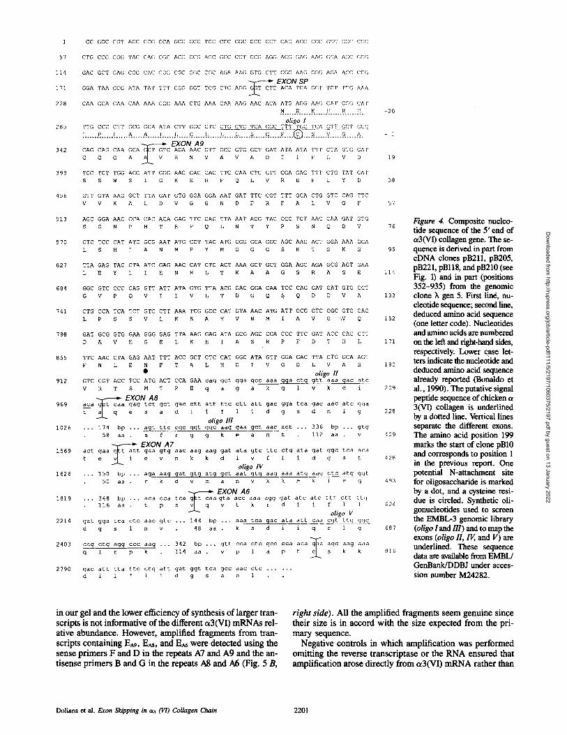
tron/exon structure of the 5'-end of chicken ot3(VI) gene was investigated by screening a chicken genomic library with synthetic oligonucleotide probes specific for sequences found in the presumed signal peptide and repeat A8 (oligonu- cleotide I and oligonucleotide HI, see Fig. 4). Overlapping genomic clones spanning a total o f * 20 kb were isolated and a partial restriction map was constructed. A more detailed analysis was performed for a 14-kb-long clone (k gen 5). Re- striction fragments were isolated from this clone by hybrid- ization to oligonucleotide probes specific for the cDNA clone pB10 (oligonucleotides I-V, see Fig. 4). By a combina- tion of restriction mapping, Southern analysis, and selective sequencing, the exon structure and the intron-exon bound- aries of the 5'-terminal part of the ot3(VI) gene were deduced (Fig. 3). Five exons were found, four of which code exactly for one type A repeat each (Fig. 3 A). The precise in- tron-exon boundaries were determined and the splice donor and acceptor sequences are shown in Fig. 3 B. Each donor and acceptor site is conventional and is in good agreement with the standard consensus motifs (Padgett et al., 1986; Krainer and Maniatis, 1988). All splice junctions are in frame and introns lie between the first and second nucleotide of a codon (phase 1 introns) (Sharp, 1981). Fig. 4 reports a composite nucleotide sequence and deduced amino acid se- quence derived from the different cDNA clones and the genomic clone k gen 5. The sequence starts with a short (266 bp) 5'-untranslated region followed by a sequence that codes for 25 amino acids characteristic of a signal peptide (van Heijne, 1986).
The NH2-terminal sequence of o~3(VI) is not known, therefore, we assume from the deduced sequence that the mature protein initiates with a glutamine as has been reported both for the chicken (Koller et al., 1989; Bonaldo et al., 1989) and the human (Chu et al., 1989) al(VI) and oe2(VI) chains. Restriction mapping, subcloning, and se- quencing of the k gen 5 genomic clone showed the existence of an additional open reading frame of 625 bp upstream to the sequences that completely matched with those of the re-
Figure 2. Northern blot analy- sis of chicken gizzard show- ing a3(VI) collagen-specific mRNA bands. Each lane was loaded with 7 #g of poly (A +)- enriched RNA. Individual strips were hybridized to [tx- 32p] dCTP-labeled eDNA frag- ments derived from clone pB10 (Bonaldo et al., 1990) and con- talning sequences specific for different type A repeats. (lane a) AS-specific probe (255-bp- long Ban II-Eco RV fragment);
(lane b) A7-specific probe (253-bp-long Neo I-Eco RV fragment); (lane c) At-specific probe (383-bp-long Eco RV-Ava I fragment); 0ane d) A3-specific probe (318-bp-long Eco RI-Bam HI frag- ment); (lane e) unrelated probe (pEX1 vector). The arrowheads in- dicate the migration of the mRNA that is not detected by A8 and A6 probes. On the right the migration of molecular weight markers is indicated in kb. Only the relevant part of the autoradiogram is shown.
Doliana et al. Exon Slu'pping in cl3 (VI) Collagen Chain 2199
Dow
nloaded from http://rupress.org/jcb/article-pdf/111/5/2197/1060375/2197.pdf by guest on 13 January 2022

A
GENE
mRNA
Esp EA9 EA8 EA7 EA6
Agl A~I ATLAS lAS I A41A31A21 A11A'31 CoL IA'2 I A'I I
Hp S X D BH N P I I I I !1 I I
1 _ _ = 1 Kb.
B INTRON
3' SPLICE JUNCTION (accepter site) i
yyyyyyyyyyynca~G G . . . . . . . . . . . . . . . . . . . . . . . . . . . . . AAGgtraqt
INTRON EXON 5' SPLICE JUNCTION
(donor site)
• CAGCAAGCAG~ aagac GlnGlnAla
• GACATCACAG~/t aatgg AspIleThr
• GTGACTGAAGgt at gt a ValThrGlu
• ACACCATCAGgt aatt c ThrProSer
. GCCCCAACAG~t a at at AlaProThr i
+++++++
ataacgaccagtctattgacctcgttctccctctcccttag~TCTCACA ..... 132 bp ....
++++++-
tgactgctagaaaatcctaaaccccatctgtttttttaaa~CTGTCAGA--'- ..... 598 bp .... ValArg
-++++++
taatactcatttaatgcttcacctttgcattctttttcaa~CTCAAGAG ..... 585 bp .... GlnAsp
++++-++
taattgacgttatattcatgtgtgtgtatgattgcttgcac TTATTGAA ..... 585 bp .... IleGln
+++++++ taatcatgatgtctcatgttattttctgtgctctaccgca~TTCAAGTA ..... 582 bp ....
i G l n V a l
consensus
ESp = 150bp
EA9 = 618 bp
EA8 = 603 bp
E A7 = 603 bp
E A6 = 600 bp
Figure 3. Sequences and physical map of the Y-end of the chicken ¢3(VI) collagen gene. (.4) Diagram of the physical map and partial restriction sites of clone )~ gen 5. Exons are indicated by open rectangles and introns by thick lines. B, Bgl I; D, Dra I; H, Hind llI; lip, Hpa I; N, Nco I; P, Pvu II; S, Sma I; X, Xba I. (B) Nucleotide sequences at the exon-intron boundaries. The splice junctions of five introns are aligned and compared to the splice consensus sequences for eukaryotic genes• Intron sequences are indicated by lowercase and exon sequences by capital letters. Deduced amino acids are indicated by the three letter code. The lariat branchpoint consensus sequence ynytray (Padgett et al., 1986) is shown by + and - . At the right the definition of the exons and their length is shown, r, purine; y, pyrimidine; n, purine or pyrimidine.
peat AS. Comparison of the deduced amino acid sequences with the sequences of the eight type A repeats o f a 3(VI) pre- viously identified (Bonaldo et al., 1990) revealed that this open reading frame is an exon precisely encoding a full type A repeat. Except for a few residues present in clone pB10 (Bonaldo et al., 1990) this repeat was not detected in any of our previous eDNA clones. With the addition of this extra type A repeat (A9), residue 1 of clone pB10 in our previous report (Bonaldo et al., 1990) becomes residue 199. Fig. 4 also shows that repeats A9, AS, A7, and A6 are encoded within single exons (EAg, EAs, EA7, and E^6), whereas the presumed signal peptide is encoded together with 62 bp of 5'-untranslated mRNA sequence by a separate exon (Esp).
u3(VI) mRNA Heterogeneity Is Due to Exon Skipping of 7~pe A Repeats In view of the heterogeneity of the c~3(VI) mRNAs, the selec- tive hybridization with the eDNA probe specific for different
repeats, the isolation of eDNA clones lacking individual type A repeats, and the demonstration that the repeats from A9 through A6 are encoded within single exons, we applied the RT/PCR amplification assay to analyze further this complex transcription unit. Using this approach together with a3(VI)- specific primers we examined the region of probable isoform variation comprised between the signal peptide and repeat A5 (Fig. 5).
Evidence both for spliced and unspliced transcripts was obtained. Amplified fragments from transcripts missing EA9 and E^8 were detected using the sense primer A in the signal peptide and the downstream antisense primers B and C in the repeats A8 and A7, respectively. Similarly, using the sense primer D in the repeat A7 and the antisense primer E in the repeat A5, an amplified fragment of 364 bp missing EA6 was detected (Fig. 5 B, left side)• Our assay conditions favor the amplification of short sequences, consequently higher Mr eDNA including the spliced exons are not visible
The Journal of Cell Biology, Volume 111, 1990 2200
Dow
nloaded from http://rupress.org/jcb/article-pdf/111/5/2197/1060375/2197.pdf by guest on 13 January 2022
]
51
114
171
228
285
342
399
456
513
570
627
684
741
798
855
912
969
1026
1569
1626
1819
2214
2403
2790
CC []GC C(]T ACC GC(] CCA GCC (]CC TCC CCC G(]C CCC (][;'I' CA(] ACC CCC (;GC [](]C CCC
CTG CCG CG(; TAC CA(] CG( ? ACC CCG AGC GGC CCT CCG AGG A(]G GAG AA(] GCA AGC f](]f]
GAC GCT CA(; C(;G CAC CGG CGC G(;C CGC A(;A AA(] GTG CTT C(;(; AA(; r.;(;(; A(;A ACC CCG
EXON SO GGA TAA GCG ATA TAT T T T CGG GGT TCG CTC AGC f f j ~ CTC ACA TCA GCT T C T TTG AAA
CAA GGA GAA CAA AAA GG(] AAA CTG AAA CAA AAG AAC ACA ATG AGG AAG CAT C(]G CAT
. . . . . .R . . . . . .K... . . [! . . . . . . % . . . . ! [ .
oligo I TTG CCC C T T CCG GCA ATA C T T GGC C T C CTG C T C TCA GGC T T T TGC TCA GTT CGT GCC
. < . . . ~ . . . . . : , . . . . . ~ . . . . ~ . . . . . : . . . . . ~ . . . . ~ . . . . . . ~ . . . . . ~ . . . . . : , . . . . . . ~ . . . . . ~ . . . . . ~ . . . . . © . . . ~ - - - . . . . . -v . . . . . -~ . . . . . ~ . ,. EXON A9
CAG CAG CAA GCA ~CT GTC AGA AAC GTT GCC GTG GCT GAT ATA ATA TTT CTA GTG GAT
Q Q Q A ~ V R N V A V A D I I F 1, V D
TCC TCT TGG AGC ATT GGG AAG GAG CAC TTC CAA CTC GTT CGA GAG TTT CTG TAT GAT
S S W S I G K E H F Q L V R E F L Y D
GTT GTA AAG GCT TTA GAT GTG GGA GGA AAT GAT TTC CGT TTT GCA CTG GTC CAG TTC
V V K A L D V G G N D F R F A L V Q F
AGC GGA AAC CCA CAC ACA GAG TTC CAG TTA AAT ACG TAC CCC TCT AAC CAA GAT GTG
S G N P H T E F Q L N T Y P S N Q D V
CTC TCC CAT ATC GCG AAT ATG CCT TAC ATG GGG GGA GGC AGC AAG ACT GGA AAA GGA
L S H I A N M P Y M G G G S K T G K G
TTA GAG TAC CTA ATC GAG AAC CAT CTC ACT AAA GCT GCT GGA AGC AGA GCG AGT GAA
L E Y L I E N H L T K A A G S R A S E
GGC GTC CCC CAG GTT ATT ATA GTG TTA ACG GAC GGA CAA TCC CAG GAT GAT GTG GCT
G V P Q V I I V L T D G Q S Q D D V A
CTG CCA TCA TCT GTC CTT AAA TCG GCC CAT GTA AAC ATG ATT GCG GTC GGC GTG CAG
L P S S V L K S A H V N M I A V G ",V Q
GAT GCG GTG GAA GGG GAG TTA AAG GAG ATA GCG AGC CGA CCC TTC GAT ACC CAC CTT
D A V E G E L K E I A S R P F D T H L
TTC AAC CTA GAG AAT TTT ACC GCT CTC CAT GGC ATA GTT GGA GAC TTA GTG GCA AGT
F N L E N F T A L H G I V G D L V A S
• oligo II GTC CGT ACC TCC ATG ACT CCA GAA cag gct gga gcc aaa gga ctg gtt aaa gac arc
V R T S M T P E q a g a k g 1 v k d i
,. E X O N A 8 aca g~t caa gag tct gct gac ctt att ttc ctt att gac gga tca gac aac atc gga
t a I - q e s a d 1 i f 1 i d g s d n i g
oligo Ill .. • 174 bp . .. agt ttc cgc ggt ggc aag gaa gct aac act ... 336 bp ... gtg
58 aa . s f r g g k e a n t 112 aa . v
EXON A7 act gaa g~t att gaa gtg aac aag aag gat ata gtc ttc ctg ata gat ggc tca aca
t e vll - i e v n k k d i v f 1 i d g s t
oligo IV •.. 150 bp ... aga aag gat gtg atg get aat gtg aag aaa atg aag ctc atg ggt
50 aa . r k d v m a n v k k m k 1 m g
... 348 bp ... aca cca tca g~t 116 aa . t p s 3_
gat gga tca ctc aac gtc ,.. 144
d g s 1 n v 4 8
EXON A6 caa gta acc aaa agg gat atc atc ttt ctt ttg
q v t k r d i i f 1 1
o1~o V ,.. aaa tca gac ata art caa cgt ttg ggg bp
aa
cag ctg agg ccc aag ... 342 bp ...
q 1 r p k 114 aa .
gac att tta ttc ctg att gat ggt tca
d i 1 f 1 i d g s
• k s d i i q r I q
gtt cca ctc gcc cca acag~aa age aag aaa v p 1 a p t c~ s k k
gcc aac ctc ......
a n 1
-2O
- ]
19
38
57
76
95
114
133
152
171
190
209
228
4O9
428
493
624
687
816
Figure 4. Composite nucleo- tide sequence of the 5' end of ~3(VI) collagen gene. The se- quence is derived in part from eDNA clones pB211, pB205, pB221, pBll8, and pB210 (see Fig• 1) and in part (positions 352-935) from the genomic clone X gen 5. First lin4, nu- cleotide sequence; second line, deduced amino acid sequence (one letter code). Nucleotides and amino acids are numbered on the letI and fight-hand sides, respectively. Lower ease let- ters indicate the nucleotide and deduced amino acid sequence already reported (Bonaldo et al., 1990). The putative signal peptide sequence of chicken o~ 3(VI) collagen is underlined by a dotted line• Vertical lines separate the different exons. The amino acid position 199 marks the start of clone pB10 and corresponds to position 1 in the previous report. One potential N-attachment site for oligosaecharide is marked by a dot, and a cysteine resi- due is circled. Synthetic oli- gonucleotides used to screen the EMBL-3 genomic library (oligo 1 and Ill) and to map the exons (oligo II, F6, and V) are underlined. These sequence data are available from EMBL/ GenBanldDDBJ under acces- sion number M24282.
in our gel and the lower efficiency of synthesis of larger tran- scripts is not informative o f the different ot3(VI) mRNAs rel- ative abundance. However, amplified fragments from tran- scripts containing EA9, E^8, and E^6 were detected using the sense primers F and D in the repeats A7 and A9 and the an- tisense primers B and G in the repeats A8 and A6 (Fig. 5 B,
right side). All the amplified fragments seem genuine since their size is in accord with the size expected from the pri- mary sequence.
Negative controls in which amplification was performed omitting the reverse transcriptase or the RNA ensured that amplification arose directly from ol3(VI) m R N A rather than
D o l i a n a et a l . Exon Skipping in oO (VI) Collagen Chain 2 2 0 1
Dow
nloaded from http://rupress.org/jcb/article-pdf/111/5/2197/1060375/2197.pdf by guest on 13 January 2022

! o t'3 o__
.
o o o e~
to
to
Fig
ure 5
. Det
ecti
on o
f mR
NA
tra
nscr
ipts
cod
ing
for
the
c~3(
VI)
isof
orm
s by
the
RT
/PC
R m
etho
d. (
A)
Lin
ear
mod
el o
f the
ct3
(VI)
mR
NA
sho
win
g th
e po
siti
on o
f the
alt
erna
tive
ly e
xpre
ssed
se
quen
ces
(sha
ded
boxe
s) a
nd o
f th
e sy
nthe
tic
olig
onuc
leot
ide
prim
ers
used
in
the
ampl
ific
atio
ns.
(B)
Am
plif
icat
ion
show
ing
the
expe
cted
and
obs
erve
d D
NA
fra
gmen
ts o
btai
ned
wit
h th
e fo
llow
ing
com
bina
tion
s of
oli
gonu
cleo
tide
s: p
rim
ers
A a
nd B
(la
nes
a an
d d)
; pr
imer
s A
and
C (
lane
b);
pri
mer
s D
and
E (
lane
c);
pri
mer
s D
and
G (
lane
s g
and
i);
prim
ers
F an
d B
(l
ane
h).
In n
egat
ive
cont
rol r
eact
ion
mix
ture
s th
e re
vers
e tr
ansc
ript
ase
(lan
e d)
or
the
tem
plat
e R
NA
(la
ne i)
wer
e om
itte
d. A
Hae
III
dig
est o
f tkX
174
DN
A w
as u
sed
as s
tand
ard
Mr m
arke
r (l
anes
e a
nd f
).
Dow
nloaded from http://rupress.org/jcb/article-pdf/111/5/2197/1060375/2197.pdf by guest on 13 January 2022
from minute amounts of contaminating plasmid cDNA since no DNA bands were detected in this case (Fig. 5 B, lanes d and h). The size of the amplified fragments excluded also the possibility that amplification arose from unprocessed RNA or genomic DNA which might contaminate the reaction.
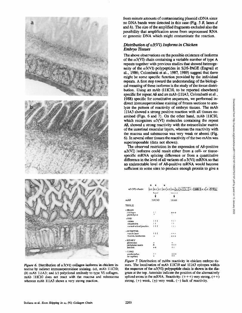
Distribution o f c~3(VI) Isoforms in Chicken Embryo Tissues
The above observations on the possible existence of isoforms of the ~3(VI) chain containing a variable number of type A repeats together with previous studies that showed heteroge- neity of the ot3(VI) polypeptides in SDS-PAGE (Engvall et al., 1986; Colombatti et al., 1987, 1989) suggest that there might be some specific function provided by the individual repeats. A first step toward the understanding of the biologi- cal meaning of these isoforms is the study of the tissue distri- bution. Using an mAb (lllC10, to be reported elsewhere) specific for repeat A8 and an mAb (111A3, Colombatti et al., 1988) specific for constitutive sequences, we performed in- direct immunoperoxidase staining of frozen sections to ana- lyze the pattern of reactivity of embryo tissues. The mAb ll lA3 showed a strong positive reaction with all tissues ex- amined (Figs. 6 and 7). On the other hand, mAb lllC10, which recognizes ~3(VI) molecules containing the repeat A8, showed a strong reactivity with the extracellular matrix of the intestinal muscular layers, whereas the reactivity with the mucosa and submucosa was very weak or absent (Fig. 6). In several other tissues the reactivity of the two mAbs was superimposable (data not shown).
The observed restriction in the expression of A8-positive a3(VI) isoforms could result either from a cell- or tissue- specific mRNA splicing difference or from a quantitative difference in the level of all variants of c~ 3(VI) mRNA so that an undetectable level of ?d-positive mRNA would become sufficient in some sites to produce enough protein to give a
Figure 6. Distribution of ~3(VI) collagen isoforms in chicken in- testine by indirect immunoperoxidase staining. (a), mAb lllC10; (b) mAb lllA3; and (c) polyclonal antibody to type VI collagen. mAb lllC10 does not react with the mucosa and submucosa whereas mAb 111A3 shows a very strong reaction.
(VI) chaba
m A B
T I S S U E
• BRAIN meninges + + + + + parenchyma
• EYE comea + + + + + + coajunctiva + + + + corneal-scleral jtulction + + + + + +
• INTESTINE muscle layer + + + + + + mucosa, submucosa - + + +
• KIDNEY glomerulus ~_ - peritubular matrix -- + + + arteriole + + + +
• LUNG parabronchus + + + + + air capillary + + +
IA9 IA9 [A7 IA6 IA 5 IA4 IA3 IA 2 IA, I~ ~ [ : ~ 1 ^' 2l~' . ~ : ~
I I 1 C 1 0 l l l A 3
Figure 7. Distribution of mAbs reactivity in chicken embryo tis- sues. The localization of mAb lllC10 and lllA3 epitopes within the sequence of the ~3(VI) polypeptide chain is shown in the dia- gram at the top. Asterisks indicate the position of the alternatively spliced exons in the mRNA. Reactivity: (+++) very strong, (++) strong, (+) weak, (+) very weak, ( - ) lack of reactivity.
Doliana et al. Exon Skipping in cO (VI) Collagen Chain 2203
Dow
nloaded from http://rupress.org/jcb/article-pdf/111/5/2197/1060375/2197.pdf by guest on 13 January 2022
positive immunoperoxidase signal. To exclude this latter possibility we incubated adjacent sections with several dilu- tions of mAb lllA3 and lllC10. In these experiments the distinct pattern of the reactivity of the two mAbs was still un- changed.
Discussion
We have used eDNA and genomic clones to study exon/intron organization of the 5'-end of chicken oe3(VI) col- lagen. The demonstration that the pattern of multiple mRNAs of this chain is at least in part the result of a mecha- nism of multiple alternative splicing of exons encoding type A repeats is a major finding of this study. In addition, the exon structure suggests that the or3 chain of type VI collagen evolved by multiple processes of gene shuttling and am- plification. Alternative splicing of ot3(VI) gene transcripts was first suggested by the observation that several eDNA clones lacked one or more sequences of •600 bp coding for individual type A repeats that were present in other eDNA clones from the same library. The oe3(VI) gene constitutes a complex transcription unit and the size of the transcripts (,,o10 kb) does not allow a fine resolution of the different mes- sages. Nevertheless, by hybridization of Northern blots with type A-specific eDNA probes, the presence of variant mRNAs that differed in size was initially demonstrated in this study. The finding that individual type A repeats are en- coded within single exons and the possible correspondence between the size difference of the various mRNAs and the size of type A-coding exons were highly suggestive that the different transcripts might Se the result of an alternative splicing mechanism involving type A repeats. Furthermore, the pattern of hybridization of the Northern blot with the type A-specific eDNA probes corresponding to the spliced A8 and ?,.6 domains suggests that certain mRNA molecules not only exclude both the exons but might involve skipping of ad- ditional exons (A9 and maybe other exons). Given the high Mf of the mRNA and the nearly identical size of the differ- ent type A repeats it is conceivable that each band of the Northern blot represents a mixture of comigrating mRNA species that have skipped different exons. The RT/PCR am- plification analysis, using various primers specific for the signal sequence and for presumed constitutive exons, yielded fragments with sizes expected if mRNA isoforms missing ei- ther one or at least two type A-encoding exons were ex- pressed. Appropriate controls excluded the possibility that cDNA clones missing individual repeats could contaminate the reaction mixture and serve as templates. Evidence for unspliced fragments also was obtained by the RT/PCR am- plification. It is conceivable that the ratio between the vari- ous ~3(VI) RNA transcripts may change depending on specific sites or physiological and pathological conditions as has been already shown for the different isoforms of another extracellular matrix glycoprotein, namely fibronectin (Zardi et al., 1987; ffrench-Constant and Hynes 1988, 1989; ffrench-Constant et al., 1989; Carnemolla et al., 1989).
Alternative splicing is an important mechanism of gene regulation and it is well documented for several proteins (Andreadis et al., 1987), including the extracellular matrix constituents fibronectin (Kornblihtt et al., 1984; Schwarz- bauer et al., 1987; Gutman and Kornblihtt, 1987), elastin (Indik et al., 1987), tenascin/cytotactin (Jones et al., 1989;
Gulcher et al., 1989), and link protein (Rhodes et al., 1988). Among collagens, apart from the c~3(VI), there is evidence that transcripts of the human ot2(VI) (Chu et al., 1989), of the human od(XRI) (Tikka et al., 1988), and of the chicken od(IX) (Nishimura et al., 1989) and o~2(I) (Bennett et al., 1989), undergo alternative splicing. The use of alternative promoters by the od(IX) collagen gene results in protein products with different sequence domains and specific tissue distribution. Regarding the other collagen genes it is not known at the moment whether the different mRNAs are translated in different proteins. At least for the o~2(I) this is not the case. Little is presently known about the mechanisms involved in the determination and regulation of the alterna- tive splicing, mainly because of the lack of suitable in vitro systems that preserve cell-specific features (Padgett et al., 1986). Only in few instances has it been possible to study the expression and processing of cell-specific splicing path- ways by transfecting different cell lines as shown for fibronectin (Baron et al., 1989). In this case it was demon- strated that all the information necessary to induce tissue- specific alternative splicing is in cis respective to the exons undergoing splicing and that trans-acting factors differen- tially expressed in the various cell lines confer the tissue- specific expression. It was beyond the purpose of the present study to obtain information on the intron sequences further upstream and downstream from the exon-intron boundaries, but from the short intron sequences available and the limited number of repeats analyzed (three conditional A9, AS, and A6 and two constitutive A7 and AS) we could not identify specific sequences that would distinguish alternatively spliced exons from constitutive exons.
Splicing of A9, AS, and A6 exons of ~3(VI) is similar to the optional skipping of ED-A and ED-B exons of fibronectin (Kornblihtt et al., 1984; Gutman and Kornhlihtt, 1987; Schwarzbauer et al., 1987). Through this mechanism several functionally appropriate o~3(VI) polypeptides can be gener- ated that have a different number of type A repeats. Indeed, heterogeneity at the protein level had been reported previ- ously for the o~3(VI) chain both in in vitro biosynthetic studies (Engvall et al., 1986; Colombatti et al., 1987; Colombatti and Bonaldo, 1987) and in vivo (Jander et al., 1984; Wu et al., 1987; Colombatfi et al., 1989). There is no direct evidence at the moment to relate the different mRNAs to the different polypeptides, although it is tempting to spec- ulate that the ladder of multiple polypeptides derives from messages that have skipped one or more exons coding for complete type A repeats. The finding that an mAb with specificity for conditional exons has a different tissue distri- bution than an mAb with specificity for constitutive exons is consistent with this notion and with the ot3(VI) polypeptide heterogeneity detected in tissue extracts (Colombatti et al., 1989). Given the fact that A8-specific mAb lllC10 detects a single epitope, it is in principle still possible that the lack of reactivity of the intestinal mucosa and submucosa with this mAb might not be the result of the local synthesis of type VI molecules with o~3 chains devoid of repeat A8, but only the consequence of epitope masking and/or interaction with different constituents of the extracellular matrix.
We have reported that type VI collagen and recombinant fusion proteins of the NH~-terminal portion of a3(VI) chain have the potential to interact under physiological con- ditions in vitro with type I collagen fibrils (Bonaldo et al.,
The Journal of Cell Biology, Volume 111, 1990 2204
Dow
nloaded from http://rupress.org/jcb/article-pdf/111/5/2197/1060375/2197.pdf by guest on 13 January 2022

1990; Russo, V., A. Appi, and A. Colombatti, manuscript in preparation). Given the widespread distribution of type VI collagen and its close vicinity to the cells, it seems reason- able to imply that the presence of multiple type A repeats modulates the interaction of type VI collagen with type I col- lagen and also with other potential ligands in the extracellu- lar matrix and at the cell surface.
We thank Dr. Sandro Poletti for his help with the immunoporoxidase tech- nique and Elisabetta Montagner for typing the manuscript.
This work was supported by a grant from the Associazione Italiana per la Rieerca sul Cancro.
Received for publication 26 February 1990 and in revised form 18 July 1990.
References
Andreadis, A., M. E. Gallego, and B. Nadal-Ginard. 1987. Generation of pro- tein isoform diversity by alternative splicing: mechanistic and biological im- plications. Annu. Rev. Cell Biol. 3:207-242.
Barone, M. V., C. Hencheliffe, F. E. Baralle, and G. Panlella. 1989. Cell type specific trans-acting factors are involved in alternative splicing of human fibronectin pre-mRNA. EMBO (Eur. Mol. Biol. Organ.) J. 8:1079-1085.
Bennett, V. D., I. M. Weiss, and S. L. Adams. 1989. Cartilage-specific 5'end of chick alpha-2(I) collagen messenger RNAs. J. Biol. Chem. 264:8402- 8409.
Biggin, M. D., T. J. Gibson, and G. F. Hoag. 1983. Buffer gradient gels and [3sS]-label as an aid to rapid DNA sequence determination. Proc. Natl. Acad Sci. USA. 80:3963-3965.
Bonaldo, P., and A. Colombatti. 1989. The carboxyl terminus of the chicken t~3 chain of collagen VI is a unique mosaic structure with glycoprotein lb- like, fibronectin type HI, and Kunitz modules. J. Biol. Chem. 264:20235- 20239.
Bonaldo, P., F. Bucciotti, and A. Colombatti. 1987. Isolation of eDNA clones corresponding to the Mr = 150,000 subunit of chick type VI collagen. Bio- chem. Biophys Res. Commun. 149:347-354.
Bonaldo, P., V. Russo, F. Bucciotti, G. M. Bressan, and A. Colombatti. 1989. cd chain of chick type VI collagen: the complete eDNA sequence reveals a hybrid molecule made of one short collagen and three yon Willebrand fac- tor type A-like domains. J. Biol. Chem. 264:5575-5580.
Bonaldo, P., V. Russo, F. Bucciotti, R. Doliana, and A. Colombatti. 1990. Structural and functional features of the chicken a3 chain indicate a bridging role for collagen VI in connective tissues. Biochemistry. 29:1245-1254.
Bressan, G. M., P. Argos, and K. K. Stanley. 1987. Repeating structure of chick tropoelastin revealed by complementary DNA cloning. Biochemistry. 26:1497-1503.
Bruns, R. 1984. Beaded filaments and long-spacing fibrils: relation to type VI collagen. J. Ultrastruct. Res. 89:136-145.
Brnns, R., W. Press, E. Engvall, R. Timpl, and J. Gross. 1986. Type VI colla- gen in extracellular 100-rim periodic filaments and fibrils: identification by immunoelectron microscopy. J. Cell Biol. 103:394--404.
Burgeson, R. E. 1988. New collagens, new concepts. Annu. Rev. Cell Biol. 4:551-577.
Carnemolla, B., E. Baiza, A. Sift, L. Zardi, M. R. Nicotra, A. Bigotti, and P. G. Natali. 1989. A tumor-associated fibronectin isoform generated by al- ternative splicing of messenger RNA precursors. J. Cell Biol. 108:1139- 1148.
Chu, M. L., K. Mann, R. Deutzmann, D. Pftbula-Conway, C. C. Hsu-Chen, M. P. Bernard, and R. Timpl. 1987. Characterization of three constituent chains of collagen type VI by peptide sequences and eDNA clones. Eur. J. Biochem. 168:309-317.
Chu, M. L., T. Pan, D. Conway, H. J. Kuo, R. W. Glanville, R. Timpl, K. Mann, and R. Deutzmarm. 1989. Sequence analysis of cO(VI) and c~2(VI) chains of human type VI collagen reveals internal triplication of globular do- mains similar to the A domains of von Willebrand factor and two c~2(VI) chain variants that differ in the carboxyl terminus. EMBO (Eur. MoL Biol. Organ.) J. 8:1939-1946.
Colombatti, A., and P. Bonaldo. 1987. Biosynthesis of chick type VI collagen. If. Processing and secretion in fibroblasts and smooth muscle cells. J. Biol. Chem. 262:14461-14466.
Colombatti, A., P. Bonaldo, K. Ainger, G. M. Bressan, and D. Volpin. 1987. Biosynthesis of chick type VI collagen. I. lntrucellular assembly and molecu- lar structure. J. Biol. Chem. 262:14454-14460.
Colombatti, A., K. Ainger, M. T. Mucignat, and P. Benaldo. 1988. Monoclo- hal antibodies for the different chains of chick type VI collagen. Collagen Relat. Res. 8:331-377.
Colombatti, A., K. Ainger and F. Colizzi. 1989. Type VI collagen: high yields of a molecule with multiple forms of or3 chain from avian and human tissues. Matrix. 9:177-185.
Engvall, E., H. Hessle, and G. Klier. 1986. Molecular assembly, secretion, and
matrix deposition of type VI collagen. J. Cell Biol. 102:703-710. ffrench-Constant, C., and R. O. Hynes. 1988. Patterns of fibronectin gene ex-
pression and spficing during cell migration in chicken embryos. DevekTp- ment (Camb.). 104:369-382.
ffrench-Conalunt, C., and R. O. Hyaes. 1989. Alternative splicing of fibronec- tin is temporally and spatially regulated in the chicken embryo. Deve/opment (Camb.). 106:375-388.
ffrench-Constant, C., L. Van De Water, H. F. Dvorak, and R. O. Hynes. 1989. Reappearance of an embryonic pattern of fibronectin spficing during wound healing in the adult rat. J. Cell Biol. 109:903-914.
Furthmayr, H., H. Wiedemnnn, R. Timpl, E. Odermatt, and J, Engel. 1983. Electro-microscopical approach to a structural model of intima collagen. Biochem. J. 211:303-311.
Gulcber, J. R., D. E. Nies, L. S. Marton, and K. Stefansson. 1989. An alterna- tively spficed region of the human hexabrachion contains a repeat of potential N-glycosylation sites. Proc. Natl. Acad. Sci. USA. 86:1588-1592.
Gutman, A., and A. R. Komblihtt. 1987. Identification of a third region of cell- specific alternative splicing in human fibrnnectin mRNA. Proc. Natl. Acad. Sci. USA. 84:7179-7182.
Indik, Z., H. Yeh, N. Ornstein-Goldstein, P. Sbeppard, N. Anderson, J. C. Rosenbloom, L. Peltonen, and J. Rosenbioom. 1987. Alternative splicing of human elastin mRNA indicated by sequence analysis of cloned genomic and complementary DNA. Proc. Natl. Acad. Sci. USA. 84:5680-5684.
Jander, R., D. Troyer, and J. Rauterberg. 1984. A collagen-like glycoprotein of the extracellular matrix is the undegraded form of type VI collagen. Bio- chemistry. 23:3675-3681.
Jones, F. S., S. Hoffrnan, B. A. Cunningham, and G. M. Edelman. 1989. A detailed structural model of cytotactin: protein homologies, alternative RNA splicing, and binding regions. Proc. Natl. Acad. Sci. USA. 86:1905-1909.
Kecne, D. R., E. Engvall, and R. W. Glanvllle. 1988. Ullrastrncture of type VI collagen in human skin and cartilage suggests an anchoring function for this filamentous network. J. Cell Biol. 107:1995-2006.
Koller, E., K. H. Wintcrhalter, and B. Triieb. 1989. The globular domains of type VI collagen are related to the collagen-binding domains of cartilage ma- trix protein and yon Willebrand factor. EMBO (Eur. MoL Biol. Organ.) J. 8:1037-1077.
Koroblihtt, A. R., K. Vibe-Petersen, and F. E. Baralle. 1984. Human fibrnnec- tin cell specific alternative mRNA splicing generates polypeptide chains differing in the number of internal repeats. Nucleic Acids Res. 12:5853~ 5868.
Krainer, A. R., and T. Maniatis. 1988. RNA splicing. In Transcription and Splicing. B. D. Hames and D. M. Glover, editors. IRL Press, Oxford. 131-143.
Maniatis, T., E. F. Ffttsch, andJ. Sambrook. 1982. Molecular Cloning: A Lab- oratory Manual. Cold Spring Harbor Press, Cold Spring Harbor, NY. 545 pp.
Messing, J. 1983. New M13 vectors for cloniag. MethodsEnzymol. 101:20-78. Nishimura, I., Y. Muragaki, and B. R. Olsen. 1989. Tissue-specific forms of
type IX collagen-proteoglycan arise from the use of two widely separated promoters. J. Biol. Chem. 264:20033-20041.
Padgett, R. A., P. J. Grabowski, M. M. Konarska, S. Seller, and P. A. Sharp. 1986. Splicing of messenger RNA precursors. Annu. Rev. Biochem. 55: 1119-1150.
Rappolee, A. D., D. Mark, M. J. Band,a, and Z. Werb. 1988. Wound macro- phages express TGF-alpha and o"e r growth factors in vivo: analysis by mRNA phenotyping. Science (Wash. DC). 241:708-712.
Rhodes, C., K. Doege, M. Sasaki, and Y. Yamade. 1988. Alternative splicing generated two different mRNA species for rat link protein. J. Biol. Chem. 263:6063-6067.
Sanger, F., S. Midden, and A. R. Coulson. 1977. DNA sequencing with chain- terminating inhibitors. Proc. Natl. Acad. Sci. USA. 74:5463-5467.
Schwarzbauer, J. E., R. E. Patel, D. Fonda, and R. O. Hynes. 1987. Multiple sites of alternative splicing in the rat fibronectin gene transcript. EMBO (Fur. Mol. Biol. Organ.)J. 6:2573-2580.
Sharp, P. A. 1981. Speculation on RNA splicing. Cell. 23:643-646. Shelton-Inloes, B. B., K. Titani, and J. E. Sadler. 1986. eDNA sequences for
human von Willebrand factor reveal five types of repeated domains and five possible protein sequence polymorphisms. Biochemistry. 25:3164-3171.
Tabor, S., and C. C. Richardson. 1987. DNA sequence analysis with modified bacteriophage T7 DNA polymerase. Proc. Natl. Acad. Sci. USA. 84:4767- 4771.
Tikka, L., T. Pihlajaniemi, P. Henttu, D. J. Prockop, and K. Tryggvason. 1988. Gene structure for the ,1 chain of a human short-chain collagen (type XIII) with alternatively spliced transcripts and translation termination codon at the 5' end of the last exon. Proc. Natl. Acad. Sci. USA 85:7491-7495.
van Heijne, G. 1986. A new method for predicting signal sequence cleavage sites. Nucleic Acids Res. 14:4683-4690.
yon der Mark, H., M. Aumailley, G. Wick, R. Fleishmajer, and R. Timpl. 1984. Immunochemistry, genuine size and tissue localization of collagen VI. Eur. J. Biochem. 142:493-502.
Wu, J. J., D. Eyre, and H. S. Slayter. 1987. Type VI collagen of the interver- tebral disc. Biochem. J. 248:373-381.
Zardi, L., B. Carnemolla, A. Sift, T. E. Petersen, G. Paolella, G. Sebastio, and F. E. Baralle. 1987. Transformed human cells produce a new fibrnnectin isoform by preferential alternative splicing of a previously unobserved exon. EMBO (Eur. Mol. Biol. Organ.) J. 6:2337-2342.
Doliana et al. Exon Skipping in a3 (VI) Collagen Chain 2205
Dow
nloaded from http://rupress.org/jcb/article-pdf/111/5/2197/1060375/2197.pdf by guest on 13 January 2022