multiparametric prostate mri - auanet.org · •to review the basic components of a...
TRANSCRIPT
Optimizing Implementation of Prostate MRI
Andrei S Purysko, M.D.
Section of Abdominal Imaging &
Nuclear Radiology Department
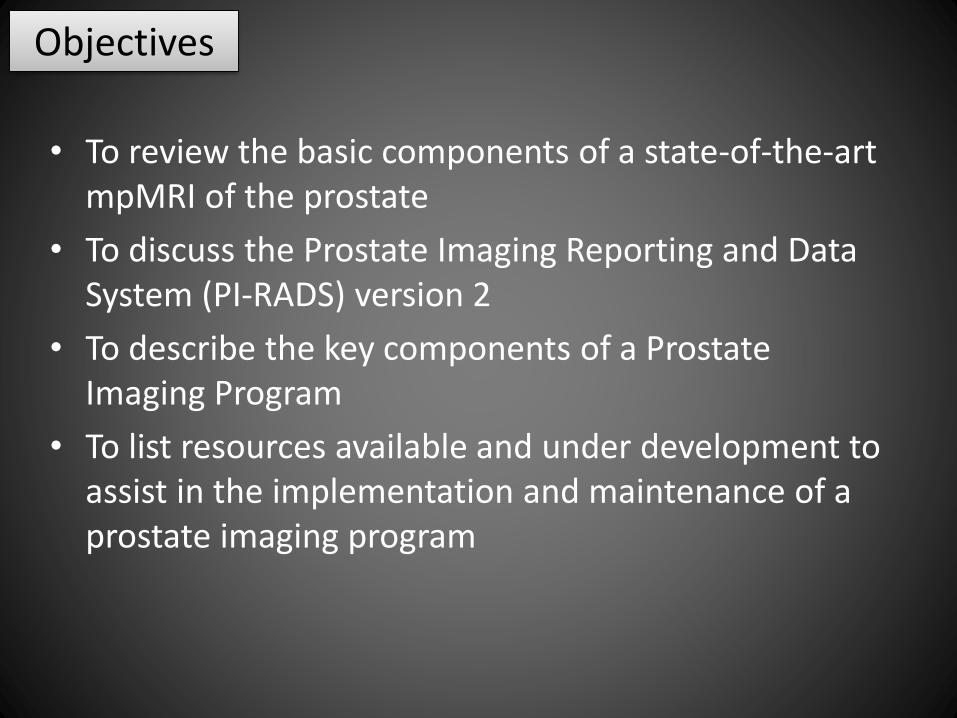
• To review the basic components of a state-of-the-art mpMRI of the prostate
• To discuss the Prostate Imaging Reporting and Data System (PI-RADS) version 2
• To describe the key components of a Prostate Imaging Program
• To list resources available and under development to assist in the implementation and maintenance of a prostate imaging program
Objectives

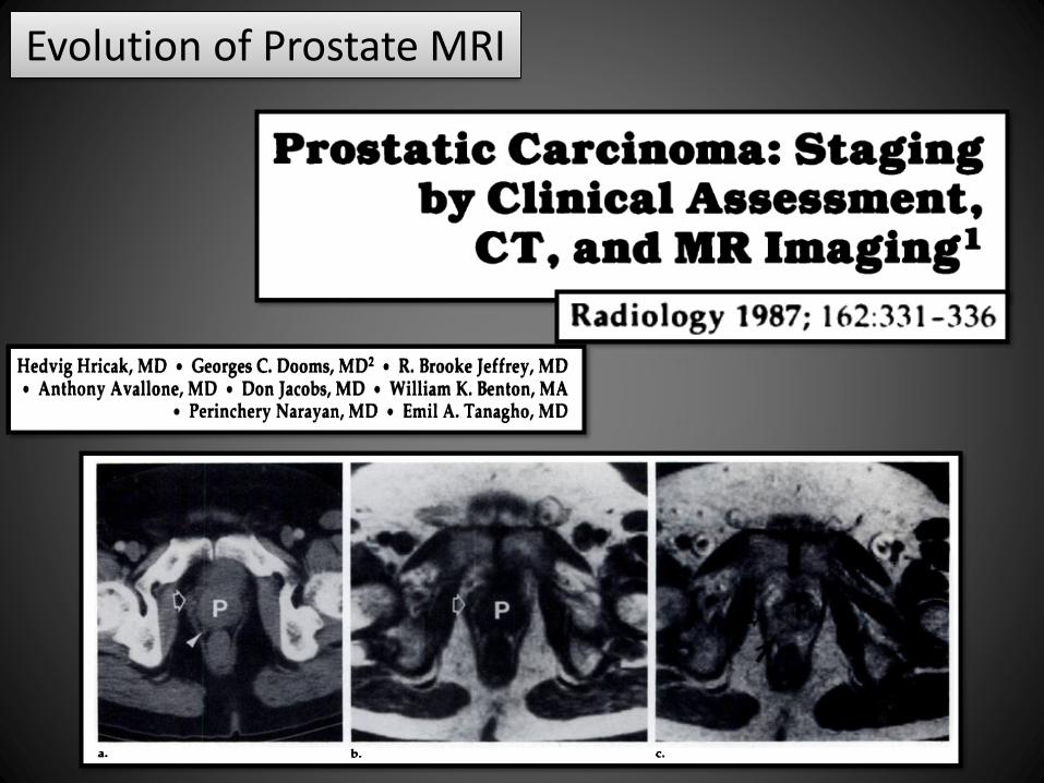
Evolution of Prostate MRI
Evolution of Prostate MRI
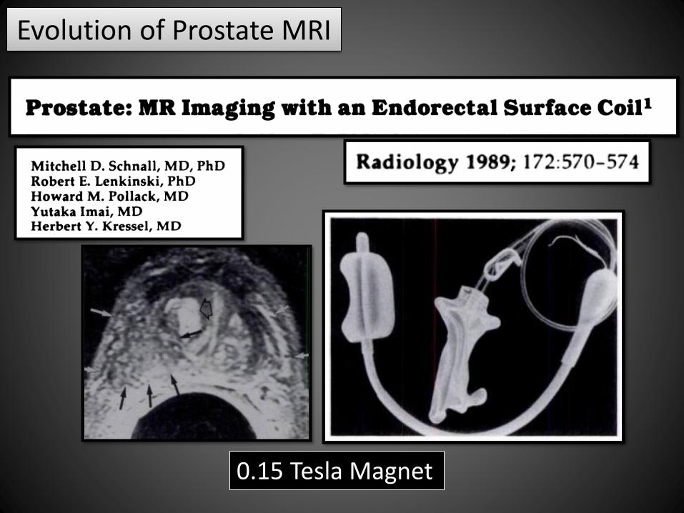
Evolution of Prostate MRI
0.15 Tesla Magnet

Evolution of Prostate MRI
• State-of-the-art technique
– 1.5 or 3.0 T
– Pelvic coil with or without ER coil
• Pulse Sequences:
– Multiplanar T2
– DCE
– DWI/ADC
– T1 small FOV
– T1 large FOV post contrast
Post-Bx “hemorrhage”
Lymph nodes/bone mets
Multiparametric

Prostate MRI Pulse Sequences
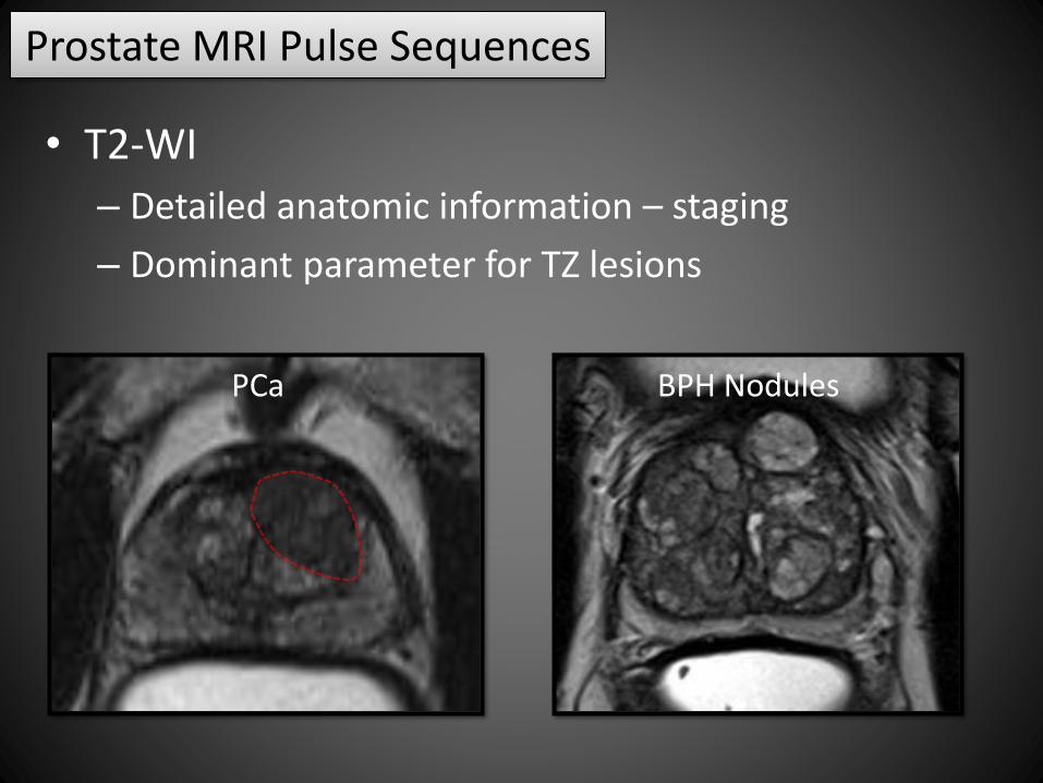
• T2-WI
– Detailed anatomic information – staging
– Dominant parameter for TZ lesions
Base Mid Apex
• T2-WI
– Detailed anatomic information – staging
– Dominant parameter for TZ lesions
PCa BPH Nodules
Prostate MRI Pulse Sequences
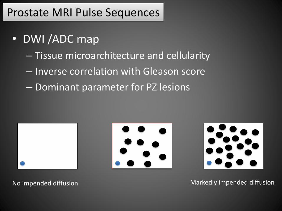
• DWI /ADC map
– Tissue microarchitecture and cellularity
– Inverse correlation with Gleason score
– Dominant parameter for PZ lesions
Prostate MRI Pulse Sequences
No impended diffusion Markedly impended diffusion

• DWI /ADC map
– Tissue microarchitecture and cellularity
– Inverse correlation with Gleason score
– Dominant parameter for PZ lesions
Prostate MRI Pulse Sequences
Gleason 3 + 3
Gleason 5 + 5
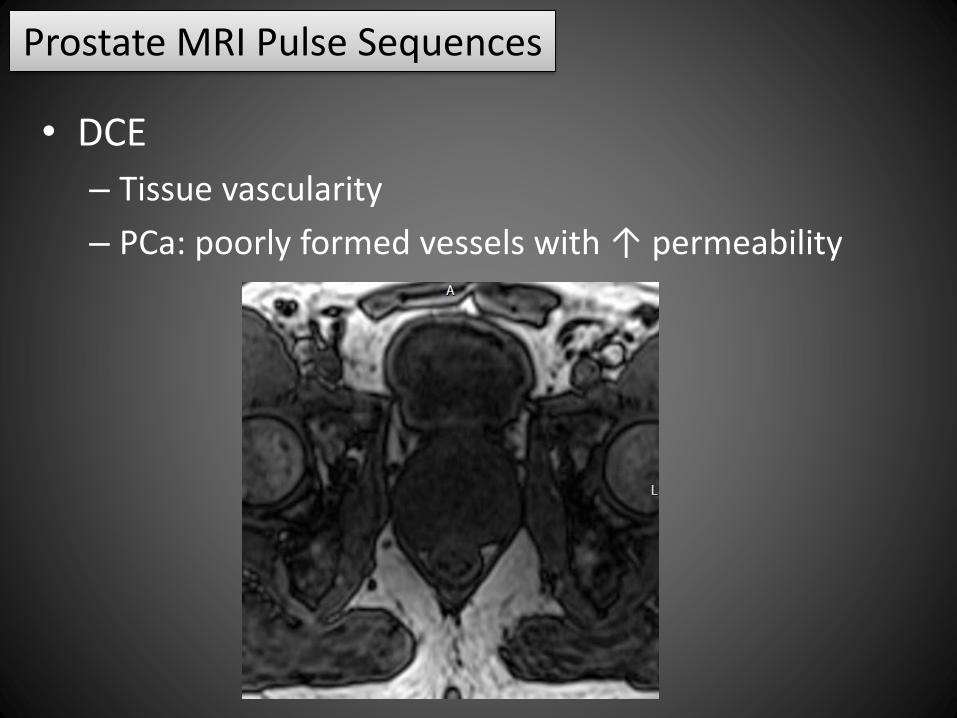
• DCE
– Tissue vascularity
– PCa: poorly formed vessels with ↑ permeability
Prostate MRI Pulse Sequences

• DCE
– Tissue vascularity
– PCa: poorly formed vessels with ↑ permeability
Prostate MRI Pulse Sequences

Prostate Imaging Reporting and Data System
• Version 1 (2012)
• AdMeTech Foundation’s International Prostate MRI Working
Group and the European Society of Urogenital Radiology (ESUR)
• Clinical guidelines for mpMRI based on evidence from the
literature and consensus expert opinion
• Included a structured reporting system (PI-RADS)

Prostate Imaging Reporting and Data System
• Version 2 (2014)
• ACR, ESUR, and the AdMeTech Foundation
• Establishing minimum acceptable technical parameters;
Standardizing radiology reports to enhance communication
among radiologists and referring physicians;
Developing assessment categories that summarize levels of
suspicion or risk for clinically significant PCa, so that they can be
used to triage patients to appropriate management;
Promoting research and quality assurance that will ultimately
lead to improvement in patient outcomes
Prostate Imaging Reporting and Data System
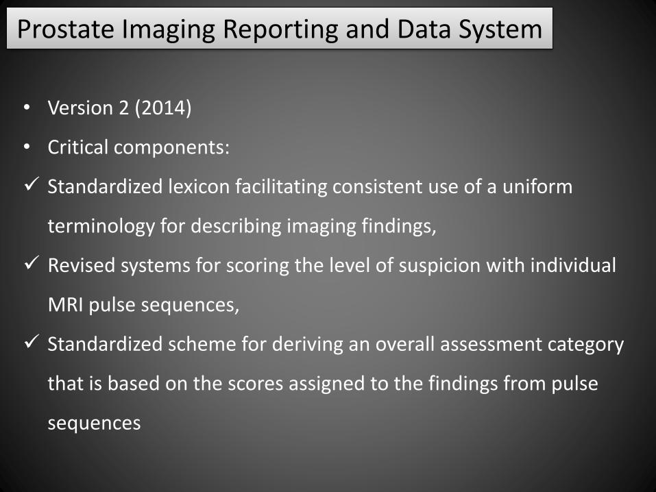
• Version 2 (2014)
• Critical components:
Standardized lexicon facilitating consistent use of a uniform
terminology for describing imaging findings,
Revised systems for scoring the level of suspicion with individual
MRI pulse sequences,
Standardized scheme for deriving an overall assessment category
that is based on the scores assigned to the findings from pulse
sequences

Prostate Imaging Reporting and Data System
PI-RADS
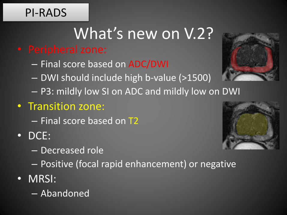
What’s new on V.2?• Peripheral zone:
– Final score based on ADC/DWI
– DWI should include high b-value (>1500)
– P3: mildly low SI on ADC and mildly low on DWI
• Transition zone:– Final score based on T2
• DCE: – Decreased role
– Positive (focal rapid enhancement) or negative
• MRSI:– Abandoned
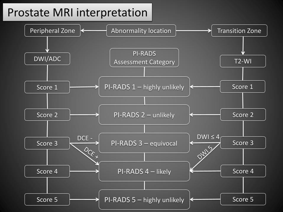
Prostate MRI interpretation
Abnormality locationPeripheral Zone Transition Zone
DWI/ADC T2-WIPI-RADS
Assessment Category
PI-RADS 1 – highly unlikelyScore 1
Score 2
Score 3
Score 4
Score 5
Score 1
Score 2
Score 3
Score 4
DCE - DWI ≤ 4
PI-RADS 2 – unlikely
PI-RADS 3 – equivocal
PI-RADS 4 – likely
PI-RADS 5 – highly unlikely Score 5
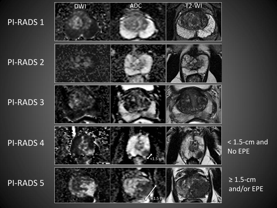
DWI ADC T2-WI
PI-RADS 1
PI-RADS 2
PI-RADS 3
PI-RADS 4
PI-RADS 5
<1.5 cm
> 1.5 cm
< 1.5-cm andNo EPE
≥ 1.5-cm and/or EPE
Prostate Imaging Reporting and Data System
• PI-RADS v2 document
• Weinreb et al. (PMID: 26427566)
• PI-RADS Atlas
• Available online
• Prostate MRI workshop
• ACR Education Center, Reston VA
• 2-day hands-on course 100+ cases and lectures

Prostate Imaging Program
• Key components for implementation:
Engaged urologist
Institutional support
Local champion in the radiology department
• “The Director of Prostate Imaging”*
*Westphalen et al. PMID 28396916
Prostate Imaging Program
• Roles of local champion
• Collaborate with urologists and other referring physicians on
institutional policies for imaging utilization;
• Informal review and formal case discussions in conferences
and tumor boards;
• Evaluation of outside imaging examinations for quality and
potential use in patient management;
• Discussion of new imaging applications;
• Troubleshooting individual cases as necessary.

Prostate Imaging Program
• Roles of local champion
• Radiology engagement
• To assist in the improvement of consistency and accuracy of
reports
• Development and use of report templates
• Training (baseline and continued)
• Feedback system

Prostate Imaging Program
• Roles of Local champion
• Technologist engagement
• Ensure consistent and adequate image quality
• Development of imaging protocols ensuring they meet or
exceed the parameters standardized by PI-RADS

Thank you!