multimodality management of soft tissue tumors in the ... · ajcc staging for soft tissue sarcoma...
TRANSCRIPT
MultimodalityManagement of Soft Tissue
Tumors in the ExtremityAimee M. Crago, MD, PhDa,b,*, Ann Y. Lee, MDa
KEYWORDS
� Soft tissue sarcoma � Extremity sarcoma � Limbs � Resection � Radiotherapy
KEY POINTS
� Work-up for an extremity mass suspicious for a soft tissue sarcoma includes cross-sectional imaging with an MRI and a core biopsy done in line with the planned incision.
� The standard for treatment of extremity soft tissue sarcomas is limb-sparing surgery with amargin of 1 to 2 cm. Overlying fascial layers (ie, muscular fascia, femoral sheath, perios-teum) are often barriers to tumor extension and are acceptable margins when major neu-rovascular or bony structures are in close proximity.
� Rates of local and distant recurrence vary by histologic subtype. These differences informsurgical margins and the use of chemotherapy and radiation.
� Radiation therapy is used to decrease rates of local recurrence in high-risk tumors. Neo-adjuvant (vs adjuvant) radiation can minimize side effects to nearby joints and normal tis-sues, but is associated with increased rates of wound complications and has equivalentrates of local control.
� Use of adjuvant chemotherapy is controversial. Neoadjuvant chemotherapy should beroutinely prescribed for high-risk, chemosensitive subtypes (ie, Ewing sarcoma and rhab-domyosarcoma). It can be selectively prescribed for moderately chemosensitive subtypesbased on other risk factors, such as size.
INTRODUCTION
Soft tissue sarcoma (STS) is a term referring to approximately 100 different subtypesof cancer.1 These diseases are rare, and as a group are diagnosed in only approxi-mately 12,000 patients in the United States each year.2 Although STS is identified in
Disclosures: The authors have no disclosures to report.Thisworkwas supportedbytheMemorial SloanKetteringCancerCenterCoreGrant (P30CA008748)and the Kristen Ann Carr Fund.a Sarcoma Disease Management Team, Department of Surgery, Memorial Sloan KetteringCancer Center, 1275 York Avenue, H1220, New York, NY 10065, USA; b Department of Surgery,Weill Cornell Medical College, 1300 York Avenue, New York, NY 10065, USA* Corresponding author. Gastric and Mixed Tumor Service, Department of Surgery, 1275 YorkAvenue, H1220, New York, NY 10065.E-mail address: [email protected]
Surg Clin N Am 96 (2016) 977–992http://dx.doi.org/10.1016/j.suc.2016.05.001 surgical.theclinics.com0039-6109/16/$ – see front matter � 2016 Elsevier Inc. All rights reserved.
Crago & Lee978
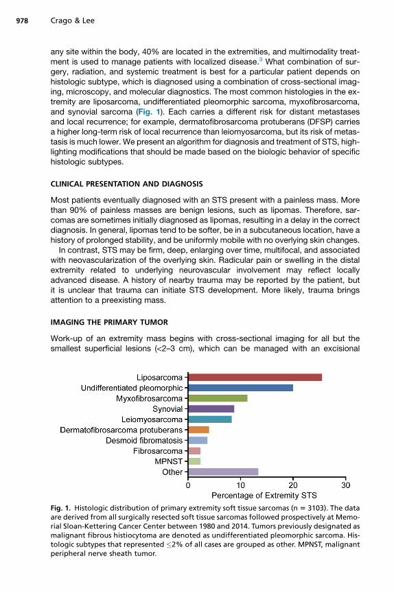
any site within the body, 40% are located in the extremities, and multimodality treat-ment is used to manage patients with localized disease.3 What combination of sur-gery, radiation, and systemic treatment is best for a particular patient depends onhistologic subtype, which is diagnosed using a combination of cross-sectional imag-ing, microscopy, and molecular diagnostics. The most common histologies in the ex-tremity are liposarcoma, undifferentiated pleomorphic sarcoma, myxofibrosarcoma,and synovial sarcoma (Fig. 1). Each carries a different risk for distant metastasesand local recurrence; for example, dermatofibrosarcoma protuberans (DFSP) carriesa higher long-term risk of local recurrence than leiomyosarcoma, but its risk of metas-tasis is much lower. We present an algorithm for diagnosis and treatment of STS, high-lighting modifications that should be made based on the biologic behavior of specifichistologic subtypes.
CLINICAL PRESENTATION AND DIAGNOSIS
Most patients eventually diagnosed with an STS present with a painless mass. Morethan 90% of painless masses are benign lesions, such as lipomas. Therefore, sar-comas are sometimes initially diagnosed as lipomas, resulting in a delay in the correctdiagnosis. In general, lipomas tend to be softer, be in a subcutaneous location, have ahistory of prolonged stability, and be uniformly mobile with no overlying skin changes.In contrast, STS may be firm, deep, enlarging over time, multifocal, and associated
with neovascularization of the overlying skin. Radicular pain or swelling in the distalextremity related to underlying neurovascular involvement may reflect locallyadvanced disease. A history of nearby trauma may be reported by the patient, butit is unclear that trauma can initiate STS development. More likely, trauma bringsattention to a preexisting mass.
IMAGING THE PRIMARY TUMOR
Work-up of an extremity mass begins with cross-sectional imaging for all but thesmallest superficial lesions (<2–3 cm), which can be managed with an excisional
Fig. 1. Histologic distribution of primary extremity soft tissue sarcomas (n 5 3103). The dataare derived from all surgically resected soft tissue sarcomas followed prospectively at Memo-rial Sloan-Kettering Cancer Center between 1980 and 2014. Tumors previously designated asmalignant fibrous histiocytoma are denoted as undifferentiated pleomorphic sarcoma. His-tologic subtypes that represented �2% of all cases are grouped as other. MPNST, malignantperipheral nerve sheath tumor.
Multimodality Management of Extremity Tumors 979
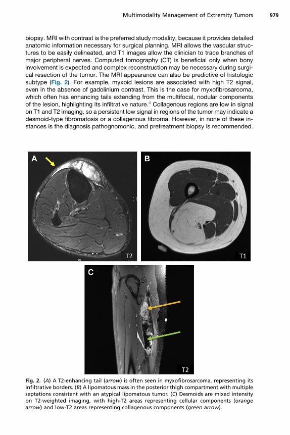
biopsy. MRI with contrast is the preferred study modality, because it provides detailedanatomic information necessary for surgical planning. MRI allows the vascular struc-tures to be easily delineated, and T1 images allow the clinician to trace branches ofmajor peripheral nerves. Computed tomography (CT) is beneficial only when bonyinvolvement is expected and complex reconstruction may be necessary during surgi-cal resection of the tumor. The MRI appearance can also be predictive of histologicsubtype (Fig. 2). For example, myxoid lesions are associated with high T2 signal,even in the absence of gadolinium contrast. This is the case for myxofibrosarcoma,which often has enhancing tails extending from the multifocal, nodular componentsof the lesion, highlighting its infiltrative nature.4 Collagenous regions are low in signalon T1 and T2 imaging, so a persistent low signal in regions of the tumor may indicate adesmoid-type fibromatosis or a collagenous fibroma. However, in none of these in-stances is the diagnosis pathognomonic, and pretreatment biopsy is recommended.
Fig. 2. (A) A T2-enhancing tail (arrow) is often seen in myxofibrosarcoma, representing itsinfiltrative borders. (B) A lipomatous mass in the posterior thigh compartment with multipleseptations consistent with an atypical lipomatous tumor. (C) Desmoids are mixed intensityon T2-weighted imaging, with high-T2 areas representing cellular components (orangearrow) and low-T2 areas representing collagenous components (green arrow).
Crago & Lee980
There is only one STS type for which imaging is considered diagnostic (and only whenreviewed by an experienced radiologist). This type is atypical lipomatous neoplasm/well-differentiated liposarcoma/dedifferentiated liposarcoma, and has a signal profilealmost identical to subcutaneous fat, but with enhancing septae and, if a dedifferen-tiated component is present, with solid nodules.5,6
BIOPSY TECHNIQUES
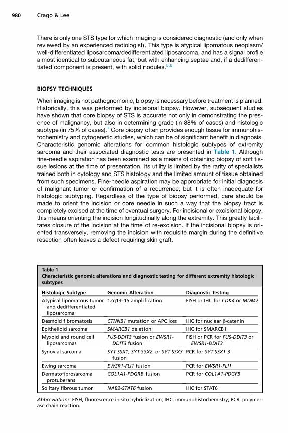
When imaging is not pathognomonic, biopsy is necessary before treatment is planned.Historically, this was performed by incisional biopsy. However, subsequent studieshave shown that core biopsy of STS is accurate not only in demonstrating the pres-ence of malignancy, but also in determining grade (in 88% of cases) and histologicsubtype (in 75% of cases).7 Core biopsy often provides enough tissue for immunohis-tochemistry and cytogenetic studies, which can be of significant benefit in diagnosis.Characteristic genomic alterations for common histologic subtypes of extremitysarcoma and their associated diagnostic tests are presented in Table 1. Althoughfine-needle aspiration has been examined as a means of obtaining biopsy of soft tis-sue lesions at the time of presentation, its utility is limited by the rarity of specialiststrained both in cytology and STS histology and the limited amount of tissue obtainedfrom such specimens. Fine-needle aspiration may be appropriate for initial diagnosisof malignant tumor or confirmation of a recurrence, but it is often inadequate forhistologic subtyping. Regardless of the type of biopsy performed, care should bemade to orient the incision or core needle in such a way that the biopsy tract iscompletely excised at the time of eventual surgery. For incisional or excisional biopsy,this means orienting the incision longitudinally along the extremity. This greatly facili-tates closure of the incision at the time of re-excision. If the incisional biopsy is ori-ented transversely, removing the incision with requisite margin during the definitiveresection often leaves a defect requiring skin graft.
Table 1Characteristic genomic alterations and diagnostic testing for different extremity histologicsubtypes
Histologic Subtype Genomic Alteration Diagnostic Testing
Atypical lipomatous tumorand dedifferentiatedliposarcoma
12q13–15 amplification FISH or IHC for CDK4 or MDM2
Desmoid fibromatosis CTNNB1 mutation or APC loss IHC for nuclear b-catenin
Epithelioid sarcoma SMARCB1 deletion IHC for SMARCB1
Myxoid and round cellliposarcomas
FUS-DDIT3 fusion or EWSR1-DDIT3 fusion
FISH or PCR for FUS-DDIT3 orEWSR1-DDIT3
Synovial sarcoma SYT-SSX1, SYT-SSX2, or SYT-SSX3fusion
PCR for SYT-SSX1-3
Ewing sarcoma EWSR1-FLI1 fusion PCR for EWSR1-FLI1
Dermatofibrosarcomaprotuberans
COL1A1-PDGRB fusion PCR for COL1A1-PDGFB
Solitary fibrous tumor NAB2-STAT6 fusion IHC for STAT6
Abbreviations: FISH, fluorescence in situ hybridization; IHC, immunohistochemistry; PCR, polymer-ase chain reaction.
Multimodality Management of Extremity Tumors 981
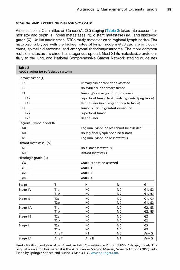
STAGING AND EXTENT OF DISEASE WORK-UP
American Joint Committee on Cancer (AJCC) staging (Table 2) takes into account tu-mor size and depth (T), nodal metastases (N), distant metastases (M), and histologicgrade (G). Unlike carcinomas, STSs rarely metastasize to regional lymph nodes. Thehistologic subtypes with the highest rates of lymph node metastasis are angiosar-coma, epithelioid sarcoma, and embryonal rhabdomyosarcoma. The more commonroute of metastasis is direct hematogenous spread. Most STSs metastasize preferen-tially to the lung, and National Comprehensive Cancer Network staging guidelines
Table 2AJCC staging for soft tissue sarcoma
Primary tumor (T)
TX Primary tumor cannot be assessed
T0 No evidence of primary tumor
T1 Tumor �5 cm in greatest dimension
T1a Superficial tumor (not involving underlying fascia)
T1b Deep tumor (involving or deep to fascia)
T2 Tumor >5 cm in greatest dimension
T2a Superficial tumor
T2b Deep tumor
Regional lymph nodes (N)
NX Regional lymph nodes cannot be assessed
N0 No regional lymph node metastasis
N1 Regional lymph node metastasis
Distant metastases (M)
M0 No distant metastasis
M1 Distant metastasis
Histologic grade (G)
GX Grade cannot be assessed
G1 Grade 1
G2 Grade 2
G3 Grade 3
Stage T N M G
Stage IA T1a N0 M0 G1, GXT1b N0 M0 G1, GX
Stage IB T2a N0 M0 G1, GXT2b N0 M0 G1, GX
Stage IIA T1a N0 M0 G2, G3T1b N0 M0 G2, G3
Stage IIB T2a N0 M0 G2T2b N0 M0 G2
Stage III T2a N0 M0 G3T2b N0 M0 G3Any T N1 M0 Any G
Stage IV Any T Any N M1 Any G
Used with the permission of the American Joint Committee on Cancer (AJCC), Chicago, Illinois. Theoriginal source for this material is the AJCC Cancer Staging Manual, Seventh Edition (2010) pub-lished by Springer Science and Business Media LLC, www.springer.com.

Crago & Lee982
generally recommend chest radiograph or CT scan to rule out distant disease. In gen-eral, the use of chest imaging can be tailored to the metastatic risk associated with agiven lesion. This risk can be estimated based on AJCC staging or, more accurately,based on prognostic nomograms, many of which are subtype-specific.8–10 Forexample, atypical lipomatous tumors do not metastasize. In general, low-gradeSTSs rarely spread; local recurrence is more common and staging can be completedwith preoperative chest radiograph. Large, high-grade undifferentiated pleomorphicsarcoma or leiomyosarcoma have rates of disease-specific death that may approach50%, and distant metastases are common, so chest CT is reasonable as a means ofdetermining extent of disease for these high-risk patients. Special consideration isgiven to myxoid/round cell liposarcoma, which has a unique pattern of spread witha propensity for metastasis to soft tissue fat pads and the spine. CT of the chest,abdomen, and pelvis is generally performed in high-risk patients, and MRI of the totalspine can be considered.11,12 PET scan is not clearly warranted for routine extent ofdisease work-up in STS; many histologic subtypes are not fluorodeoxyglucose-sensitive, and PET is less sensitive than CT for identifying subcentimeter pulmonarynodules.
SURGICAL APPROACH
Benign soft tissue tumors can often be observed, and tumors of a few types, such asnodular fasciitis, may spontaneously regress. However, for intermediate and malig-nant subtypes of soft tissue tumors, surgery has been considered the gold standardof treatment. Historically, STS of the extremity was treated with amputation. However,in a randomized clinical trial of 43 patients who received adjuvant chemotherapy andunderwent limb-sparing surgery followed by radiation or amputation (2:1 randomiza-tion), only 15% of patients undergoing limb-sparing experienced local recurrences,and the two treatment groups did not differ in 5-year disease-specific or overall sur-vival.13 For this reason, limb-sparing procedures are now the standard for treatmentof extremity STS. Initially, this was performed by resecting the entire involved muscle,but the current standard for most STS histologies is resection of a 1-cm margin. Forsuperficial lesions, the underlying muscular fascia is removed with the specimen. Simi-larly, for intramuscular tumors, the fascial barrier between muscle bodies or compart-ments can provide an adequate barrier and should be resected with the specimen.The key principles of surgery for extremity sarcomas are summarized in Box 1.In the extremity, extent of resection can sometimes be limited by adjacent neuro-
vascular bundles. Generally, the STS is resected away from a neurovascular bundle
Box 1
Principles of surgery in extremity soft tissue sarcoma
� Core biopsy should be done in line with the planned surgical incision and then excised at thetime of definitive resection.
� Plan for 1-cm margin except for the infiltrative subtypes (myxofibrosarcoma and DFSP),which require a 2-cm margin.
� Superficial sarcomas should be excised with the underlying fascia. For deep/intramusculartumors, the fascia between muscle bodies or compartments provides a good barrier totumor extension and should be resected with the specimen.
� Skeletonize vessels and motor nerves unless encased by high-grade sarcoma. Low-gradetumors can be bivalved around critical structures to minimize morbidity.
Multimodality Management of Extremity Tumors 983
with the overlying fascial layer (eg, femoral sheath) or the perineurium so as to optimizemargins. Encasement of a major neurovascular structure by a low-grade STS is gener-ally managed by bivalving the tumor to minimize morbidity. Encasement by a high-grade lesion may necessitate resection of a major neurovascular structure. Arterialreconstruction can be planned when the artery is involved; venous reconstruction isgenerally unsuccessful and is often deferred. If the patient develops venous conges-tion, compression and elevation are prescribed to minimize morbidity as alternativeroutes of venous drainage develop.Patients should be carefully counseled regarding expected results of a nerve
resection. Sciatic or peroneal resection is generally well tolerated, but results infoot drop and requires ankle bracing. Interruption of the tibial branches of the sciaticcauses paresthesias on the plantar aspect of the foot, and patients should beinstructed to monitor their feet routinely for trauma that may occur secondary to aninsensate foot.14 Femoral nerve injury results in instability of the knee and may neces-sitate bracing, particularly in older patients, and patients are at increased risk offracture in the long term.15 Resection of one of the three major nerves in the upperextremity generally allows some retention of function, but may necessitate bracingof the limb, result in significant paresthesias, or limit opposition. Advancedmaneuvers, such as tendon transfers, are considered in collaboration with a handspecialist.Special consideration should be given to tumors associated with high rates of local
recurrence, mainly myxofibrosarcoma, DFSP, and desmoid-type fibromatosis.16–18
Both myxofibrosarcoma and DFSP have microscopic components that extend out-ward from the visible tumor. In the context of myxofibrosarcoma, these may be visibleon MRI as enhancing tails (see Fig. 2A), and 2-cm margins should be planned circum-ferentially around the dominant nodules and these tails. In the context of DFSP, areasonable margin is 2 cm around visible disease. In a retrospective analysis of 206DFSPs, approximately 85% of surgeries planned with 1- to 2-cm margins resultedin complete microscopic resection.19 Both DFSP with fibrosarcomatous degenerationand myxofibrosarcoma may invade through fascial margins, so these planes shouldnot be considered as adequate alternatives to the full width of the margin as theycan be in most tumor histologies.Desmoid-type fibromatosis was historically treated like a low-grade fibrosarcoma
andmanaged with aggressive surgical extirpation. However, surgery is currently beingused less aggressively for all but the smallest extremity desmoids that can be removedwith wide margins, for three reasons. First, related metastases have never been iden-tified for desmoids. Second, extremity desmoids have high rates of local recurrence(>50%–60% in some series),20–22 and repeated surgeries or amputation cause signif-icant morbidity. Finally, over time, many desmoids remain stable in size or regress.23
Observation is a reasonable alternative, or in the context of symptoms, systemic ther-apy can be considered. Traditional agents (ie, doxorubicin or vincristine/methotrexatecombinations) and targeted therapies (ie, sorafenib and notch inhibitors) show re-sponses and alleviate symptoms in clinical trials, so they are being used increasinglyin management of patients with high-risk desmoids.24–26 Aggressive surgery is lessand less frequently considered for some other histologic subtypes for which, as fordesmoids, rates of local or regional recurrence are high even after complete micro-scopic resection, progression follows an indolent course, and surgery is associatedwith significant morbidity. These include multifocal epithelioid hemangioendotheliomain a single extremity and tenosynovial giant cell tumors, which can be infiltrative le-sions affecting entire muscle compartments and may be responsive to tyrosine kinaseinhibitors.

Crago & Lee984
Lymph node metastases occur in less than 5% of STS so that routine sentinel lymphnode biopsy or nodal dissection is not performed. When clinically positive nodes arepresent, prognosis is poor, however. In these instances, radical lymphadenectomy forisolated regional nodal recurrence is associated with improved survival. The histologicsubtypes most frequently associated with lymph node metastases are angiosarcoma,rhabdomyosarcoma, clear cell, and epithelioid sarcoma where rates of nodal metasta-ses are approximately 25%.27–29 In these histologies, it has been proposed that sentinellymph node biopsy may be of prognostic or therapeutic benefit. However, the role ofsentinel lymph node biopsy is unclear because prospective studies have only demon-strated a 5% to 7% rate of occult lymph node metastases in patients with these higherrisk subtypes and no survival advantage has been observed in patients undergoing theprocedure.30,31 The small number of patients in these studies makes it difficult to drawany definitive conclusions, but most benefit obtained from sentinel node biopsy is likelyrelated to improved prognostication as opposed to true therapeutic benefit, and we donot routinely recommend the procedure even in high-risk histologies.
CLINICAL OUTCOMES
Rates of local and distant recurrence differ by histologic subtype. For extremity STS,distant metastases are the primary cause of sarcoma-specific death. This is incontrast to retroperitoneal sarcomas, where local recurrence can cause significantmorbidity and even disease-specific death. The local recurrence–free survival anddistant recurrence–free survival of the six most common subtypes of extremity STSare shown in Fig. 3. Highlighted by these survival curves is the high rate of local recur-rence for myxofibrosarcoma, high rate of early distant recurrence of undifferentiatedpleomorphic sarcoma, and particularly low rate of distant recurrence for DFSP.Even within the group of liposarcomas there are differences, with pleomorphic liposar-comas and round cell liposarcomas having a notably higher risk of distant recurrence.These differences in local and distant recurrence help inform patient management andfollow-up. General risk factors for sarcoma-related death are size greater than 5 cm,age greater than 50 years, deep location, high grade, and incomplete gross resection.
MULTIMODALITY TREATMENT
Adjuvant radiation and chemotherapy are considered in conjunction with surgery toprevent local or distant recurrence. Although adjuvant and neoadjuvant radiation
0 5 10 15 20 25 300
25
50
75
100
Local Recurrence-Free Survival
Time (years)
Perc
ents
urvi
val
Liposarcoma
Undifferentiated pleomorphic sarcomaSynovial sarcoma
Myxofibrosarcoma
Leiomyosarcoma
DFSP
00
25
50
75
100
Distant Recurrence-free survival
Time (years)
Perc
ents
urvi
val
5 10 15 20 25 30
Fig. 3. Local recurrence-free survival and distant recurrence-free survival for the most com-mon subtypes of extremity sarcoma (n 5 2498).
Multimodality Management of Extremity Tumors 985
have relatively clear indications, the role of adjuvant chemotherapy is controversial,with wide differences in treatment strategies used even across highly specialized sar-coma centers.
Adjuvant and Neoadjuvant Radiation
The role of local radiation to prevent local recurrence has been defined in a range ofclinical trials and prospective studies. Adjuvant radiation was used in conjunctionwith limb-sparing surgery in the National Institutes of Health–led trial defining amputa-tion as unnecessary in routine management of STS. A second randomized trial, againled by investigators at the National Institutes of Health, examined whether adjuvant ra-diation affects patient outcomes. Ninety-one patients were randomized to undergoadjuvant radiation versus observation after their surgery. Local recurrence rateswere lower in patients receiving adjuvant radiation than in those on observation andoverall survival did not significantly differ. Similar findings were recorded in subsetsof patients with low-grade and high-grade tumors, and these results were durablewhen reanalyzed with a median follow-up of 18 years.32,33 Analogous findings wereobserved when patients were randomized to receive brachytherapy versus no furthertherapy after limb-sparing surgery; specifically, lower rates of local recurrence wereobserved in patients receiving radiation than in those treated with surgery alone. Inthis context, however, the observed benefit was restricted to patients with high-grade lesions. Again, radiation was not associated with better overall or disease-specific survival.34
Because adjuvant radiation has not been shown to improve rates of overall sur-vival, the risks associated with radiation should be considered before recommendingthe treatment of a given patient. Radiation can result in postoperative wound com-plications, radiation-associated fracture, fibrosis in nearby joints, neuritis, and sec-ondary sarcomas. For this reason, when the baseline risk of local recurrence issmall or a local recurrence could be easily salvaged with secondary surgeries, adju-vant radiation is not advised. A prospective study of patients with T1 STS treatedwith surgery alone showed that forgoing radiation seemed to be safe in most cases.At a median follow-up of 75 months, 5-year local recurrence rates after R0 resection(n 5 74) were only 8%.35 In low-risk lesions, such as these, most clinicians woulddefer the use of radiation unless local recurrence would be salvageable onlywith a morbid procedure. Previous studies have identified age greater than50,36,37 microscopically positive margins,36–41 high grade,39–42 deep location,37,40
and recurrent tumors36,37 as risk factors for local recurrence. Many of these factors,and atypical lipomatous tumor histology (a positive prognostic factor), have been in-tegrated into a nomogram that estimates a patient’s risk of local recurrence after sur-gery alone and can therefore be used to predict whether the patient is likely to benefitfrom adjuvant treatment.41
Radiation is administered in either the adjuvant or the neoadjuvant setting; to deter-mine which is appropriate for the individual patient, the clinician should carefullyconsider the risks and benefits of each regimen (Box 2). These risks and benefitswere defined in a randomized trial of 94 patients reported by O’Sullivan and col-leagues.43 Participants received either 50 Gy preoperatively with a 5-Gy postoperativeboost to the tumor bed or 66 Gy administered postoperatively. At a median follow-upof 3.3 years, the two groups had equivalent rates of local recurrence. However, signif-icant wound complications (defined as requiring operative intervention, prolongedpacking, or invasive procedures to minimize complications) were more frequent inthose who received neoadjuvant radiation than in those who received their treatmentin the adjuvant setting (35% vs 17%; P 5 .01). This difference was exclusively related
Box 2
Advantages of neoadjuvant versus adjuvant radiation therapy
Neoadjuvant
Decreased joint toxicity
Decreased field size and radiation dose
Decreased surgical field
Avoid radiating complex reconstruction
Adjuvant
Fewer wound complications
Crago & Lee986
to high rates of wound complications in the thigh (affecting 45% of patients with tu-mors resected from the upper leg).Despite its association with wound complications in the thigh, neoadjuvant radiation
can have significant benefit compared with adjuvant radiation. Standard delivery of ra-diation involves radiating a field that extends 1.5 cm radially and 4 cm proximally anddistally from the site of the tumor or surgical bed.43 Because the tumor is resected witha margin of normal tissue where possible, a larger volume of normal tissue must beincluded in the radiation field if radiation is given to the surgical bed as opposed tothe tumor in situ. In addition, adjuvant radiation is generally given to doses of 66 Gyas opposed to preoperative doses that are maximally 55 Gy. Neoadjuvant radiationis, therefore, theoretically able to minimize risks of radiation-associated side effects,such as neuritis. Long-term follow-up of patients in the O’Sullivan and colleagues43
study has demonstrated concrete reductions in joint fibrosis, which was noted in31% of patients who received neoadjuvant radiation and 48% of patients whoreceived adjuvant radiation (P5 .07). In addition, patients receiving neoadjuvant treat-ment tended to have less edema (15% vs 23%) and less joint stiffness (18% vs23%).44 Implementation of newer modes of radiation delivery, such as image-guided intensity-modulated radiotherapy (IMRT) or proton beam therapy, may mini-mize nonspecific injury to normal margins and minimize side effects further. IMRT issimilar to conventional radiation therapy for minimizing local recurrence, and in aninitial IMRT series postoperative edema was 11% and joint fibrosis was 5.6%, fre-quencies lower than in historical control subjects. No joint fractures were reported.45
Given the increased availability of modalities, such as IMRT and proton beam ther-apy, most institutions now routinely use neoadjuvant radiation therapy for patients athigh risk of local recurrence. In our own case, because of the persistent increase inwound complications related to neoadjuvant treatment and the increased need forcomplex wound closures that may complicate reresection in the event of local recur-rence, we prescribe neoadjuvant radiation selectively. It is prescribed for tumors neara joint, preferentially in the upper extremity and in instances where locally advanceddisease may necessitate extensive resection. For tumors in the upper thigh, wherewound complications are highest, we tend to resect first and treat with radiation inthe adjuvant setting. Radiation should not be prescribed in the neoadjuvant settingif preoperative biopsy is inconclusive regarding grade and/or histology, because radi-ation would constitute overtreatment if the tumor is low risk or benign.
Adjuvant and Neoadjuvant Chemotherapy
Recommendations regarding the prescription of adjuvant chemotherapy in patientswith STS of the extremity vary greatly, even among high-volume specialty centers.
Multimodality Management of Extremity Tumors 987
This variability comes from the range of results in randomized clinical trials and the lim-itations associated with these trials in general. Early trials examined small cohorts ofpatients undergoing surgery (with or without radiation), who were randomized to un-dergo adjuvant systemic treatment versus observation. In an early study, 88 patientswith FNCLCC grade II or III STS were randomized to systemic therapy (epirubicin withor without ifosfamide) versus no systemic therapy. Although the systemic therapygroup had significantly better recurrence-free survival (44% vs 69% at 5 years;P5 .01), no difference in overall survival was identified.46 Several subsequent studies,generally randomizing patients to observation versus doxorubicin/ifosfamide-basedregimens, had similar outcomes with no consistent evidence that systemic regimensimprove overall or disease-specific survival.The most recent of these randomized trial reports was an European Organization for
Research and Treatment of Cancer (EORTC)-sponsored trial (EORTC 62931).36 Pa-tients with grade II or III STS (n 5 351) were randomized to either five cycles of doxo-rubicin and ifosfamide or to no chemotherapy following treatment of local disease. Inthis trial, no difference was observed between the two groups in either relapse-freesurvival or overall survival. As part of the study, however, the investigators performeda meta-analysis of data presented in previously reported clinical trials and their owndata for a combined 1071 patients receiving adjuvant chemotherapy and 1074observed following treatment of local disease. Although 46.5% of patients who didnot receive adjuvant chemotherapy died during follow-up, only 41.4% of patients inadjuvant arms died (hazard ratio, 0.86; P5 .02), suggesting an absolute risk reductionof approximately 5% associated with adjuvant chemotherapy. Although the paucity ofsurvival benefit has led many groups to argue against the routine use of adjuvantchemotherapy, the meta-analysis result, combined with the limitations of publishedtrials (specifically the heterogeneity of tumor types eligible for entrance), has led othersto argue that at least a subset of patients may have more substantial benefit fromtreatment than that observed in the cohort as a whole.47
The argument in favor of chemotherapy for subsets of patients with sarcoma hasbeen further bolstered by subset analysis of the patient cohort enrolled on EORTC62931. In ad hoc analyses, the patients whose outcomes tended to be most improvedin the adjuvant chemotherapy arm were those patients with tumors that were grade III,located in the limb (vs trunk or central site), and greater than or equal to 10 cm in diam-eter. These subset analyses did not reach statistical significance, but results leaveopen the possibility that these clinical characteristics may define cohorts who wouldreceive the most benefit from adjuvant chemotherapy.47 Retrospective series havegiven further credence to this hypothesis, particularly those examining patients withsingle histologic subtype of STS with known sensitivity to chemotherapy. An exampleis a review of 255 patients treated for localized synovial sarcoma. Data for patientswho did not receive adjuvant chemotherapy were used to contract a nomogram basedon tumor size and site, among other variables. Patients treated with adjuvant chemo-therapy, however, had significantly better 3-year disease-specific survival than pre-dicted by the nomogram.9 Another study suggested that improved outcomes inpatients with pleomorphic or round cell liposarcoma are associated with receipt ofadjuvant ifosfamide-based chemotherapy.48
In general, our own practice has been to consider neoadjuvant chemotherapy forhigh-risk patients. The neoadjuvant setting allows for observation of the tumorin situ and for early discontinuation of the treatment if there is no evidence of response.An additional criterion for chemotherapy is the relative chemosensitivity of the STSsubtype (Table 3). For example, patients with rhabdomyosarcoma and Ewing sar-coma have risks of sarcoma-specific death that can be more than 50%, and these

Table 3Risk of distant metastases and chemosensitivity for high-grade extremity sarcomas
Chemosensitivity
Low Moderate High
Risk of distantmetastases
Low Dedifferentiatedliposarcoma
Myxofibrosarcoma
Moderate Undifferentiatedpleomorphic sarcoma
LeiomyosarcomaHigh Round cell liposarcoma
Pleomorphic liposarcomaAngiosarcomaSynovial sarcoma
Ewing sarcomaRhabdomyosarcoma
Crago & Lee988
tumors are sensitive to chemotherapy; therefore, in our practice, all of these patientsreceive neoadjuvant systemic therapy. Moderately chemosensitive histologies aretreated with chemotherapy when a high-risk tumor is more than 5 cm in size (eg, sy-novial sarcoma), a moderate-risk tumor is more than 8 cm (eg, undifferentiated pleo-morphic sarcoma), and a low-risk tumor is more than 10 cm (eg, myxofibrosarcoma).Chemoresistant histologies, such as dedifferentiated liposarcoma ,are not treated withneoadjuvant chemotherapy.
ISOLATED LIMB PERFUSION
Limb-sparing surgery is the standard of care for extremity sarcoma; however, resec-tion of some extremity sarcomas requires an amputation because of involvement ofmajor neurovascular bundles or multifocality. Isolated limb perfusion has been usedin patients that would otherwise require an amputation in an attempt to convert themto a limb-sparing operation. Isolated limb perfusion with melphalan alone had limitedsuccess49; however combination treatment with tumor necrosis factor-a has beenshown to have clinical response and limb salvage rates of 70% to 80%.50–55 Thisfinding has not significantly altered therapeutic paradigms in the United States,however, because tumor necrosis factor-a is not available in North Americancenters.
FOLLOW-UP
After resection and adjuvant therapy for localized STS, the National ComprehensiveCancer Network guidelines generally recommend follow-up every 3 to 6 months for2 to 3 years and every 6 to 12 months thereafter, depending on stage. Patients withtumors of higher metastatic risk generally undergo chest radiograph or chest CT. Inpractice, low-grade tumors and small, high-grade tumors may have minimal risk ofdistant metastases and are initially followed every 6 to 12 months. More frequent sur-veillance is undertaken for patients with large, high-grade tumors identified in the deepcompartments of the extremity. Care should be taken for certain histologies to ensurethat screening examinations are tailored to the unique patterns of spread. Forexample, round cell liposarcoma has a propensity to metastasize to fat pads, so afterresection, CT of the abdomen and pelvis may be considered in addition to chestscans. Local recurrence can generally be detected by physical examination, althoughMRI is considered for deep lesions and those with infiltrative histologies, such as myx-ofibrosarcoma that may not be palpable early.

Multimodality Management of Extremity Tumors 989
SUMMARY
Work-up of a mass suspicious for STS starts with a detailed history and physical,followed by cross-sectional imaging (MRI preferred) and a well-planned core needlebiopsy for pathologic diagnosis. Rates of local recurrence, distant metastasis (typi-cally to lungs), and sarcoma-specific survival vary significantly among different histo-logic subtypes, and these differences help inform multimodality treatment planning.The standard of care for extremity sarcoma is limb-sparing surgery with a margin of
1 to 2 cm (depending on histology), with fascial boundaries providing an acceptablemargin to limit morbidity of the resection when major neurovascular or bony structuresare in close proximity. Radiation therapy should be considered for tumors with a highrisk of local recurrence. The timing of radiation is best determined by weighing theincreased risk of wound complications from neoadjuvant radiation against theincreased risk of side effects to surrounding tissues and joints from adjuvant radiation.The use of adjuvant chemotherapy is controversial; however, there are relative indica-tions for neoadjuvant chemotherapy for chemosensitive subtypes with moderate orhigh risk of distant metastases. Currently there are approximately 100 histologic sub-types of STS with variable biology, and these nuances in the therapeutic algorithmhighlight the importance of patients being evaluated and managed by a multidisci-plinary team with experience and expertise in sarcoma.
ACKNOWLEDGMENTS
The authors thank Janet Novak for editorial assistance.
REFERENCES
1. Fletcher CDM, Bridge JA, Hogendoorn PCW, et al. WHO classification of tumoursof soft tissue and bone. 4th edition. Lyon (France): IARC; 2013.
2. Siegel RL, Miller KD, Jemal A. Cancer statistics, 2015. CA Cancer J Clin 2015;65(1):5–29.
3. Brennan MF, Antonescu CR, Moraco N, et al. Lessons learned from the study of10,000 patients with soft tissue sarcoma. Ann Surg 2014;260(3):416–21 [discus-sion: 421–2].
4. Lefkowitz RA, Landa J, Hwang S, et al. Myxofibrosarcoma: prevalence and diag-nostic value of the “tail sign” on magnetic resonance imaging. Skeletal Radiol2013;42(6):809–18.
5. Jelinek JS, Kransdorf MJ, Shmookler BM, et al. Liposarcoma of the extremities:MR and CT findings in the histologic subtypes. Radiology 1993;186(2):455–9.
6. Murphey MD, Arcara LK, Fanburg-Smith J. From the archives of the AFIP: imag-ing of musculoskeletal liposarcoma with radiologic-pathologic correlation. Radio-graphics 2005;25(5):1371–95.
7. Heslin MJ, Lewis JJ, Woodruff JM, et al. Core needle biopsy for diagnosis of ex-tremity soft tissue sarcoma. Ann Surg Oncol 1997;4(5):425–31.
8. Eilber FC, Brennan MF, Eilber FR, et al. Validation of the postoperative nomogramfor 12-year sarcoma-specific mortality. Cancer 2004;101(10):2270–5.
9. Canter RJ, Qin LX, Maki RG, et al. A synovial sarcoma-specific preoperativenomogram supports a survival benefit to ifosfamide-based chemotherapy andimproves risk stratification for patients. Clin Cancer Res 2008;14(24):8191–7.
10. Dalal KM, Kattan MW, Antonescu CR, et al. Subtype specific prognostic nomo-gram for patients with primary liposarcoma of the retroperitoneum, extremity, ortrunk. Ann Surg 2006;244(3):381–91.
Crago & Lee990
11. Hoffman A, Ghadimi MP, Demicco EG, et al. Localized and metastatic myxoid/round cell liposarcoma: clinical and molecular observations. Cancer 2013;119(10):1868–77.
12. Schwab JH, Boland PJ, Antonescu C, et al. Spinal metastases from myxoid lip-osarcoma warrant screening with magnetic resonance imaging. Cancer 2007;110(8):1815–22.
13. Rosenberg SA, Tepper J, Glatstein E, et al. The treatment of soft-tissue sarcomasof the extremities: prospective randomized evaluations of (1) limb-sparing sur-gery plus radiation therapy compared with amputation and (2) the role of adju-vant chemotherapy. Ann Surg 1982;196(3):305–15.
14. Brooks AD, Gold JS, Graham D, et al. Resection of the sciatic, peroneal, or tibialnerves: assessment of functional status. Ann Surg Oncol 2002;9(1):41–7.
15. Jones KB, Ferguson PC, Deheshi B, et al. Complete femoral nerve resection withsoft tissue sarcoma: functional outcomes. Ann Surg Oncol 2010;17(2):401–6.
16. Mentzel T, Calonje E, Wadden C, et al. Myxofibrosarcoma. Clinicopathologicanalysis of 75 cases with emphasis on the low-grade variant. Am J Surg Pathol1996;20(4):391–405.
17. Huang HY, Lal P, Qin J, et al. Low-grade myxofibrosarcoma: a clinicopathologicanalysis of 49 cases treated at a single institution with simultaneous assessmentof the efficacy of 3-tier and 4-tier grading systems. Hum Pathol 2004;35(5):612–21.
18. Fields RC, Hameed M, Qin LX, et al. Dermatofibrosarcoma protuberans (DFSP):predictors of recurrence and the use of systemic therapy. Ann Surg Oncol 2011;18(2):328–36.
19. Farma JM, Ammori JB, Zager JS, et al. Dermatofibrosarcoma protuberans: howwide should we resect? Ann Surg Oncol 2010;17(8):2112–8.
20. Crago AM, Denton B, Salas S, et al. A prognostic nomogram for prediction ofrecurrence in desmoid fibromatosis. Ann Surg 2013;258(2):347–53.
21. Merchant NB, Lewis JJ, Woodruff JM, et al. Extremity and trunk desmoid tumors:a multifactorial analysis of outcome. Cancer 1999;86(10):2045–52.
22. Salas S, Dufresne A, Bui B, et al. Prognostic factors influencing progression-freesurvival determined from a series of sporadic desmoid tumors: a wait-and-seepolicy according to tumor presentation. J Clin Oncol 2011;29(26):3553–8.
23. Fiore M, Rimareix F, Mariani L, et al. Desmoid-type fibromatosis: a front-line con-servative approach to select patients for surgical treatment. Ann Surg Oncol2009;16(9):2587–93.
24. de Camargo VP, Keohan ML, D’Adamo DR, et al. Clinical outcomes of systemictherapy for patients with deep fibromatosis (desmoid tumor). Cancer 2010;116(9):2258–65.
25. Gounder MM, Lefkowitz RA, Keohan ML, et al. Activity of sorafenib against des-moid tumor/deep fibromatosis. Clin Cancer Res 2011;17(12):4082–90.
26. Messersmith WA, Shapiro GI, Cleary JM, et al. A phase I, dose-finding study inpatients with advanced solid malignancies of the oral gamma-secretase inhibitorPF-03084014. Clin Cancer Res 2015;21(1):60–7.
27. Fong Y, Coit DG, Woodruff JM, et al. Lymph node metastasis from soft tissue sar-coma in adults. Analysis of data from a prospective database of 1772 sarcomapatients. Ann Surg 1993;217(1):72–7.
28. Riad S, Griffin AM, Liberman B, et al. Lymph node metastasis in soft tissue sar-coma in an extremity. Clin Orthop Relat Res 2004;(426):129–34.
29. Behranwala KA, A’Hern R, Omar AM, et al. Prognosis of lymph node metastasis insoft tissue sarcoma. Ann Surg Oncol 2004;11(7):714–9.
Multimodality Management of Extremity Tumors 991
30. Maduekwe UN, Hornicek FJ, Springfield DS, et al. Role of sentinel lymph nodebiopsy in the staging of synovial, epithelioid, and clear cell sarcomas.Ann Surg Oncol 2009;16(5):1356–63.
31. Andreou D, Boldt H, Werner M, et al. Sentinel node biopsy in soft tissue sarcomasubtypes with a high propensity for regional lymphatic spread: results of a largeprospective trial. Ann Oncol 2013;24(5):1400–5.
32. Yang JC, Chang AE, Baker AR, et al. Randomized prospective study of thebenefit of adjuvant radiation therapy in the treatment of soft tissue sarcomas ofthe extremity. J Clin Oncol 1998;16(1):197–203.
33. Beane JD, Yang JC, White D, et al. Efficacy of adjuvant radiation therapy in thetreatment of soft tissue sarcoma of the extremity: 20-year follow-up of a random-ized prospective trial. Ann Surg Oncol 2014;21(8):2484–9.
34. Pisters PW, Harrison LB, Leung DH, et al. Long-term results of a prospective ran-domized trial of adjuvant brachytherapy in soft tissue sarcoma. J Clin Oncol1996;14(3):859–68.
35. Pisters PW, Pollock RE, Lewis VO, et al. Long-term results of prospective trial ofsurgery alone with selective use of radiation for patients with T1 extremity andtrunk soft tissue sarcomas. Ann Surg 2007;246(4):675–81 [discussion: 681–2].
36. Pisters PW, Leung DH, Woodruff J, et al. Analysis of prognostic factors in 1,041patients with localized soft tissue sarcomas of the extremities. J Clin Oncol1996;14(5):1679–89.
37. Gronchi A, Casali PG, Mariani L, et al. Status of surgical margins and prognosis inadult soft tissue sarcomas of the extremities: a series of patients treated at a sin-gle institution. J Clin Oncol 2005;23(1):96–104.
38. Stojadinovic A, Leung DH, Hoos A, et al. Analysis of the prognostic significanceof microscopic margins in 2,084 localized primary adult soft tissue sarcomas.Ann Surg 2002;235(3):424–34.
39. Trovik CS, Bauer HC, Alvegard TA, et al. Surgical margins, local recurrence andmetastasis in soft tissue sarcomas: 559 surgically-treated patients from the Scan-dinavian Sarcoma Group Register. Eur J Cancer 2000;36(6):710–6.
40. Coindre JM, Terrier P, Bui NB, et al. Prognostic factors in adult patients with locallycontrolled soft tissue sarcoma. A study of 546 patients from the French Federa-tion of Cancer Centers Sarcoma Group. J Clin Oncol 1996;14(3):869–77.
41. Cahlon O, Brennan MF, Jia X, et al. A postoperative nomogram for local recur-rence risk in extremity soft tissue sarcomas after limb-sparing surgery withoutadjuvant radiation. Ann Surg 2012;255(2):343–7.
42. Fabrizio PL, Stafford SL, Pritchard DJ. Extremity soft-tissue sarcomas selectivelytreated with surgery alone. Int J Radiat Oncol Biol Phys 2000;48(1):227–32.
43. O’Sullivan B, Davis AM, Turcotte R, et al. Preoperative versus postoperativeradiotherapy in soft-tissue sarcoma of the limbs: a randomised trial. Lancet2002;359(9325):2235–41.
44. Davis AM, O’Sullivan B, Turcotte R, et al. Late radiation morbidity followingrandomization to preoperative versus postoperative radiotherapy in extremitysoft tissue sarcoma. Radiother Oncol 2005;75(1):48–53.
45. O’Sullivan B, Griffin AM, Dickie CI, et al. Phase 2 study of preoperative image-guided intensity-modulated radiation therapy to reduce wound and combinedmodality morbidities in lower extremity soft tissue sarcoma. Cancer 2013;119(10):1878–84.
46. Petrioli R, Coratti A, Correale P, et al. Adjuvant epirubicin with or without ifosfa-mide for adult soft-tissue sarcoma. Am J Clin Oncol 2002;25(5):468–73.
Crago & Lee992
47. Woll PJ, Reichardt P, Le Cesne A, et al. Adjuvant chemotherapy with doxorubicin,ifosfamide, and lenograstim for resected soft-tissue sarcoma (EORTC 62931): amulticentre randomised controlled trial. Lancet Oncol 2012;13(10):1045–54.
48. Eilber FC, Eilber FR, Eckardt J, et al. The impact of chemotherapy on the survivalof patients with high-grade primary extremity liposarcoma. Ann Surg 2004;240(4):686–95 [discussion: 695–7].
49. Krementz ET, Carter RD, Sutherland CM, et al. Chemotherapy of sarcomas of thelimbs by regional perfusion. Ann Surg 1977;185(5):555–64.
50. Eggermont AM, Schraffordt Koops H, Lienard D, et al. Isolated limb perfusionwith high-dose tumor necrosis factor-alpha in combination with interferon-gamma and melphalan for nonresectable extremity soft tissue sarcomas: a multi-center trial. J Clin Oncol 1996;14(10):2653–65.
51. Eggermont AM, Schraffordt Koops H, Klausner JM, et al. Isolated limb perfusionwith tumor necrosis factor and melphalan for limb salvage in 186 patients withlocally advanced soft tissue extremity sarcomas. The cumulative multicenterEuropean experience. Ann Surg 1996;224(6):756–64 [discussion: 764–5].
52. Deroose JP, Eggermont AM, van Geel AN, et al. Long-term results of tumor necro-sis factor alpha- and melphalan-based isolated limb perfusion in locallyadvanced extremity soft tissue sarcomas. J Clin Oncol 2011;29(30):4036–44.
53. Rastrelli M, Campana LG, Valpione S, et al. Hyperthermic isolated limb perfusionin locally advanced limb soft tissue sarcoma: a 24-year single-centre experience.Int J Hyperthermia 2016;32(2):165–72.
54. Bhangu A, Broom L, Nepogodiev D, et al. Outcomes of isolated limb perfusion inthe treatment of extremity soft tissue sarcoma: a systematic review. Eur J SurgOncol 2013;39(4):311–9.
55. Bonvalot S, Laplanche A, Lejeune F, et al. Limb salvage with isolated perfusion forsoft tissue sarcoma: could less TNF-alpha be better? Ann Oncol 2005;16(7):1061–8.