multifocal tdcs targeting the resting state motor network ... · pdf filetdcs-induced electric...
TRANSCRIPT

Contents lists available at ScienceDirect
NeuroImage
journal homepage: www.elsevier.com/locate/neuroimage
Multifocal tDCS targeting the resting state motor network increases corticalexcitability beyond traditional tDCS targeting unilateral motor cortex
D.B. Fischera,b,⁎, P.J. Frieda, G. Ruffinic,d, O. Ripollesd, R. Salvadord,g, J. Banusd,h,W.T. Ketchabawa, E. Santarnecchia, A. Pascual-Leonea, M.D. Foxa,e,f,⁎⁎
a Berenson-Allen Center for Noninvasive Brain Stimulation, Division of Cognitive Neurology, Department of Neurology, Harvard Medical School and BethIsrael Deaconess Medical Center, 330 Brookline Ave, Boston, MA 02215, United Statesb Harvard Medical School, 25 Shattuck St., Boston, MA 02115, United Statesc Starlab Barcelona, Avda. Tibidabo 47 bis, 08035 Barcelona, Spaind Neuroelectrics Corporation, 14th Floor, 1 Broadway, Cambridge, MA 02142, United Statese Department of Neurology, Harvard Medical School and Massachusetts General Hospital, 175 Cambridge Street – Suite 300, Boston, MA 02114, USAf Athinoula A. Martinos Center for Biomedical Imaging, Massachusetts General Hospital, 149 3th Street, Charlestown, MA 02129, USAg IBEB, Faculdade de Ciências, Universidade de Lisboa, Portugalh Universitat Pompeu Fabra, DTIC, Barcelona, Spain
A R T I C L E I N F O
Keywords:FMRIResting stateFunctional connectivityTranscranial direct current stimulationNetwork stimulation
A B S T R A C T
Scientists and clinicians have traditionally targeted single brain regions with stimulation to modulate brainfunction and disease. However, brain regions do not operate in isolation, but interact with other regions throughnetworks. As such, stimulation of one region may impact and be impacted by other regions in its network. Herewe test whether the effects of brain stimulation can be enhanced by simultaneously targeting a region and itsnetwork, identified with resting state functional connectivity MRI. Fifteen healthy participants received twotypes of transcranial direct current stimulation (tDCS): a traditional two-electrode montage targeting a singlebrain region (left primary motor cortex [M1]) and a novel eight-electrode montage targeting this region and itsassociated resting state network. As a control, 8 participants also received multifocal tDCS mismatched to thisnetwork. Network-targeted tDCS more than doubled the increase in left M1 excitability over time compared totraditional tDCS and the multifocal control. Modeling studies suggest these results are unlikely to be due totDCS effects on left M1 itself, however it is impossible to completely exclude this possibility. It also remainsunclear whether multifocal tDCS targeting a network selectively modulates this network and which regionswithin the network are most responsible for observed effects. Despite these limitations, network-targeted tDCSappears to be a promising approach for enhancing tDCS effects beyond traditional stimulation targeting a singlebrain region. Future work is needed to test whether these results extend to other resting state networks andenhance behavioral or therapeutic effects.
Introduction
Scientists and clinicians have traditionally used brain stimulation,both invasive and noninvasive, to target single regions in order tomodulate brain function and disease (Nitsche and Paulus, 2000;Nitsche et al., 2003; Fox et al., 2014; Wang et al., 2014; Eldaiefet al., 2013; Brunoni et al., 2012). Stimulating primary motor cortex,for example, can increase corticospinal excitability (Nitsche andPaulus, 2000; Horvath et al., 2014) and may improve motor symptoms
in stroke and Parkinson's Disease (Horvath et al., 2014; Schlaug et al.,2008; Broeder et al., 2015). However, brain regions do not operate inisolation; they communicate with other regions, through excitatory andinhibitory interactions, in distributed brain networks (Siegel et al.,2015; Yeo et al., 2011; Sporns et al., 2004; Grefkes and Fink, 2014;Grefkes et al., 2008). It is likely that these network-level interactionsare influenced by stimulation of a single site, and influence the impactof stimulation at that site (Fox et al., 2014; Wang et al., 2014; Chenet al., 2013; Volz et al., 2015; Nettekoven et al., 2015; Polanía et al.,
http://dx.doi.org/10.1016/j.neuroimage.2017.05.060Received 12 March 2017; Received in revised form 8 May 2017; Accepted 27 May 2017
⁎ Corresponding author at: 45 Green St., Apt B2, Brookline, MA 02446, United States.⁎⁎ Corresponding author at: 330 Brookline Ave., Kirstein Building KS 151, Boston, MA 02215, United States.E-mail addresses: [email protected] (D.B. Fischer), [email protected] (M.D. Fox).
Abbreviations: EEG, electroencephalography; FDI, first dorsal interosseus muscle; fMRI, functional magnetic resonance imaging; M1, primary motor cortex; MEP, motor evokedpotential; tDCS, transcranial direct current stimulation; TMS, transcranial magnetic stimulation
NeuroImage 157 (2017) 34–44
Available online 29 May 20171053-8119/ © 2017 Elsevier Inc. All rights reserved.
MARK

2011). For example, transcranial magnetic stimulation (TMS) admi-nistered to brain regions connected to primary motor cortex altersmotor cortex excitability and its response to a subsequent TMS pulse(Kujirai et al., 1993; Koch et al., 2007; Pinto and Chen, 2001). If onesimultaneously stimulated multiple brain regions connected to primarymotor cortex, this could result in a larger effect on motor cortexexcitability than stimulating motor cortex alone.
One type of brain stimulation potentially well-suited for targeting adistributed brain network is transcranial direct current stimulation(tDCS) (Ruffini et al., 2014; M.A. Nitsche and Paulus, 2000; Polaníaet al., 2011; Polanía et al., 2012; Kuo et al., 2013). Traditionally, tDCSinvolves two electrodes placed on the scalp surface: a positively chargedanode (thought to enhance excitability of underlying cortex) and anegatively charged cathode (thought to suppresses excitability ofunderlying cortex) (Nitsche et al., 2008). More recently, high-densitymulti-electrode tDCS arrays have become available (Caparelli-Daqueret al., 2012) and could be used to simultaneously stimulate multiple
regions of a distributed brain network (Ruffini et al., 2014). Onepopular tool for visualizing brain networks is resting state functionalconnectivity MRI (rs-fcMRI), which identifies functionally associatedbrain areas through synchronous oscillations of spontaneous brainactivity (Yeo et al., 2011; Fox et al., 2005; Fox and Raichle, 2007).Recently, we presented an algorithm for determining the optimalplacement and current output of multifocal tDCS electrodes to besttarget a spatially distributed resting state network (Ruffini et al., 2014).However, this algorithm was based on several assumptions regardingtDCS-induced electric fields, the neurophysiological effect of theseelectric fields, and the interactions between brain regions within aresting state network (Ruffini et al., 2014). Whether a multifocal tDCSarray targeting a brain network results in different neurophysiologicaleffects compared to traditional tDCS targeting a single brain regionremains to be tested experimentally.
Here, we conduct the first test of network-targeted stimulation,using multifocal tDCS to simultaneously stimulate left primary motor
Fig. 1. Experimental design. Motor evoked potentials (MEPs) were assessed at baseline, and after tDCS at regular intervals (A). MEPs were elicited with neuro-navigated robotic TMS tooptimize accuracy/precision and minimize bias (B). For all sessions, electrodes were placed in identical locations, with electrical current applied through the electrodes in one of threeconfigurations (C): traditional tDCS (N = 15), network tDCS (N = 15), and network-mismatch tDCS (N = 8). Anodal stimulation is represented in red, cathodal stimulation in blue.
D.B. Fischer et al. NeuroImage 157 (2017) 34–44
35
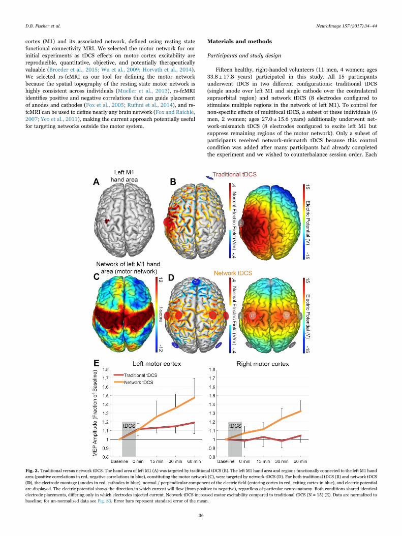
cortex (M1) and its associated network, defined using resting statefunctional connectivity MRI. We selected the motor network for ourinitial experiments as tDCS effects on motor cortex excitability arereproducible, quantitative, objective, and potentially therapeuticallyvaluable (Broeder et al., 2015; Wu et al., 2009; Horvath et al., 2014).We selected rs-fcMRI as our tool for defining the motor networkbecause the spatial topography of the resting state motor network ishighly consistent across individuals (Mueller et al., 2013), rs-fcMRIidentifies positive and negative correlations that can guide placementof anodes and cathodes (Fox et al., 2005; Ruffini et al., 2014), and rs-fcMRI can be used to define nearly any brain network (Fox and Raichle,2007; Yeo et al., 2011), making the current approach potentially usefulfor targeting networks outside the motor system.
Materials and methods
Participants and study design
Fifteen healthy, right-handed volunteers (11 men, 4 women; ages33.8 ± 17.8 years) participated in this study. All 15 participantsunderwent tDCS in two different configurations: traditional tDCS(single anode over left M1 and single cathode over the contralateralsupraorbital region) and network tDCS (8 electrodes configured tostimulate multiple regions in the network of left M1). To control fornon-specific effects of multifocal tDCS, a subset of these individuals (6men, 2 women; ages 27.0 ± 15.6 years) additionally underwent net-work-mismatch tDCS (8 electrodes configured to excite left M1 butsuppress remaining regions of the motor network). Only a subset ofparticipants received network-mismatch tDCS because this controlcondition was added after many participants had already completedthe experiment and we wished to counterbalance session order. Each
Fig. 2. Traditional versus network tDCS. The hand area of left M1 (A) was targeted by traditional tDCS (B). The left M1 hand area and regions functionally connected to the left M1 handarea (positive correlations in red, negative correlations in blue), constituting the motor network (C), were targeted by network tDCS (D). For both traditional tDCS (B) and network tDCS(D), the electrode montage (anodes in red, cathodes in blue), normal / perpendicular component of the electric field (entering cortex in red, exiting cortex in blue), and electric potentialare displayed. The electric potential shows the direction in which current will flow (from positive to negative), regardless of particular neuroanatomy. Both conditions shared identicalelectrode placements, differing only in which electrodes injected current. Network tDCS increased motor excitability compared to traditional tDCS (N = 15) (E). Data are normalized tobaseline; for un-normalized data see Fig. S3. Error bars represent standard error of the mean.
D.B. Fischer et al. NeuroImage 157 (2017) 34–44
36
session was held on separate days at least 48 h apart. Session order wascounter-balanced between participants, including those that receivedall three stimulation conditions. Each session consisted of a baselinemeasurement of motor excitability, application of tDCS, and reassess-ment of motor excitability at regular intervals (Fig. 1). We screenedparticipants for adverse effects of brain stimulation at the start and endof each session. This study was approved by the Beth Israel DeaconessMedical Center Institutional Review Board, and written informedconsent was obtained from all participants.
tDCS intervention
In each session, we applied traditional tDCS, network tDCS ornetwork-mismatch tDCS (see montage details below). For all condi-tions, we delivered tDCS through the Starstim tDCS system(Neuroelectrics, Barcelona, Spain), using 3.14 cm2 Ag/AgCl gelledelectrodes placed into holes of a neoprene cap corresponding to theinternational 10/10 EEG system, with the central Cz position aligned tothe vertex of the head. For all three conditions, we placed electrodes atC1, C2, C3, C4, Fz, P3, P4, T8 and Fp2, although only a subset of theseelectrodes was used in any one stimulation condition. As such, the cap,gel and electrode placement were identical for all conditions; only theamount of current delivered through each electrode varied. Currentwas delivered through each electrode via a wireless neurostimulator.For all conditions, tDCS was applied for 10 min, preceded by a 30 sramp up period and followed by a 30 s ramp down period. Mildtingling, warmth and/or itching were experienced in all conditions.
Traditional tDCS montageFor traditional tDCS, we delivered a total of 2 mA: 2 mA through C3
(the left M1 anode) and −2 mA through Fp2 (the contralateralsupraorbital cathode) (Fig. 2B, Fig. S1). We intentionally placedelectrodes at standard EEG electrode positions (e.g. C3) instead ofindividualized positions (e.g. over the motor hotspot), for threereasons. First, this C3-FP2 montage has been used in prior studies totarget left M1 (Hanley et al., 2015; Noetscher et al., 2014; Laakso et al.,2015). Second, standardized positioning facilitates modeling of theassociated electric fields (Miranda et al., 2013) and thus our optimiza-tion algorithm for matching a tDCS configuration to a functionalconnectivity map (Ruffini et al., 2014). Finally, our goal was to test ageneralizable approach to network-targeted stimulation that couldpotentially be applied to any cortical network, and individualizedhotspots are difficult to identify outside of the motor system.
Network tDCS montage. Generating our network tDCS montageconsisted of two main steps: 1) identifying the left M1 resting statefunctional connectivity (rs-fcMRI) map and 2) finding an electrodemontage that best matches this network map.
First, we used rs-fcMRI to identify the brain network associatedwith the left M1 hand area. To do so, we defined a region of interest(ROI) in the left M1 hand area: we used a 6 mm radius sphere based onROI sizes in prior rs-fcMRI experiments (Fox et al., 2005), andcentered the sphere on Montreal Neurological Institute (MNI) coordi-nates from a prior study of fMRI activation for right hand movements(x=−41, y=−20, z=62; Fig. 2A) (Buckner et al., 2011). We then usedthis left M1 ROI as a “seed region” to generate a rs-fcMRI map, usingan existing rs-fcMRI dataset from 98 healthy, right handed individuals(48 males, ages 22 ± 3.2 years). Details of this dataset and itsassociated processing have been reported previously (Fox et al.,2012a). Briefly, subjects were instructed to rest quietly in an MRIscanner while spontaneous fluctuations in brain activity were mea-sured. Rs-fcMRI preprocessing steps included spatial smoothing with a6 mm full-width at half-maximum Gaussian kernel, temporal filtering(0.009–0.08 Hz) and the removal of the following variables by regres-
sion: standard six movement parameters, mean whole brain signal,mean brain signal within the lateral ventricles, and the mean signalwithin a deep white matter ROI. The first temporal derivatives of theseregressors were included within the linear model to account for time-shifted versions of spurious variance. Seed-based functional connectiv-ity analysis was performed by extracting spontaneous fluctuations infMRI activity from the left M1 hand ROI, then computing the Pearson'scorrelation between this extracted time course and the time course ofall other brain voxels. Results were combined across the 98 subjects byconverting r values to Fisher z values and performing a random effectsanalysis (Student's T-test).
The result of this analysis is a map of resting state functionalconnectivity with the left M1 hand region. The map includes positivecorrelations over the bilateral primary motor cortex, as reportedpreviously (Yeo et al., 2011; Biswal et al., 1995; Fox and Raichle,2007), and negative correlations over the prefrontal and parietalcortices (Fig. 2C). The functional significance of negatively correlatedbrain regions remains a matter of debate (Fox et al., 2009; Murphyet al., 2009), but may reflect brain regions with opposing functions(Fox et al., 2005). Peak coordinates in this rs-fcMRI map wereidentified using an automated peak search algorithm with a t scorethreshold of > 4.25 or < −0.4.25 (Table S1).
Based on this left M1 rs-fcMRI map, we then used an algorithm(Ruffini et al., 2014) to identify an 8 electrode configuration (based onthe 10/10 EEG system) that would produce an electric field optimallymatched to this network map (StimWeaver, Neuroelectrics). Thedetails of this algorithm and the finite element model upon which itis based have been published previously (Ruffini et al., 2014; Mirandaet al., 2013). Briefly, MRI data from the publically available Colin27brain was segmented into 5 different tissues (scalp, skull, cerebrospinalfluid, grey matter and white matter; Fig. S2) using freeware tools(Freesurfer, www.freesurfer.net; ITK-Snap, http://www.itksnap.org/;and SPM 8, http://www.fil.ion.ucl.ac.uk/spm/ with the toolbox MARS,http://neuralengr.com/mars/). The segmentation masks wereprocessed for continuity and smoothed using scripts written inMatlab (www.mathworks.com). The electrodes were modeled ascylinders with a radius of 1 cm and a thickness of 3 mm,representing the conductive gel beneath the electrode cap. A volumeFE mesh with ~4.8 million tetrahedral second order elements wascreated using Iso2Mesh (http://iso2mesh.sourceforge.net/), a Matlabtoolbox. The mesh was imported into Comsol (www.comsol.com)where the E-field calculations were performed. All tissues wereconsidered homogeneous and isotropic.
A genetic algorithm was then used to identify the location ofelectrodes within the 10-10 EEG grid system, and the current toadminister through each electrode, to best match the spatial topogra-phy of the rs-fcMRI motor network (Ruffini et al., 2014). Thisalgorithm matched the components of the electric field entering thecortex at a normal (i.e., perpendicular) angle (thought to be excitatory)to positive correlations in the motor network map, and components ofthe electric field exiting the cortex at a normal angle (thought to beinhibitory) to the negative correlations. The ideal solution was the onethat minimized error relative to no intervention (ERNI), a goodness offit measure (Ruffini et al., 2014). The tDCS algorithm was constrainedby safety parameters, in which the total injected current could notexceed 4 mA, and no more than 2 mA could be injected per electrode,consistent with the safety limits of the Starstim device. The algorithmidentified the optimal electrode configuration and amperage to be:872 uA from C1, 888 uA from C2, 1135 uA from C3, 922 uA from C4,−1843 uA from Fz, −1121uA from P3, −1036uA from P4, and 183uAfrom T8 (4 mA total; Fig. 2D).
Network-mismatch tDCS montage. As a control multifocalstimulation condition, network-mismatch tDCS was configured tokeep left M1 stimulation similar to or higher than that of networktDCS, while opposing the remaining motor network. The network-
D.B. Fischer et al. NeuroImage 157 (2017) 34–44
37

mismatch configuration controlled for the higher total injected current(4 mA) of network tDCS, and for potential multifocal scalp sensations.We maintained the currents at C1 and C3, and for the remaining 6electrodes, reversed the polarity from that of network tDCS, such thatanodes became cathodes (suppressing positively correlated nodes), andcathodes became anodes (exciting negative correlated nodes). Toensure current conservation, we adjusted the currents for thesereversed electrodes, maintaining proportions between cathodes andbetween non-left M1 anodes (i.e., C2, C4 and T8 were proportionatelyscaled from network tDCS, as were Fz, P3 and P4). Thus, network-mismatch tDCS was delivered as follows: 872 uA from C1, −1782 uAfrom C2, 1135 uA from C3, −1851uA from C4, 919 uA from Fz, 559 uAfrom P3, 515 uA from P4, and −367uA from T8 (4 mA total; Fig. 3B).
We generated all electric field and electric potentials for figures withmodeling software (Neuroelectrics Instrument Controller,Neuroelectrics).
Cortical excitability measures
We assessed how each tDCS configuration affected cortical excit-ability by applying TMS to left and right M1 to elicit muscle contrac-tions from the contralateral hand. The strength of the muscle contrac-tion was recorded as a MEP, the amplitude of which reflects corticalexcitability from the targeted M1 region (Bestmann and Krakauer,2015).
We used a figure-of-8 TMS coil (Model Cool-B65; MagPro X100,MagVenture, Atlanta, Georgia, USA) to find left and right hand motorhotspots, defined as the optimal cortical locations to elicit MEPs fromthe contralateral first dorsal interosseus muscle (FDI). We held theTMS coil tangential to the head, with the handle rotated approximately45° away from the midsagittal plane, and delivered single pulses atsuprathreshold intensity in ~1 cm intervals. MEPs were measuredusing surface electromyography, with gelled electrodes positioned overthe belly and tendon of the left and right FDI muscle, and recorded at4 kHz with a band-pass filter of 0.3–1000 Hz, a 5 mV range, a 200 mstimespan of 1024 samples, and a 60 Hz notch and mains filter (Scope,
Fig. 3. Network versus network-mismatch tDCS. The electrode configuration and electric potentials of network tDCS (A) and network-mismatch tDCS (B), with anodes in red andcathodes in blue. Both conditions shared identical electrode placements, differing only in injected current. Network tDCS increased motor excitability, whereas network-mismatch tDCSdid not (n = 8) (C). Data are normalized to baseline; for un-normalized data see Fig. S4. Error bars represent standard error of the mean.
D.B. Fischer et al. NeuroImage 157 (2017) 34–44
38
PowerLab 4/25 T, Ad instruments, Colorado Springs, Colorado, USA).The location and position of the left and right hand motor hotspotswere recorded using a neuronavigation system (Localite, SanktAugustin, Germany) (Santarnecchi et al., 2014). Because most partici-pants did not have their own MRI, a standard MRI brain atlas(MNI152) was used to record the motor hotspots. The use of a standardbrain with neuronavigation allows for reproducible positioning of theTMS coil, but does not allow one to relate the hotspot to individualanatomy. Four participants had their own MRI from participation in aprior experiment, and in these cases the participant's own MRI wasused for neuronavigation.
After identifying the motor hotspots, we applied the tDCS cap withthe stimulation electrodes inserted, and administered electricallyconductive gel beneath each electrode with a syringe. We then used aTMS-robot (Axilum, Strasbourg, France) to position the TMS coil overthe previously defined left and right stimulation sites at the previouslydefined orientations (Supplemental Video 1), the first use of suchtechnology to assess tDCS effects. The robotic TMS not only optimizedstimulation accuracy and precision (Ginhoux et al., 2013; Richter et al.,2013), which can improve MEP measurements (Toschi et al., 2009),but also eliminated any bias associated with hand-held TMS adminis-tration. The TMS-robot held the same TMS coil model used to find theoptimal stimulation sites. We adjusted the force sensitivity of the TMS-robot to accommodate the cap and electrodes beneath the coil. TMSwas applied over/through the tDCS cap as in prior neuronavigatedTMS/tDCS experiments to avoid introducing error into the neurona-vigation (Santarnecchi et al., 2014). Specifically, placing and removingthe tDCS cap between TMS measurements could displace the subjecttracker, resulting in small offsets in the TMS coil position.
Supplementary material related to this article can be found onlineat doi:10.1016/j.neuroimage.2017.05.060.
Using the robot-administered TMS, resting motor thresholds werecomputed for both the left and right M1, defined as the minimum TMSintensity required to elicit a peak-to-peak MEP amplitude ≥50 uV in atleast 50% of 10 consecutive trials. TMS intensity was increased to 120%of the resting motor threshold, then adjusted to elicit MEPs of 1–1.5 mV (or until maximum stimulator output was reached). Five of our15 participants required maximum stimulator output at some pointduring the experiment (3 required max output for both traditional andnetwork tDCS, 1 for network tDCS only, and 1 for traditional tDCSonly).
To assess baselines, we used the TMS-robot to deliver 60 singlepulses of TMS at the previously determined intensities to each the leftand right motor cortices. Baselines were collected in 2 sets of 30,alternating between the left and right hemispheres. All TMS pulseswere administered from an automated system that randomly jitteredthe TMS pulse intervals, such that pulses occurred anywhere between 5and 10 s apart. For all MEPs, we instructed participants to keep theireyes open and hands relaxed. EMG activity was monitored in real timeto ensure muscles were relaxed prior to each TMS assessment. If EMGactivity was present, subject's hands were repositioned and relaxationinstructions were repeated. We reassessed cortical excitability fromboth hemispheres immediately after tDCS was complete, 15 min after,30 min after and 60 min after. At each time point, we delivered 30,jittered, single pulses of TMS to the left, then the right, motor cortices.We recorded the peak-to-peak amplitude of each MEP. In rare cases (< 5% of all blocks), muscle activation was observed between TMSpulses during MEP acquisition. In such cases, the TMS assessment wasstopped, all MEPs for that time point were deleted, and 30 MEPs werere-acquired after repeating relaxation instructions. These events almostalways occurred during baseline (pre-tDCS) assessment as subjectslearned proper hand positioning and relaxation. Participants usuallyremained seated between measurement periods, but were permitted tostand, walk, and use the bathroom if necessary.
Data analysis
For each participant, tDCS session, and hemisphere, MEPs wereaveraged at each time point. The first MEP for each time point wasexcluded, but all other MEPs were included with no post-hoc proces-sing or exclusions. To assess differences between stimulation condi-tions, considering all time points and both hemispheres, we used amixed effect linear regression model (SAS Institute Inc., Cary, NorthCarolina, USA). This approach was selected over the more commonanalysis of variance (ANOVA) because the linear regression model 1)accounts for the continuity of time (i.e. time points occurring in aspecific order) and 2) better accounts for inter-subject variability inbaseline MEP (Fitzmaurice et al., 2011). Our linear model included arandom intercept for each participant, and fixed effects terms for maineffects of baseline MEP, hemisphere, condition (traditional tDCS vsnetwork tDCS vs network-mismatch tDCS), time (minutes from tDCS)and the interactions of time x hemisphere, time x condition, and time xhemisphere x condition. Baseline MEP was included as a fixed effectrather than just another time point so tDCS response would be assessedrelative to this baseline. This is similar to MEP normalization oftenused in tDCS analysis, i.e. computing the ratio between post tDCS andbaseline MEPs (M.A. Nitsche and Paulus, 2000; Horvath et al., 2014).However, our approach has the advantage of accounting for variance intDCS response as a function of baseline MEP, a relationship which hasbeen previously observed (Wiethoff et al., 2014; Tremblay et al., 2016).Including baseline MEP as a fixed effect controls for this variance,allowing us to better identify differences between stimulation condi-tions. The same model parameters were used to compare traditionaltDCS to network tDCS (in all 15 participants), network tDCS tonetwork-mismatch tDCS (in the subset of 8 participants) and tradi-tional tDCS to network-mismatch tDCS (in the subset of 8 partici-pants). All tDCS conditions could not be incorporated simultaneouslyinto a single model, as each contained different numbers of partici-pants. We evaluated significant effects post hoc using stratifiedanalyses, entering data from each hemisphere and each conditionseparately into the linear model. All statistics were computed using theraw MEP data, which were found to be normally distributed (more sothan data normalized to baselines) and thus appropriate for the abovestatistical analysis. For display purposes, each MEP time point wasexpressed as a ratio to its respective baseline. Such normalizationallows for easier visual comparison between conditions and conformsto convention in the tDCS literature (Lang et al., 2004). Time coursesfrom the raw MEP data are presented as supplemental material.
In a post-hoc analysis, we tested whether differences in muscleactivity just prior to each TMS pulse could explain our MEP results. Wefocused our analysis on the most relevant conditions and time points(traditional tDCS versus network tDCS effects in left M1, baselineversus 60 min post-tDCS). The root-mean-square (RMS) of the EMGfor the 50 msec prior to each TMS pulse was computed. Pre-triggerEMG was averaged for the 60 baseline pulses for each subject and the30 pulses at 60 min post-tDCS. Time points and conditions werecompared using paired t-tests (two-tailed).
Post-hoc modeling of tDCS and individual differences
To aid in interpretation of results, we performed several modelinganalyses. First, we used our Colin27 head model to estimate the electricfield (both total magnitude and normal component) for each of ourthree tDCS montages. The goodness of fit between our target network(i.e., the rs-fcMRI motor network) and the electric field produced byeach of our three tDCS montages was computed using two goodness-of-fit measures as in prior work (Ruffini et al., 2014): the error relative tono intervention (ERNI), and the weighted cross correlation coefficient(WCC). A closer match between the electric field and the target networkresults in a smaller / more negative ERNI and a WCC closer to 1. Toestimate the local effects of our tDCS montages on left M1 itself, we
D.B. Fischer et al. NeuroImage 157 (2017) 34–44
39

computed the electric field (total and normal component) within ourleft M1 ROI, defined by the same 6 mm cortical sphere used to generatethe rs-fcMRI motor network.
To ensure that these modeling results, based on a head model froma standard reference brain and group-averaged rs-fcMRI data, general-ize to individual participants, we repeated all analyses using indivi-dualized MRI data from 4 participants in the present experiment.These four participants completed an MRI scan (GE 3 T HDX scanner)including structural and rs-fcMRI sequences as part of a separate priorexperiment. T1-weighted structural images were acquired via a 3D-turbo field echo sequence (TE= 2.9 ms, flip angle=15°, 0.94 × 0.94 ×1 mm voxels). Each subject completed three 6.4 min long resting statefMRI scans (124 volumes, TR=3200 ms, TE=30 ms, flip angle=90°,3.75 × 3.75 × 3 mm voxels). BOLD imaging employed fat saturation tominimize signal loss and Array Spatial Sensitivity Encoding Technique(ASSET) with acceleration factor 2 to minimize geometric distortion.During the resting state fMRI scans the subjects were asked to lay asstill as possible and stare at a fixation cross.
Anatomical MRI data was used to generate four individualizedfinite element models using the same procedure described above forthe Colin27 brain (Fig. S2) (Ruffini et al., 2014; Miranda et al., 2013).Rs-fcMRI data was processed using the procedures described above,and individualized rs-fcMRI motor networks were identified for thesefour participants. Finally, individualized left M1 ROIs were definedalong the pre-central gyrus based on the anatomical location of thehand knob. Using this individualized data, the electric field intensitywithin the left M1 ROI, and the fit between the tDCS electric fields andrs-fcMRI motor network, were calculated for each of the four partici-pants.
Results
We compared traditional two-electrode tDCS targeting the left M1(Fig. 2A, B) to “network” tDCS targeting the left M1 and its associatedresting state network (Fig. 2C, D). Network tDCS increased corticalexcitability beyond that seen with traditional tDCS (Fig. 2E, Fig. S3).Statistical analysis revealed significant main effects of time (b = 0.019,t(218) = 7.72, p < 0.0001), condition (b = −0.22, t(218) = −2.45, p <0.05), and hemisphere (b = −0.27, t(218) = −2.97, p < 0.01), with a
significant time x condition x hemisphere interaction (b = 0.012, t(218)= 3.68, p < 0.001).
Focusing specifically on left M1, network tDCS induced a greaterrise in excitability over time (condition x time interaction in left M1, b= −0.02, t(101) = −4.89, p < 0.0001), with a trend towards a maineffect of condition (b = −0.23, t(101) = −1.83, p = 0.07). At the lasttime point, one hour after stimulation, the increase in left M1excitability following network tDCS was more than double the increasefollowing traditional tDCS (47% vs 19% increase) and still rising(Fig. 2E).
In right M1, there was a similar trend towards greater excitabilityfollowing network tDCS (main effect of condition: b = −0.23, t(101) =−1.80, p = 0.08), but no significant condition x time interaction (b =−0.01, t(101) = −1.28, p = 0.20).
Stratified by condition, there was a main effect of hemisphere inboth traditional tDCS (b = −0.27, t(101) = −2.42, p < 0.05) andnetwork tDCS (b = −0.29, t(101) = −2.24, p < 0.05), whereby bothconditions increased left M1 excitability above right M1 excitability.Network tDCS also increased cortical excitability more steeply in leftM1 than in right M1 (time x hemisphere interaction in network tDCS, b= −0.01, t(101) = −3.29, p < 0.01). A full report of statistical outputsfor traditional and network tDCS can be found in Table S2.
In the subset of participants who underwent the control experi-ment, we compared network tDCS to network-mismatch tDCS.Network-mismatch tDCS was designed to maintain left M1 stimulationand total injected current of network tDCS, but oppose rather thanbolster the remaining motor network (Fig. 3A, B). Network-mismatchtDCS did not induce the same elevation in left M1 excitability asnetwork tDCS (Fig. 3C, Fig S4). Statistically, there was a significantmain effect of condition, whereby network-mismatch tDCS resulted incortical excitability below that of network tDCS (b = −0.33, t(113) =−2.96, p < 0.005); there were no significant main effects of time orhemisphere, nor were there significant interactions. Considering eachhemisphere individually, a main effect of condition was present in rightM1 (b = −0.41, t(52) = −2.29, p < 0.05), which was expected given thereversed polarity of stimulation there, but also in left M1 (b = −0.29,t(52) = −2.31, p < 0.05), where the delivered stimulation was nearlyidentical between conditions.
Comparing network-mismatch tDCS to traditional tDCS (Fig. S5),
Fig. 4. Individualized motor networks and electric fields. The motor network (A) and modeled electric fields for our three tDCS montages (B) were determined using subject-specificMRI data for a subset of 4 participants. Results from one representative subject are shown here (see Fig. S6, Fig. S7 for other subjects). Both the normal component (top row) and thetotal magnitude (bottom row) of the modeled electric field are displayed. The fit between each subject's motor network and their individualized electric fields was computed using aweighted cross-correlation coefficient (WCC), averaged across the four participants (C). The normal component of the electric field from network tDCS best matches the motor network.
D.B. Fischer et al. NeuroImage 157 (2017) 34–44
40
there was a significant main effect of condition, with network-mis-match tDCS suppressing cortical excitability below that of traditionaltDCS (b = 0.26, t(113) = 2.27, p < 0.05). This effect was significant inleft M1 alone (b = 0.30, t(52) = 2.05, p < 0.05), with a trend towardssignificance in the right M1 alone (b = 0.27, t(52) = 1.85, p = 0.07).Thus, network-mismatch tDCS not only failed to elevate left M1excitability to the same extent as network tDCS, but it effectivelysuppressed the elevation in left M1 excitability normally induced bytraditional tDCS. A full report of statistical outputs for network-mismatch tDCS and its comparisons can be found in Table S3.
There was no carry over of excitability changes between sessions.Specifically, there were no significant differences in baseline excitabilityin the LM1 (p = 0.89) or RM1 (p = 0.88) between the first and secondsessions, even if network tDCS was performed first (LM1, p = 0.88;RM1, p = 0.63). There was no difference in muscle activity prior to theTMS pulses at time points showing the largest MEP effects. Specifically,pre-trigger EMG activity in LM1 was similar at baseline versus 60 minpost-tDCS for both the traditional tDCS (0.035 mV vs 0.032 mV, p =0.56) and network tDCS conditions (0.027 mV vs 0.031 mV, p = 0.35)and was similar at 60 min post-tDCS directly comparing the traditionaland network tDCS conditions (0.032 mV vs 0.031 mV, p = 0.90).
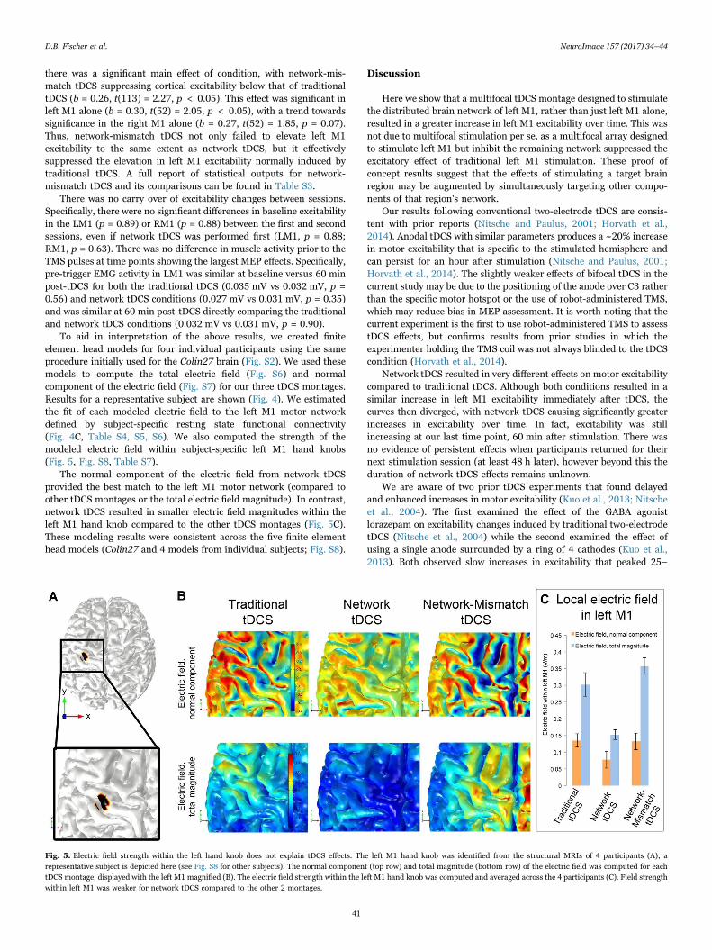
To aid in interpretation of the above results, we created finiteelement head models for four individual participants using the sameprocedure initially used for the Colin27 brain (Fig. S2). We used thesemodels to compute the total electric field (Fig. S6) and normalcomponent of the electric field (Fig. S7) for our three tDCS montages.Results for a representative subject are shown (Fig. 4). We estimatedthe fit of each modeled electric field to the left M1 motor networkdefined by subject-specific resting state functional connectivity(Fig. 4C, Table S4, S5, S6). We also computed the strength of themodeled electric field within subject-specific left M1 hand knobs(Fig. 5, Fig. S8, Table S7).
The normal component of the electric field from network tDCSprovided the best match to the left M1 motor network (compared toother tDCS montages or the total electric field magnitude). In contrast,network tDCS resulted in smaller electric field magnitudes within theleft M1 hand knob compared to the other tDCS montages (Fig. 5C).These modeling results were consistent across the five finite elementhead models (Colin27 and 4 models from individual subjects; Fig. S8).
Discussion
Here we show that a multifocal tDCS montage designed to stimulatethe distributed brain network of left M1, rather than just left M1 alone,resulted in a greater increase in left M1 excitability over time. This wasnot due to multifocal stimulation per se, as a multifocal array designedto stimulate left M1 but inhibit the remaining network suppressed theexcitatory effect of traditional left M1 stimulation. These proof ofconcept results suggest that the effects of stimulating a target brainregion may be augmented by simultaneously targeting other compo-nents of that region's network.
Our results following conventional two-electrode tDCS are consis-tent with prior reports (Nitsche and Paulus, 2001; Horvath et al.,2014). Anodal tDCS with similar parameters produces a ~20% increasein motor excitability that is specific to the stimulated hemisphere andcan persist for an hour after stimulation (Nitsche and Paulus, 2001;Horvath et al., 2014). The slightly weaker effects of bifocal tDCS in thecurrent study may be due to the positioning of the anode over C3 ratherthan the specific motor hotspot or the use of robot-administered TMS,which may reduce bias in MEP assessment. It is worth noting that thecurrent experiment is the first to use robot-administered TMS to assesstDCS effects, but confirms results from prior studies in which theexperimenter holding the TMS coil was not always blinded to the tDCScondition (Horvath et al., 2014).
Network tDCS resulted in very different effects on motor excitabilitycompared to traditional tDCS. Although both conditions resulted in asimilar increase in left M1 excitability immediately after tDCS, thecurves then diverged, with network tDCS causing significantly greaterincreases in excitability over time. In fact, excitability was stillincreasing at our last time point, 60 min after stimulation. There wasno evidence of persistent effects when participants returned for theirnext stimulation session (at least 48 h later), however beyond this theduration of network tDCS effects remains unknown.
We are aware of two prior tDCS experiments that found delayedand enhanced increases in motor excitability (Kuo et al., 2013; Nitscheet al., 2004). The first examined the effect of the GABA agonistlorazepam on excitability changes induced by traditional two-electrodetDCS (Nitsche et al., 2004) while the second examined the effect ofusing a single anode surrounded by a ring of 4 cathodes (Kuo et al.,2013). Both observed slow increases in excitability that peaked 25–
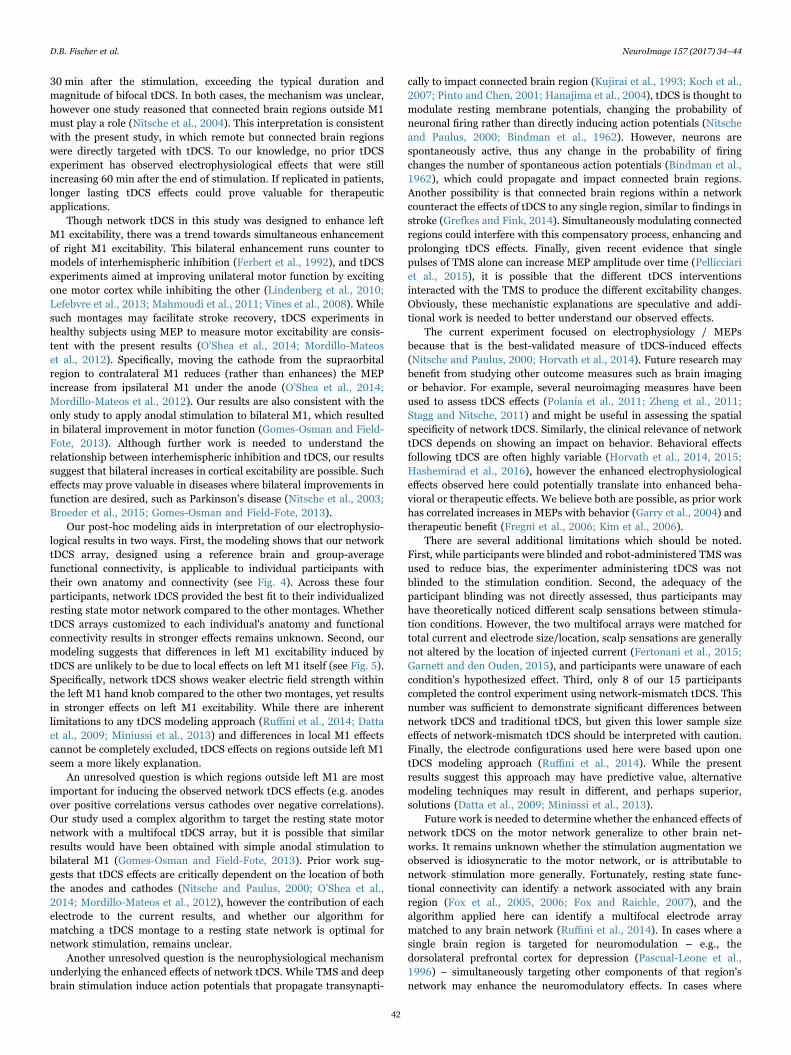
Fig. 5. Electric field strength within the left hand knob does not explain tDCS effects. The left M1 hand knob was identified from the structural MRIs of 4 participants (A); arepresentative subject is depicted here (see Fig. S8 for other subjects). The normal component (top row) and total magnitude (bottom row) of the electric field was computed for eachtDCS montage, displayed with the left M1 magnified (B). The electric field strength within the left M1 hand knob was computed and averaged across the 4 participants (C). Field strengthwithin left M1 was weaker for network tDCS compared to the other 2 montages.
D.B. Fischer et al. NeuroImage 157 (2017) 34–44
41
30 min after the stimulation, exceeding the typical duration andmagnitude of bifocal tDCS. In both cases, the mechanism was unclear,however one study reasoned that connected brain regions outside M1must play a role (Nitsche et al., 2004). This interpretation is consistentwith the present study, in which remote but connected brain regionswere directly targeted with tDCS. To our knowledge, no prior tDCSexperiment has observed electrophysiological effects that were stillincreasing 60 min after the end of stimulation. If replicated in patients,longer lasting tDCS effects could prove valuable for therapeuticapplications.
Though network tDCS in this study was designed to enhance leftM1 excitability, there was a trend towards simultaneous enhancementof right M1 excitability. This bilateral enhancement runs counter tomodels of interhemispheric inhibition (Ferbert et al., 1992), and tDCSexperiments aimed at improving unilateral motor function by excitingone motor cortex while inhibiting the other (Lindenberg et al., 2010;Lefebvre et al., 2013; Mahmoudi et al., 2011; Vines et al., 2008). Whilesuch montages may facilitate stroke recovery, tDCS experiments inhealthy subjects using MEP to measure motor excitability are consis-tent with the present results (O’Shea et al., 2014; Mordillo-Mateoset al., 2012). Specifically, moving the cathode from the supraorbitalregion to contralateral M1 reduces (rather than enhances) the MEPincrease from ipsilateral M1 under the anode (O’Shea et al., 2014;Mordillo-Mateos et al., 2012). Our results are also consistent with theonly study to apply anodal stimulation to bilateral M1, which resultedin bilateral improvement in motor function (Gomes-Osman and Field-Fote, 2013). Although further work is needed to understand therelationship between interhemispheric inhibition and tDCS, our resultssuggest that bilateral increases in cortical excitability are possible. Sucheffects may prove valuable in diseases where bilateral improvements infunction are desired, such as Parkinson's disease (Nitsche et al., 2003;Broeder et al., 2015; Gomes-Osman and Field-Fote, 2013).
Our post-hoc modeling aids in interpretation of our electrophysio-logical results in two ways. First, the modeling shows that our networktDCS array, designed using a reference brain and group-averagefunctional connectivity, is applicable to individual participants withtheir own anatomy and connectivity (see Fig. 4). Across these fourparticipants, network tDCS provided the best fit to their individualizedresting state motor network compared to the other montages. WhethertDCS arrays customized to each individual's anatomy and functionalconnectivity results in stronger effects remains unknown. Second, ourmodeling suggests that differences in left M1 excitability induced bytDCS are unlikely to be due to local effects on left M1 itself (see Fig. 5).Specifically, network tDCS shows weaker electric field strength withinthe left M1 hand knob compared to the other two montages, yet resultsin stronger effects on left M1 excitability. While there are inherentlimitations to any tDCS modeling approach (Ruffini et al., 2014; Dattaet al., 2009; Miniussi et al., 2013) and differences in local M1 effectscannot be completely excluded, tDCS effects on regions outside left M1seem a more likely explanation.
An unresolved question is which regions outside left M1 are mostimportant for inducing the observed network tDCS effects (e.g. anodesover positive correlations versus cathodes over negative correlations).Our study used a complex algorithm to target the resting state motornetwork with a multifocal tDCS array, but it is possible that similarresults would have been obtained with simple anodal stimulation tobilateral M1 (Gomes-Osman and Field-Fote, 2013). Prior work sug-gests that tDCS effects are critically dependent on the location of boththe anodes and cathodes (Nitsche and Paulus, 2000; O’Shea et al.,2014; Mordillo-Mateos et al., 2012), however the contribution of eachelectrode to the current results, and whether our algorithm formatching a tDCS montage to a resting state network is optimal fornetwork stimulation, remains unclear.
Another unresolved question is the neurophysiological mechanismunderlying the enhanced effects of network tDCS. While TMS and deepbrain stimulation induce action potentials that propagate transynapti-
cally to impact connected brain region (Kujirai et al., 1993; Koch et al.,2007; Pinto and Chen, 2001; Hanajima et al., 2004), tDCS is thought tomodulate resting membrane potentials, changing the probability ofneuronal firing rather than directly inducing action potentials (Nitscheand Paulus, 2000; Bindman et al., 1962). However, neurons arespontaneously active, thus any change in the probability of firingchanges the number of spontaneous action potentials (Bindman et al.,1962), which could propagate and impact connected brain regions.Another possibility is that connected brain regions within a networkcounteract the effects of tDCS to any single region, similar to findings instroke (Grefkes and Fink, 2014). Simultaneously modulating connectedregions could interfere with this compensatory process, enhancing andprolonging tDCS effects. Finally, given recent evidence that singlepulses of TMS alone can increase MEP amplitude over time (Pellicciariet al., 2015), it is possible that the different tDCS interventionsinteracted with the TMS to produce the different excitability changes.Obviously, these mechanistic explanations are speculative and addi-tional work is needed to better understand our observed effects.
The current experiment focused on electrophysiology / MEPsbecause that is the best-validated measure of tDCS-induced effects(Nitsche and Paulus, 2000; Horvath et al., 2014). Future research maybenefit from studying other outcome measures such as brain imagingor behavior. For example, several neuroimaging measures have beenused to assess tDCS effects (Polanía et al., 2011; Zheng et al., 2011;Stagg and Nitsche, 2011) and might be useful in assessing the spatialspecificity of network tDCS. Similarly, the clinical relevance of networktDCS depends on showing an impact on behavior. Behavioral effectsfollowing tDCS are often highly variable (Horvath et al., 2014, 2015;Hashemirad et al., 2016), however the enhanced electrophysiologicaleffects observed here could potentially translate into enhanced beha-vioral or therapeutic effects. We believe both are possible, as prior workhas correlated increases in MEPs with behavior (Garry et al., 2004) andtherapeutic benefit (Fregni et al., 2006; Kim et al., 2006).
There are several additional limitations which should be noted.First, while participants were blinded and robot-administered TMS wasused to reduce bias, the experimenter administering tDCS was notblinded to the stimulation condition. Second, the adequacy of theparticipant blinding was not directly assessed, thus participants mayhave theoretically noticed different scalp sensations between stimula-tion conditions. However, the two multifocal arrays were matched fortotal current and electrode size/location, scalp sensations are generallynot altered by the location of injected current (Fertonani et al., 2015;Garnett and den Ouden, 2015), and participants were unaware of eachcondition's hypothesized effect. Third, only 8 of our 15 participantscompleted the control experiment using network-mismatch tDCS. Thisnumber was sufficient to demonstrate significant differences betweennetwork tDCS and traditional tDCS, but given this lower sample sizeeffects of network-mismatch tDCS should be interpreted with caution.Finally, the electrode configurations used here were based upon onetDCS modeling approach (Ruffini et al., 2014). While the presentresults suggest this approach may have predictive value, alternativemodeling techniques may result in different, and perhaps superior,solutions (Datta et al., 2009; Miniussi et al., 2013).
Future work is needed to determine whether the enhanced effects ofnetwork tDCS on the motor network generalize to other brain net-works. It remains unknown whether the stimulation augmentation weobserved is idiosyncratic to the motor network, or is attributable tonetwork stimulation more generally. Fortunately, resting state func-tional connectivity can identify a network associated with any brainregion (Fox et al., 2005, 2006; Fox and Raichle, 2007), and thealgorithm applied here can identify a multifocal electrode arraymatched to any brain network (Ruffini et al., 2014). In cases where asingle brain region is targeted for neuromodulation – e.g., thedorsolateral prefrontal cortex for depression (Pascual-Leone et al.,1996) – simultaneously targeting other components of that region'snetwork may enhance the neuromodulatory effects. In cases where
D.B. Fischer et al. NeuroImage 157 (2017) 34–44
42

functions better localize to networks than to single regions, such asattention (Gitelman et al., 1999; Posner, 2012) and memory (Iidakaet al., 2005), or when treating brain diseases associated with networkdysfunction, such as Alzheimer's Disease (Fox et al., 2014; Greiciuset al., 2004) and depression (Fox et al., 2014; Broyd et al., 2009;Mayberg, 2009), network-targeted neuromodulation may prove parti-cularly valuable.
In conclusion, our results add to data suggesting that brainconnectivity, especially resting state functional connectivity, may beuseful for guiding noninvasive brain stimulation (Wang et al., 2014;Chen et al., 2013; Volz et al., 2015; Fox et al., 2014, 2012b). NetworktDCS to the motor network appears to enhance the electrophysiologicaleffects of regional stimulation to M1 alone. Further investigation isnecessary to determine the mechanism and generalizability of thisresult. Whether network tDCS will help align neuromodulation with thenetwork-based focus of contemporary neuroscience can be tested infuture experiments aimed at modulating cognition, behavior, or diseaselocalizing to brain networks.
Funding
This work was supported by the Howard Hughes Medical Institute,Parkinson's Disease Foundation, Dystonia Medical ResearchFoundation, NIH/National Institute of Neurological Disorders andStroke (grant number K23NS083741), Sidney R. Baer Jr.Foundation, NIH (grant numbers R21 NS082870, R01HD069776,R01NS073601, R21 MH099196, R21 NS085491, R21 HD07616),and Harvard Catalyst | The Harvard Clinical and TranslationalScience Center (NCRR and the NCATS NIH, UL1 RR025758).
Competing Interests
GR is a cofounder, OR is a software development manager, and APLserves on the scientific advisory board of Neuroelectrics, whichproduces the brain stimulation device and software used in this study.APL serves on the scientific advisory board for Axilum Robotics (whichmanufactures the TMS robot used in this study), and is listed as aninventor on several issued and pending patents on the real-timeintegration of TMS with EEG and MRI. MDF is listed as an inventoron pending patents on combining TMS and fMRI. GR, OR, APL andMDF are listed inventors on a filed patent for multifocal tCS.
Appendix A. Supporting information
Supplementary data associated with this article can be found in theonline version at doi:10.1016/j.neuroimage.2017.05.060.
References
Bestmann, Sven, Krakauer, John W., 2015. The Uses and Interpretations of theMotor‑evoked Potential for Understanding Behaviour. Exp. Brain Res. 233 (3),679–689.
Bindman, Lynn J., Lippold, O.C.J., Redfearn, J.W.T., 1962. Long-Lasting Changes in theLevel of the Electrical Activity of the Cerebral Cortex Produced by PolarizingCurrents. Nature 196, 584–585.
Biswal, Bharat B., Yetkin, F.Z., Haughton, V.M., Hyde, J.S., 1995. Functionalconnectivity in the motor cortex of resting human brain using echo-planar MRI.Magn. Reson. Med.: Off. J. Soc. Magn. Reson. Med. / Soc. Magn. Reson. Med. 34 (4),537–541.
Broeder, Sanne, Nackaerts, Evelien, Heremans, Elke, Vervoort, Griet, Meesen, Raf,Verheyden, Geert, Nieuwboer, Alice, 2015. Transcranial Direct Current Stimulationin Parkinson's Disease: neurophysiological Mechanisms and Behavioral Effects.Neurosci. Biobehav. Rev. 57, 105–117.
Broyd, Samantha J., Demanuele, Charmaine, Debener, Stefan, Helps, Suzannah K.,James, Christopher J., Sonuga-Barke, Edmund J.S., 2009. Default-Mode BrainDysfunction in Mental Disorders: a Systematic Review. Neurosci. Biobehav. Rev. 33(3), 279–296.
Brunoni, Andre Russowsky, Ferrucci, Roberta, Fregni, Felipe, Sergio Boggio, Paulo,Priori, Alberto, 2012. Transcranial direct current stimulation for the treatment ofmajor depressive disorder: a summary of preclinical, clinical and translationalfindings. Progress. Neuro-Psychopharmacol. Biol. Psychiatry 39 (1), 9–16.
Buckner, R.L., Krienen, F.M., Castellanos, A., Diaz, J.C., Yeo, B.T.T., 2011. Theorganization of the human cerebellum estimated by intrinsic functional Connectivity.J. Neurophysiol. 106 (5), 2322–2345.
Caparelli-Daquer, EgasM., Zimmermann, TrelawnyJ., Mooshagian, Eric, Parra, LucasC.,Rice, JustinK., Datta, Abhishek, Bikson, Marom, Wassermann, EricM., 2012. A PilotStudy on Effects of 4×1 High-Definition tDCS on Motor Cortex Excitability.Conference Proceedings: … Annual International Conference of the IEEEEngineering in Medicine and Biology Society. IEEE Engineering in Medicine andBiology Society. Annual Conference 2012: 735–38.
Chen, Ashley C., Oathes, Desmond J., Chang, Catie, Bradley, Travis, Zhou, Zheng-Wei,Williams, Leanne M., Glover, Gary H., Deisseroth, Karl, Etkin, Amit, 2013. Causalinteractions between fronto-parietal central executive and default-mode networks inhumans. Proc. Natl. Acad. Sci. 110 (49), 19944–19949.
Datta, Abhishek, Bansal, Varun, Diaz, Julian, Patel, Jinal, Reato, Davide, Bikson, Marom,2009. Gyri-Precise Head Model of Transcranial Direct Current Stimulation:improved Spatial Focality Using a Ring Electrode versus Conventional RectangularPad. Brain Stimul. 2 (4), 201–207.
Eldaief, M.C., Press, D.Z., Pascual-Leone, A., 2013. Trancranial Magnetic stimulation inneurology: a review of established and proespective applications. Neurol. Clin. Pract.3 (6), 519–526.
Ferbert, A., Priori, A., Rothwell, J.C., Day, B.L., Colebatch, J.G., Marsden, C.D., 1992.Interhemispheric inhibition of the human motor Cortex. J. Physiol. 453, 525–546.
Fertonani, Anna, Ferrari, Clarissa, Miniussi, Carlo, 2015. What Do you feel if i applytranscranial electric stimulation? Safety, sensations and secondary induced Effects.Clin. Neurophysiol. 126 (11), 2181–2188.
Fitzmaurice, Garrett M., Laird, Nan M., Ware, James H., 2011. Applied LongitudinalAnalysis 2nd ed. Wiley, Hoboken.
Fox, Michael D., Buckner, Randy L., Liu, Hesheng, Chakravarty, M. Mallar, Lozano,Andres M., Pascual-Leone, Alvaro, 2014. Resting-State Networks Link Invasive andNoninvasive Brain Stimulation across Diverse Psychiatric and Neurological Diseases.Proc. Natl. Acad. Sci. USA 111 (41), E4367–E4375.
Fox, Michael D., Buckner, Randy L., White, Matthew P., Greicius, Michael D., Pascual-Leone, Alvaro, 2012a. Efficacy of Transcranial Magnetic Stimulation Targets forDepression Is Related to Intrinsic Functional Connectivity with the SubgenualCingulate. Biol. Psychiatry 72 (7), 595–603.
Fox, Michael D., Corbetta, Maurizio, Snyder, Abraham Z., Vincent, Justin L., Raichle,Marcus E., 2006. Spontaneous Neuronal activity distinguishes human dorsal andventral attention systems. Proc. Natl. Acad. Sci. USA 103 (26), 10046–10051.
Fox, Michael D., Halko, Mark A., Eldaief, Mark C., Pascual-Leone, Alvaro, 2012b.Measuring and Manipulating Brain Connectivity with Resting State FunctionalConnectivity Magnetic Resonance Imaging (fcMRI) and Transcranial MagneticStimulation (TMS)NeuroImage 62, 2232–2243.
Fox, Michael D., Raichle, Marcus E., 2007. Spontaneous fluctuations in brain activityobserved with functional magnetic resonance imaging. Nat. Rev. Neurosci. 8 (9),700–711.
Fox, Michael D., Snyder, Abraham Z., Vincent, Justin L., Corbetta, Maurizio, Van Essen,David C., Raichle, Marcus E., 2005. The human brain is intrinsically organized intodynamic, anticorrelated functional networks. Proc. Natl. Acad. Sci. USA 102 (27),9673–9678.
Fox, Michael D., Zhang, Dongyang, Snyder, Abraham Z., Raichle, Marcus E., 2009. Theglobal signal and observed anticorrelated resting state brain networks. J.Neurophysiol. 101 (6), 3270–3283.
Fregni, Felipe, Boggio, Paulo S., Santos, Marcelo C., Lima, Moises, Vieira, Adriana L.,Rigonatti, Sergio P., Teresa, M., Silva, A., Barbosa, Egberto R., Nitsche, Michael A.,Pascual-Leone, Alvaro, 2006. Noninvasive cortical stimulation with transcranialdirect current stimulation in Parkinson's disease. Mov. Disord.: Off. J. Mov. Disord.Soc. 21 (10), 1693–1702.
Garnett, Emily O., den Ouden, Dirk-Bart, 2015. Validating a Sham condition for use inhigh definition transcranial direct current Stimulation. Brain Stimul. 8 (3), 551–554.
Garry, Michael I., Kamen, Gary, Nordstrom, Michael A., 2004. Hemispheric Differencesin the relationship between corticomotor excitability changes following a fine-motortask and motor Learning. J. Neurophysiol. 91 (4), 1570–1578.
Ginhoux, R., Renaud, P., Zorn, L., Goffin, L., Bayle, B., Foucher, J., Lamy, J., Armspach,J.P., de Mathelin, M., 2013. A Custom Robot for Transcranial Magnetic Stimulation:First Assessment on Healthy Subjects. Engineering in Medicine and Biology Society(EMBC), 2013 In: Proceedings of the 35th Annual International Conference of theIEEE 2013 (January): 5352–5355.
Gitelman, D.R., Nobre, A.C., Parrish, T.B., LaBar, K.S., Kim, Y.H., Meyer, J.R., Mesulam,M.M., 1999. A large-scale distributed network for covert spatial attention. Brain 122(6), 1093–1106.
Gomes-Osman, J., Field-Fote, E.C., 2013. Bihemispheric anodal corticomotorstimulation using transcranial direct current stimulation improves bimanual typingtask performance. J. Mot. Behav. 45 (4), 361–367.
Grefkes, Christian, Fink, Gereon R., 2014. Connectivity-based approaches in stroke andrecovery of function. Lancet Neurol. 13 (2), 206–216.
Grefkes, Christian, Nowak, Dennis A., Eickhoff, Simon B., Dafotakis, Manuel, Küst, Jutta,Karbe, Hans, Fink, Gereon R., 2008. Cortical connectivity after subcortical strokeassessed with functional magnetic resonance imaging. Ann. Neurol. 63 (2), 236–246.
Greicius, Michael D., Srivastava, Gaurav, Reiss, Allan L., Menon, Vinod, 2004. Default-mode network activity distinguishes Alzheimer's disease from healthy aging:evidence from functional MRI. Proc. Natl. Acad. Sci. USA 101 (13), 4637–4642.
Hanajima, R., Ashby, P., Lozano, A.M., Lang, A.E., Chen, R., 2004. Single PulseStimulation of the Human Subthalamic Nucleus Facilitates the Motor Cortex at ShortIntervals. J. Neurophysiol. 92 (3), 1937–1943.
Hanley, Claire J., Singh, Krish D., McGonigle, David J., 2015. Transcranial modulation ofbrain oscillatory responses: a concurrent tDCS-MEG investigation. NeuroImage 140,
D.B. Fischer et al. NeuroImage 157 (2017) 34–44
43

20–32.Hashemirad, Fahimeh, Zoghi, Maryam, Fitzgerald, Paul B., Jaberzadeh, Shapour, 2016.
The Effect of anodal transcranial direct current stimulation on motor sequencelearning in healthy individuals: a systematic review and meta-analysis. Brain Cogn.102, 1–12.
Horvath, Jared Cooney, Forte, Jason D., Carter, Olivia, 2014. Evidence That transcranialdirect current stimulation (tDCS) generates little-to-no reliable neurophysiologiceffect beyond MEP amplitude modulation in healthy human subjects: a systematicreview. Neuropsychologia 68, 213–236.
Horvath, J.C., Forte, Jason D., Carter, Olivia, 2015. Quantitative Review Finds NoEvidence of Cognitive Effects in Healthy Populations from Single-SessionTranscranial Direct Current Stimulation (tDCS). Brain Stimul., 1–16.
Iidaka, T., Matsumoto, A., Nogawa, J., Yamamoto, Y., Sadato, N., 2005. FrontoparietalNetwork Involved in Successful Retrieval from Episodic Memory. Spatial andTemporal Analyses Using fMRI and ERP. Cereb. Cortex 16 (9), 1349–1360.
Kim, Yun Hee, You, Sung H., Ko, Myoung Hwan, Park, Ji. Won, Lee, Kwang Ho, Jang,Sung Ho, Yoo, Woo Kyoung, Hallett, Mark, 2006. Repetitive transcranial magneticstimulation-induced corticomotor excitability and associated motor skill acquisitionin chronic stroke. Stroke 37 (6), 1471–1476.
Koch, Giacomo, Fernandez Del Olmo, Miguel, Cheeran, Binith, Ruge, Diane, Schippling,Sven, Caltagirone, Carlo, Rothwell, John C., 2007. Focal stimulation of the posteriorparietal cortex increases the excitability of the ipsilateral motor cortex. J. Neurosci.27 (25), 6815–6822.
Kujirai, T., Caramia, M.D., Rothwell, J.C., Day, B.L., Thompson, P.D., Ferbert, A., Wroe,S., Asselman, P., Marsden, C.D., 1993. Corticocortical inhibition in human motorcortex. J. Physiol. 471, 501–519.
Kuo, Hsiao-I., Bikson, Marom, Datta, Abhishek, Minhas, Preet, Paulus, Walter, Kuo,Min-Fang, Nitsche, Michael A., 2013. Comparing Cortical Plasticity Induced byConventional and High-Definition 4×1 Ring tDCS: a Neurophysiological Study.Brain Stimul. 6 (4), 644–648.
Laakso, Ilkka, Tanaka, Satoshi, Koyama, Soichiro, Santis, Valerio De, Hirata, Akimasa,2015. Inter-Subject Variability in Electric Fields of Motor Cortical tDCS. BrainStimul. 8 (5), 906–913.
Lang, N., Nitsche, M.A., Paulus, W., Rothwell, J.C., Lemon, R.N., 2004. Effects oftranscranial direct current stimulation over the human motor cortex on corticospinaland transcallosal excitability. Exp. Brain Res. 156 (4), 439–443.
Lefebvre, S., Laloux, P., Peeters, A., Desfontaines, P., Jamart, J., Vandermeeren, Y., 2013.Dual-tDCS Enhances Online Motor Skill Learning and Long-Term Retention inChronic Stroke Patients. Front. Human. Neurosci. 6, 1–17.
Lindenberg, R., Renga, V., Zhu, L.L., Nair, D., Schlaug, G., 2010. Bihemispheric brainstimulation facilitates motor recovery in chronic stroke patients. Neurology 75 (24),2176–2184.
Mahmoudi, Hooman, Haghighi, Afshin Borhani, Petramfar, Peyman, Jahanshahi,Sepehr, Salehi, Zahra, Fregni, Felipe, 2011. Transcranial direct current stimulation:electrode montage in stroke. Disabil. Rehabil. 33, 1383–1388.
Mayberg, Helen S., 2009. Targeted electrode-based modulation of neural circuits fordepression. J. Clin. Investig. 119 (4), 717–725.
Miniussi, Carlo, Harris, Justin A., Ruzzoli, Manuela, 2013. Modelling non-invasive brainstimulation in cognitive neuroscience. Neurosci. Biobehav. Rev. 37 (8), 1702–1712.
Miranda, Pedro Cavaleiro, Mekonnen, Abeye, Salvador, Ricardo, Ruffini, Giulio, 2013.The electric field in the cortex during transcranial current stimulation. NeuroImage70, 48–58.
Mordillo-Mateos, Laura, Turpin-Fenoll, Laura, Millán-Pascual, Jorge, Núñez-Pérez,Natalia, Panyavin, Ivan, Gómez-Argüelles, José Maria, Botia-Paniagua, Enrique,Foffani, Guglielmo, Lang, Nicolas, Oliviero, Antonio, 2012. Effects of simultaneousbilateral tDCS of the human motor cortex. Brain Stimul. 5 (3), 214–222.
Mueller, Sophia, Wang, Danhong, Fox, Michael D., Yeo, B.T. Thomas, Sepulcre, Jorge,Sabuncu, Mert R., Shafee, Rebecca, Lu, Jie, Liu, Hesheng, 2013. Individualvariability in functional connectivity architecture of the human brain. Neuron 77 (3),586–595.
Murphy, Kevin, Birn, Rasmus M., Handwerker, Daniel A., Jones, Tyler B., Bandettini,Peter A., 2009. The impact of Global signal regression on resting state correlations:are anti-correlated networks introduced? NeuroImage 44 (3), 893–905.
Nettekoven, Charlotte, Volz, Lukas J., Leimbach, Martha, Pool, Eva-Maria, Rehme, AnneK., Eickhoff, Simon B., Fink, Gereon R., Grefkes, Christian, 2015. Inter-individualvariability in cortical excitability and motor network connectivity Following multipleblocks of rTMS. NeuroImage 118, 209–218.
Nitsche, M.A., Paulus, W., 2000. Excitability changes induced in the human motor cortexby weak transcranial direct current stimulation. J. Physiol. 527 (3), 633–639.
Nitsche, M.A., Paulus, W., 2001. Sustained excitability elevations induced by transcranialDC motor cortex stimulation in humans. Neurology 57 (10), 1899–1901.
Nitsche, Michael A., Liebetanz, David, Schlitterlau, Anett, Henschke, Undine, Fricke,Kristina, Frommann, Kai, Lang, Nicolas, Henning, Stefan, Paulus, Walter, Tergau,Frithjof, 2004. GABAergic modulation of DC stimulation-induced motor cortexexcitability shifts in humans. Eur. J. Neurosci. 19 (10), 2720–2726.
Nitsche, Michael A., Cohen, Leonardo G., Wassermann, Eric M., Priori, Alberto, Lang,Nicolas, Antal, Andrea, Paulus, Walter, et al., 2008. Transcranial direct current
stimulation: state of the art 2008. Brain Stimul. 1 (3), 206–223.Nitsche, Michael A., Schauenburg, Astrid, Lang, Nicolas, Liebetanz, David, Exner,
Cornelia, Paulus, Walter, Tergau, Frithjof, 2003. “Facilitation of implicit motorlearning by Weak transcranial direct current stimulation of the primary motor cortexin the human. J. Cogn. Neurosci. 15 (1998), 619–626.
Noetscher, Gregory M., Yanamadala, Janakinadh, Makarov, Sergey N., Pascual-Leone,Alvaro, 2014. Comparison of Cephalic and Extracephalic Montages for TranscranialDirect Current Stimulation – a Numerical Study. IEEE Trans. Biomed. Eng. 61 (9),2488–2498.
O’Shea, Jacinta, Boudrias, Marie H.élène, Stagg, Charlotte Jane, Bachtiar, Velicia,Kischka, Udo, Blicher, Jakob Udby, Johansen-Berg, Heidi, 2014. Predictingbehavioural response to TDCS in chronic motor stroke. NeuroImage 85, 924–933.
Pascual-Leone, Alvaro, Rubio, Belen, Pallardó, Federico, Catalá, Maria Dolores, 1996.Rapid-rate transcranial magnetic stimulation of left dorsolateral prefrontal cortex indrug-resistant depression. Lancet 348 (9022), 233–237.
Pellicciari, Maria Concetta, Miniussi, Carlo, Ferrari, Clarissa, Koch, Giacomo, Bortoletto,Marta, 2015. Ongoing cumulative Effects of single TMS pulses on corticospinalexcitability: an Intra- and Inter-block investigation. Clin. Neurophysiol. 127 (1),621–628.
Pinto, A.D., Chen, R., 2001. Suppression of the motor cortex by magnetic stimulation ofthe cerebellum. Exp. Brain Res. 140 (4), 505–510.
Polanía, Rafael, Nitsche, Michael A., Paulus, Walter, 2011. Modulating functionalconnectivity patterns and topological functional organization of the human brainwith transcranial direct current stimulation. Human. Brain Mapp. 32 (8),1236–1249.
Polanía, Rafael, Paulus, Walter, Nitsche, Michael A., 2012. Modulating cortico-striataland thalamo-cortical functional connectivity with transcranial direct currentstimulation. Human. Brain Mapp. 33 (10), 2499–2508.
Posner, Michael I., 2012. Imaging attention networks. NeuroImage 61 (2), 450–456.Richter, Lars, Trillenberg, Peter, Schweikard, Achim, Schlaefer, Alexander, 2013.
Stimulus Intensity for Hand Held and Robotic Transcranial Magnetic Stimulation.Brain Stimul. 6 (3), 315–321.
Ruffini, Giulio, Fox, Michael D., Ripolles, Oscar, Miranda, Pedro Cavaleiro, Pascual-Leone, Alvaro, 2014. Optimization of multifocal transcranial current stimulation forweighted cortical pattern targeting from realistic modeling of electric fields.NeuroImage 89, 216–225.
Santarnecchi, Emiliano, Feurra, Matteo, Barneschi, Federico, Acampa, Maurizio, Bianco,Giovanni, Cioncoloni, David, Rossi, Alessandro, Rossi, Simone, 2014. Time Course ofCorticospinal Excitability and Autonomic Function Interplay during and FollowingMonopolar tDCS. Front. Psychiatry 5 (86), 1–11.
Schlaug, G., Renga, V., Nair, D., 2008. Transcranial Direct Current Stimulation in StrokeRecovery. Arch. Neurol. 65 (12), 1571–1576.
Siegel, Markus, Buschman, Timothy J., Miller, Earl K., 2015. Cortical information flowduring flexible sensorimotor decisions. Science 348 (6241), 1352–1355.
Sporns, O., Chialvo, D., Kaiser, M., Hilgetag, C., 2004. Organization, development andfunction of complex brain networks. Trends Cogn. Sci. 8 (9), 418–425.
Stagg, Charlotte J., Nitsche, Michael A., 2011. Physiological basis of transcranial directcurrent stimulation. Neuroscientist 17 (1), 37–53.
Toschi, Nicola, Welt, Tobias, Guerrisi, Maria, Keck, Martin E., 2009. Transcranialmagnetic stimulation in heterogeneous brain tissue: clinical impact on focality,reproducibility and true sham stimulation. J. Psychiatr. Res. 43 (3), 255–264.
Tremblay, Sara, Larochelle-Brunet, F.élix, Lafleur, Louis-Philippe, Mouderrib, Sofia El,Lepage, Jean-François, Théoret, Hugo, 2016. Systematic assessment of duration andintensity of anodal transcranial direct current stimulation on primary motor cortexexcitability. Eur. J. Neurosci. 44 (5), 2184–2190.
Vines, Bradley W., Cerruti, Carlo, Schlaug, Gottfried, 2008. Dual-hemisphere tDCSfacilitates greater improvements for healthy subjects' non-dominant hand comparedto uni-hemisphere stimulation. BMC Neurosci. 9, 103.
Volz, Lukas J., Hamada, Masashi, Rothwell, John C., Grefkes, Christian, 2015. WhatMakes the Muscle Twitch: Motor System Connectivity and TMS-Induced Activity.Cereb. Cortex 25 (9), 2346–2353.
Wang, Jane X., Rogers, L.M., Gross, E.Z., Ryals, A.J., Dokucu, M.E., Brandstatt, K.L.,Hermiller, M.S., Voss, J.L., 2014. Targeted enhancement of cortical-hippocampalbrain networks and associative memory. Science 345 (6200), 1054–1057.
Wiethoff, Sarah, Hamada, Masashi, Rothwell, John C., 2014. Variability in response totranscranial direct current stimulation of the motor cortex. Brain Stimul. 7 (3),468–475. http://dx.doi.org/10.1016/j.brs.2014.02.003.
Wu, Tao, Wang, Liang, Chen, Yi, Zhao, Cheng, Li, Kuncheng, Chan, Piu, 2009. Changes offunctional connectivity of the motor network in the resting state in Parkinson'sdisease. Neurosci. Lett. 460 (1), 6–10.
Yeo, B.T. Thomas, Krienen, Fenna M., Sepulcre, Jorge, Sabuncu, Mert R., Lashkari,Danial, Hollinshead, Marisa, Roffman, Joshua L., et al., 2011. The organization ofthe human cerebral cortex estimated by Intrinsic functional connectivity. J.Neurophysiol. 106, 1125–1165.
Zheng, Xin, Alsop, David C., Schlaug, Gottfried, 2011. Effects of transcranial directcurrent stimulation (tDCS) on human regional cerebral blood flow. NeuroImage 58(1), 26–33.
D.B. Fischer et al. NeuroImage 157 (2017) 34–44
44