multicompartmentalized polymersomes for … polymersomes for selective encapsulation of...
TRANSCRIPT

Multicompartmentalized polymersomes for selective encapsulation of
biomacromolecules
Supplementary Information
Zhikang Fu, Mirjam Andreasson Ochsner, Hans‐Peter M. de Hoog, Nikodem Tomczak, Madhavan Nallani
Materials & Methods
Poly [(2‐methyloxazoline) ‐poly‐ (dimethylsiloxane)‐ poly‐ (2‐methyloxazoline)] (PMOXA12‐PDMS55 ‐
PMOXA12) ABA tri‐block copolymer was obtained from the group of Prof. Wolfgang Meier, University of
Basel, Switzerland. Poly[styrene‐b‐poly (L‐isocyanoalanine (2‐thiophen‐3‐yl‐ethyl) amide)] (PS40‐PIAT50)
diblock copolymer was synthesized as described previously.1 Biotin conjugated green fluorescent protein
(GFP‐Biotin) was obtained from Dr. Emma Luong Van, Institute of Materials Research and Engineering,
A*STAR, Singapore, and cyanine‐5 conjugated Immunoglobin G (Cy5‐IgG) was bought from Chemicon
International. Calcein and phosphate buffered saline (PBS, 10X, pH 7.4) were purchased from Sigma
Aldrich and Invitrogen (Gibco) respectively. Absolute Ethanol was bought from Fisher and
tetrahydrofuran (THF) was purchased from Tedia.
Preparation of ABA polymersomes
ABA polymersomes were prepared by film rehydration. 5.0 mg of ABA polymer was dissolved in 200 μL
of ethanol and dried slowly under a stream of nitrogen in a conical bottom tube to form a polymer film.
The film was further dried for at least 4 h under a constant nitrogen stream. Subsequently, 1.0 ml of 10%
GFP or 30 mM calcein in PBS was added to the tube and stirred gently for at least 18 h to rehydrate the
film and allow spontaneous formation of polymer vesicles, obtaining a uniformly turbid suspension. The
resulting suspension was first extruded through 0.45 μm and 0.22 μm PVDF syringe filters (Millipore)
successively, then through a 100 nm filter using a mini‐extruder (Avanti Polar Lipids). Non‐encapsulated
molecules were removed by dialysis (MWCO 50 kDa, Spectra/Por® 7, Spectrum Laboratories) against PBS
for 24 h.
Preparation of PS‐PIAT polymersomes
PS‐PIAT polymersomes were formed by direct dissolution. PS‐PIAT polymer (0.5 mg) was dissolved in
500 μL of THF and added dropwise to 2.5 ml of PBS containing 60 μg of Cy5‐IgG. The mixture was
subsequently left at room temperature for at least 12 h. The resulting suspension was filtered 8 times
Supplementary Material (ESI) for Chemical CommunicationsThis journal is (c) The Royal Society of Chemistry 2011
through centrifuge filters with 100 nm cut‐off (Ultrafree‐MC (PVDF), Amicon, Millipore) at 3000 rpm for
10 min each time (MiniSpin® plus, Eppendorf) to remove the non‐encapsulated molecules.
Preparation of multicompartmentalized polymersomes
For formation of multicompartmentalized vesicles, 500 μL of purified ABA polymer vesicle solution was
added to 2.0 ml of PBS containing 60 μg of Cy5‐IgG. Next, 500 μL of 1.0 mg/ml PS‐PIAT polymer in THF
was added dropwise to the solution and left to sit at room temperature for at least 12 h. The suspension
was then filtered using centrifugal filters with 100 nm cut‐off as described previously.
TEM measurements
Transmission electron microscopy (TEM) imaging was performed with a Philips CM300 FEGTEM. The
samples were prepared by dispensing a 15 µl drop of vesicle suspension on a copper grid followed by
the removal of excess solution with filter paper after 30 min of incubation.
DLS measurements
Dynamic light scattering (DLS) measurements for individual ABA vesicles were carried out with
Brookhaven BI‐APD at a 90˚ angle with 633 nm laser wavelength. DLS measurements for individual PS‐
PIAT vesicles and multicompartmentalized vesicles were carried out at a 90˚ angle with 488 nm laser
wavelength. All measurements were analyzed using CONTIN analysis.
Time‐resolved scanning confocal fluorescence microscopy imaging
Fluorescence images were obtained using a time‐resolved scanning confocal microscope MicroTime 200
(PicoQuant, Berlin). The microscope was equipped with a 100x objective (Plan‐Apo, NA = 1.4, optimized
for 400‐850 nm), nanosecond pulsed laser light sources emitting at 470 (LDH‐D‐C‐470, PicoQuant, Berlin)
and 640 nm (LDH‐D‐C‐640, PicoQuant, Berlin), suitable optical filters and dichroic mirrors, and avalanche
photodiodes as photon detectors.
The samples for microscopy were prepared by adding 500 μL of diluted vesicle (1:100) solution onto a
glass cover slip for few seconds to allow the vesicles to adhere onto the surface. Excess solution was
removed by a pipette. The concentration of the vesicle solutions were adjusted so that a surface
coverage of less than 10% was obtained, resulting in an average vesicle separation above the optical
diffraction limit imposed by the microscope imaging system. The excitation power was adjusted
depending on the concentration of the chromophores in the vesicles to minimize photobleaching.
Supplementary Material (ESI) for Chemical CommunicationsThis journal is (c) The Royal Society of Chemistry 2011

Flow Cytometry Measurements
Polymersomes were analyzed using BD FACSCalibur (without sorter). Calcein was detected using a
530±30 nm bandpass filter. Cy5‐IgG was detected using a 650 nm long pass filter. Data was presented as
a two dimensional dot plot between calcein and Cy5‐IgG using forward‐ and side‐angle scatter (FSC/SSC)
gating to exclude larger particles and noise from the system.
Supplementary Material (ESI) for Chemical CommunicationsThis journal is (c) The Royal Society of Chemistry 2011
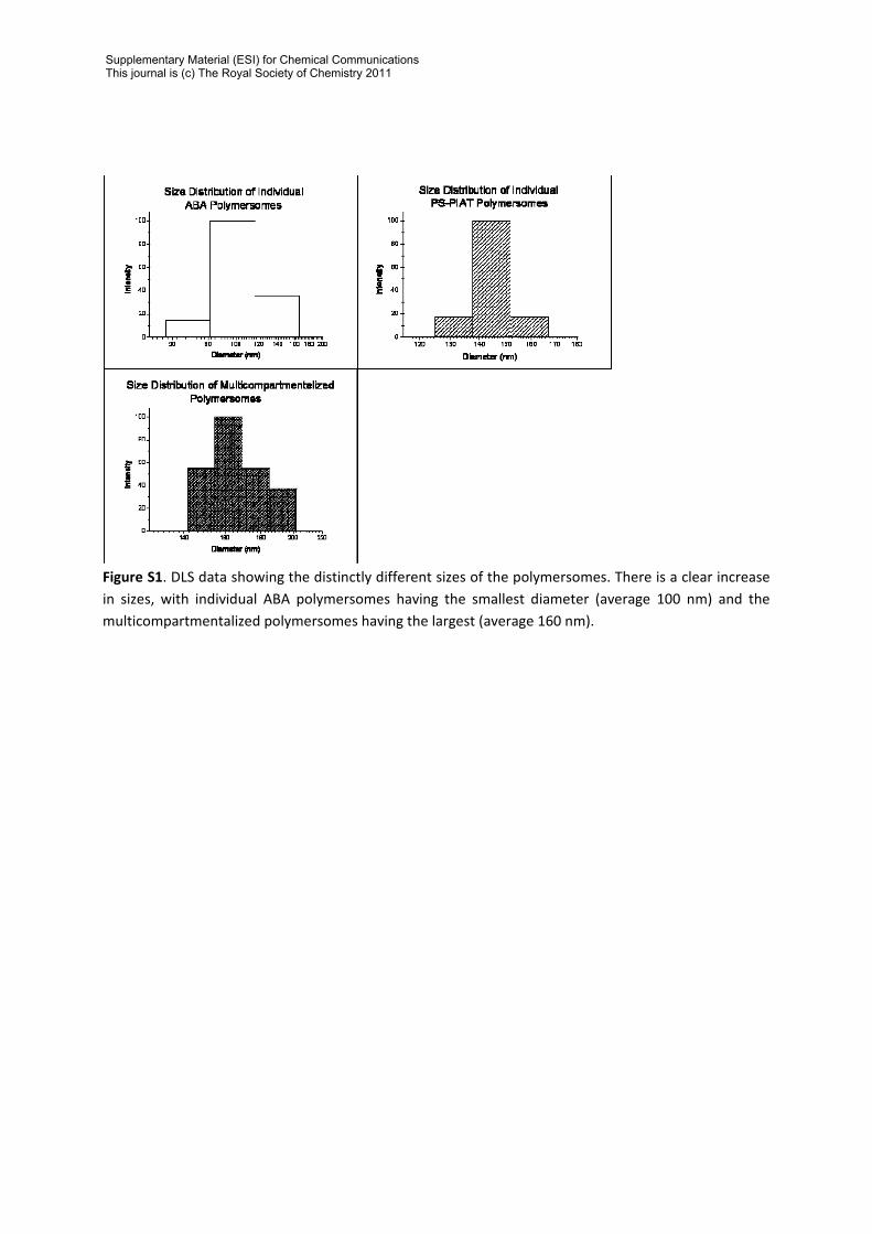
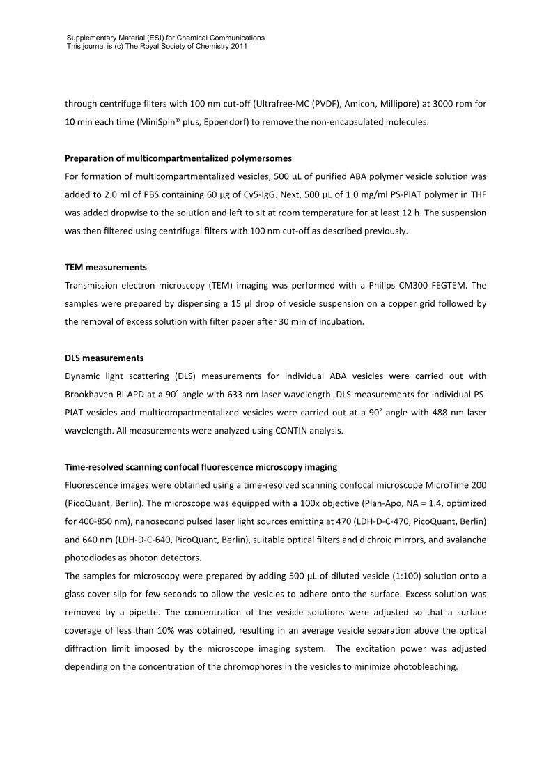
Figure S1. DLS data showing the distinctly different sizes of the polymersomes. There is a clear increase in sizes, with individual ABA polymersomes having the smallest diameter (average 100 nm) and the multicompartmentalized polymersomes having the largest (average 160 nm).
Supplementary Material (ESI) for Chemical CommunicationsThis journal is (c) The Royal Society of Chemistry 2011
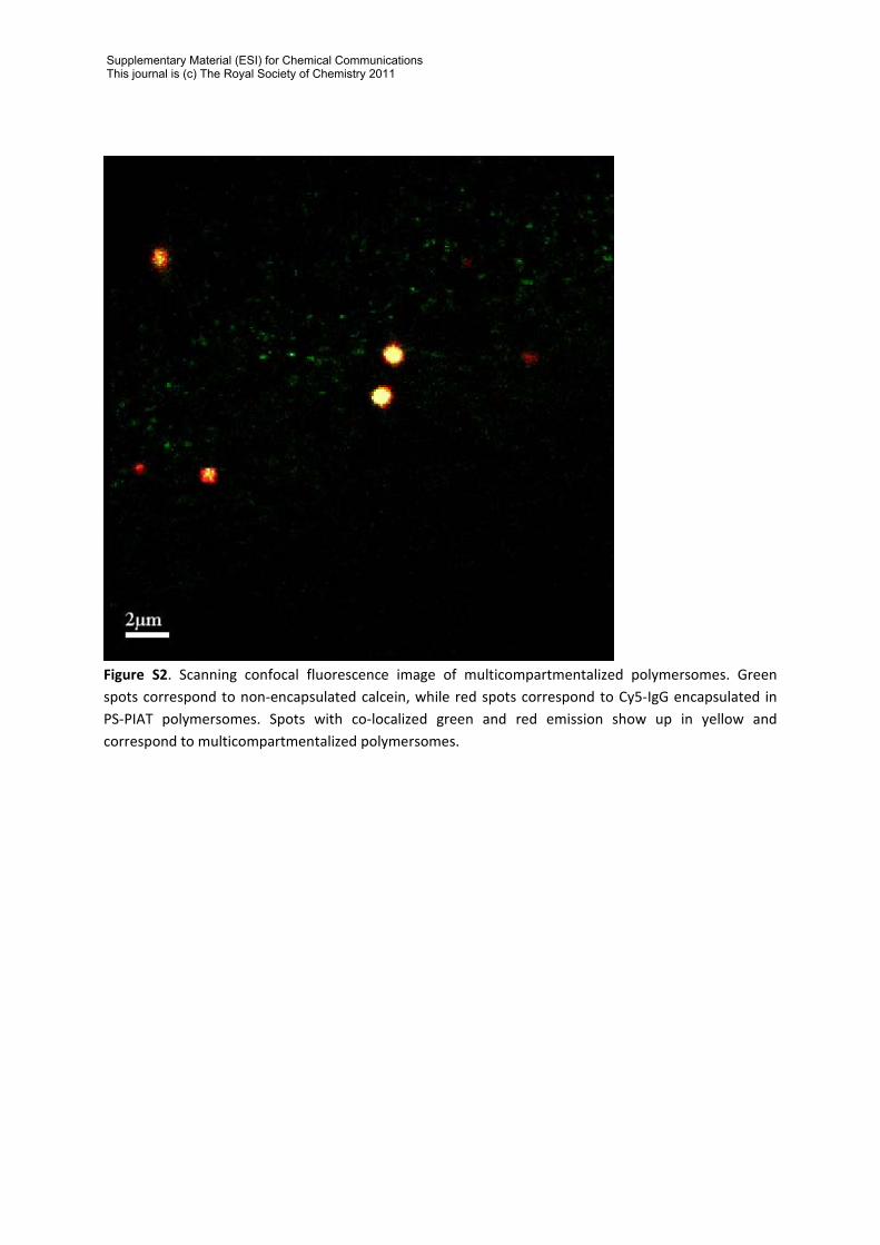
Figure S2. Scanning confocal fluorescence image of multicompartmentalized polymersomes. Green spots correspond to non‐encapsulated calcein, while red spots correspond to Cy5‐IgG encapsulated in PS‐PIAT polymersomes. Spots with co‐localized green and red emission show up in yellow and correspond to multicompartmentalized polymersomes.
Supplementary Material (ESI) for Chemical CommunicationsThis journal is (c) The Royal Society of Chemistry 2011
Figure S3. Flow cytometry data of multicompartmentalized polymersomes. (a) Dot plot from dispersions of multicompartmentalized polymersomes without any encapsulated fluorophores. FSC/SSC gating was applied (region R1) to exclude larger particles and noise. (b) Dot plot of FL1 (530±30 nm for calcein detection) against FL4 (650 nm for Cy5‐IgG detection) after the gating. (c) Dot plot of multicompartmentalized polymersomes encapsulating calcein (ABA compartment) and Cy5‐IgG (PS‐PIAT compartment) where gate R1 was applied to the FSC/SSC dot plot. Calcein was used instead of GFP as it gives a larger signal. (d) Dot plot of FL1/FL4 after gating, clearly showing in the upper right quadrant the presence of polymersomes encapsulating both calcein and Cy5‐IgG. The fraction of multicompartmentalized polymersomes with respect to the total population of polymersomes is 45%. The absence of ABA polymersomes encapsulating calcein, as observed by flow cytometry, is likely to be the result of the centrifugation process, where the size cut‐off of the filters used was similar to the diameter of the ABA polymersomes (100 nm).
Supplementary Material (ESI) for Chemical CommunicationsThis journal is (c) The Royal Society of Chemistry 2011

References
1. H.‐P. M. d. Hoog, D. M. Vriezema, M. Nallani, S. Kuiper, J. J. L. M. Cornelissen, A. E. Rowan and R. J. M. Nolte, Soft Matter, 2008, 4, 1003.
Supplementary Material (ESI) for Chemical CommunicationsThis journal is (c) The Royal Society of Chemistry 2011