mucroporin-m1 inhibits hepatitis b virus replication by activating
TRANSCRIPT
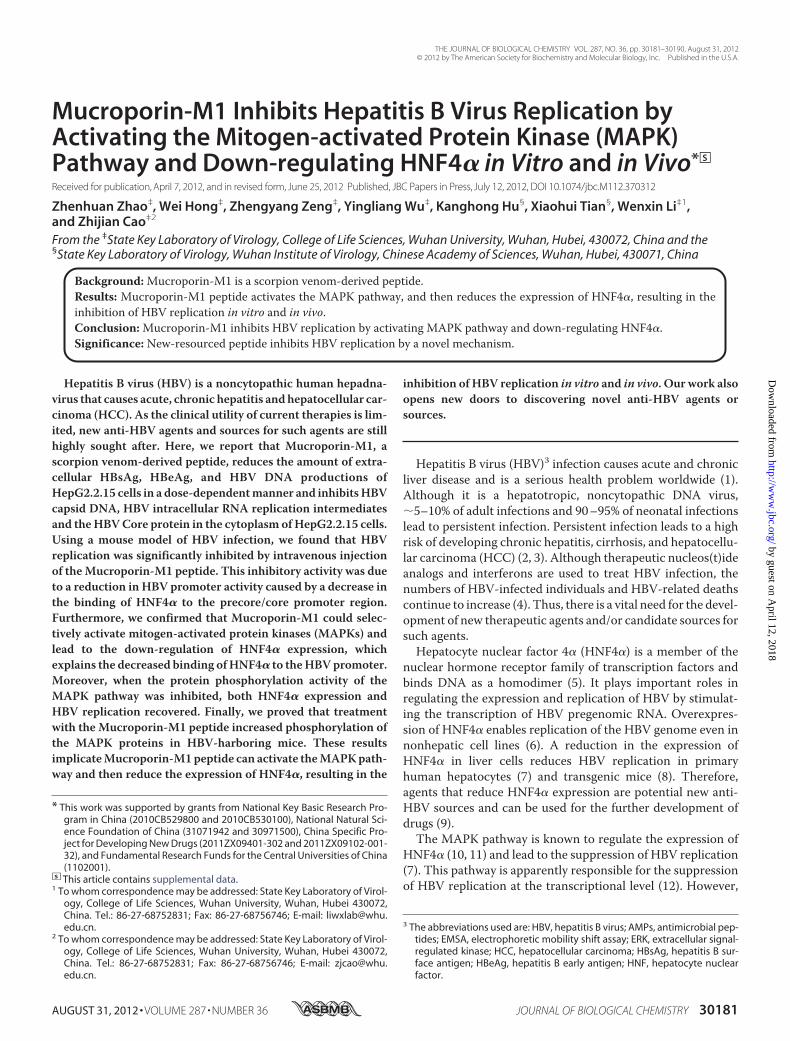
Mucroporin-M1 Inhibits Hepatitis B Virus Replication byActivating the Mitogen-activated Protein Kinase (MAPK)Pathway and Down-regulating HNF4� in Vitro and in Vivo*□S
Received for publication, April 7, 2012, and in revised form, June 25, 2012 Published, JBC Papers in Press, July 12, 2012, DOI 10.1074/jbc.M112.370312
Zhenhuan Zhao‡, Wei Hong‡, Zhengyang Zeng‡, Yingliang Wu‡, Kanghong Hu§, Xiaohui Tian§, Wenxin Li‡1,and Zhijian Cao‡2
From the ‡State Key Laboratory of Virology, College of Life Sciences, Wuhan University, Wuhan, Hubei, 430072, China and the§State Key Laboratory of Virology, Wuhan Institute of Virology, Chinese Academy of Sciences, Wuhan, Hubei, 430071, China
Background:Mucroporin-M1 is a scorpion venom-derived peptide.Results: Mucroporin-M1 peptide activates the MAPK pathway, and then reduces the expression of HNF4�, resulting in theinhibition of HBV replication in vitro and in vivo.Conclusion:Mucroporin-M1 inhibits HBV replication by activating MAPK pathway and down-regulating HNF4�.Significance: New-resourced peptide inhibits HBV replication by a novel mechanism.
Hepatitis B virus (HBV) is a noncytopathic human hepadna-virus that causes acute, chronic hepatitis andhepatocellular car-cinoma (HCC). As the clinical utility of current therapies is lim-ited, new anti-HBV agents and sources for such agents are stillhighly sought after. Here, we report that Mucroporin-M1, ascorpion venom-derived peptide, reduces the amount of extra-cellular HBsAg, HBeAg, and HBV DNA productions ofHepG2.2.15 cells in a dose-dependentmanner and inhibitsHBVcapsid DNA, HBV intracellular RNA replication intermediatesand theHBVCore protein in the cytoplasm ofHepG2.2.15 cells.Using a mouse model of HBV infection, we found that HBVreplication was significantly inhibited by intravenous injectionof theMucroporin-M1 peptide. This inhibitory activity was dueto a reduction in HBV promoter activity caused by a decrease inthe binding of HNF4� to the precore/core promoter region.Furthermore, we confirmed that Mucroporin-M1 could selec-tively activate mitogen-activated protein kinases (MAPKs) andlead to the down-regulation of HNF4� expression, whichexplains the decreased binding ofHNF4� to theHBVpromoter.Moreover, when the protein phosphorylation activity of theMAPK pathway was inhibited, both HNF4� expression andHBV replication recovered. Finally, we proved that treatmentwith the Mucroporin-M1 peptide increased phosphorylation ofthe MAPK proteins in HBV-harboring mice. These resultsimplicateMucroporin-M1peptide can activate theMAPKpath-way and then reduce the expression of HNF4�, resulting in the
inhibition of HBV replication in vitro and in vivo. Our work alsoopens new doors to discovering novel anti-HBV agents orsources.
Hepatitis B virus (HBV)3 infection causes acute and chronicliver disease and is a serious health problem worldwide (1).Although it is a hepatotropic, noncytopathic DNA virus,�5–10% of adult infections and 90–95% of neonatal infectionslead to persistent infection. Persistent infection leads to a highrisk of developing chronic hepatitis, cirrhosis, and hepatocellu-lar carcinoma (HCC) (2, 3). Although therapeutic nucleos(t)ideanalogs and interferons are used to treat HBV infection, thenumbers of HBV-infected individuals and HBV-related deathscontinue to increase (4). Thus, there is a vital need for the devel-opment of new therapeutic agents and/or candidate sources forsuch agents.Hepatocyte nuclear factor 4� (HNF4�) is a member of the
nuclear hormone receptor family of transcription factors andbinds DNA as a homodimer (5). It plays important roles inregulating the expression and replication of HBV by stimulat-ing the transcription of HBV pregenomic RNA. Overexpres-sion of HNF4� enables replication of the HBV genome even innonhepatic cell lines (6). A reduction in the expression ofHNF4� in liver cells reduces HBV replication in primaryhuman hepatocytes (7) and transgenic mice (8). Therefore,agents that reduce HNF4� expression are potential new anti-HBV sources and can be used for the further development ofdrugs (9).The MAPK pathway is known to regulate the expression of
HNF4� (10, 11) and lead to the suppression of HBV replication(7). This pathway is apparently responsible for the suppressionof HBV replication at the transcriptional level (12). However,
* This work was supported by grants from National Key Basic Research Pro-gram in China (2010CB529800 and 2010CB530100), National Natural Sci-ence Foundation of China (31071942 and 30971500), China Specific Pro-ject for Developing New Drugs (2011ZX09401-302 and 2011ZX09102-001-32), and Fundamental Research Funds for the Central Universities of China(1102001).
□S This article contains supplemental data.1 To whom correspondence may be addressed: State Key Laboratory of Virol-
ogy, College of Life Sciences, Wuhan University, Wuhan, Hubei 430072,China. Tel.: 86-27-68752831; Fax: 86-27-68756746; E-mail: [email protected].
2 To whom correspondence may be addressed: State Key Laboratory of Virol-ogy, College of Life Sciences, Wuhan University, Wuhan, Hubei 430072,China. Tel.: 86-27-68752831; Fax: 86-27-68756746; E-mail: [email protected].
3 The abbreviations used are: HBV, hepatitis B virus; AMPs, antimicrobial pep-tides; EMSA, electrophoretic mobility shift assay; ERK, extracellular signal-regulated kinase; HCC, hepatocellular carcinoma; HBsAg, hepatitis B sur-face antigen; HBeAg, hepatitis B early antigen; HNF, hepatocyte nuclearfactor.
THE JOURNAL OF BIOLOGICAL CHEMISTRY VOL. 287, NO. 36, pp. 30181–30190, August 31, 2012© 2012 by The American Society for Biochemistry and Molecular Biology, Inc. Published in the U.S.A.
AUGUST 31, 2012 • VOLUME 287 • NUMBER 36 JOURNAL OF BIOLOGICAL CHEMISTRY 30181
by guest on April 12, 2018
http://ww
w.jbc.org/
Dow
nloaded from
sources or agents that activate the MAPK pathway are rarelystudied for their anti-HBV effects.Antimicrobial peptides (AMPs) are important for the anti-
microbial efficacy of phagocytes. It has previously beenreported that AMPs can activate MAPKs (13). Some AMPshave been shown to be effective against viral pathogens throughdifferent mechanisms (14–17). However, little is known aboutthe relationship of theMAPK signaling pathway andAMP anti-viral activities.Here, we found that an antimicrobial peptide, Mucropo-
rin-M1 (16), activated the MAPKs extracellular signal-regu-lated kinase 1/2 (ERK1/2) and c-Jun N-terminal kinase (JNK)and subsequently inhibited the expression of HNF4�. As aresult of HNF4� down-regulation, the transcriptional activityof theHBVpromoterwas significantly reduced.When theHBVRNA transcript was reduced by the Mucroporin-M1 peptide,production of HBV DNA and proteins also decreased. Using amouse model of HBV infection (18), we evaluated the expres-sion of the HBV Core antigen in hepatocytes by immunohisto-chemical staining and determined the presence of HBsAg andHBeAg in the blood by ELISA. The Mucroporin-M1 peptide-treated group showed a lower HBV viral load in both the hepa-tocytes and blood than did the untreated group. Moreover, wefound that the Mucroporin-M1 peptide also activated MAPKsin mouse hepatocytes, similar to the results from human hepa-toma cells. These data suggest that a natural animal-derivedpeptide, Mucroporin-M1, inhibits HBV replication by activat-ing the MAPK pathway and then down-regulating HNF4�expression in vitro and in vivo.
EXPERIMENTAL PROCEDURES
Chemical Synthesis—The Ctri10036, Ctri10033, Ctry2801,Ctriporin, and Mucroporin-M1 peptides were from the scor-pion venom peptide library that was recently characterized byour group and were synthesized at purities of �95% by GLBiochem Ltd. (China).Cell Culture—HepG2.2.15 cells were cultured at 37 °C in a
humidified 5% CO2/air atmosphere in Dulbecco’s modifiedEagle’s medium supplemented with 10% (v/v) fetal calf serum,50 units/ml penicillin, and 50 �g/ml streptomycin.Reagents—Mitogen-activated protein kinases (MAPK) in-
hibitors PD98059, SB203580, and SP600125 were purchasedfrom Promega (Promega, Madison, WI).Cytotoxicity—Cytotoxicity was measured by a 3-(4,5-dim-
ethylthiazol-2-yl)-2,5-diphenyltetrazolium bromide (MTT)assay.HepG2.2.15 cellswere seeded in 96-well plates at 104 cellsper well and grown to confluence in DMEM containing 10%FCS. Peptides were added to the wells at different concentra-tions. After 48 h of incubation, 10 �l of MTT solution wasadded to each well, and the plates were incubated for 2 to 4 h in5%CO2 at 37 °C. The plates were then gently swirled for 10minat room temperature to dissolve the precipitate, and theabsorbance was measured at a wavelength of 550 nm.Quantification of HBsAg, HBeAg, and HBV DNA in the Cul-
ture Medium—Cells were seeded in 24-well plates at a densityof 8 � 104 cells/well in DMEM containing 10% FCS. After 12 hof incubation, the cells were treated with various concentra-tions ofMucroporin-M1 for 2 days. The HBsAg and HBeAg in
the culture medium were measured using an enzyme-linkedimmunoassay (ELISA) kit. HBV DNA was measured by real-time PCR according to the manufacturer’s instructions (Qia-gen, Valencia, CA).HBV RNA, Core Protein, and Replicative DNA Analyses—
HepG2 or Hep2.2.15 cells were seeded in 6-well culture platesat a density of 5 � 105 cells per well. At 12 h after seeding,Mucroporin-M1 was added to the cell cultures, and cells werefed with fresh medium for another 2 days. As a control, 10 �M
3TC was added to the cell cultures for 2 days. Cells were col-lected for Southern, Northern and Western blot analyses forviral DNA, RNA, and protein, respectively. For Southern blotanalysis, viral capsid DNAwas detected as previously described(19). Radioactively 32P-labeled probes prepared from full-length HBV genomic DNA were generated by using the Redi-prime labeling kit (Amersham Biosciences) as described by themanufacturer. For Northern blot analysis, total RNA was iso-lated by Trizol (Invitrogen) following the manufacturer’sinstructions. The prehybridization and hybridization were per-formed identically to the Southern blot analysis. For Westernblot analysis, 40 �g of sample was electrophoresed and trans-ferred to a nitrocellulose membrane (Millipore, Bedford, MA).Themembranewas probed using a polyclonal antibody specificfor HBV Core antigen (Dako-Cytomation, Carpinteria, CA).Anti-HBV Activity Analysis in Vivo—All animal studies were
approved by the Institutional Animal Care and Use CommitteeatWuhan University. Amouse model of acute hepatitis B virusinfection was used in this study. A total of 20 �g of pUC-HBV1.3 was injected into the tail veins of 6–9-week-oldBALB/c mice in a volume of saline equivalent to 8% of the bodymass of each mouse (e.g. 1.6 ml for a mouse of 20 g). The totalvolume was delivered within 5–8 s. The second day of plasmidinjection, Mucroporin-M1 was administered into the tail veinsat 12.5 mg/kg. Sera and livers were collected on the third day.Viremia was measured by an ELISA similar to the method usedin HepG2.2.15 cells. The HBV Core protein was visualized byimmunohistochemical staining of tissues fixed in zinc-bufferedformalin using anti-core polyclonal rabbit antibody.HBV Promoter Luciferase Reporter Assay—The promoter
regions of the genes encoding the HBV Core (nucleotides (nt)1603–1819), X (nt 935–1361), preS (nt 2700–2830), or S (nt2950–3174) were cloned upstream of the luciferase gene of thepGL3-basic vector. The mutated Core promoter sequence wasobtained by converting the 13-nucleotide HNF4 binding sitesequence (between 1662 and 1674) from ggactcttggact tocgctagcctcgta as described previously (20). And the mutatedCore sequence was constructed into pGL3-basic vector.HepG2.2.15 cells were transiently transfected with the
reporter vector in a 48-well plate by using Lipofectamine 2000(Invitrogen) according to the manufacturer’s instructions.Twelve hours after transfection, peptides were added to themedium, and cells were incubated for 2 days (similar to an anti-viral assay). Transcriptional activity was determined by meas-uring luciferase activity in a multiwell plate luminometer usingthe Luciferase Reporter Assay System (Promega).EMSA—Same as the anti-HBV activity analysis in vitro,
Mucroporin-M1 was added to the cell cultures for 2 days inEMSA. Nuclear extracts were prepared with NE-PER extrac-
Anti-HBV Activity and Mechanism of the Peptide Mucroporin-M1
30182 JOURNAL OF BIOLOGICAL CHEMISTRY VOLUME 287 • NUMBER 36 • AUGUST 31, 2012
by guest on April 12, 2018
http://ww
w.jbc.org/
Dow
nloaded from
tion reagent and were stored at �80 °C. The protein contentwasmeasured with a BCA protein assay. Then, nuclear extractswere incubatedwith biotin-labeled oligonucleotides in gel-shiftbinding buffer for 30 min at room temperature. Samples wereseparated using 5% native polyacrylamide gels followed bychemiluminescent detection. Competition assays were per-formed by incubating the nuclear extracts with unlabeled oli-gonucleotides on ice for 25 min before the addition of the bio-tin-labeled probe. The sequences of the oligomers used wereas follows: HNF4, 5�-GAGGACTCTTGGACTCTCA-3� (nt1660–1678); HNF3, 5�-TCAAAGACTGTGTGTTTAA-GGAC-3� (nt 1710–1732); FTF, 5�-AATGTCAACGAC-CGACCTTGAGG-3� (nt 1681–1703).Quantification of Gene Expression by Real-time Reverse
Transcription Polymerase Chain Reaction—Same as the anti-HBV activity analysis in vitro, Mucroporin-M1 was added tothe cell cultures for 2 days in qPCR. Total RNA was extractedusing Trizol reagent and was transcribed into cDNA using theFirst-Strand Synthesis Supermix (Invitrogen). Real-time PCRswere performed using the SYBR green PCR assay and an ABI7500 system according to the manufacturer’s instruction. FormRNA detection, HNF4� and GAPDH primer sets were used:HNF4� forward primer, 5�-GAGTGGGCCAAGTACA-3�;HNF4� reverse primer, 5�-GGCTTTGAGGTAGGCATA-3�;GAPDH forward primer, 5�-CAAGAAGGTGGTGAAGCAG-3�;GADPH reverse primer, 5�-AGGTGGAGGAGTGGGTG-3�).HNF4� and MAPK Protein Analyses—HNF4� and MAPK
pathway proteins were separated by SDS-PAGE and were ana-lyzed byWestern blotting. The primary antibodies usedwere asfollows: rabbit polyclonal anti-ERK1/2, rabbitmonoclonal anti-phospho-ERK1/2, rabbit monoclonal anti-p38, rabbit poly-clonal anti-phospho-p38, mouse monoclonal anti-phospho-SAPK-JNK (Cell Signaling Technology, Beverly, MA), rabbitmonoclonal anti-HNF4� (Abcam, Cambridge, UK), rabbitmonoclonal anti-�-tubulin and mouse polyclonal anti-�-actin(Santa Cruz Biotechnology).
RESULTS
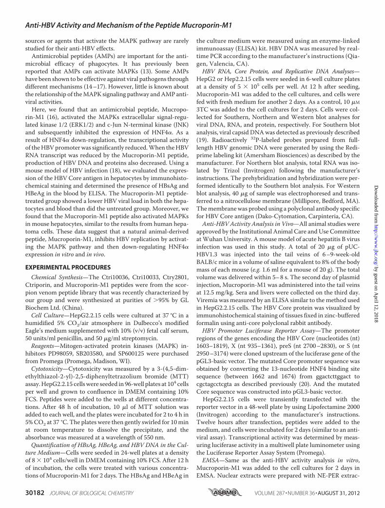
Screening of Anti-HBVAgents from Scorpion VenomPeptides—Cationic host defense peptides from scorpion venomous glandswere recently characterized by our group. Mucroporin-M1(21), Ctriporin (22) and other venom-derived peptides weresynthesized at a purity of �95% by GL Biochem Ltd. (China).Their molecular weights, as measured byMS, matched the cal-culated molecular weights of the amidated peptides. Sequencealignments of Mucroporin-M1 with other antimicrobial pep-tides were performed using ClustalX and BioEdit (Fig. 1A).Peptides from the scorpion venom were screened for the
capacity to inhibit HBV replication in the HepG2.2.15 cell line.Peptide was added to the cells to a final concentration of 25�M,and the amount of HBsAg and HBeAg present in the culturemedium were tested using an ELISA. Incubation with theMucroporin-M1 peptide resulted in an �80% reduction in theamount ofHBeAg and a 70% reduction in the amount ofHBsAgpresent in the culture medium compared with cultures notexposed to the peptide, and incubation with the Ctry2801 pep-tide resulted in an �40% reduction in the amount of HBsAg inthe culture medium. The other peptides had little effect on the
amount of secretedHBsAg andHBeAg (Fig. 1B).We found thatthe Mucroporin-M1 peptide had the most effective inhibitoryactivity against the production of HBeAg and HBsAg. Accord-ingly, we chose the Mucroporin-M1 peptide to study further.Anti-HBV Effects of Mucroporin-M1 at Noncytotoxic
Concentrations—The cytotoxicity of the peptide onHepG2.2.15 cells was tested using an MTT assay. The concen-tration of Mucroporin-M1 that inhibited 50% of cell growth(CC50) was 87 �M. When the peptide concentration was lessthan 25�M, the viability of the peptide-treated cells was greaterthan 90%, indicating that 25 �M or less of the Mucroporin-M1peptide was minimally cytotoxic to cells.The effect of Mucroporin-M1 on HBV in HepG2.2.15 cells
was assessed. HepG2.2.15 is a cell line that has been stablytransfected with the HBV genome. The cells were cultured inthe presence of 2-fold serial dilutions of the Mucroporin-M1peptide for 2 days. Inhibitory effects on the production of extra-cellular HBsAg andHBeAgwere determined by ELISA, and theamount of extracellular HBV progeny DNA was assessed byreal-time PCR. The data showed that theMucroporin-M1 pep-tide inhibited the expression of HBsAg and HBeAg in a dose-dependent manner (Fig. 2A). The IC50 values of Mucropo-rin-M1 against HBsAg and HBeAg production were 20.6 and4.9�M, respectively. The production ofHBVprogenyDNAwasalso inhibited in a dose-dependentmanner byMucroporin-M1,with an IC50 of 11 �M (Fig. 2B). Southern, Northern, andWest-ernblot analyseswereused formeasuring intracellularHBVDNA,RNA, and Core protein levels, respectively, after treatment withtheMucroporin-M1peptide. The various formsof theHBV intra-
FIGURE 1. Screening of anti-HBV agents from scorpion venom peptides.A, sequence alignments of Mucroporin-M1 and its related peptides. Thesequence alignments of Mucroporin-M1 with other antimicrobial peptideswere performed using ClustalX and BioEdit. The residues shaded in the samecolor are highly conserved sites; the residues in similar color are less con-served sites; and the residues without a background color are highly variablesites. B, inhibitory activity of scorpion venom peptides on HBsAg and HBeAgproduction in HepG2.2.15 cells. Peptides were tested at a concentration of 25�M for inhibitory activity against HBV replication, as assessed by ELISA.
Anti-HBV Activity and Mechanism of the Peptide Mucroporin-M1
AUGUST 31, 2012 • VOLUME 287 • NUMBER 36 JOURNAL OF BIOLOGICAL CHEMISTRY 30183
by guest on April 12, 2018
http://ww
w.jbc.org/
Dow
nloaded from

cellularDNA replication intermediateswere potently inhibited byMucroporin-M1 in a concentration-dependentmanner, as exem-plified in HepG2.2.15 cells. When used as a positive control, 3TCinhibited HBV DNA synthesis effectively (Fig. 2D). HBV RNAexpression was also potently inhibited by Mucroporin-M1 inHepG2.2.15 cells. In contrast, viral RNA levels were unchangedafter 3TC treatment, as expected (Fig. 2E). HBV Core proteinexpression was also inhibited in a dose-dependent manner byMucroporin-M1 in HepG2.2.15 cells. Again, 3TC did not inhibitviral Core protein synthesis, as expected (Fig. 2C). The resultsshowed that the Mucroporin-M1 peptide had anti-HBV activity,and the anti-HBVmechanismof theMucroporin-M1peptidewasdifferent from that of 3TC.Inhibitory Activity of Mucroporin-M1 against HBV in Vivo—
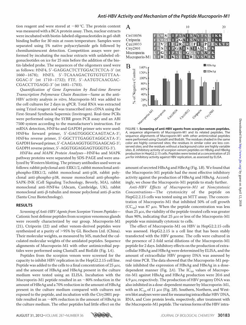
As Mucroporin-M1 had anti-HBV activity in hepatoma cells,we further examined its anti-HBV activity in an HBV infectionmouse model. After hydrodynamic injection of the pUC18-HBV1.3 plasmid, the secretion of viral antigens into the bloodwasmonitored at day 2. Three treatments were examined (n� 7miceper group). HBsAg accumulated to an average concentration of21.6 PEIU/ml in the untreatedmice, whereas the concentration ofHBsAg in the blood of the Mucroporin-M1-treated mice was 9.4PEIU/ml. Similarly to HBsAg production, the amount of HBeAg
decreased from 10.4 PEIU/ml in the untreated group to 4.8PEIU/ml in the Mucroporin-M1-treated group (Fig. 3, A and B).Themice thatwerenotadministeredpUC18-HBV1.3didnothavedetectable viral antigens in their blood.The livers of the mice were also examined for HBV Core
protein by immunohistochemical staining. The frequency ofHBV Core protein-positive hepatocytes was 4% � 2% inMucroporin-M1-treated mice compared with 13% � 3% inuntreated mice (Fig. 3, C and D). Thus, the Mucroporin-M1peptide inhibited HBV replication in mouse hepatocytes andreduced HBV antigen secretion in mouse blood.Inhibition of HBV Promoter Activity by Reducing the Interac-
tion of HNF4� with HBV Promoters—As the results aboveshowed, Mucroporin-M1 reduced the HBV RNA transcriptlevels, leading to reduced HBV DNA and protein production.These results suggested that the active target of Mucropo-rin-M1 is the viral RNA transcription step and not the HBVDNApolymerase, unlike other anti-HBVnucleos(t)ide analogs.Therefore, we constructed plasmids containing promoters forthe four different HBV transcripts (preC/Cp, Xp, pSp, or Sp)followed by the luciferase reporter gene to examine the effect ofMucroporin-M1 on HBV promoter activity. After transienttransfection of the plasmids into HepG2.2.15 cells, Mucropo-
FIGURE 2. Anti-HBV activity of Mucroporin-M1 peptide in HepG2.2.15 cells. A, HBsAg and HBeAg expression in the culture medium of HepG2.2.15 aftertreatment with Mucroporin-M1. Extracellular HBsAg and HBeAg production decreased in a dose-dependent manner after treatment with the indicatedconcentrations of Mucroporin-M1. B, HBV DNA analysis of the culture medium of HepG2.2.15 after treatment by Mucroporin-M1. Extracellular HBV DNAmeasured by real-time PCR decreased in a dose-dependent manner after treatment with the different concentrations of Mucroporin-M1. C, Western blotanalysis of HBV Core protein synthesis in HepG2.2.15 cells treated by Mucroporin-M1. Mucroporin-M1 inhibited HBV Core protein synthesis, but 3TC did not.D, inhibition of intracellular HBV DNA in HepG2.2.15 cells treated by Mucroporin-M1. Intracellular relaxed circle (RC), double strand (DS), and single strand (SS)DNA synthesis was reduced after treatment with the indicated concentrations of Mucroporin-M1 or 3TC using Southern blot analysis. E, inhibition of HBV RNAsynthesis in the HepG2.2.15 cells treated by Mucroporin-M1. Mucroporin-M1 inhibited HBV RNA production in a dose-dependent manner, but 3TC did not.
Anti-HBV Activity and Mechanism of the Peptide Mucroporin-M1
30184 JOURNAL OF BIOLOGICAL CHEMISTRY VOLUME 287 • NUMBER 36 • AUGUST 31, 2012
by guest on April 12, 2018
http://ww
w.jbc.org/
Dow
nloaded from
rin-M1was added to the cell cultures, and viral promoter activ-ity was examined (supplemental data). The data showed thatthe HBV four promoter activities were partially inhibited byMucroporin-M1 peptide, where the most significant one wasHBV Core promoters. Core/precore promoter-driven lucifer-ase expression decreased to 20% of the negative control levelafter Mucroporin-M1 peptide treatment, but the CMV pro-moter was not affected (Fig. 4A).It has been reported that hepatocyte nuclear transcriptional
factors, togetherwith viral proteins, bind to theHBVpromotersand modulate viral promoter activity (23–26). DNA oligonu-cleotides corresponding to the HBV precore/core promoter/Enh II sequence HNF4, HNF3, and Fetoprotein transcriptionfactor (FTF) were synthesized and biotin-labeled for electro-phoretic mobility-shift assays (EMSAs). Nuclear extracts fromHepG2.2.15 cells treated with 25 �M Mucroporin-M1 or leftuntreated were incubated with the probes to determinewhetherMucroporin-M1 alters the binding of nuclear proteins.Binding to the HNF4 probe was significantly decreased aftertreatment with 25 �M Mucroporin-M1 (Fig. 4B). The bindingto the HNF3 DNA probe was decreased after treatment withthe Mucroporin-M1 peptide, but the difference was not signif-icant. However, the binding of the nuclear extract to FTF didnot yield a distinctive shift (data not shown).
To further confirm whether Mucroporin-M1 specificallyreduced HNF4a binding HBV Core promoter, we constructedthe mutated Core promoter with a mutant HNF4 binding site.The results of luciferase activities showed that the activity ofHBV Core promoter with a mutant HNF4 binding site wasalmost not inhibited by Mucroporin-M1 treatment (Fig. 4C).The data suggests that theHNF4 binding site specifically playedan important role in the HBV inhibitory activity of Mucropo-rin-M1 peptide.Mucroporin-M1 Down-regulates HNF4� and Then Inhibits
HBV Progeny DNA Expression by Activating MAPKs—Todetermine whether the decreased binding observed in theEMSAswas due to a reduction inHNF4�, quantitative PCR andWestern blot analyses were performed. As shown in Fig. 4, Dand E, the reduction inHNF4� expression inevitably resulted in adecrease in the binding of HNF4� to the HBV precore/core pro-moter, which explained the inhibitory activity of the Mucropo-rin-M1 peptide on HBV transcription. The decrease in HNF4�expression inHepG2.2.15 cells could also explain the inhibition ofother viral protein promoters (pSp, Sp, andXp) thatwas observed,because all of these viral protein promoters have HNF4�-bindingsites. Taken together, Mucroporin-M1 down-regulated theexpression of HNF4�, inhibited HBV promoter activity, and fur-ther reduced the levels of the HBV transcript.
FIGURE 3. Anti-HBV activity of Mucroporin-M1 in HBV-infected mice. A, inhibitory activity of Mucroporin-M1 against HBsAg in the blood of the HBV-infectedmice. Mucroporin-M1 reduced the expression of HBsAg in the blood of HBV-infected mice by ELISA. B, inhibitory activity of Mucroporin-M1 against HBeAg inthe blood of the HBV-infected mice. Mucroporin-M1 reduced the expression of HBeAg in the blood of HBV-infected mice by ELISA. C and D, inhibitory effect onHBV Core protein expression in the hepatocytes of HBV-infected mice treated with Mucroporin-M1. Mucroporin-M1 decreased the expression of HBV Coreprotein in the hepatocytes of HBV-infected mice treated by Mucroporin-M1, as measured by an immunohistochemical staining assay.
Anti-HBV Activity and Mechanism of the Peptide Mucroporin-M1
AUGUST 31, 2012 • VOLUME 287 • NUMBER 36 JOURNAL OF BIOLOGICAL CHEMISTRY 30185
by guest on April 12, 2018
http://ww
w.jbc.org/
Dow
nloaded from
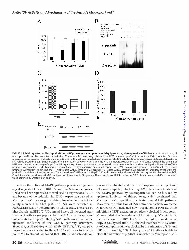
Because the activated MAPK pathway proteins exogenoussignal regulated kinase (ERK) 1/2 and Jun N-terminal kinase(JNK) have been reported to controlHNF4� expression (10, 11)and because of the reduction in HNF4� expression caused byMucroporin-M1, we sought to determine whether the MAPKfamily members ERK1/2, p38, and JNK were activated inHepG2.2.15 cells by the Mucroporin-M1 peptide. The levels ofphosphorylated ERK1/2, JNK, and p38 were all increased aftertreatment with 25 �M peptide, but the MAPK pathways werenot activated in HepG2 cells (Fig. 5A). Furthermore, when theupstream inhibitors of the MAPK pathway (PD98059,SP600125, or SB203580), which inhibit ERK1/2, JNK, and p38,respectively, were added to HepG2.2.15 cells prior to Mucro-porin-M1 treatment, we found that ERK1/2 phosphorylation
was mostly inhibited and that the phosphorylation of p38 andJNK was completely blocked (Fig. 5B). Thus, the activation ofthe MAPK pathway by Mucroporin-M1 can be blocked byupstream inhibitors of this pathway, which confirmed thatMucroporin-M1 specifically activates the MAPK pathway.Moreover, the inhibition of JNK activation partially overcameMucroporin-M1-mediated down-regulation of HNF4�, whileinhibition of ERK activation completely blocked Mucroporin-M1-mediated down-regulation of HNF4� (Fig. 5C). Similarly,the detection of HBV DNA in the culture medium ofHepG2.2.15 by real-time PCR showed that the anti-HBV activ-ity ofMucroporin-M1was blocked by the inhibition of JNK andERK activation (Fig. 5D). Although the p38 inhibitor is able toblock the activation of p38 that ismediated byMucroporin-M1,
FIGURE 4. Inhibitory effect of Mucroporin-M1 on HBV promoter transcriptional activity by reducing the expression of HNF4�. A, inhibitory activity ofMucroporin-M1 on HBV promoter transcription. Mucroporin-M1 selectively inhibited the HBV promoter (preC/Cp) but not the CMV promoter. Data arepresented as the means of triplicate experiments (each with duplicate samples) normalized to vehicle-treated cells. Error bars represent standard deviations.NC, vehicle-treated cells. B, EMSA analysis of the interaction between HNF4� and the HBV promoters. Mucroporin-M1 significantly reduced the binding ofHNF4� to the HBV promoter (preC/Cp). C, inhibitory activity of Mucroporin-M1 on the mutated Core promoter without HNF4a binding site. The activity of Corepromoter with a mutant HNF4 binding site was not affected by 25 �M Mucroporin-M1 peptide. wild, Wild type of Core promoter. mut, Mutant type of Corepromoter without HNF4a binding site. �, Not-treated with Mucroporin-M1 peptide. �, Treated with Mucroporin-M1 peptide. D, inhibitory effect of Mucro-porin-M1 on HNF4� mRNA expression. The expression of HNF4� in the HepG2.2.15 cells treated with Mucroporin-M1 was quantified by real-time PCR.E, inhibitory effect of Mucroporin-M1 on the expression of the HNF4� protein. The expression of HNF4� in the HepG2.2.15 cells treated with Mucroporin-M1was quantified by Western blot analysis.
Anti-HBV Activity and Mechanism of the Peptide Mucroporin-M1
30186 JOURNAL OF BIOLOGICAL CHEMISTRY VOLUME 287 • NUMBER 36 • AUGUST 31, 2012
by guest on April 12, 2018
http://ww
w.jbc.org/
Dow
nloaded from

it cannot abolish Mucroporin-M1-mediated down-regulationofHNF4� and the anti-HBV activity ofMucroporin-M1 (Fig. 5,C andD). In HBV-harboringmice, the levels of phosphorylatedMAPK proteins were examined byWestern blot analysis. Sim-ilar to the results in hepatoma cells, Mucroporin-M1 activatedERK1/2 and JNK and reduced HBV Core expression in HBV-harboring mice, but not activated ERK1/2 and JNK in the nor-mal mice (Fig. 6).These results demonstrated that the Mucroporin-M1 pep-
tide selectively activated ERK1/2 and JNK and subsequentlydown-regulatedHNF4� expression, leading to the inhibition ofHBV replication in both HepG2.2.15 cells and HBV-infectedmice (Fig. 7).
DISCUSSION
As the increase in resistance to nucleos(t)ide analogs rendersthese drugs less potent, and the use of interferons is limitedbecause of their side effects. The need for potent new anti-HBVagents with differentmechanisms of action prompted us to test
our scorpion venom-derived peptides for anti-HBV activity.Recently, new compounds targeting different stages of theHBVlife cycle have been reported to successfully manage chronicHBV infection. The heteroaryldihydropyrimidines (HAPs) area new class of antivirals that inhibit the production of HBVvirions by binding to the HBV Core protein and causing itsdegradation, which subsequently inhibits nucleocapsid forma-tion (27). Helioxanthins decrease the amount of host hepato-cyte nuclear transcription factors required for the initiation ofviral transcription (9, 28). Amyristoylated peptide derived fromthe large HBV envelope protein (Pre S1) blocks virus entry tohepatocytes in vitro and in vivo (29, 30). However, no researchhas yet reported an agent that activates theMAPKpathway andinhibits HBV replication. Our study examined the Mucropo-rin-M1 peptide from scorpion venom, which activatedMAPKsin HBV-harboring cells, reduced HNF4� expression and abol-ished HBV replication.Recently, our group found that Mucroporin-M1 inhibited
RNA viruses, including measles, SARS-CoV and influenza
FIGURE 5. MAP kinase pathways involved in the anti-HBV activity of Mucroporin-M1 peptide in vitro. A, activation of the ERK, JNK and p38 pathways byMucroporin-M1. pERK, ERK, pJNK, JNK, phosphorylated p38 and p38 from HepG2.2.15 cells and HepG2 cells, with or without treatment of Mucroporin-M1, wereanalyzed by Western blot analysis. �, Not-treated with Mucroporin-M1. �, Treated with Mucroporin-M1. B, inhibitory effect of MAP kinase inhibitors on theMAP kinase activation activity of the peptide Mucroporin-M1. The pERK, pJNK, and phosphorylated p38-producing cells were untreated or treated with 50 �M
PD98059 (inhibiting pERK), SP600125 (inhibiting pJNK) or SB203580 (inhibiting phosphorylated p38) for 30 min prior to stimulation with Mucroporin-M1. TheMAP kinase inhibitors blocked Mucroporin-M1-induced activation of the MAPKs. C, inhibitory effect of MAP kinase inhibitors on the inhibitory activity ofMucroporin-M1 peptide on HNF4� expression. Twenty-microgram quantities of nuclear proteins from HepG2.2.15 cells (mock) without Mucroporin-M1treatment or Mucroporin-M1-treated (�) or preincubation with PD98059, SP600125, and SB203580 before Mucroporin-M1 treatment were analyzed byWestern blot for HNF4�. D, inhibitory effect of MAP kinase inhibitors on the anti-HBV activity of Mucroporin-M1. HBV progeny DNA in the culture medium ofHepG2.2.15 was quantified by real-time PCR.
Anti-HBV Activity and Mechanism of the Peptide Mucroporin-M1
AUGUST 31, 2012 • VOLUME 287 • NUMBER 36 JOURNAL OF BIOLOGICAL CHEMISTRY 30187
by guest on April 12, 2018
http://ww
w.jbc.org/
Dow
nloaded from

H5N1 viruses (16). Additionally, it has been reported that sim-ilar peptides inhibit other RNA viruses (14, 17, 31). These anti-viral compounds were thought to function by disturbing theviral membrane. Thus, this antiviral mechanism was not effec-tive against DNA viruses and virally infected cells. As a startingpoint of our study, we found that the Mucroporin-M1 peptideinhibited HBV replication in HepG2.2.15 cells, which is a cellline stably transfected with the HBV genome. This result sug-gested that Mucroporin-M1 uses a different strategy to inhibitHBV replication. As a similar venom-derived peptide (from beevenom) has been reported to activate MAPKs (13, 32), a path-way that directly participates in the inhibition of HBV replica-tion (7, 12), we sought to determine the relationships betweenthe Mucroporin-M1 peptide, the MAPK pathway and HBVreplication. Our data clearly demonstrated that Mucropo-rin-M1 inhibited HBV replication by activating MAPKs. First,Mucroporin-M1 increased the levels of phosphorylatedERK1/2 and JNK and simultaneously inhibited HBV replica-tion. Second, when the specific inhibitors of ERK1/2 and JNKwere added, both ERK1/2 and JNK phosphorylation and theanti-HBV activity of Mucroporin-M1 were blocked. Further-more, the levels of phosphorylated MAPK proteins were alsoincreased in HBV-infected mice with Mucroporin-M1 treat-
FIGURE 6. MAPK pathways involved in the anti-HBV activity of thepeptide Mucroporin-M1 in vivo. The ERK and JNK MAP kinase pathwayswere activated in HBV-infected mice treated with an intravenous injectionof Mucroporin-M1. Additionally, the expression of the HBV Core proteinwas also significantly reduced in mouse hepatocytes after intravenousinjection of Mucroporin-M1. Group B, the mice treated by an intravenousinjection of saline. Group M, the mice treated by an intravenous injectionof Mucroporin-M1 peptide. Group P, the mice treated by both HBV plasmidand saline. Group T, the mice treated by both HBV plasmid and Mucropo-rin-M1 peptide.
FIGURE 7. Hypothetical model of the anti-HBV activity of the Mucroporin-M1 peptide. Mucroporin-M1 activates the MAPK pathway (ERK, JNK, andp38) and subsequently down-regulates the expression of HNF4�, resulting in a reduction in the interaction between HNF4� and the HBV promoters.Mucroporin-M1 diminishes HBV replication by blocking HBV RNA expression. These findings indicate that the ERK and JNK pathways regulate theexpression of HNF4� but that the p38 pathway does not, which suggests that the Mucroporin-M1 peptide may be a good molecular probe for studyingthe MAPK pathway.
Anti-HBV Activity and Mechanism of the Peptide Mucroporin-M1
30188 JOURNAL OF BIOLOGICAL CHEMISTRY VOLUME 287 • NUMBER 36 • AUGUST 31, 2012
by guest on April 12, 2018
http://ww
w.jbc.org/
Dow
nloaded from
ment. Following MAPK activation in mouse hepatocytes, HBVreplication was also inhibited by Mucroporin-M1. These dataproved that the Mucroporin-M1 peptide inhibited HBV repli-cation by activating MAPKs. It is well known that the MAPKpathway plays an important role in innate immunity. Pathogensstimulate and activate the MAPK pathway, which inducesinnate immune responses. Some AMPs participate in the acti-vation of the MAPK pathway (33, 34). Therefore, MAPK path-way activation byAMPsmay be a key part of the innate immuneresponse against pathogens.In this study, we also found that HBV promoter activity was
reduced byMucroporin-M1peptide treatment. Thiswas due toa decrease in the binding of HNF4� to HBV promoters. Whenthe wild type Core promoter was mutated to the mutant CorepromoterwithoutHNF4 binding site, we found that themutantpromoter activity was not reduced byMucroporin-M1 peptide.Furthermore, the Western blot and quantitative PCR resultsdemonstrated that the expression of HNF4� was inhibited byMucroporin-M1. The inhibition of HNF4� expression byMucroporin-M1 treatment was blocked by the specific inhibi-tors of ERK1/2 or JNK, which suggested that Mucroporin-M1inhibited the expression of HNF4� by activating ERK1/2 orJNK. This result confirmed that the expression of HNF4� wasregulated by the ERK and JNK pathways (10, 35). Interestingly,p38was also activated byMucroporin-M1, but it did not impactHNF4� expression. These data may suggest that the p38 cas-cade does not participate in the regulation of HNF4� expres-sion. Another group has also reported that a p38 inhibitor doesnot regulate the expression of HNF4�, regardless of the activa-tion state of p38 (35).HNF4� binds the proximal regulatory element of the nucleo-
capsid promoter and induces the expression of the 3.5-kb HBVpregenomic RNA in cell culture. When we artificially reducedHNF4� by treatment with Mucroporin-M1, HBV RNA tran-scripts, DNAreplication intermediates, and protein productionwere decreased in vitro. This phenomenon has also beenobserved in mice (8, 36) and PHH (7). HNF4� modulates hep-atocyte gene expression in hepatoma cells and has been utilizedby HBV replication in patients (6). The expression of HNF4�was significantly higher in patients with severe hepatitis B thanin those with chronic hepatitis B (37). Down-regulation ofHNF4� expressionmay be used as amethod or target for reduc-ing acute liver damage caused by HBV infection. Althoughknocking out HNF4� affects embryonic viability in mice andHNF4� has been found to disrupt the expression ofmany genesinvolved in most aspects of mature hepatocyte function inadults (38), HNF4� is still considered to be an anti-HBV target(9). In our study, Mucroporin-M1 peptide could not activatethe phosphorylation of ERKs, p38, and JNK in normal mice,consistent with the results of the cultured HepG2 cells. Thisselective activation of MAPK pathways contributes to reducethe adverse effect of persistent activation of MAPK pathway.Moreover, we tested that a triple intravenous dose of theMucroporin-M1peptide did not influencemouse survival (datanot shown). Clearly, further evaluation of the safety of peptideadministration is needed. Recently, a gene chip-based studyfound that HNF4� might be involved in the regulation of theinflammatory response in the liver (39). Thus, a modest reduc-
tion inHNF4� expression in hepatocytesmight not only reduceHBV replication but also improve the prognosis of HBVinfection.
REFERENCES1. McMahon, B. J. (2005) Epidemiology and natural history of hepatitis B.
Seminars Liver Disease 25, 3–82. Torres, H. A., and Davila, M. (2012) Reactivation of hepatitis B virus and
hepatitis C virus in patients with cancer.Nat. Rev. Clin. Oncol. 9, 156–1663. Bertoletti, A., and Gehring, A. J. (2006) The immune response during
hepatitis B virus infection. J. Gen. Virol. 87, 1439–14494. Centers forDiseaseControl and Prevention (CDC) (2012) Surveillance for
chronic hepatitis B virus infection-New York City, June 2008–November2009.Morb. Mortal. Wkly. Rep. 61, 6–9
5. Dhe-Paganon, S., Duda, K., Iwamoto, M., Chi, Y. I., and Shoelson, S. E.(2002) Crystal structure of the HNF4� ligand binding domain in complexwith endogenous fatty acid ligand. J. Biol. Chem. 277, 37973–37976
6. Long, Y., Chen, E., Liu, C., Huang, F., Zhou, T., He, F., Liu, L., Liu, F., andTang, H. (2009) The correlation of hepatocyte nuclear factor 4� and 3�
with hepatitis B virus replication in the liver of chronic hepatitis B patients.J. Viral Hepatitis 16, 537–546
7. Hösel, M., Quasdorff, M., Wiegmann, K., Webb, D., Zedler, U., Broxter-mann, M., Tedjokusumo, R., Esser, K., Arzberger, S., Kirschning, C. J.,Langenkamp, A., Falk, C., Büning, H., Rose-John, S., and Protzer, U. (2009)Not interferon, but interleukin-6 controls early gene expression in hepa-titis B virus infection. Hepatology 50, 1773–1782
8. Li, L., Oropeza, C. E., Sainz, B., Jr., Uprichard, S. L., Gonzalez, F. J., andMcLachlan, A. (2009) Developmental regulation of hepatitis B virus bio-synthesis by hepatocyte nuclear factor 4�. PloS ONE 4, e5489
9. Ying, C., Li, Y., Leung, C.H., Robek,M.D., andCheng, Y. C. (2007)Uniqueantiviral mechanism discovered in anti-hepatitis B virus research with anatural product analogue. Proc. Natl. Acad. Sci. U. S. A. 104, 8526–8531
10. Hatzis, P., Kyrmizi, I., and Talianidis, I. (2006) Mitogen-activated proteinkinase-mediated disruption of enhancer-promoter communication inhib-its hepatocyte nuclear factor 4� expression.Mol. Cell. Biol. 26, 7017–7029
11. de Boussac, H., Ratajewski, M., Sachrajda, I., Köblös, G., Tordai, A., Pu-laski, L., Buday, L., Váradi, A., and Arányi, T. (2010) The ERK1/2-hepato-cyte nuclear factor 4� axis regulates human ABCC6 gene expression inhepatocytes. J. Biol. Chem. 285, 22800–22808
12. Zheng, Y., Li, J., Johnson, D. L., andOu, J. H. (2003) Regulation of hepatitisB virus replication by the ras-mitogen-activated protein kinase signalingpathway. J. Virol. 77, 7707–7712
13. Wang, C., Chen, T., Zhang, N., Yang, M., Li, B., Lü, X., Cao, X., and Ling,C. (2009) Melittin, a major component of bee venom, sensitizes humanhepatocellular carcinoma cells to tumor necrosis factor-related apoptosis-inducing ligand (TRAIL)-induced apoptosis by activatingCaMKII-TAK1-JNK/p38 and inhibiting I�B� kinase-NF�B. J. Biol. Chem. 284, 3804–3813
14. VanCompernolle, S. E., Taylor, R. J., Oswald-Richter, K., Jiang, J., Youree,B. E., Bowie, J. H., Tyler, M. J., Conlon, J. M., Wade, D., Aiken, C., Der-mody, T. S., KewalRamani, V. N., Rollins-Smith, L. A., and Unutmaz, D.(2005) Antimicrobial peptides from amphibian skin potently inhibit hu-man immunodeficiency virus infection and transfer of virus from den-dritic cells to T cells. J. Virol. 79, 11598–11606
15. Lorin, C., Saidi, H., Belaid, A., Zairi, A., Baleux, F., Hocini, H., Bélec, L.,Hani, K., and Tangy, F. (2005) The antimicrobial peptide dermaseptin S4inhibits HIV-1 infectivity in vitro. Virology 334, 264–275
16. Li, Q., Zhao, Z., Zhou, D., Chen, Y., Hong,W., Cao, L., Yang, J., Zhang, Y.,Shi, W., Cao, Z., Wu, Y., Yan, H., and Li, W. (2011) Virucidal activity of ascorpion venom peptide variant mucroporin-M1 against measles, SARS-CoV and influenza H5N1 viruses. Peptides 32, 1518–1525
17. Yan, R., Zhao, Z., He, Y.,Wu, L., Cai, D., Hong,W.,Wu, Y., Cao, Z., Zheng,C., and Li, W. (2011) A new natural �-helical peptide from the venom ofthe scorpion Heterometrus petersii kills HCV. Peptides 32, 11–19
18. Yang, P. L., Althage, A., Chung, J., and Chisari, F. V. (2002) Hydrodynamicinjection of viral DNA: a mouse model of acute hepatitis B virus infection.Proc. Natl. Acad. Sci. U. S. A. 99, 13825–13830
19. Feng, H., Beck, J., Nassal, M., and Hu, K. H. (2011) A SELEX-screened
Anti-HBV Activity and Mechanism of the Peptide Mucroporin-M1
AUGUST 31, 2012 • VOLUME 287 • NUMBER 36 JOURNAL OF BIOLOGICAL CHEMISTRY 30189
by guest on April 12, 2018
http://ww
w.jbc.org/
Dow
nloaded from
aptamer of human hepatitis B virus RNA encapsidation signal suppressesviral replication. PloS ONE 6, e27862
20. Raney, A. K., Johnson, J. L., Palmer, C. N., and McLachlan, A. (1997)Members of the nuclear receptor superfamily regulate transcription fromthe hepatitis B virus nucleocapsid promoter. J. Virol. 71, 1058–1071
21. Dai, C., Ma, Y., Zhao, Z., Zhao, R., Wang, Q., Wu, Y., Cao, Z., and Li, W.(2008)Mucroporin, the first cationic host defense peptide from the venomof Lychas mucronatus. Antimicrobial. Agents Chemother. 52, 3967–3972
22. Fan, Z., Cao, L., He, Y., Hu, J., Di, Z., Wu, Y., Li, W., and Cao, Z. (2011)Ctriporin, a new anti-methicillin-resistant Staphylococcus aureus peptidefrom the venom of the scorpion Chaerilus tricostatus. AntimicrobialAgents Chemotherapy 55, 5220–5229
23. Zheng, Y., Li, J., and Ou, J. H. (2004) Regulation of hepatitis B virus corepromoter by transcription factors HNF1 and HNF4 and the viral X pro-tein. J. Virol. 78, 6908–6914
24. Yu,X., andMertz, J. E. (2003)Distinctmodes of regulation of transcriptionof hepatitis B virus by the nuclear receptors HNF4� and COUP-TF1.J. Virol. 77, 2489–2499
25. Tang, H., and McLachlan, A. (2001) Transcriptional regulation of hepati-tis B virus by nuclear hormone receptors is a critical determinant of viraltropism. Proc. Natl. Acad. Sci. U. S. A. 98, 1841–1846
26. Ishida, H., Ueda, K., Ohkawa, K., Kanazawa, Y., Hosui, A., Nakanishi, F.,Mita, E., Kasahara, A., Sasaki, Y., Hori, M., and Hayashi, N. (2000) Identi-fication of multiple transcription factors, HLF, FTF, and E4BP4, control-ling hepatitis B virus enhancer II. J. Virol. 74, 1241–1251
27. Deres, K., Schröder, C. H., Paessens, A., Goldmann, S., Hacker, H. J.,Weber, O., Krämer, T., Niewöhner, U., Pleiss, U., Stoltefuss, J., Graef, E.,Koletzki, D., Masantschek, R. N., Reimann, A., Jaeger, R., Gross, R., Beck-ermann, B., Schlemmer, K.H., Haebich, D., andRübsamen-Waigmann,H.(2003) Inhibition of hepatitis B virus replication by drug-induced deple-tion of nucleocapsids. Science 299, 893–896
28. Li, Y., Fu, L., Yeo, H., Zhu, J. L., Chou, C. K., Kou, Y. H., Yeh, S. F., Gullen,E., Austin, D., and Cheng, Y. C. (2005) Antiviral Chem. Chemother. 16,193–201
29. Gripon, P., Cannie, I., and Urban, S. (2005) Efficient inhibition of hepatitisB virus infection by acylated peptides derived from the large viral surfaceprotein. J. Virol. 79, 1613–1622
30. Petersen, J., Dandri, M., Mier,W., Lütgehetmann,M., Volz, T., vonWeiz-
säcker, F., Haberkorn, U., Fischer, L., Pollok, J. M., Erbes, B., Seitz, S., andUrban, S. (2008) Prevention of hepatitis B virus infection in vivo by entryinhibitors derived from the large envelope protein. Nat. Biotechnol. 26,335–341
31. Kovalchuk, L. V., Gankovskaya, L. V., Gankovskaya, O. A., and Lavrov,V. F. (2007) Herpes simplex virus: treatment with antimicrobial peptides.Adv. Exp. Med. Biol. 601, 369–376
32. Chen, H. S., He, X., Qu, F., Kang, S. M., Yu, Y., Liao, D., and Lu, S. J. (2009)Differential roles of peripheral mitogen-activated protein kinase signaltransduction pathways in bee venom-induced nociception and inflamma-tion in conscious rats. J. Pain 10, 201–207
33. Niyonsaba, F., Ushio, H., Nagaoka, I., Okumura, K., and Ogawa, H. (2005)The human�-defensins (-1, -2, -3, -4) and cathelicidin LL-37 induce IL-18secretion through p38 and ERK MAPK activation in primary humankeratinocytes. J. Immunol. 175, 1776–1784
34. Kim, C., Gajendran, N., Mittrücker, H. W., Weiwad, M., Song, Y. H.,Hurwitz, R., Wilmanns, M., Fischer, G., and Kaufmann, S. H. (2005) Hu-man alpha-defensins neutralize anthrax lethal toxin and protect against itsfatal consequences. Proc. Natl. Acad. Sci. U. S. A. 102, 4830–4835
35. Mogilenko,D.A., Dizhe, E. B., Shavva, V. S., Lapikov, I. A., Orlov, S. V., andPerevozchikov, A. P. (2009) Role of the nuclear receptors HNF4�, PPAR�,and LXRs in the TNF�-mediated inhibition of human apolipoprotein A-Igene expression in HepG2 cells. Biochemistry 48, 11950–11960
36. Wang, S. H., Yeh, S. H., Lin, W. H., Yeh, K. H., Yuan, Q., Xia, N. S., Chen,D. S., and Chen, P. J. (2012) Estrogen receptor-� represses transcription ofHBV genes via interaction with hepatocyte nuclear factor 4�. Gastroen-terology 142, 989–998
37. Chen, E. Q., Sun, H., Feng, P., Gong, D. Y., Liu, C., Bai, L., Yang,W. B., Lei,X. Z., Chen, L. Y., Huang, F. J., and Tang, H. (2012) Study of the expressionlevels of Hepatocyte nuclear factor 4� and 3� in patients with differentoutcome of HBV infection. Virol. J. 9, 23
38. Hayhurst, G. P., Lee, Y. H., Lambert, G., Ward, J. M., and Gonzalez, F. J.(2001)Hepatocyte nuclear factor 4� (nuclear receptor 2A1) is essential formaintenance of hepatic gene expression and lipid homeostasis.Mol. Cell.Biol. 21, 1393–1403
39. Wang, Z., Bishop, E. P., and Burke, P. A. (2011) Expression profile analysisof the inflammatory response regulated by hepatocyte nuclear factor 4�.BMC Genomics 12, 128
Anti-HBV Activity and Mechanism of the Peptide Mucroporin-M1
30190 JOURNAL OF BIOLOGICAL CHEMISTRY VOLUME 287 • NUMBER 36 • AUGUST 31, 2012
by guest on April 12, 2018
http://ww
w.jbc.org/
Dow
nloaded from

Tian, Wenxin Li and Zhijian CaoZhenhuan Zhao, Wei Hong, Zhengyang Zeng, Yingliang Wu, Kanghong Hu, Xiaohui
in Vivo and in VitroαMitogen-activated Protein Kinase (MAPK) Pathway and Down-regulating HNF4
Mucroporin-M1 Inhibits Hepatitis B Virus Replication by Activating the
doi: 10.1074/jbc.M112.370312 originally published online July 12, 20122012, 287:30181-30190.J. Biol. Chem.
10.1074/jbc.M112.370312Access the most updated version of this article at doi:
Alerts:
When a correction for this article is posted•
When this article is cited•
to choose from all of JBC's e-mail alertsClick here
Supplemental material:
http://www.jbc.org/content/suppl/2012/07/12/M112.370312.DC1
http://www.jbc.org/content/287/36/30181.full.html#ref-list-1
This article cites 39 references, 20 of which can be accessed free at
by guest on April 12, 2018
http://ww
w.jbc.org/
Dow
nloaded from