mucin-producing cystic neoplasms of the liver: current ...current update on nomenclature,...
TRANSCRIPT
Mucin-Producing Cystic Neoplasms of the Liver: Current Update on Nomenclature, Pathogenesis &
Cross-Sectional Imaging Features
Venkat Katabathina, Hardik Shah, *Christine Menias,
**Kenichi Harada, ***Kazuto Kozaka, ^Varaha
Tammisetti & #Srinivasa R Prasad
Depts. of Radiology
University of Texas HSC at San Antonio, *Mayo Clinic at
Scottsdale, **Kanazawa University Hospital, ^University of
Texas HSC at Houston & # MD Anderson Cancer Center
No Financial Disclosures

Review the updated nomenclature of the mucin-producing cystic neoplasms of the liver
Discuss the pathogenesis & pathological features with special emphasis on evolving tumor entity – ‘intraductal papillary neoplasms of the bile duct’
Describe CT and MR imaging findings of these tumors & how to differentiate them from non-neoplastic cystic lesions & cystic metastases
Aims / Objectives

• Biliary cystadenoma and cystadenocarcinoma are the blanket words
that have been used to describe mucin-producing cystic neoplasms of
the liver
• Recent advances in pathology have led to their updated classification
into two different groups that differ in clinical, histological ,
radiological features & prognosis: Hepatic mucinous cystic
neoplasms (MCN) & Intraductal papillary neoplasms of bile duct
(IPNB)
Introduction
• Preoperative differentiation
between MCN and IPNB is
important; in addition, they
should be differentiated
from non-neoplastic cystic
lesions & cystic metastases
• Imaging plays a pivotal
role in diagnosis, surgical
planning & follow-up.
Hepatic MCN
IPNB
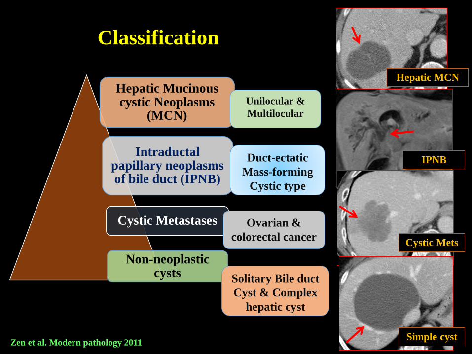
Classification
Hepatic Mucinous cystic Neoplasms
(MCN)
Intraductal papillary neoplasms of bile duct (IPNB)
Cystic Metastases
Non-neoplastic cysts
Duct-ectatic
Mass-forming
Cystic type
Unilocular &
Multilocular
Ovarian &
colorectal cancer
Solitary Bile duct
Cyst & Complex
hepatic cyst
Hepatic MCN
IPNB
Cystic Mets
Simple cystZen et al. Modern pathology 2011

Cyst forming
Mass forming
Duct ectatic
Mucin producing cystic neoplasms of liver
Intraductal paillary neoplasm of bile duct
(IPNB)
Mucinous Cystic Neoplasm (MCN)
Sub epithelial ovarian stroma
PresentAbsent
Bile duct communication
Absent
Hepatic MCN vs. IPNB: How Do They Differ?
Present
• Subepithelial Ovarian-Like Stroma – Hepatic MCN
• Bile duct Communication – IPNB Zen et al. Histopathology 2014

• Cyst forming epithelial neoplasm composed of mucin producing epithelium
& associated with ovarian-type subepithelial stroma.
• About 5 % of hepatic cystic lesions; mostly in females, 90 % are
histologically benign; central liver location is more common
• Previously classified as cystadenoma & cystadenocarcinoma; now the
synonymous terms used are noninvasive and invasive MCN respectively
Hepatic MCN
• Develops from endodermal immature stroma or primary yolk sac cells
implanted during embryogenesis. The prevalence of hepatic mucinous
cystic neoplasm in segment IV may support an implant origin as
hamartomatous lesions commonly develop in segment IV
• Expression of estrogen & progesterone receptors in ovarian-like
subepithelial stroma also supports a putative role for female hormones in
the tumorigenesis.
Pathogenesis
Zen et al. Modern pathology 2011
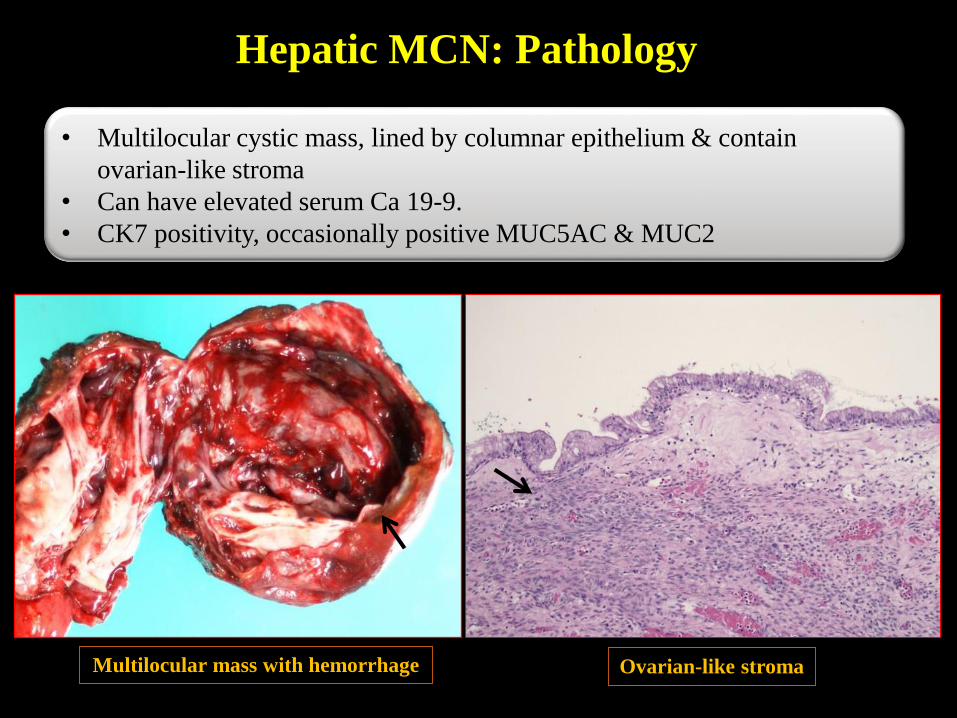
Hepatic MCN: Pathology
• Multilocular cystic mass, lined by columnar epithelium & contain
ovarian-like stroma
• Can have elevated serum Ca 19-9.
• CK7 positivity, occasionally positive MUC5AC & MUC2
Ovarian-like stromaMultilocular mass with hemorrhage

Hepatic MCN: Imaging Findings
• Multiloculated cystic mass with
enhancing septations & rare solid
components
• Upstream biliary dilatation is
uncommon
• No bile ductal communication or
intraductal filling defects
• No downstream ductal dilatation

Hepatic MCN: Imaging Findings
Characteristic MRI findings of hepatic MCN in two different patients
Despite of large size, no
significant biliary
dilatation & no bile duct
communication

Biliary papilloma &
papillomatosis
Intraductal type of
cholangio carcinoma
Papillary type of bile
duct carcinoma
Mucin-producing bile duct
tumor
Intraductal Papillary Neoplasm
of the Bile ducts (IPNB)
IPNB
Macroscopically identifiable papillary tumor within the bile ducts
May or may not have
mucin production
Bile ductPapillary
mass
Ohtsuka et al. International Journal of Hepatology 2014

• Most commonly seen in 50-70 years age group without gender
predilection
• Intermittent abdominal pain, jaundice and cholangitis are the most
common clinical manifestations
• Intrahepatic bile duct is the most common site of origin for IPNB
(69%) followed by extrahepatic bile duct (22%) & hilar location(9%)
• Hepatolithiasis increases the risk of IPNB, which explains it’s
relative increased incidence in Asian countries with higher incidence
of hepatolithiasis and Clonorchiasis
IPNB
Classic (polypoidal type) Cyst-forming type

IPNB: Pathogenesis
Develop from stem cells in bile
ductules, bile duct epithelium or
peribiliary glands
Dilated ductswith
papillary mass
Mass formation
Predominant epithelial
proliferation
Predominantmucus
secretion
Superficial spread along
biliary epithelium
Dilated ducts filledwith mucin
without mass
Interplay of two factors: Epithelial proliferation &
mucin secretion decides the formation of mass forming,
predominant duct ectatic & cystic types of IPNB
• Mucin-forming
• Duct-ectasia without
identifiable mass
• Cystic type
Lim et al. AJR 2011

IPNB arising for biliary
ductal epithelium
Intraductal papillary mass
Mucinoverproduction with increased
intraductal pressure
Cystic or aneurysmal
biliary ductal dilatation
IPNB arising from peribiliary
glands
Blockage of conduit between
the gland and bile duct
Progressive dilatation ofgland with
mucin
Cystic or diverticulum like dilatation
Cystic
variant of
IPNB
Cystic IPNB: Pathogenesis
Lim et al. AJR 2011
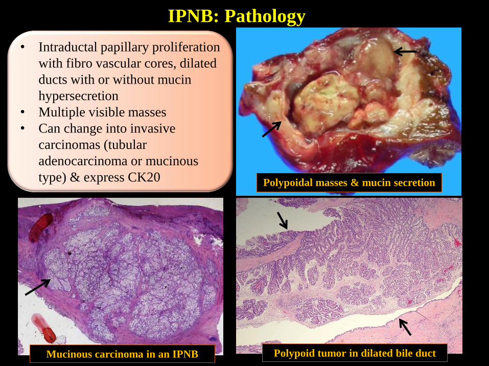
IPNB: Pathology
• Intraductal papillary proliferation
with fibro vascular cores, dilated
ducts with or without mucin
hypersecretion
• Multiple visible masses
• Can change into invasive
carcinomas (tubular
adenocarcinoma or mucinous
type) & express CK20 Polypoidal masses & mucin secretion
Mucinous carcinoma in an IPNB Polypoid tumor in dilated bile duct

IPNB: Imaging Findings
• Intraductal filling defect with papillary mass
• Ductal dilatation– downstream as well as upstream ductal
dilatation, can be segmental, lobar and diffuse depending
on the location
• Crowded bile ducts with hepatic parenchymal atrophy
• Multilocualr cystic mass with ‘bunch of grapes’
appearance; enhancing mural nodule
• Ductal communication
Mass forming IPNB

Duct ectatic
without visible mass
• Predominant segmental or lobar ductal dilatation
• No visible mass
• Hepatic segmental or lobar atrophy
IPNB: Imaging Classification
Mass forming IPNB
Cystic IPNB
Duct-ectatic IPNB
Lim et al. AJR 2008 & AJR 2011Yoon HJ et al. Abdominal Radiology 2013

Mass-Forming IPNB
Bile ductal dilation with enhancing intraductal masses

Duct-ectatic IPNB
Abundant mucus in the bile ducts
Diffusely dilated bile ducts
without any identifiable
intraductal mass

Cystic IPNB
Cystic lesions with bile ductal communication & biliary dilatation
Eovist MRI clearly shows
intrabiliary extension of tumor

MCN vs. IPNB: How Do They Differ on Imaging?
• Multiple intracystic septations
• Minimal mural nodularity; rare
solid components
• No bile duct dilatation /
communication
• Multilocular cystic lesion.
• Enhancing mural nodule .
• Presence of bile duct
dilatation & communication
MCN Cystic IPNB

Complex Hepatic Cyst
• Most common non-neoplastic
cystic lesions of liver present, seen
in 2.5% of population
• About 10% of MCNs manifest as
unilocular cystic lesions, which are
difficult to differentiate from
solitary simple cysts
Solitary Bile Duct Cyst
• Simple cyst with multiple episodes
of infection and/or hemorrhagic
resulting in minimally thickened
septations & echogenic debris
• Mimic hepatic MCN
Complex Hepatic cyst
Solitary Bile Duct cyst
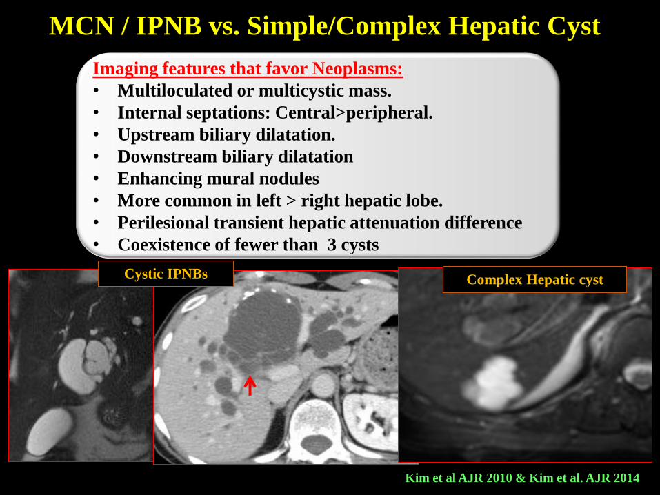
MCN / IPNB vs. Simple/Complex Hepatic Cyst
Imaging features that favor Neoplasms:
• Multiloculated or multicystic mass.
• Internal septations: Central>peripheral.
• Upstream biliary dilatation.
• Downstream biliary dilatation
• Enhancing mural nodules
• More common in left > right hepatic lobe.
• Perilesional transient hepatic attenuation difference
• Coexistence of fewer than 3 cysts
Kim et al AJR 2010 & Kim et al. AJR 2014
Complex Hepatic cystCystic IPNBs

Mucin-Producing Cystic Metastases
Abundant production of mucin by acinar tissues &
glandular structures in mucinous adenocarcinomas
Metastases from adenocarcinomas of
the ovary and colon/rectum
Cystic Metastases mimic MCN/IPNBs
Mucin-Producing
Cystic Metastases
from Colon Cancer
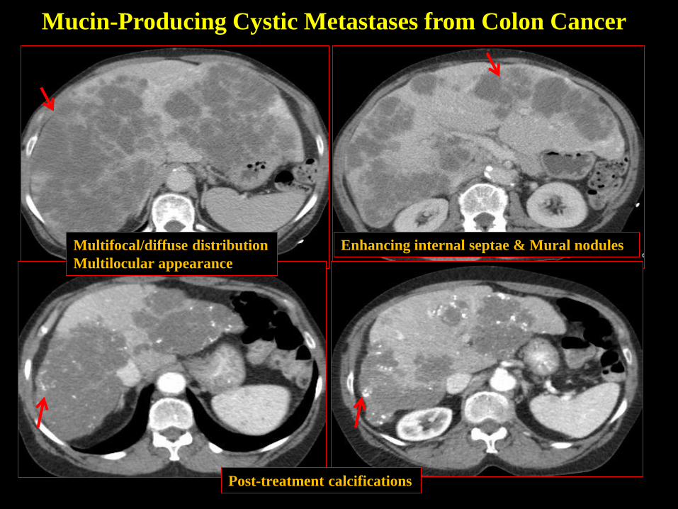
Multifocal/diffuse distribution
Multilocular appearance
Enhancing internal septae & Mural nodules
Post-treatment calcifications
Mucin-Producing Cystic Metastases from Colon Cancer

• Enucleation or segmental liver
resection
• Noninvasive MCN has a propensity
for recurrence & malignant
transformation
• Treatment should therefore be
complete surgical resection
MCN
• Segmental liver resection due to
propensity for superficial spread
along the bile duct & recurrence
after aspiration or enucleation
Management
IPNB
Watchful waiting may be one of the treatments options for MCN, unlike in IPNB where surgery is the only treatment option owing to the high probability of malignancy
Solitary bile duct cysts / complex hepatic cysts are managed conservatively or by aspiration followed by sclerotherapy or fenestration, and therefore misdiagnosis of MCN/IPNB as SBC / complex cyst may lead to inappropriate management & recurrence.

MCN / IPNB: Complications & Prognosis
• Obstructive jaundice & cholangitis
• Intraperitoneal rupture, hemorrhage within cyst or compression of
portal vein.
• Malignant transformation: Risk is as high as 15 % in MCN &
40-80 % IPNB; Always treat IPNB as malignant lesion.
• Recurrence: Fenestration, aspiration, sclerosis, internal drainage,
marsupialization, or partial resection with or without cavity ablation
can result in recurrence rate as high as 80% to 90% in both tumors
• 5 year survival rate of 80 % after complete resection of tumor with
negative margins in IPNB.
Cholangiocarcinoma developing in
mass forming IPNB
Cholangiocarcinoma developing in
cystic IPNB

Mucinous adenocarcinoma developing in IPNB
High-grade dysplasia in IPNB

Mucinous cystic neoplasms of liver are divided into MCN & IPNB on the basis of sub-epithelial ovarian stroma (MCN) and bile ductal communication (IPNB)
In every case of cystic hepatic lesion, look for biliary dilatation and ductal communication, otherwise diagnosis of IPNB can be missed !!
Radiologists should be aware of differentiating imaging features between MCN and IPNB, as there are significant differences in surgical management, follow-up and prognosis
Mucinous cystic neoplasms of liver should also be differentiated from non-neoplastic cystic lesions and complex hepatic cysts.
Cross sectional imaging modalities like CT, MRI & MRCP play an important role in preoperative diagnosis & differentiation between these entities
Summary

Venkata S Katabathina, MD
Associate Professor
Section of Abdominal Imaging
Department of Radiology
University of Texas Health Science Center at San Antonio
7703 Floyd Curl Dr, MC 7800
San Antonio, Texas 78229
Email: [email protected]
Author Correspondence Information
Thank You !