muc1 (episialin) expression in non-small cell lung cancer is
TRANSCRIPT

J Clin Pathol 1998;51:667-671
MUC 1 (episialin) expression in non-small celllung cancer is independent ofEGFR andc-erbB-2 expression and correlates with poor
survival in node positive patients
Francesca Guddo, Alexandra Giatromanolaki, Michael I Koukourakis, Clotilde Reina,Antonio-Maurizio Vignola, George Chlouverakis, John Hilkens, Kevin C Gatter,Adrian L Harris, Giovanni Bonsignore
Institute ofRespiratoryPathophysiology,Consiglio Nazionaledelle Ricerche,Palermo, ItalyF GuddoC ReinaA-M VignolaG Bonsignore
Department ofRadiotherapy/Oncology,Laboratory of CancerBiology, andDepartment ofBiostatistics,University Hospital ofIraklion-University ofCrete, Iraklion, Crete,GreeceA GiatromanolakiM I KoukourakisG Chlouverakis
Department ofTumour Biology, TheNetherlands CancerInstitute, Amsterdam,The NetherlandsJ Hilkens
Department ofCellular Science andICRF MedicalOncology Unit, OxfordRadcliffe Hospital,Headington, Oxford,UKK C GatterA L Harris
Correspondence to:Dr Michael I Koukourakis,Tumour and AngiogenesisResearch Group, 18Dimokratias Avenue, Iraklion71306, Crete, Greece.
Accepted for publication23 April 1998
AbstractAim-To examine tumour samples inumu-nohistochemically for MUCI (episialin),epidermal growth factor receptor(EGFR), and c-erbB-2, since the disrup-tion of the cell-cell adhesion system byMUCI and the c-erbB oncoprotein familyis known to be important in the develop-ment of metastasis in human cancers.Methods-93 tumour samples from pa-tients with early stage non-small cell lungcancer treated with surgery alone wereexamined for episialin, EGFR, andc-erbB-2.Results-Episialin depolarised expressiondid not correlate with any of the histo-pathological variables examined (T,Nstage, grade, histology, Ki67 proliferationindex). No correlation was observed be-tween episialin and EGFR or c-erbB-2expression. Survival analysis showed thatepisialin depolarised expression corre-lated with poor prognosis (p = 0.003),especially in squamous cell cases (p =0.0003). Episialin expression defined agroup of patients with poor prognosis inthe node positive category (p = 0.003). Inmultivariate analysis episialin was themost significant independent prognosticfactor (p = 0.007), followed by N stage (p =0.04).Conclusions-Depolarised expression ofepisialin is associated with poor outcomein early stage non-small cell lung cancer.Despite the similar acdvity on the cad-herin cell-cell adhesion system, the ex-pression of episialin and c-erbBoncoproteins is likely to be activatedwithin different pathogenic pathways.( Clin Pathol 1998;51:667-67 1)
Keywords: lung cancer; episialin; c-erbB-2
For a cancer to invade adjacent structures andto migrate to distant organs, complex mecha-nisms are required. Angiogenic factor secretionby tumour and stromal cells, endothelial cellmigration and vessel formation, and expressionof proteins involved in the cell-cell andcell-matrix adhesion disruption or cell motilityare essential for the tumour to grow, invade,and metastasise.' 2The cadherin-catenin cell-cell adhesion sys-
tem is well known to be involved in animal
morphogenesis and the maintenance ofnormaladhesion between cells.3 The epidermal growthfactor receptor (EGFR) and c-erbB-2 trans-membrane oncoproteins (members of the erboncoprotein family) interact with the cadherin-catenin complex and may inhibit cell adhesionfunction, thus enhancing cancer cell invasivepotential.4 Episialin, also known as MUC1 (orPEM, CA-15-3 antigen, or EMA) is anothertransmembrane protein shown in vitro toreduce E-cadherin mediated cell-cell adhesionby steric hindrance.6 Episialin is a glycoproteinexpressed at the apical side ofnormal glandularepithelial cells. In cancer cells, depolarisedexpression throughout the entire cell surfacehas been observed.7
In this study we examined the depolarisedexpression ofMUC 1 glycoprotein immunohis-tochemically in a series of early stage operablenon-small cell lung cancers. We report correla-tions with histopathological variables, Ki67mitotic index, EGFR, and c-erbB-2, as well assurvival.
MethodsWe examined 93 tumour surgical samples frompatients with early operable (Tl,2-NO,1staged8) non-small cell lung cancer. All patientswere treated with surgery alone, without radio-therapy or chemotherapy. Histological diagno-sis, grading, and N stage was done onhaematoxylin and eosin stained sections. Ofthese, 35 were adenocarcinomas and 58squamous cell lung carcinomas. Follow upranged from 45 to 74 months (median 62).
EPISIALIN IMMUNOHISTOCHEMISTRYEpisialin MUC1 expression was assessed onparaffin embedded material with the 214D4MoAb (IgGl) using an avidin-biotin compleximmunoperoxidase technique. This antibodyrecognises the protein backbone of episialin.Sections were dewaxed and rehydrated, andtreated for 10 minutes with 3% H202 to limitendogenous peroxidase activity. Samples werethen washed three times in 50 mM Tris-HCl/150 mM NaCl, pH 7.6 (TBS), and non-specific binding was blocked in normal rabbitserum (30 minutes, 1:20; Dako, Copenhagen,Denmark) in TBS, and incubated with themonoclonal antibody 214D4 (mouse IgGl,diluted 1:32 000) overnight at 4°C. Thesections were washed thoroughly in TBS andincubated with biotin conjugated rabbit
667

Guddo, Giatromanolaki, Koukourakis, et al
A
B~
Figure 1 (A) A lung adenocarcinoma with polarised MUCI expression. (B) A 1
squamous cell carcinoma with depolarised circumferential cytoplasmic and membraMUCI expression.
antimouse immunoglobulin antibodminutes, 1:200, Dako), followed by an
biotin-peroxidase complex (30 mv
1:100, Dako). Finally, the sectionsincubated with 3-4-3-4-diaminobenzi(chromogen for five minutes and c
stained with haematoxylin. Omissionprimary antibody was used for ncontrol.
100 rU,
>
a)U1)
CO01)
4-
cLC.)
80
60
In normal epithelium, episialin shows a
polarised pattern of immunoreactivity. Posi-tive "polarised pattern" of expression isdefined when episialin is localised in the cyto-plasmic vacuoles or in the cell membrane atthe apical site of cells. The circumferentialcytoplasmic and membrane immunoreactivity,never seen in normal cells, was recorded as
"depolarised" episialin expression (fig 1). Thepercentage of cancer cells with episialin depo-larised expression was recorded. This allowedanalysis using MUC1 as a continues variable.Cases were also divided into two groups
following the percentage of cells with depolar-ised expression, thus positive (depolarisedexpression in more than 25% of cells) andnegative (0-25%). The 25% used cut off pointwas the mean percentage of cells with episialindepolarised expression, but also the point thatdefined the group of patients with the highestpercentage of deaths at the time of analysis(fig 2).
OTHER IMMUNOHISTOCHEMICAL STUDIES
C-erbB-2 oncoprotein expression was assessedwith the monoclonal antibody NCL-CBl 1(Novocastra Laboratories, Newcastle upon
Tyne, UK), which recognises the internaldomain of the c-erbB-2 protein amino acidsequence. Staining was done with a previouslydescribed indirect immunoperoxidasetechnique.9 For a positive control we used a
breast carcinoma with 15-fold amplification ofthe c-erbB-2 gene and as a negative theprimary antibody was omitted. In a previousstudy we showed that membrane staining was
impossible to assess in non-small cell lungcancer.9 C-erbB-2 expression was separately
'ung assessed for cytoplasmic and membrane pat-
inous terns of reactivity. Two groups were consideredfor cytoplasmic staining: the group with
ly (30 positive reactivity (strong staining intensity in
avidin- Table 1 Correlation between episialin depolarised
linutes, expression and histological and patient variables in 93
were non-small cell lung cancer cases
dine as
ounter-of the
legative
.
40
20
p = 0.01
D 20 40 60 80
Per cent of cells with MUC1depolarised expression
100
Figure 2 Linear regression of the percentage of deaths insequential groups ofpatients defined by increasing cut offpercentages of depolarised episialin expression in 93non-small cell lung cancer patients.
Mean % positiveVariable cells (SD) 95% CI p Value
Histology SC 31.2 (31) 23 to 39 0.81AC 29.7 (30) 19 to 40
T stage TI 33.8 (31) 22 to 45 0.48T2 29.0 (30) 21 to 36
N stage NO 30.3 (30) 22 to 38 0.88NI 31.2 (32) 20 to 42
Grade I/II 28.0 (32) 18 to 37 0.44III 32.9 (29) 24 to 41
Ki67 L'M 31.2 (31) 24 to 38 0.67H 27.2 (29) 12 to 43
c-erbB-2 neg/weak 28.5 (31) 21 to 35 0.31pos 35.9 (29) 23 to 48
EGFR neg 36.6 (36) 18 to 54 0.57pos 31.7 (30) 23 to 39
Age <65years 32.2 (32) 23 to 41 0.55>64 years 28.4 (28) 19 to 37
Sex Female 38.5 (32) 22 to 54 0.23Male 28.8 (30) 21 to 35
AC, adenocarcinoma; CI, confidence interval; SC, squamouscell carcinoma.
0'1
668
7
MUCI expression in non-small cell lung cancer
EGFR1 murine monoclonal antibody wasused to identify EGFR by means of an indirectimmunoperoxidase technique'" on 5 gmcryostat sections. Samples with negative (-) orweak (+) staining intensity were consideredas negative, while moderately (++) orstrongly (+++) positive were considered aspositive.
Proliferative index was assessed with themonoclonal antibody Ki67." Frozen materialwas taken from two separate areas of thetumour and the Ki67 assessment was based onthe average value. Three groups were consid-ered, based on the percent of stained nuclei:0-10% = low proliferative index, 10-40% =medium, and > 40% = high.
500 1000 1500 2000Time (days)
100
80
40
B M UC1 negative (3i- - -- MUC1 positive (2E
. ~~~~~~~p_0.00031
Sqaoscelcne
zu _
0 500 1000 1500
30 pt8 pts
2000Time (days)
- MUC1 negative (23 pts)|V |---- MUC1 positive (12 pts)|
IP07
I
~~~~~~ ------:
A,er; r ec l~~~~~~~~~~~~~~~~~~~~~~~~~~~~~~~~~~~~~~~~~~~~~~~~~~~~~~~~~~~~~~~
40
Aaenocarcinomas
500 1000 1500 2000
20
0
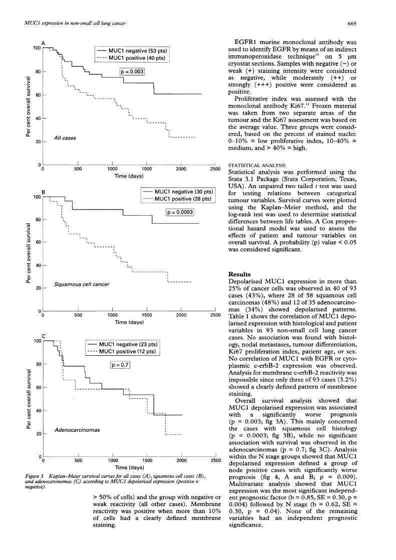
Time (days)Figure 3 Kaplan-Meier survival curves for all cases (A), squamous cell cases (B),and adenocarcinomas (C) according to MUCI depolarised expression (positive vnegative).
> 50% of cells) and the group with negatiiweak reactivity (all other cases). Membreactivity was positive when more thanof cells had a clearly defined membstaining.
25|00STATISTICAL ANALYSISStatistical analysis was performed using theStata 3.1 Package (Stata Corporation, Texas,USA). An unpaired two tailed t test was used
ts) for testing relations between categoricals) tumour variables. Survival curves were plotted
using the Kaplan-Meier method, and thelog-rank test was used to determine statisticaldifferences between life tables. A Cox propor-tional hazard model was used to assess theeffects of patient and tumour variables onoverall survival. A probability (p) value < 0.05was considered significant.
ResultsDepolarised MUC1 expression in more than25% of cancer cells was observed in 40 of 93cases (43%), where 28 of 58 squamous cellcarcinomas (48%) and 12 of 35 adenocarcino-mas (34%) showed depolarised patterns.
2500 Table 1 shows the correlation ofMUC1 depo-larised expression with histological and patientvariables in 93 non-small cell lung cancercases. No association was found with histol-ogy, nodal metastases, tumour differentiation,Ki67 proliferation index, patient age, or sex.No correlation of MUC 1 with EGFR or cyto-plasmic c-erbB-2 expression was observed.Analysis for membrane c-erbB-2 reactivity wasimpossible since only three of 93 cases (3.2%)showed a clearly defined pattern of membranestaining.
Overall survival analysis showed thatMUC1 depolarised expression was associatedwith a significantly worse prognosis(p = 0.003; fig 3A). This mainly concernedthe cases with squamous cell histology(p = 0.0003; fig 3B), while no significantassociation with survival was observed in theadenocarcinomas (p = 0.7; fig 3C). Analysis
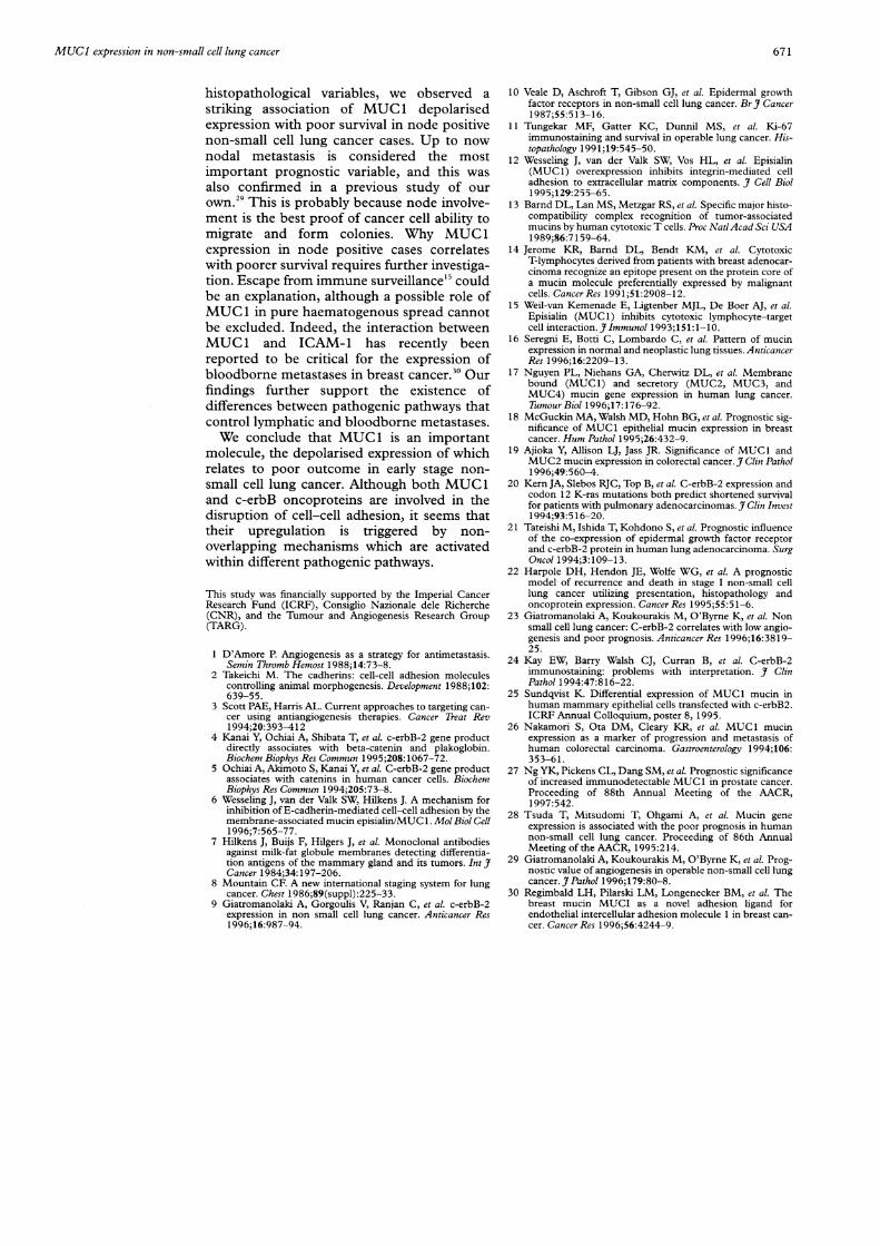
- within the N stage groups showed that MUC 12500 depolarised expression defined a group of
node positive cases with significantly worseprognosis (fig 4, A and B; p = 0.009).Multivariate analysis showed that MUC 1expression was the most significant independ-
ve or ent prognostic factor (b = 0.85, SE = 0.30, p =)rane 0.004) followed by N stage (b = 0.62, SE =10% 0.30, p = 0.04). None of the remaining)rane variables had an independent prognostic
significance.
A100
CUO
C,,
CU
0CU
a-0
40 _-
20 H
1-
All cases
'0-
Ca)aC0L
C-
coCUCU
0
C.)
0D
(lv
t
669
MUC1 negative (53 pts)MUC1 positive (40 pts)
p = 0.003]-: I---I
--I---I
80
60
60
,)A
c100
80
60

670 Guddo, Giatromanolaki, Koukourakis, et al
10c
40 -
20 H
00
NO stage
500 1000 1500 2000Time (days)
100
40
20
(V
3 MUC1 negative (20 pts)lHL ~~~~--- MUC1 positive (15 pts)|
1L IP=0091I
,IIi.
..
--E~~~~~~~~~~~~~~~~~~~~~~~~~~~~~~
Nl stage
500 1000 1500 2000Time (days)
Figure 4 Kaplan-Meier survival cases for NO stage (A) and Nl stage (B) groupsaccording to MUCI depolarised expression (positive v negative).
DiscussionSeveral in vitro studies suggest that MUexpression by cancer cells is an imporicomponent of biochemical events that ensmetastasisation. Wesseling et al showed IMUC1 inhibits the E-cadherin mediated c
cell adhesion system, the length of ML;ectodomain being the dominant factorinhibition to occur.6 High MUC1 levelsreduce the integrin mediated cell adhesioithe extracellular matrix.'2 Although nrestricted T cell cytotoxicity to the m
histocompatibility complex elicited by MLis reported,'3 '4 MUC 1 expressing cells are
susceptible to T cell mediated lysis, whichcontribute to cancer cell escape from immsurveillance. "
In this study we examined immunohichemically the depolarised expressionMUC 1 in a series of non-small cell lung cai
patients treated with surgery alone. Depcised MUC1 expression in more than 25°/cells was observed in about 45% of cases. Flevels of MUC1 mRNA in lung c'ancer
been recently reported.'6 In a study by Nguet al,7 high concentrations of MUC1 mR
were found in normal lung tissue and adeno-carcinomas, while MUC 1 levels were notincreased in squamous cell cancer. In our studywe examined the depolarised expression ofMUC 1 and found no difference betweensquamous cell carcinomas and adenocarcino-mas. Disturbance of localisation rather than themRNA expression level may more accuratelyreflect the aberrant function of the MUC 1membrane bounding mucin.We also observed no correlation between
MUC 1 expression in lung cancer and histo-pathological variables including N stage, differ-entiation, and proliferation index. In breastcancer,"8 depolarised MUC 1 expression hasbeen associated with node involvement andpositive oestrogen receptors. However, no cor-relation of MUC 1 with T stage or grade was
2500 observed. A significant association betweenMUC 1 expression and node involvement hasalso been reported in colorectal cancer.'9The c-erb protein family is well known to
disrupt the cadherin mediated cell adhesionsystem. In three studies of non-small cell lungcancer, c-erbB-2 expression was found to beassociated with a worse outcome.20 22 In a pre-vious study we showed that c-erbB-2 defined apoorer survival in cases with lowvascularisation.23 Although membrane stainingis generally accepted to be functionally relevantto tumour pathogenesis, only Kern's study20was based on membrane staining. The studiesby Tateishi et al,2' Harpole et al,22 andGiatromanolaki et afP did not recognise aclearly defined pattern of membrane staining,the whole analysis been founded on cytoplas-mic reactivity. Moreover, in a study by Kay etal,24 although membrane positivity was foundin 7-21% ofbreast, bladder, and renal cases, no
2500 membrane reactivity was identified insquamous cell carcinomas, colon adenocarci-nomas, and other tumours. Thus it seems thatassessment of membrane reactivity cannotalways be applied. Only 3.2% of our casesexpressed a clearly defined pattern of c-erbB-2
JC1 membrane staining. Further staining of ourtant cases with another c-erbB-2 antibody (Dako)able using the APAAP technique confirmed thatthat membrane staining assessment is not feasibleell- in non-small cell lung cancer (data not shown).JC 1 In the present study we examined possiblefor cooperation of the MUC 1 protein with the
also EGFR and c-erbB-2 oncoproteins. Althoughn to an unpublished study reported downregulationLon- ofMUC 1 after transfection of mammary can-.ajor cer cells with c-erbB-2,2' we failed to confirm aJC 1 similar association. This may show that in lungless cancer, although MUC1 and c-erbB proteinsmay mediate cell-cell adhesion disruption, theune regulation of their expression is controlled by
different mechanisms.isto- Although several reports show a correlationof of MUC 1 expression with survival in breast,-cercolon, and prostate cancer,'8 26 27 the role oflar- MUC 1 expression in lung cancer has not been
/o of clarified. A preliminary report from Japan inUigh 38 non-small cell lung cancer cases revealed ahave possible association of MUC 1 expressioniyen with poor survival.28 In our study, despitetNA the absence of MUC 1 association with
u
C-
a)
0U)
C.)
U,01)
U1)
0
U1)0
a)0~
. , \A - MuCl negative (33 pts)
MUC1 positive (25 pts)
p = 0.1.D
--l------------
:----I0
I-----
8(
6(
-------
I- ,
80
60
I-
MUCI expression in non-small cell lung cancer
histopathological variables, we observed astriking association of MUC 1 depolarisedexpression with poor survival in node positivenon-small cell lung cancer cases. Up to nownodal metastasis is considered the mostimportant prognostic variable, and this wasalso confirmed in a previous study of ourown.29 This is probably because node involve-ment is the best proof of cancer cell ability tomigrate and form colonies. Why MUC 1expression in node positive cases correlateswith poorer survival requires further investiga-tion. Escape from immune surveillance'" couldbe an explanation, although a possible role ofMUC 1 in pure haematogenous spread cannotbe excluded. Indeed, the interaction betweenMUC 1 and ICAM-1 has recently beenreported to be critical for the expression ofbloodborne metastases in breast cancer.30 Ourfindings further support the existence ofdifferences between pathogenic pathways thatcontrol lymphatic and bloodborne metastases.We conclude that MUC1 is an important
molecule, the depolarised expression of whichrelates to poor outcome in early stage non-small cell lung cancer. Although both MUC1and c-erbB oncoproteins are involved in thedisruption of cell-cell adhesion, it seems thattheir upregulation is triggered by non-overlapping mechanisms which are activatedwithin different pathogenic pathways.
This study was financially supported by the Imperial CancerResearch Fund (ICRF), Consiglio Nazionale dele Richerche(CNR), and the Tumour and Angiogenesis Research Group(TARG).
1 D'Amore P. Angiogenesis as a strategy for antimetastasis.Semin Thromb Hemost 1988;14:73-8.
2 Takeichi M. The cadherins: cell-cell adhesion moleculescontrolling animal morphogenesis. Development 1988;102:639-55.
3 Scott PAE, Harris AL. Current approaches to targeting can-cer using antiangiogenesis therapies. Cancer Treat Rev1994;20:393-412
4 Kanai Y, Ochiai A, Shibata T, et al. c-erbB-2 gene productdirectly associates with beta-catenin and plakoglobin.Biochem Biophys Res Commun 1995;208:1067-72.
5 Ochiai A, Akimoto S, Kanai Y, et al. C-erbB-2 gene productassociates with catenins in human cancer cells. BiochemBiophys Res Commun 1994;205:73-8.
6 Wesseling J, van der Valk SW, Hilkens J. A mechanism forinhibition of E-cadherin-mediated cell-cell adhesion by themembrane-associated mucin episialin/MUC 1. Mol Biol Cell1996;7:565-77.
7 Hilkens J, Buijs F, Hilgers J, et al. Monoclonal antibodiesagainst milk-fat globule membranes detecting differentia-tion antigens of the mammary gland and its tumors. Int JCancer 1984;34:197-206.
8 Mountain CF. A new international staging system for lungcancer. Chest 1986;89(suppl) :225-33.
9 Giatromanolaki A, Gorgoulis V, Ranjan C, et al. c-erbB-2expression in non small cell lung cancer. Anticancer Res1996;16:987-94.
10 Veale D, Aschroft T, Gibson GJ, et al. Epidermal growthfactor receptors in non-small cell lung cancer. Br 7 Cancer1987;55:513-16.
11 Tungekar MF, Gatter KC, Dunnil MS, et al. Ki-67immunostaining and survival in operable lung cancer. His-topathology 1991;19:545-50.
12 Wesseling J, van der Valk SW, Vos HL, et al. Episialin(MUC 1) overexpression inhibits integrin-mediated celladhesion to extracellular matrix components. _7 Cell Biol1995;129:255-65.
13 Barnd DL, Lan MS, Metzgar RS, et al. Specific major histo-compatibility complex recognition of tumor-associatedmucins by human cytotoxic T cells. Proc Natl Acad Sci USA1989;86:7159-64.
14 Jerome KR, Barnd DL, Bendt KM, et al. CytotoxicT-lymphocytes derived from patients with breast adenocar-cinoma recognize an epitope present on the protein core ofa mucin molecule preferentially expressed by malignantcells. Cancer Res 1991;51:2908-12.
15 Weil-van Kemenade E, Ligtenber MJL, De Boer AJ, et al.Episialin (MUC 1) inhibits cytotoxic lymphocyte-targetcell interaction. Jimmunol 1993;151:1-10.
16 Seregni E, Botti C, Lombardo C, et al. Pattern of mucinexpression in normal and neoplastic lung tissues. AnticancerRes 1996;16:2209-13.
17 Nguyen PL, Niehans GA, Cherwitz DL, et al. Membranebound (MUC 1) and secretory (MUC2, MUC3, andMUC4) mucin gene expression in human lung cancer.Tumour Biol 1996;17:176-92.
18 McGuckin MA, Walsh MD, Hohn BG, et al. Prognostic sig-nificance of MUC 1 epithelial mucin expression in breastcancer. Hum Pathol 1995;26:432-9.
19 Ajioka Y, Allison LJ, Jass JR. Significance of MUC1 andMUC2 mucin expression in colorectal cancer. 7 Clin Pathol1996;49:560-4.
20 Kern JA, Slebos RJC, Top B, et al. C-erbB-2 expression andcodon 12 K-ras mutations both predict shortened survivalfor patients with pulmonary adenocarcinomas. _7 Clin Invest1994;93:516-20.
21 Tateishi M, Ishida T, Kohdono S, et al. Prognostic influenceof the co-expression of epidermal growth factor receptorand c-erbB-2 protein in human lung adenocarcinoma. SurgOncol 1994;3:109-13.
22 Harpole DH, Hendon JE, Wolfe WG, et al. A prognosticmodel of recurrence and death in stage I non-small celllung cancer utilizing presentation, histopathology andoncoprotein expression. Cancer Res 1995;55:51 -6.
23 Giatromanolaki A, Koukourakis M, O'Byrne K, et al. Nonsmall cell lung cancer: C-erbB-2 correlates with low angio-genesis and poor prognosis. Anticancer Res 1996;16:3819-25.
24 Kay EW, Barry Walsh CJ, Curran B, et al. C-erbB-2immunostaining: problems with interpretation. .7 ClinPathol 1994:47:816-22.
25 Sundqvist K. Differential expression of MUC1 mucin inhuman mammary epithelial cells transfected with c-erbB2.ICRF Annual Colloquium, poster 8, 1995.
26 Nakamori S, Ota DM, Cleary KR, et al. MUC 1 mucinexpression as a marker of progression and metastasis ofhuman colorectal carcinoma. Gastroenterology 1994;106:353-61.
27 Ng YK, Pickens CL, Dang SM, et al. Prognostic significanceof increased immunodetectable MUC 1 in prostate cancer.Proceeding of 88th Annual Meeting of the AACR,1997:542.
28 Tsuda T, Mitsudomi T, Ohgami A, et al. Mucin geneexpression is associated with the poor prognosis in humannon-small cell lung cancer. Proceeding of 86th AnnualMeeting of the AACR, 1995:214.
29 Giatromanolaki A, Koukourakis M, O'Byrne K, et al. Prog-nostic value of angiogenesis in operable non-small cell lungcancer.J Pathol 1996;179:80-8.
30 Regimbald LH, Pilarski LM, Longenecker BM, et al. Thebreast mucin MUCI as a novel adhesion ligand forendothelial intercellular adhesion molecule 1 in breast can-cer. Cancer Res 1996;56:4244-9.
671