msc enumeration kit human
TRANSCRIPT
For 25 tests Order no. 130-106-646
MSC Enumeration Kithuman
Miltenyi Biotec B.V. & Co. KG Friedrich-Ebert-Straße 68, 51429 Bergisch Gladbach, GermanyPhone +49 2204 8306-0, Fax +49 2204 85197
[email protected], www.miltenyibiotec.com
140
-00
4-7
78
.02
Unless otherwise specifi cally indicated,
Miltenyi Biotec products and services are for research use only
and not for diagnostic or therapeutic use.
1. DescriptionContents
2 3140-004-778.02 140-004-778.02
Contents
1. Description
1.1 Principle of the assay
1.2 Background information
1.3 Application
1.4 Description of kit components
1.5 Reagents and instruments requirements
2. Protocols
2.1 Immunofl uorescent labeling
2.2 Flow cytometric acquisition and data analysis using
the MACSQuant® Analyzer 10
2.3 Flow cytometric acquisition and data analysis using
fl ow cytometer other than MACSQuant® Analyzer 10
3. Appendix
4. References
1. Description
Components 250 μL MSC Staining Cocktail, human:
cocktail of fl uorochrome-conjugated monoclonal
antibodies: CD45-FITC (clone: 5B1, isotype: mouse
IgG2a), CD235a (Glycophorin A)-VioBlue® (clone:
REA175, isotype: recombinant human IgG1),
CD271 (LNGFR)-PE (clone: Me20.4-1H.4, isotype:
mouse IgG1).
250 μL Anti-MSCA-1 (W8B2)-APC, human:
anti-MSCA-1 (W8B2) monoclonal antibody (clone:
W8B2, isotype: mouse IgG1) conjugated to APC.
250 μL MSC Isotype Cocktail, human:
cocktail of Mouse IgG1-APC (clone: IS5-21F5.12.2)
and f luorochrome-conjugated monoclonal
antibodies: CD45-FITC (clone: 5B1, isotype: mouse
IgG2a), CD235a (Glycophorin A)-VioBlue (clone:
REA175, isotype: recombinant human IgG1),
CD271 (LNGFR)-PE (clone: Me20.4-1H.4, isotype:
mouse IgG1).
5 mL Red Blood Cell Lysis Solution (10×)
1 mL FcR Blocking Reagent, human
500 μL 7-AAD Solution (52.5 μg/mL)
Capacity For 25 tests with 200 μL human cell sample per test.
Product format MSC Staining Cocktail, Anti-MSCA-1 (W8B2)-
APC, MSC Isotype Cocktail, and FcR Blocking
1. Description1. Description
4 5140-004-778.02 140-004-778.02
Reagent are supplied in buff er containing stabilizer
and 0.05% sodium azide.
Storage Store all components protected from light at 2–8 °C.
Do not freeze. Th e expiration date is indicated on
the vial label.
1.1 Principle of the assay
The MSC Enumeration Kit has been designed for the quantification of
mesenchymal stromal cells (MSCs) in human bone marrow samples,
based on the expression of CD271 (LNGFR) and Anti-MSCA-1 (W8B2).
The kit allows the identification of CD45+ leukocytes and CD271
(LNGFR)+/Anti-MSCA-1 (W8B2)+ MSCs, which are also CD45dim.
CD235a (Glycophorin A) is used to exclude remaining red blood cells,
which were not completely lysed by the Red Blood Cell Lysing Solution.
For one test, two 100 μL samples of bone marrow are needed.
The MSCs are detected by staining one 100 μL sample with the
MSC Staining Cocktail as well as with Anti-MSCA-1 (W8B2)-APC.
Subsequently, erythrocytes are lysed using the Red Blood Cell Lysis
Solution.
Additionally, a 100 μL control sample is stained with the MSC Isotype
Cocktail. This is required for the proper setting of relevant gates during
flow cytometry analysis.
The FcR Blocking Reagent increases the specificity of immunofluorescent
staining.
Dead cells are excluded from the analysis by adding of DNA stain
7-aminoactinomycin D (7-AAD). 7-AAD diffuses through the cell
membrane of dead cells and intercalates with their DNA.
Finally, cells are analyzed with flow cytometers able to detect 4-color
fluorescence (488 nm argon laser, 635 nm red diode laser, optional:
405 nm violet laser). The use of the MACSQuant® Flow Cytometers
enable an automated and accurate cell count. If using flow cytometers
other than the MACSQuant Flow Cytometer, it is necessary to determine
the absolute cell count by analysis of commercially available counting
particles such as present in BD Trucount™ Tubes.
1.2 Background information
The large scale isolation and expansion of MSCs from human bone
marrow is time consuming, cost effective, and highly dependent on the
quality of the starting material. Therefore it is desirable to qualify the
bone marrow source with regard to MSC content. The common way
to quantify MSCs within human blood sources is the colony forming
unit-fibroblast (CFU-F) assay, which is time consuming, dependent on
serum lots, and plating densities, as well as considerably subjective in
defining and scoring colonies. It was shown that Anti-MSCA-1 (W8B2)
only detects CD271 (LNGFR)bright cells and only these cells give rise to
CFU-F1. A close linear relationship was observed between the number
of CFU-F colonies counted manually after 14 days of culture and the
number of CD271 (LNGFR)bright cells per milliliter of aspirate2.
1.3 Application
● Enumeration of MSCs in human bone marrow samples
1. Description1. Description
6 7140-004-778.02 140-004-778.02
Red Blood Cell Lysis Solution
Upon incubation of the cells in 1× Red Blood Cell Lysis Solution
erythrocytes are selectively lysed. Dilute Red Blood Cell Lysis Solution
(10×) 1:10 with double-distilled water (ddH2O).
1.5 Reagents and instruments requirements
● Double-distilled water (ddH2O)
● 5 mL tubes
● Micropipettes with tips
● Centrifuge (2–8 °C)
● Vortex mixer
● Rotation device for tubes: MACSmix™ Tube Rotator (# 130-090-753)
● Th ree-laser fl ow cytometer and soft ware (405 nm, 488 nm, and
638 nm laser), e.g., MACSQuant Analyzer 10 (# 130-096-343) with
MACSQuantify™ Soft ware.
▲ Note: Alternatively, a dual-laser fl ow cytometer and soft ware (488 nm and 638 nm
laser) can be used. Exclusion of remaining red blood cells using CD235a is then not
possible.
● (Optional) BD Trucount Absolute Counting Tubes (# 340334)
● (Optional) MACS® SmartStrainers (100 μm) (# 130-098-463)
● (Optional) MACS Comp Bead Kit, anti-mouse Igκ (# 130-097-900)
● (Optional) MACS Comp Bead Kit, anti-REA (# 130-104-693)
1.4 Description of kit components
MSC Staining Cocktail
Components of the MSC Staining Cocktail, human
Antibody Fluorochrome Specifi city
CD45 FITC Leukocytes
CD235a (Glycophorin A) VioBlue Erythrocytes
CD271 (LNGFR) PE MSCs, lymphocyte progenitors
Fluorescein (FITC), Phycoerythrin (PE)
Anti-MSCA-1 (W8B2)-APC
Antibody Fluorochrome Specifi city
Anti-MSCA-1 (W8B2) APC MSCs
Allophycocyanin (APC)
MSC Isotype Cocktail
Components of the MSC Isotype Cocktail, human
Antibody Fluorochrome Specifi city
CD45 FITC Leukocytes
CD235a (Glycophorin A) VioBlue Erythrocytes
CD271 (LNGFR) PE MSCs, lymphocyte progenitors
Mouse IgG1 APC Isotype control

2. Protocol2. Protocol
8 9140-004-778.02 140-004-778.02
2. Protocols
Th e following protocol has been optimized for mesenchymal stromal
cell (MSC) enumeration in a human bone marrow sample. For best
results use starting material directly aft er aspiration.
▲ Bone marrow samples have to be supplemented with an anticoagulant,
for example, heparin.
▲ Bone marrow samples should be stored at room temperature under
agitation until analysis.
▲ When using bone marrow aspirate pass the material through a
MACS SmartStrainer (100 μm).
▲ Dilute Red Blood Cell Lysis Solution (10×) 1:10 with double-distilled
water (ddH2O), for example, dilute 200 μL of Red Blood Cell Lysis
Solution (10×) with 1800 μL of ddH2O. Do not use deionized water!
Store the prepared 1× Red Blood Cell Lysis Solution at room temperature.
Discard unused solution at the end of the day.
2.1 Immunofl uorescent labeling
▲ Volumes given below are for one test with two 100 μL samples ofbone marrow.
1. Label two tubes with A (control sample) and B (MSC sample).
▲ Note: When using other fl ow cytometer than the MACSQuant Flow Cytometers
use two BD Trucount Absolute Counting Tubes.
2. Carefully pipette 100 μL of well-mixed human bone marrow to the
bottom of each tube.
▲ Note: For each sample tube use a fresh micropipette tip.
3. Add 20 μL FcR Blocking Solution into each tube.
4. Add 10 μL of MSC Isotype Cocktail into tube A and 10 μL of MSC
Staining Cocktail as well as 10 μL of Anti-MSCA-1 (W8B2)-APC
into tube B.
5. Immediately vortex thoroughly and incubate for 10 minutes at
2–8 °C in the dark.
6. Add 860 μL of 1× Red Blood Cell Lysis Solution into tube A and
850 μL in tube B.
7. Immediately vortex and incubate for 10 minutes at room
temperature in the dark using the MACSmix Tube Rotator.
8. Add 10 μL of the 7-AAD Solution into each tube.
9. Directly proceed with fl ow cytometric analysis.
▲ Note: Analysis should be performed within 20 minutes aft er staining.
2. Protocol2. Protocol
10 11140-004-778.02 140-004-778.02
2.2 Flow cytometric acquisition and data analysis using the
MACSQuant® Analyzer 10
Before starting to acquire the samples make sure that the MACSQuant®
Analyzer 10 fl ow cytometer has been carefully cleaned in order to allow
the detection of rare MSCs.
Instrument setting and data acquisition
Choose an instrument setting for standard four-color cell analysis of
human blood leukocytes. A proper instrument setting can be reached
by adjusting parameters with separate FITC-, PE-, VioBlue-, and
APC-conjugated antibodies or by using the MACS Comp Bead Kit,
anti-mouse Igκ or the MACS Comp Bead Kit, anti-REA.
▲ Exclude debris from data acquisition by an appropriate value for the
threshold on forward scatter (FSC).
Creation of dot plots
For data acquisition and analysis create the following dot plots (region
settings are shown in the examples below):
A Plot: Forward scatter (FSC) versus side scatter (SSC)
B Plot: 7-AAD Solution versus SSC
C Plot: FSC versus CD235a (Glycophorin A)-VioBlue
D Plot: CD45-FITC versus CD271 (LNGFR)-PE
E Plot: Mouse IgG1-APC versus CD271 (LNGFR)-PE and Anti-MSCA-1
(W8B2)-APC versus CD271 (LNGFR)-PE
Defi ne the experiment as follows:
Experiment setup
Rack type Chill 5 Tube Rack, choose 2 positions
Sample ID 1. position: Control sample
2. position: MSC sample
Flow rate medium
Mode standard
Uptake volume 450 μL
Sample volume 1 mL
Instrument setting PBMC compensation (needs to be performed before data anaylsis)*
Annotations 1. position: CD45-FITC, CD235a (Glycophorin A)-VioBlue, CD271 (LNGFR)-PE, Mouse IgG1-APC, 7-AAD Solution
2. position: CD45-FITC, CD235a (Glycophorin A)-VioBlue, CD271 (LNGFR)-PE, Anti-MSCA-1 (W8B2)-APC, 7-AAD Solution
* Please refer to the user guide of the MACSQuantify Soft ware for detailed instructions.
2.2.1 Flow cytometric data acquisition

2. Protocol2. Protocol
12 13140-004-778.02 140-004-778.02
B Plot: 7-AAD Solution versus SSC (gated on P1)
Draw region P2 to exclude dead cells.
10³-10
10¹ 10²0
25
50
75
100
1
7-AAD
Sid
e s
catt
er
P2
C Plot: FSC versus CD235a (Glycophorin A)-VioBlue (gated on P1/P2)
Draw region P3 to exclude CD235a+ erythrocytes.
10³
-10
10¹
10²
0
250 500 750 1000
1
Forward scatter
CD
23
5a
-Vio
Blu
e
P3
▲ Th e gating strategy below describes the fl ow cytometric analysis of a
bone marrow sample using the MACSQuantify Soft ware.
▲ Perform the analysis of the MSC sample stained with the MSC
Staining Cocktail in the same way as the analysis of the control sample
stained with the MSC Control Cocktail.
MSC sample
A Plot: FSC versus SSC (no gate)
Draw region P1 to exclude debris and platelets.
Forward scatter
Sid
e s
catt
er
P1
2.2.2 Flow cytometric analysis
2. Protocol2. Protocol
14 15140-004-778.02 140-004-778.02
D Plot: CD45-FITC versus CD271 (LNGFR)-PE (gated on P1/P2/P3)
Defi ne region P4 thereby including all CD271 (LNGFR)+ cells.
10³-101
10¹ 10²0
10³
10²
10¹
CD45-FITC
CD
27
1 (
LN
GF
R)-
PE
-1 1
P4
E Plot: Mouse IgG1-APC/Anti-MSCA-1 (W8B2)-APC versus CD271
(LNGFR)-PE (gated on P1/P2/P3/P4)
Copy all gates into the control sample.
Defi ne region P5 with the control sample to include all Anti-MSCA-1
(W8B2)-APC+ events within the MSC sample. It is important to set gate
P5 as close as possible to the Anti-MSCA-1 (W8B2)– events to include all
Anti-MSCA-1 (W8B2)+ and Anti-MSCA-1 (W8B2)dim+ cells.
Control sample MSC sample
10³-101
10¹ 10²0
10³
10²
10¹
Mouse IgG1-APC/Anti-MSCA-1-APC
CD
27
1 (
LN
GF
R)-
PE
-1 1
P50.00%-2
1
2.22/mL
10³-101
10¹ 10²0
10³
10²
10¹
Mouse IgG1-APC/Anti-MSCA-1-APC
CD
27
1 (
LN
GF
R)-
PE
-1 1
P50.02%-2
175
389/mL
2. Protocol2. Protocol
16 17140-004-778.02 140-004-778.02
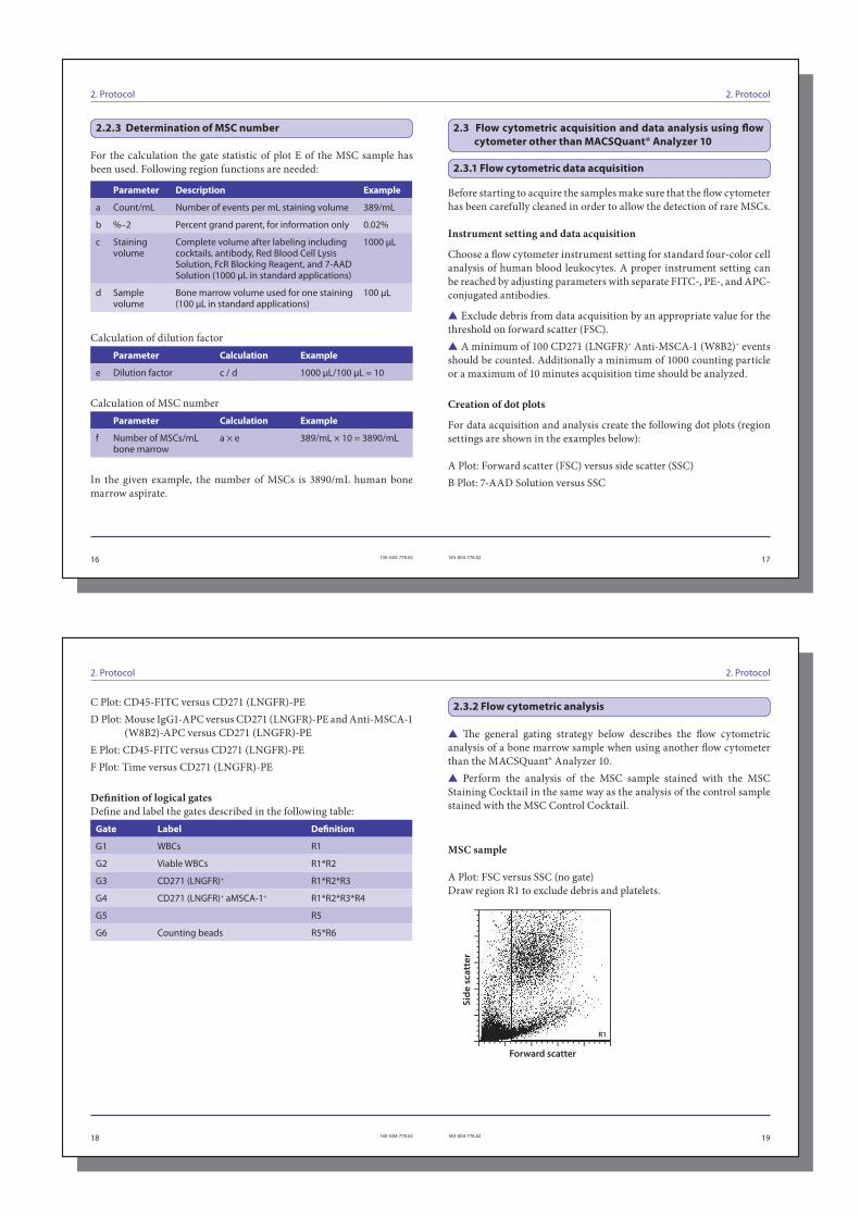
For the calculation the gate statistic of plot E of the MSC sample has
been used. Following region functions are needed:
Parameter Description Example
a Count/mL Number of events per mL staining volume 389/mL
b %–2 Percent grand parent, for information only 0.02%
c Staining volume
Complete volume after labeling including cocktails, antibody, Red Blood Cell Lysis Solution, FcR Blocking Reagent, and 7-AAD Solution (1000 μL in standard applications)
1000 μL
d Sample volume
Bone marrow volume used for one staining (100 μL in standard applications)
100 μL
Calculation of dilution factor
Parameter Calculation Example
e Dilution factor c / d 1000 μL/100 μL = 10
Calculation of MSC number
Parameter Calculation Example
f Number of MSCs/mL bone marrow
a × e 389/mL × 10 = 3890/mL
In the given example, the number of MSCs is 3890/mL human bone
marrow aspirate.
2.3 Flow cytometric acquisition and data analysis using fl ow
cytometer other than MACSQuant® Analyzer 10
Before starting to acquire the samples make sure that the fl ow cytometer
has been carefully cleaned in order to allow the detection of rare MSCs.
Instrument setting and data acquisition
Choose a fl ow cytometer instrument setting for standard four-color cell
analysis of human blood leukocytes. A proper instrument setting can
be reached by adjusting parameters with separate FITC-, PE-, and APC-
conjugated antibodies.
▲ Exclude debris from data acquisition by an appropriate value for the
threshold on forward scatter (FSC).
▲ A minimum of 100 CD271 (LNGFR)+ Anti-MSCA-1 (W8B2)+ events
should be counted. Additionally a minimum of 1000 counting particle
or a maximum of 10 minutes acquisition time should be analyzed.
Creation of dot plots
For data acquisition and analysis create the following dot plots (region
settings are shown in the examples below):
A Plot: Forward scatter (FSC) versus side scatter (SSC)
B Plot: 7-AAD Solution versus SSC
2.3.1 Flow cytometric data acquisition
2.2.3 Determination of MSC number
2. Protocol2. Protocol
18 19140-004-778.02 140-004-778.02
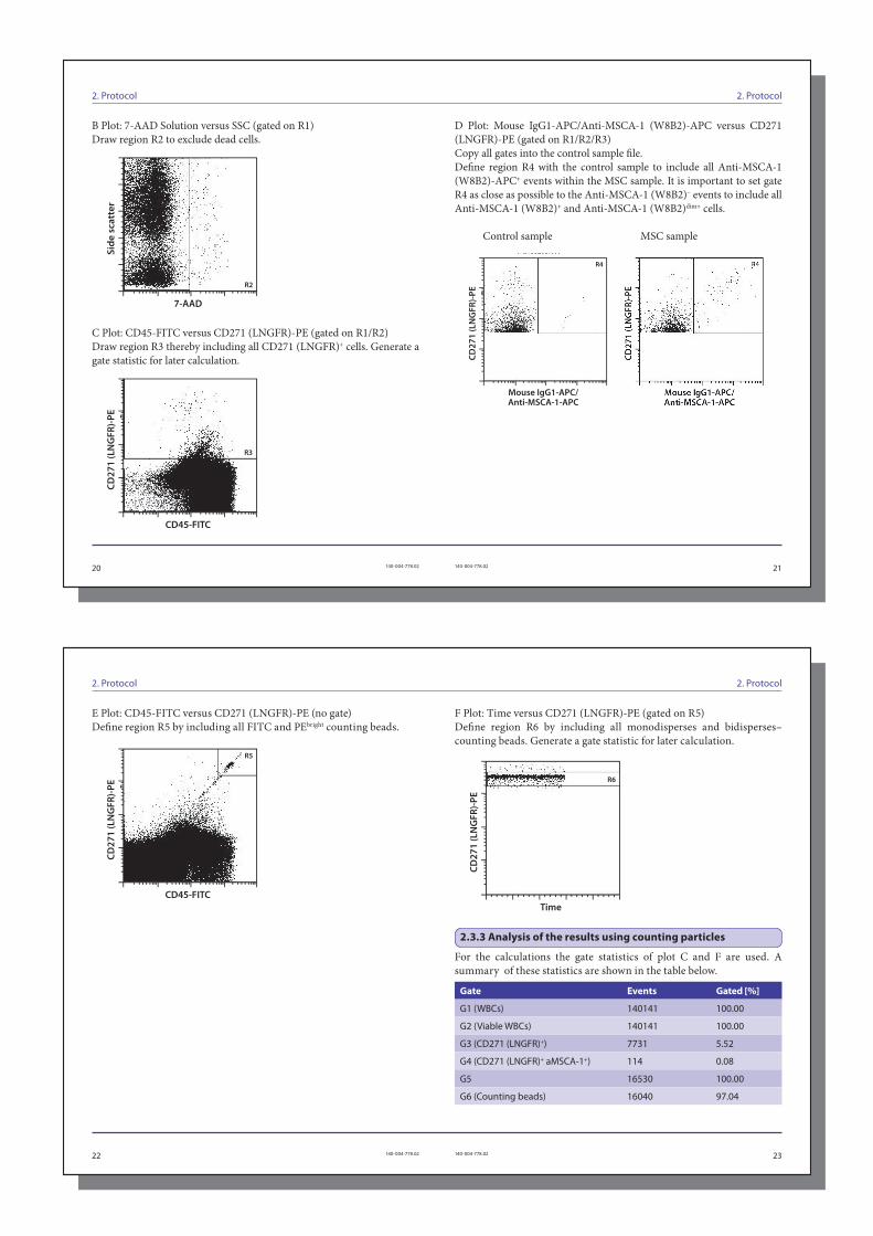
C Plot: CD45-FITC versus CD271 (LNGFR)-PE
D Plot: Mouse IgG1-APC versus CD271 (LNGFR)-PE and Anti-MSCA-1
(W8B2)-APC versus CD271 (LNGFR)-PE
E Plot: CD45-FITC versus CD271 (LNGFR)-PE
F Plot: Time versus CD271 (LNGFR)-PE
Defi nition of logical gates
Defi ne and label the gates described in the following table:
Gate Label Defi nition
G1 WBCs R1
G2 Viable WBCs R1*R2
G3 CD271 (LNGFR)+ R1*R2*R3
G4 CD271 (LNGFR)+ aMSCA-1+ R1*R2*R3*R4
G5 R5
G6 Counting beads R5*R6
▲ Th e general gating strategy below describes the fl ow cytometric
analysis of a bone marrow sample when using another fl ow cytometer
than the MACSQuant® Analyzer 10.
▲ Perform the analysis of the MSC sample stained with the MSC
Staining Cocktail in the same way as the analysis of the control sample
stained with the MSC Control Cocktail.
MSC sample
A Plot: FSC versus SSC (no gate)
Draw region R1 to exclude debris and platelets.
Forward scatter
Sid
e s
catt
er
R1
2.3.2 Flow cytometric analysis
2. Protocol2. Protocol
20 21140-004-778.02 140-004-778.02
B Plot: 7-AAD Solution versus SSC (gated on R1)
Draw region R2 to exclude dead cells.
7-AAD
Sid
e s
catt
er
R2
C Plot: CD45-FITC versus CD271 (LNGFR)-PE (gated on R1/R2)
Draw region R3 thereby including all CD271 (LNGFR)+ cells. Generate a
gate statistic for later calculation.
CD45-FITC
CD
27
1 (
LN
GF
R)-
PE
R3
D Plot: Mouse IgG1-APC/Anti-MSCA-1 (W8B2)-APC versus CD271
(LNGFR)-PE (gated on R1/R2/R3)
Copy all gates into the control sample fi le.
Defi ne region R4 with the control sample to include all Anti-MSCA-1
(W8B2)-APC+ events within the MSC sample. It is important to set gate
R4 as close as possible to the Anti-MSCA-1 (W8B2)– events to include all
Anti-MSCA-1 (W8B2)+ and Anti-MSCA-1 (W8B2)dim+ cells.
Control sample MSC sample
Mouse IgG1-APC/Anti-MSCA-1-APC
CD
27
1 (
LN
GF
R)-
PE
R4
2. Protocol2. Protocol
22 23140-004-778.02 140-004-778.02
E Plot: CD45-FITC versus CD271 (LNGFR)-PE (no gate)
Defi ne region R5 by including all FITC and PEbright counting beads.
R5
CD45-FITC
CD
27
1 (
LN
GF
R)-
PE
F Plot: Time versus CD271 (LNGFR)-PE (gated on R5)
Defi ne region R6 by including all monodisperses and bidisperses–
counting beads. Generate a gate statistic for later calculation.
Time
CD
27
1 (
LN
GF
R)-
PE
R6
For the calculations the gate statistics of plot C and F are used. A
summary of these statistics are shown in the table below.
Gate Events Gated [%]
G1 (WBCs) 140141 100.00
G2 (Viable WBCs) 140141 100.00
G3 (CD271 (LNGFR)+) 7731 5.52
G4 (CD271 (LNGFR)+ aMSCA-1+) 114 0.08
G5 16530 100.00
G6 (Counting beads) 16040 97.04
2.3.3 Analysis of the results using counting particles

3. Appendix2. Protocol
24 25140-004-778.02 140-004-778.02
Calculation of dilution factor
Th e dilution factor is calculated by dividing the staining volume (c,
complete volume aft er labeling) by the sample volume (d, bone marrow
volume used for one staining). Refer to section 2.2.3 for details.
Parameter Calculation Example
Dilution factor c / d 1000 μL / 100 μL = 10
Calculation of MSCs number
Parameter Calculation
Number of MSCs/mL bone marrow
G4 × total counting beads per tube*
G6× dilution factor
Example:
114 × 50950
16040×10 = 3621/mL
* As indicated in the manufacturer`s instruction.
In the given example, the number of MSCs is 3621/mL human bone
marrow aspirate.
3. Appendix
MSCA-1 (W8B2) is identical to the tissue non-specifi c alkaline
phosphatase (TNAP). It was demonstrated that only MSCA-1+ but not
MSCA-1– bone marrow cells express the enzymatically active form of
the TNAP. In addition, it was shown in diff erent bone marrow sources
that only MSCA-1+ cells gave rise to MSCs3 as defi ned by the ISCT
consortium4. Nevertheless there are further MSC specifi c antibodies
which can be used in combination with CD271 (LNGFR) for MSC
detection and enumeration. Th e following table lists some of these
markers with suggested titers that can be used within this assay instead
of Anti-MSCA-1 (W8B2)-APC.
Specifi city Titer Synonym Order number
CD73-APC 1:40 ecto-5'-nucleotidase # 130-095-183
CD90-APC 1:40 Thy-1 # 130-095-402
CD140b-APC 1:10 PDGF-receptor # 130-105-280
4. References
26 27140-004-778.02 140-004-778.02
4. References
1. Bühring, H. J. (2007) Novel markers for the prospective isolation of human MSC.
Ann. N. Y. Acad. Sci. 6: 262–271.
2. Cuthbert, R. (2012) Single-platform quality control assay to quantify multipotential
stromal cells in bone marrow aspirates prior to bulk manufacture or direct
therapeutic use. Cytotherapy 14( 4):431–440.
3. Sobiesiak, M. et al. (2010) Th e mesenchymal stem cell antigen MSCA-1 is identical to
tissue non-specifi c alkaline phosphatase. Stem Cells Dev. 19(5): 669–677.
4. Dominici, M. (2006) Minimal criteria for defi ning multipotent mesenchymal stromal
cells. Th e International Society for Cellular Th erapy position statement. Cytotherapy
8 (4): 315–317.
Refer to www.miltenyibiotec.com for all data sheets and protocols.
Miltenyi Biotec provides technical support worldwide. Visit
www.miltenyibiotec.com for local Miltenyi Biotec Technical Support
contact information.
WarningsReagents contain sodium azide. Under acidic conditions sodium azide yields hydrazoic
acid, which is extremely toxic. Azide compounds should be diluted with running water
before discarding. Th ese precautions are recommended to avoid deposits in plumbing
where explosive conditions may develop.

28 29140-004-778.02 140-004-778.02
LicensesTh is product and/or its use may be covered by one or more pending or issued patents
and/or may have certain limitations. Certain uses may be excluded by separate terms
and conditions. Please contact your local Miltenyi Biotec representative or visit Miltenyi
Biotec’s website at www.miltenyibiotec.com for more information.
Th e purchase of this product conveys to the customer the non-transferable right to use
the purchased amount of the product in research conducted by the customer (whether
the customer is an academic or for-profi t entity). Th is product may not be further sold.
Additional terms and conditions (including the terms of a Limited Use Label License) may
apply.
CUSTOMER’S USE OF THIS PRODUCT MAY REQUIRE ADDITIONAL LICENSES
DEPENDING ON THE SPECIFIC APPLICATION. THE CUSTOMER IS SOLELY
RESPONSIBLE FOR DETERMINING FOR ITSELF WHETHER IT HAS ALL
APPROPRIATE LICENSES IN PLACE. Miltenyi Biotec provides no warranty that
customer’s use of this product does not and will not infringe intellectual property rights
owned by a third party. BY USE OF THIS PRODUCT, THE CUSTOMER AGREES TO BE
BOUND BY THESE TERMS.
TrademarksMACS, MACSmix, MACSQuant, MACSQuantify, the Miltenyi Biotec logo, and VioBlue
are registered trademarks or trademarks of Miltenyi Biotec and/or its affi liates in various
countries worldwide. All other trademarks mentioned in this publication are the property
of their respective owners and are used for identifi cation purposes only.
Copyright © 2021 Miltenyi Biotec and/or its affi liates. All rights reserved.
Legal notices
Limited product warrantyMiltenyi Biotec B.V. & Co. KG and/or its affi liate(s) warrant this product to be free from
material defects in workmanship and materials and to conform substantially with Miltenyi
Biotec’s published specifi cations for the product at the time of order, under normal use
and conditions in accordance with its applicable documentation, for a period beginning
on the date of delivery of the product by Miltenyi Biotec or its authorized distributor and
ending on the expiration date of the product’s applicable shelf life stated on the product
label, packaging or documentation (as applicable) or, in the absence thereof, ONE (1)
YEAR from date of delivery (“Product Warranty”). Miltenyi Biotec’s Product Warranty is
provided subject to the warranty terms as set forth in Miltenyi Biotec’s General Terms and
Conditions for the Sale of Products and Services available on Miltenyi Biotec’s website at
www.miltenyibiotec.com, as in eff ect at the time of order (“Product Warranty”). Additional
terms may apply. BY USE OF THIS PRODUCT, THE CUSTOMER AGREES TO BE
BOUND BY THESE TERMS.
THE CUSTOMER IS SOLELY RESPONSIBLE FOR DETERMINING IF A PRODUCT
IS SUITABLE FOR CUSTOMER’S PARTICULAR PURPOSE AND APPLICATION
METHODS.
Technical informationTh e technical information, data, protocols, and other statements provided by Miltenyi
Biotec in this document are based on information, tests, or experience which Miltenyi
Biotec believes to be reliable, but the accuracy or completeness of such information is not
guaranteed. Such technical information and data are intended for persons with knowledge
and technical skills suffi cient to assess and apply their own informed judgment to the
information. Miltenyi Biotec shall not be liable for any technical or editorial errors or
omissions contained herein.
All information and specifi cations are subject to change without prior notice. Please
contact Miltenyi Biotec Technical Support or visit www.miltenyibiotec.com for the most
up-to-date information on Miltenyi Biotec products.
30 31140-004-778.02 140-004-778.02