mri nephrology 2017
TRANSCRIPT

BYMOHAMED ABOU EL-GHAR
UROLOGY & NEPHROLOGY CENTERMANSOURA UNIVERSITY
Basics of MRI in Nephrology
What is MRI
MRI is a machine creates a magnetic field, sends radio waves through your body, and then measures the response with a computer.
This creates an image or picture of the inside of your body that is much clearer than can be obtained with most other methods.
MRI
The potential advantages of MRI for evaluating urinary tract abnormalities are:
No ionizing radiation. Multiplanar capabilities. Excellent anatomic resolution
and soft tissue contrast.
Before MRI
Cardiac pacemaker or implantable defibrillator.
Catheter that has metal components.
Metallic prosthesis.A ferromagnetic metal
vascular clip.An implanted or external
medication pump.A cochlear implant.A neurostimulation system.
MRI EXAMINATION TECHNIQUES
• high magnetic field (1.5 – 3 Tesla)
• high performance gradients
• phased-array coil → high SNR & small FOV

MRI EXAMINATION TECHNIQUES
T2 WEIGHTED IMAGING
• simple cysts• complicated cysts • angiomyolipoma (AML)• hematoma, aneurysm• infectious mass• renal cell carcinoma (RCC)
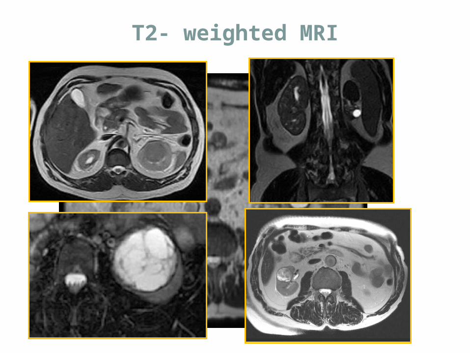
T2- weighted MRI
• SPIN ECHO SEQUENCE• IN PHASE AND OPPOSED PHASE SEQUENCE• 3D GRE SEQUENCE
MRI EXAMINATION TECHNIQUES
T1 WEIGHTED IMAGING
• complicated cysts (hemorrhage, infections, septations)
• AML• melanin-containing lesions• proteinaceous mucin containing lesions• Adrenal mass characterization
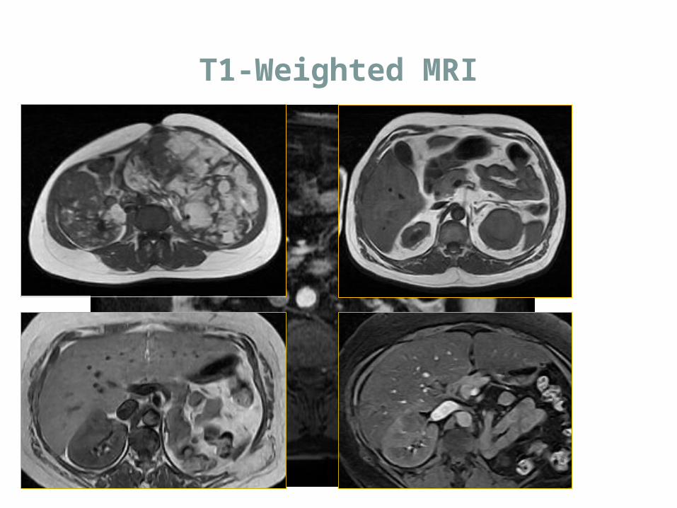
T1-Weighted MRI
3D GRE sequence
HIGH SPATIAL RESOLUTIONHIGH TEMPORAL RESOLUTION
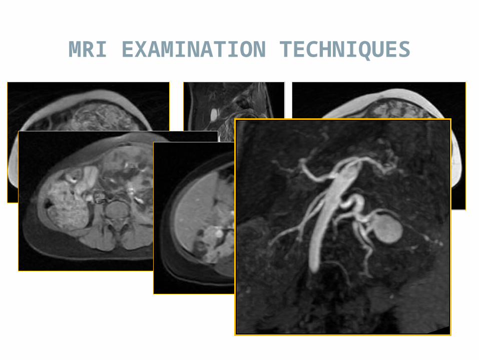
MRI EXAMINATION TECHNIQUES
• Single shot or multishot• Full coverage of the kidney in one 18-23
s breathhold
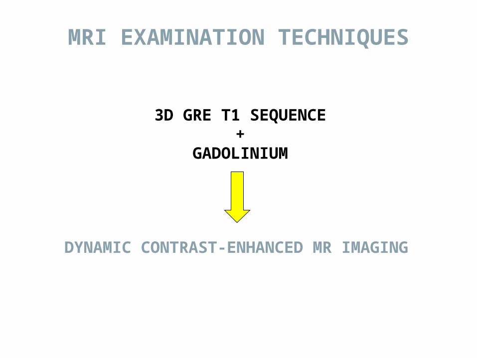
3D GRE T1 SEQUENCE+
GADOLINIUM
DYNAMIC CONTRAST-ENHANCED MR IMAGING
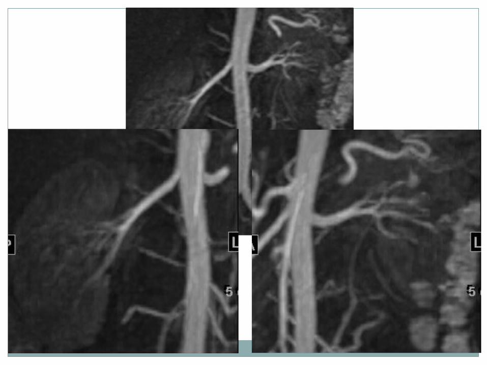
MRI EXAMINATION TECHNIQUES

MRI EXAMINATION TECHNIQUES

MRI EXAMINATION TECHNIQUES

MRI EXAMINATION TECHNIQUES
MRI EXAMINATION TECHNIQUES
MRU
MRU is the most important technique in uroradiology.
It has a good diagnostic value in virtually all kinds of urinary tract disorders.
MRU can reduce the need for radiation exposure and invasive procedure.

MRU Examination Techniques
T2-Weighted ( STATIC - FLUID) MR Urography.
* Multi-slice MRU * Single slice MRU T1-Weighted (EXCRETORY) MR
Urography.

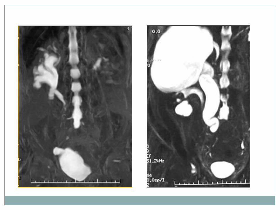
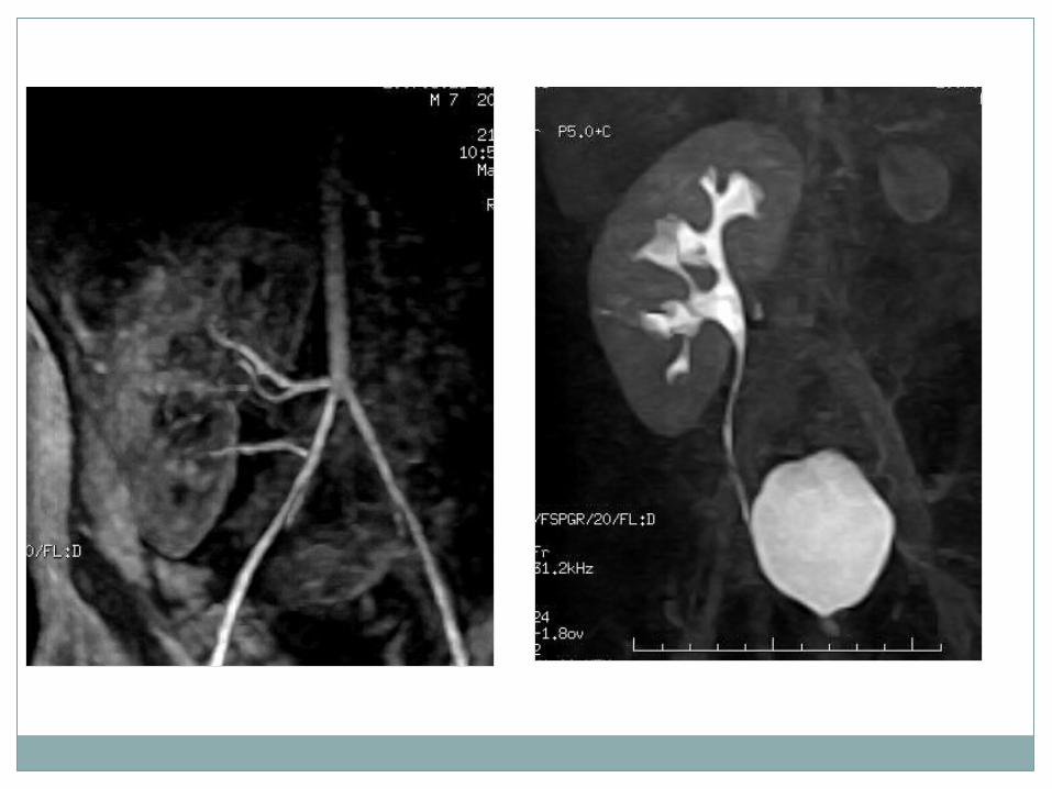
T2-W(static –Fluid) MRU
In static- fluid MRU, Heavily T2w Turbo spin echo (TSE) sequences are used to obtain water images of the urinary tract.
It is used to image fluid filled cavities such as hydronephrosis.
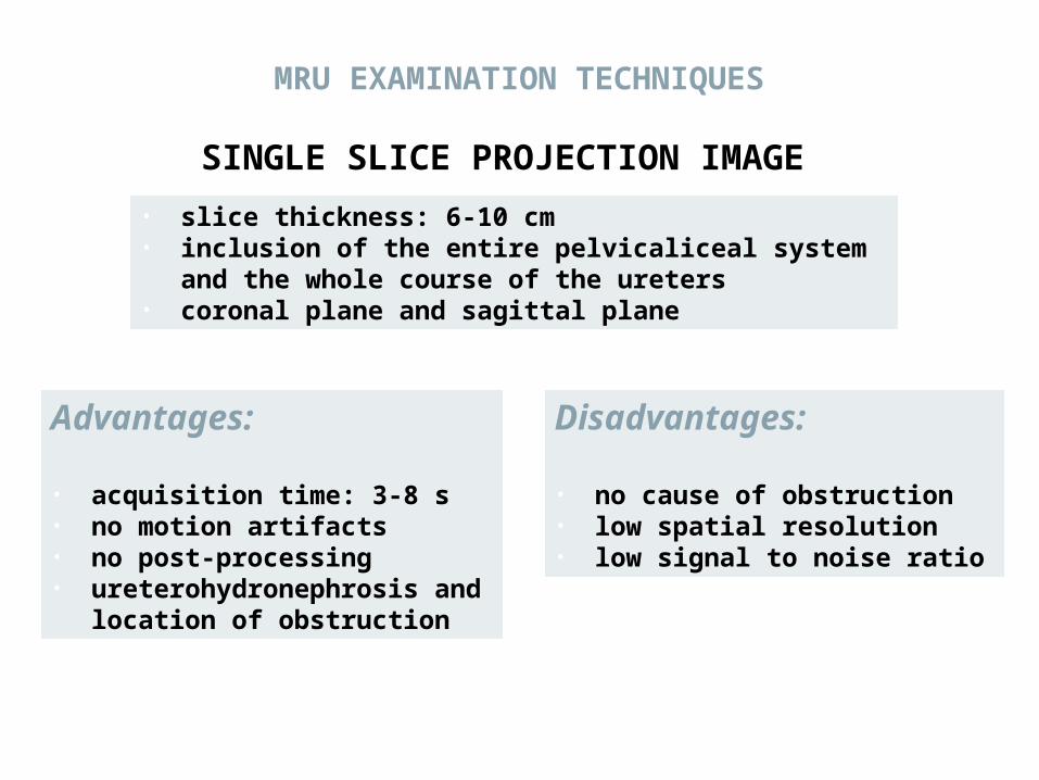
• slice thickness: 6-10 cm• inclusion of the entire pelvicaliceal system and
the whole course of the ureters• coronal plane and sagittal plane
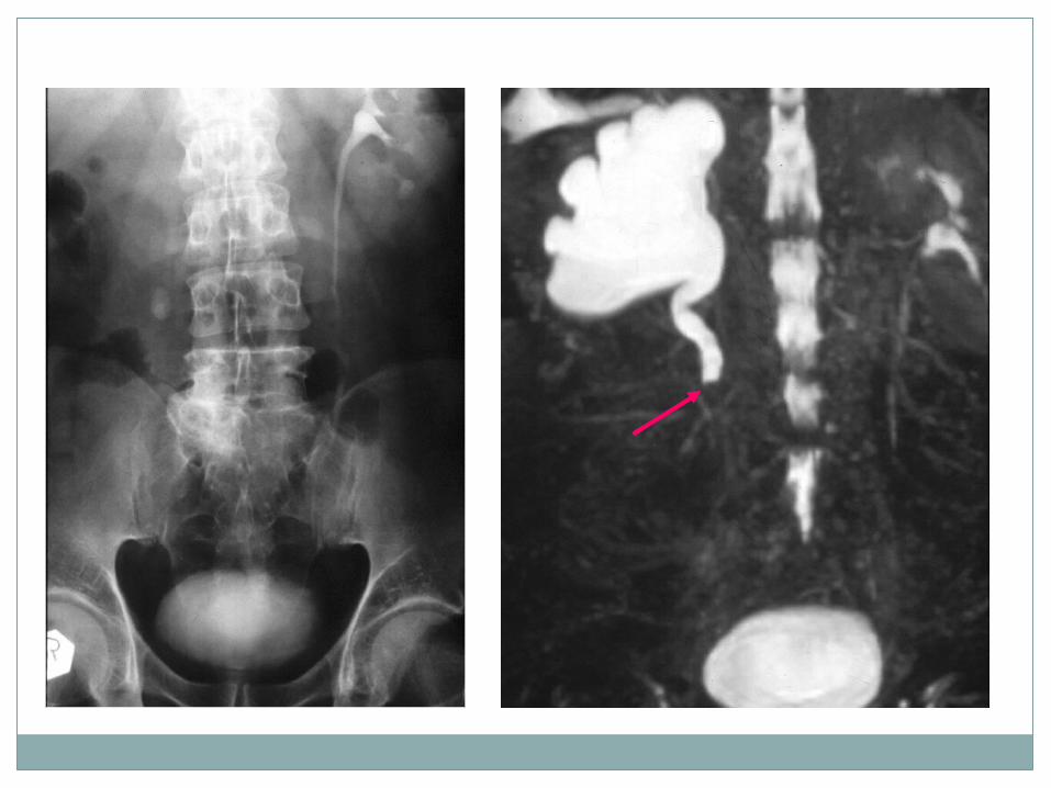
SINGLE SLICE PROJECTION IMAGE
Advantages:
• acquisition time: 3-8 s• no motion artifacts• no post-processing• ureterohydronephrosis and
location of obstruction
Disadvantages:
• no cause of obstruction• low spatial resolution• low signal to noise ratio
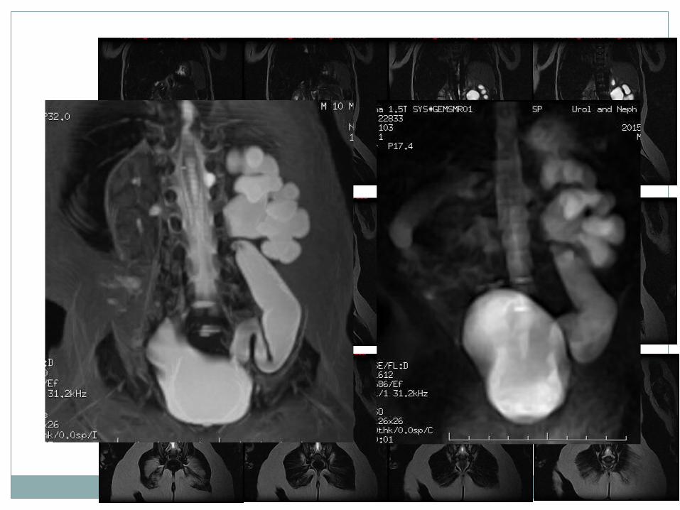
MRU EXAMINATION TECHNIQUES

MULTISLICE TECHNIQUE
• overlapping slices• section thickness: less than 5 mm• post-processing: MIP images
Advantages• reduced partial volume
averaging• small pathological details
Disadvantages• more time consuming• superimposing
extraurinary fluid
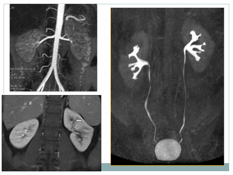
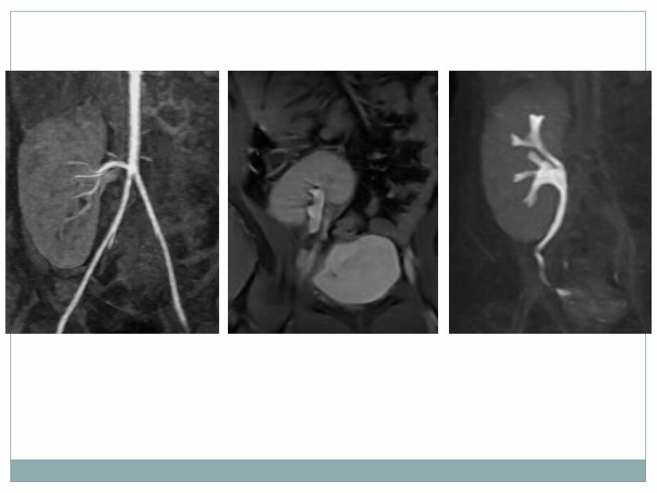
MRU EXAMINATION TECHNIQUES
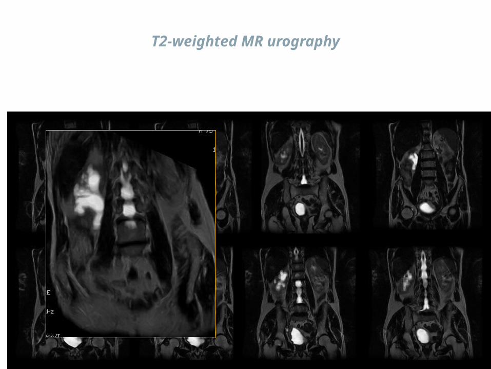
T2-weighted MR urography
T1-w excretory MR Urography
Excretory MRU imitates the conventional IVU.
Gd-Enhanced urine is imaged with use of fast T1-w GRE sequences.
Low –molecular –weight Gd have demonstrated a good safety profile at standard clinical dose.


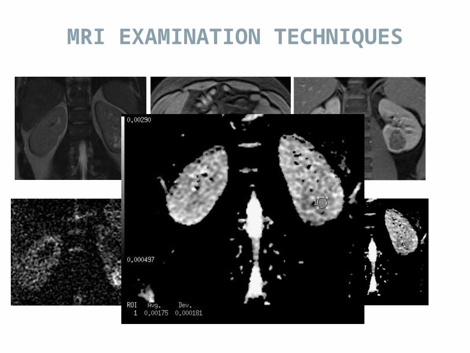
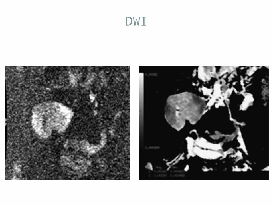
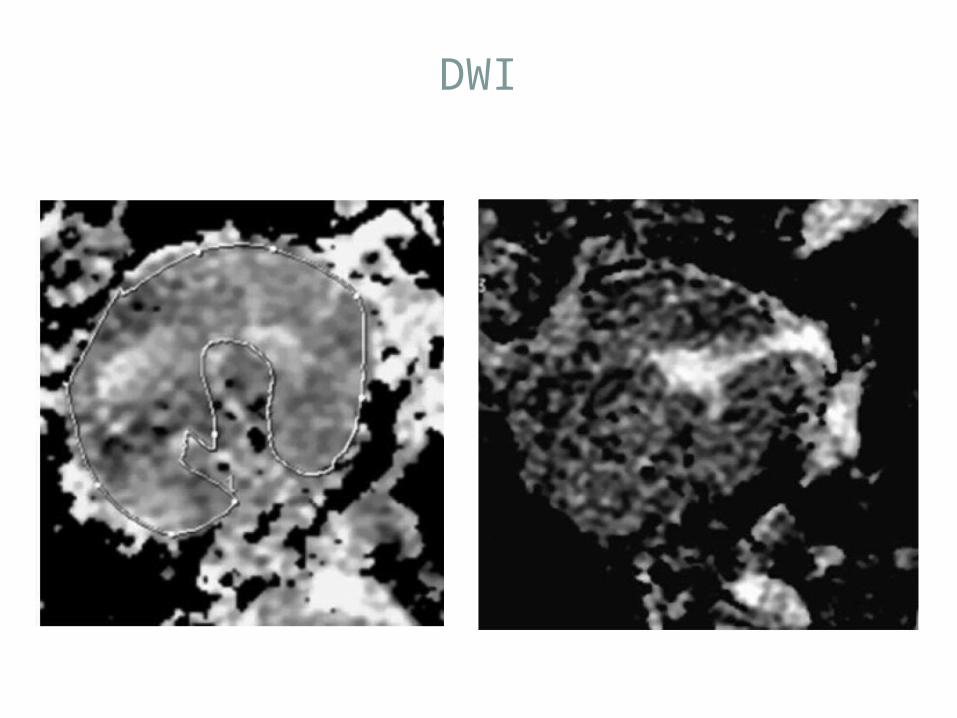
Diffusion-weighted imaging (DWI)• MR diffusion-weighted imaging (DWI) provides information on
the velocity and direction of movement of the water molecules in tissue under influence of a diffusion gradient
• The velocity and direction of the diffusion movement of the water molecules can be quantified by means of the apparent diffusion coefficient (ADC)
Koh DM et al, AJR (2007)
MRI EXAMINATION TECHNIQUES

Diffusion-weighted imaging (DWI)Restricted diffusion: *Malignancy (increased number of cells ) *Ischemia (cytotoxic edema) *Abscess (increased viscosity)
MRI EXAMINATION TECHNIQUES
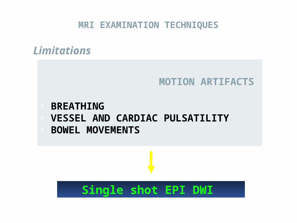
Limitations
MOTION ARTIFACTS
• BREATHING• VESSEL AND CARDIAC PULSATILITY• BOWEL MOVEMENTS
Single shot EPI DWI
MRI EXAMINATION TECHNIQUES
MRI EXAMINATION TECHNIQUES
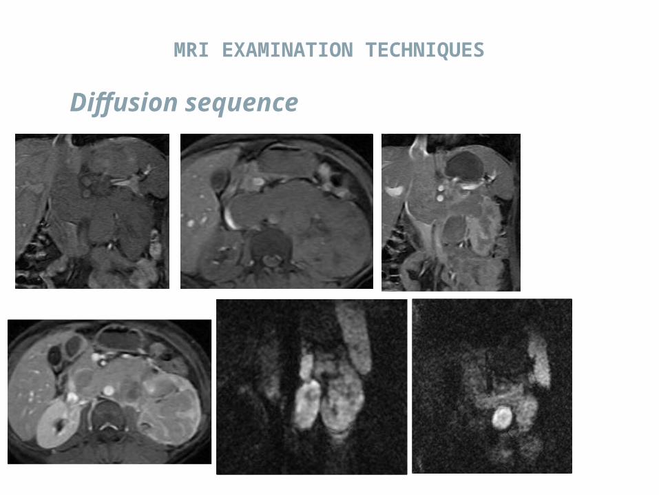
Diffusion sequence
MRI EXAMINATION TECHNIQUES

MRI EXAMINATION TECHNIQUES
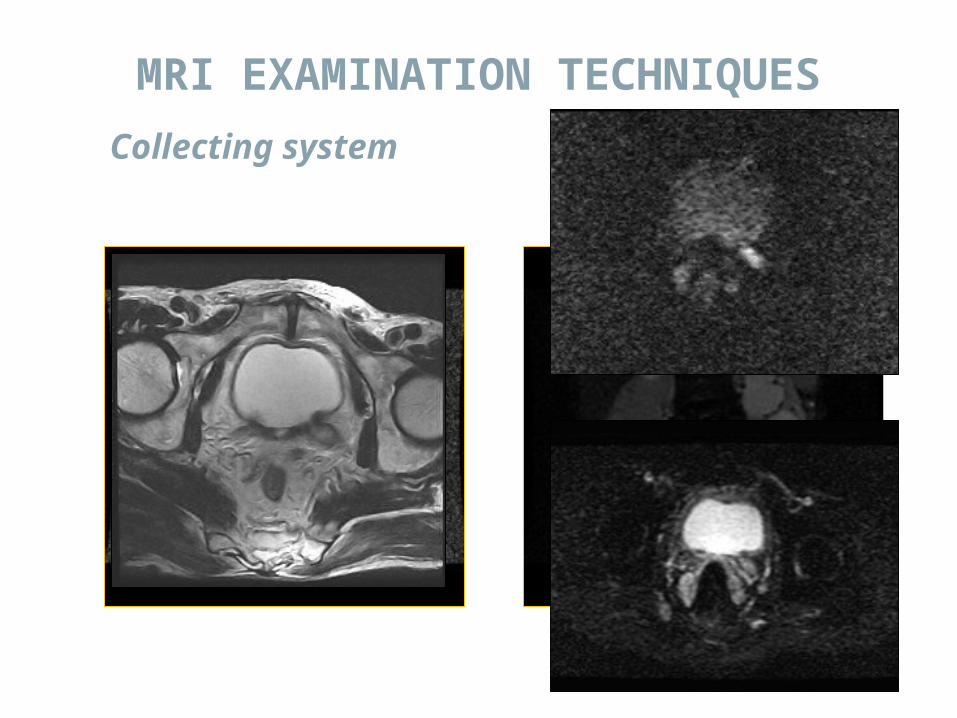
Collecting system
MRI EXAMINATION TECHNIQUES
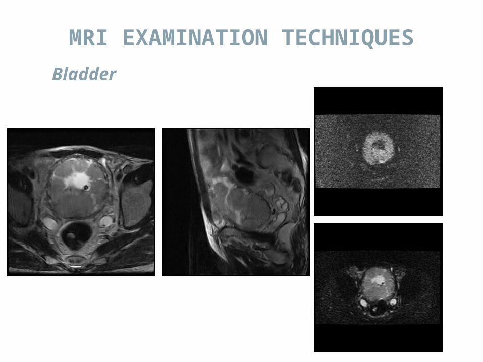
Bladder
MRI EXAMINATION TECHNIQUES

All In Approach
Preoperative assessment of potential live kidney donor.
Basal study of transplanted kidney.Pelviureteral junction obstruction.Nephron sparing surgery.

MR Urography
Obstructed or non-obstructed?Urothelial lesions.Cause of obstruction.Congenital Anomalies.

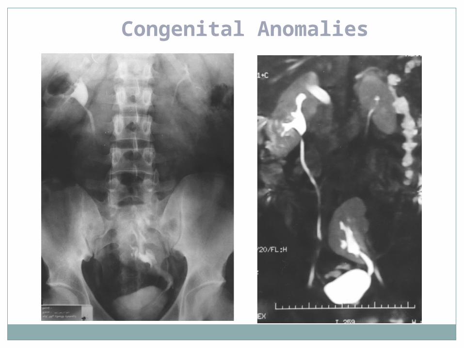
Congenital Anomalies
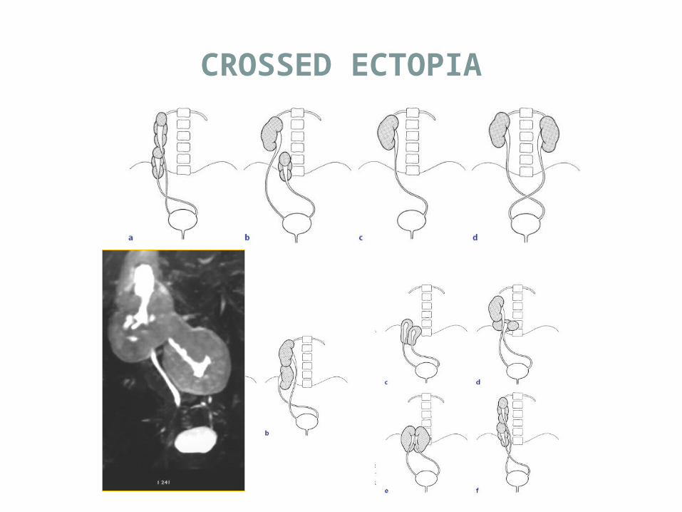
CROSSED ECTOPIA

Ectopic kidney
-Simple renal ectopy refers to a kidney that remains in the ipsilateral retroperitoneal space.
-The most common position is in the pelvis or sacral region below the aortic bifurcation.
-Crossed renal ectopia with fusion occurs in 85%, without fusion in less than 10%.
ANOMALIES OF POSITION

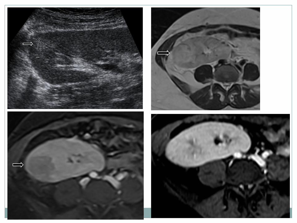
RENAL DYSPLASIA
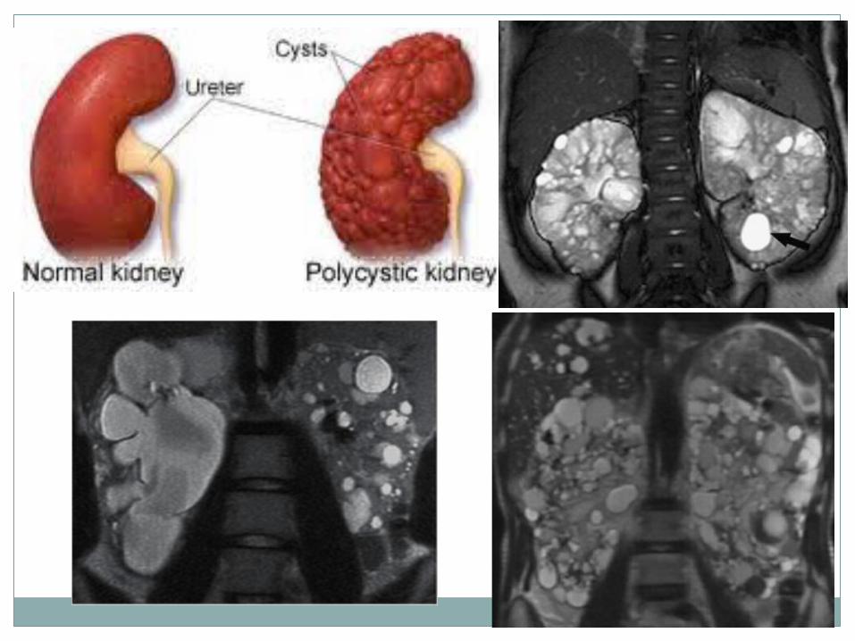
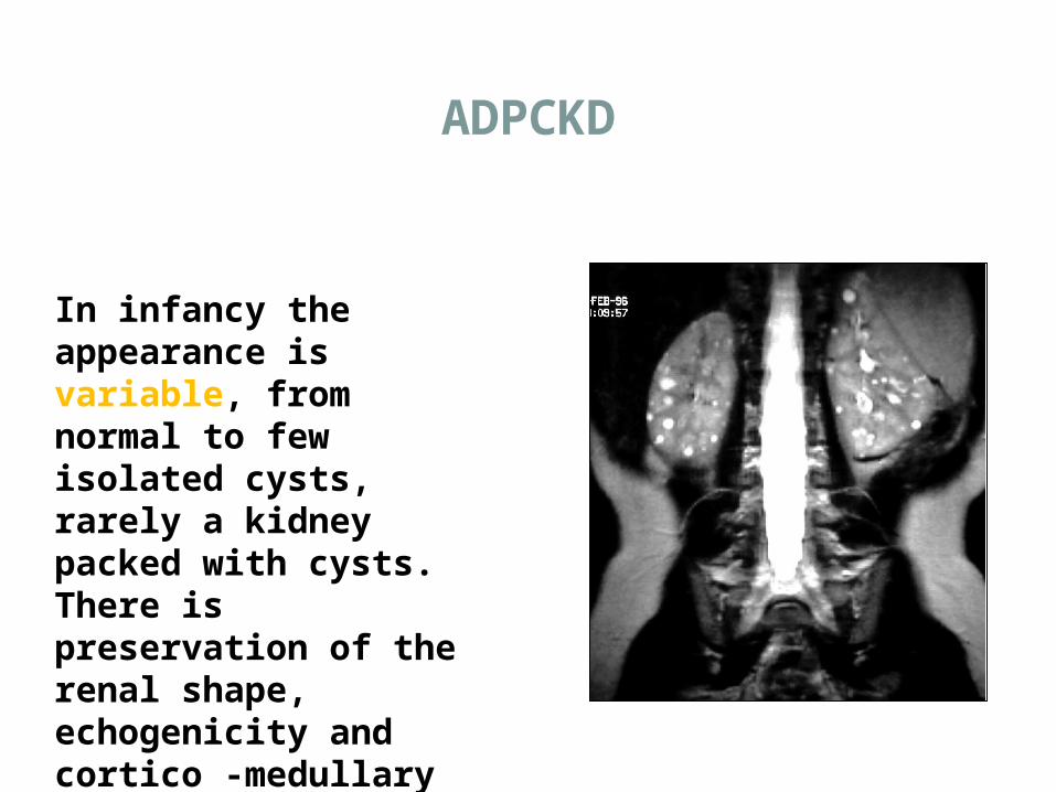
In infancy the appearance is variable, from normal to few isolated cysts, rarely a kidney packed with cysts. There is preservation of the renal shape, echogenicity and cortico -medullary differentiation.
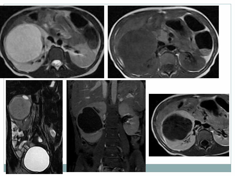
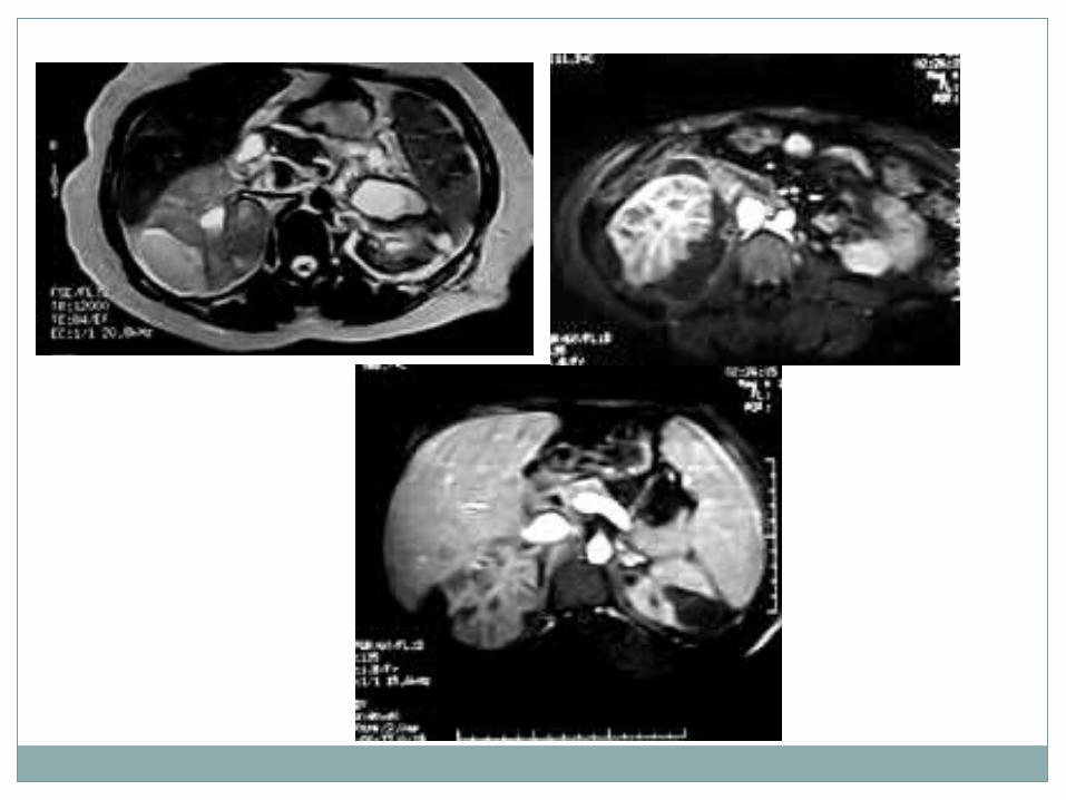
ADPCKD

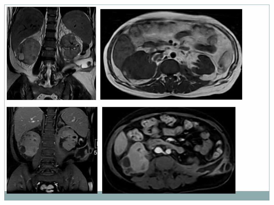
ARPCKD
Kidneys: the dilated tubules are responsible for the appearance.- Kidneys enlargment.-Diffuse increase
echogenicity and hyperechoic foci on US.
-Low attenuation with striate pattern on CT.
-Diffuse increase signal intensity in T2 w Images on MRI.
-Macro cyst in varying patterns can be present.
Liver: dilatation of the bile ducts.
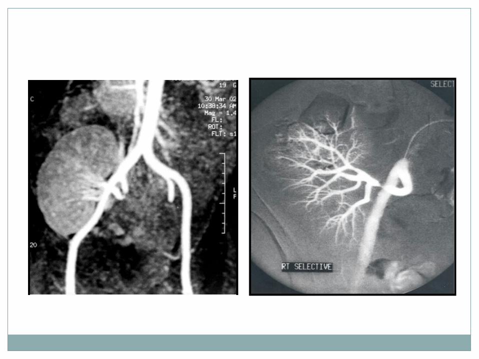
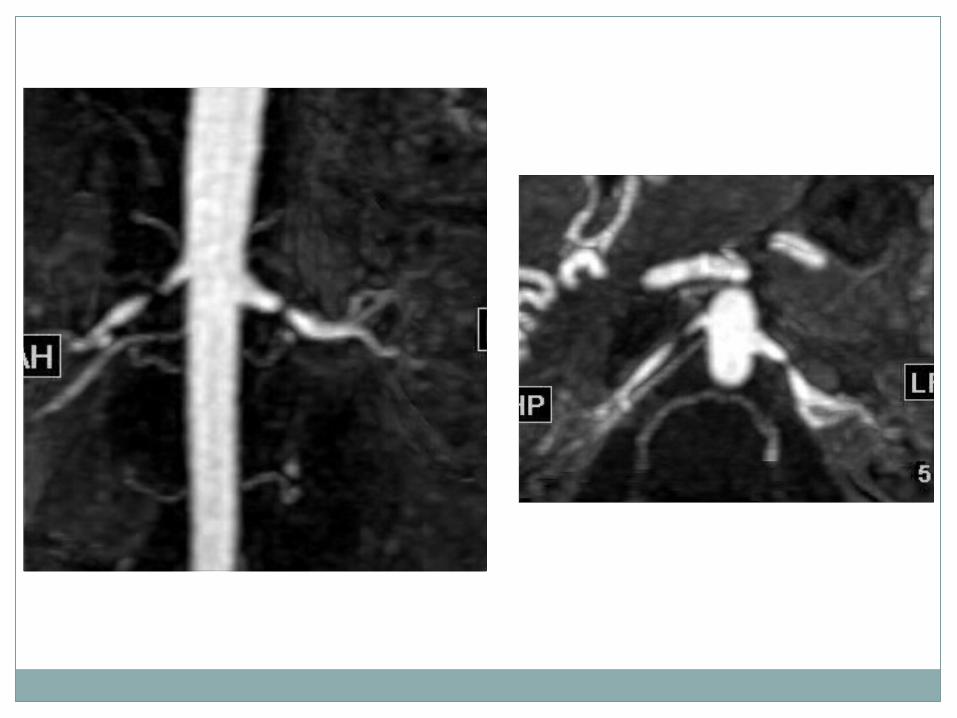
Vascular Lesions
Renal artery stenosis.Renal aneurysm.Renal AV malformation…..etc.

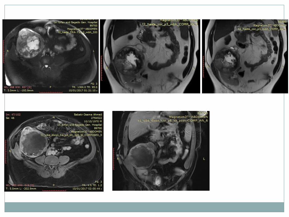
Inflammatory Lesions
Diagnosis of different inflammatory lesions.
Extension and other organ involvement.Follow up after management.

Kidney Transplant Complications
Urinary leakage.Urinary obstruction.Renal parenchyma.


DWI
DWI

NSF
Nephrogenic systemic fibrosis (NSF) is a relatively uncommon condition in which fibrous plaques develop in the dermis and, often, in deeper connective tissues.
Reported cases have occurred almost exclusively in patients with severe renal disease, and almost all have been associated with prior use of gadolinium-containing MRI contrast agents.
The disease is often disabling, no proven treatments exist.

Clinical features of NSF
Onset: From the day of exposure for up to 2–3 months Initially– Pain– Pruritus– Swelling– Erythema– Usually starts in the legs Later– Thickened skin and subcutaneous tissues — ‘woody’ texture and brawny
plaques– Fibrosis of internal organs, e.g. muscle, diaphragm, heart, liver, lungs Result– Contractures– Cachexia– Death, in a proportion of patients
Who is at RISK
Whilst cases have occurred in patients with either acute or chronic renal failure.
Most have been in patients with chronic and severe kidney disease (CKD Stage 4 & 5, glomerular filtration rate (GFR) < 30 ml/ min/1.73 m2); most have been on dialysis.
At lower risk Patients with CKD 3 (GFR 30-59ml/min)
Not at risk of NSF Patients with stable GFR > 60 ml/min
Take care
Children under one year of age, have a physiologically low GFR yet no case of NSF has been reported in a patient under the age of 6 years.
In lactating patients, the proportion entering the breast milk is very small (1% of the injected dose), and very little of this is actually absorbed. Hence the risk to the child would appear negligible.
Lactating women: Stop breastfeeding for 24 hours and discard the milk.
Pregnant women: Can be used to give essential diagnostic information.
High Risk Patients
The minimum adequate dose of gadolinium is used. Restrict dose to 0.1 mmol/kg and avoid repeat scans.
Consider immediate post-scan hemodialysis. A single conventional hemodialysis session will
remove 75% of the free Gadolinium – a 2nd treatment 93% and a 3rd treatment 98% of a dose.
If the patient has severe renal failure, but is not receiving hemodialysis, the possibility of commencing hemodialysis will need individual consideration.
ESUR Guidelines, 8.1 Contrast Media Guidelines
Never deny a patient a clinically well- indicated enhanced MRI examination.
In all patients use the smallest amount of contrast medium necessary for a diagnostic result.
Future prospective MRI Techniques
BOLD.ASLDTIDynamic MRI.
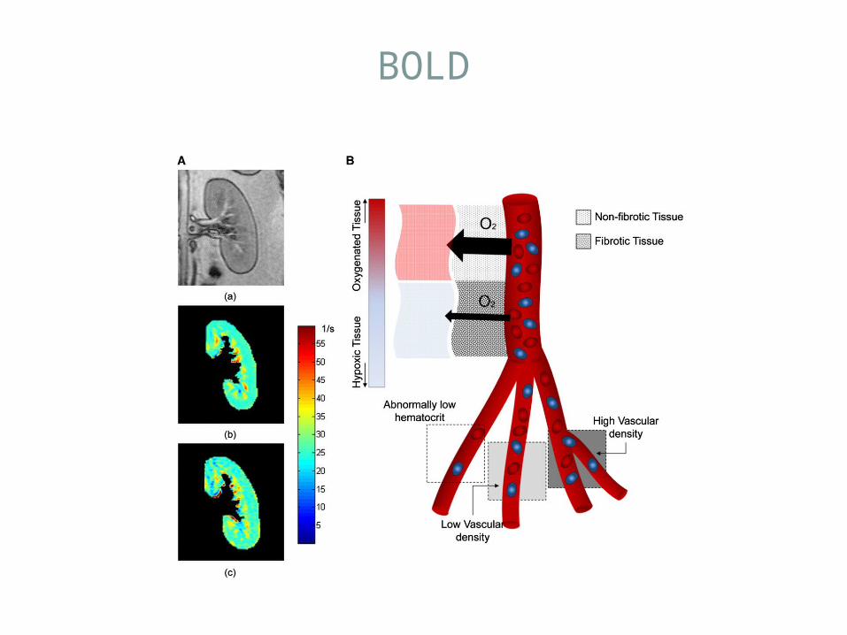
BOLD
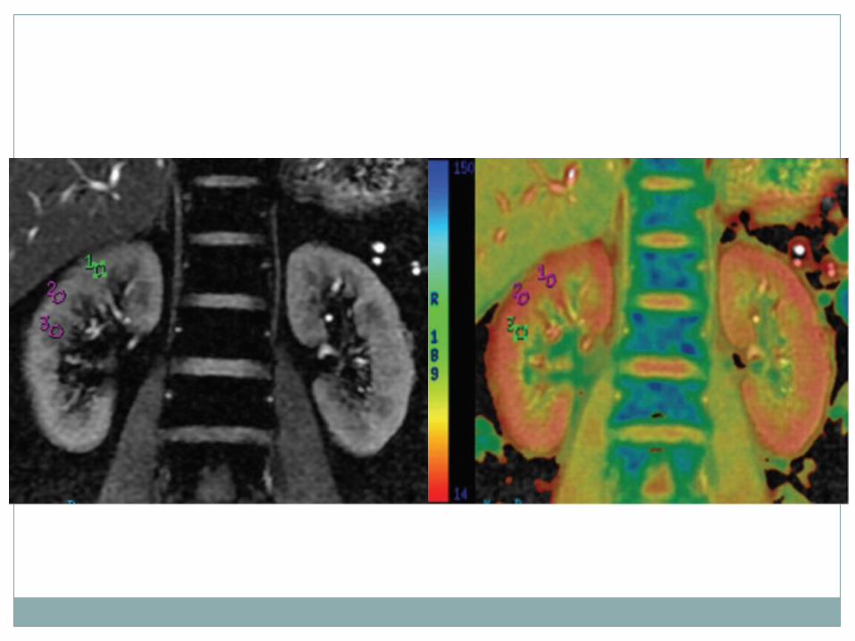

ASL
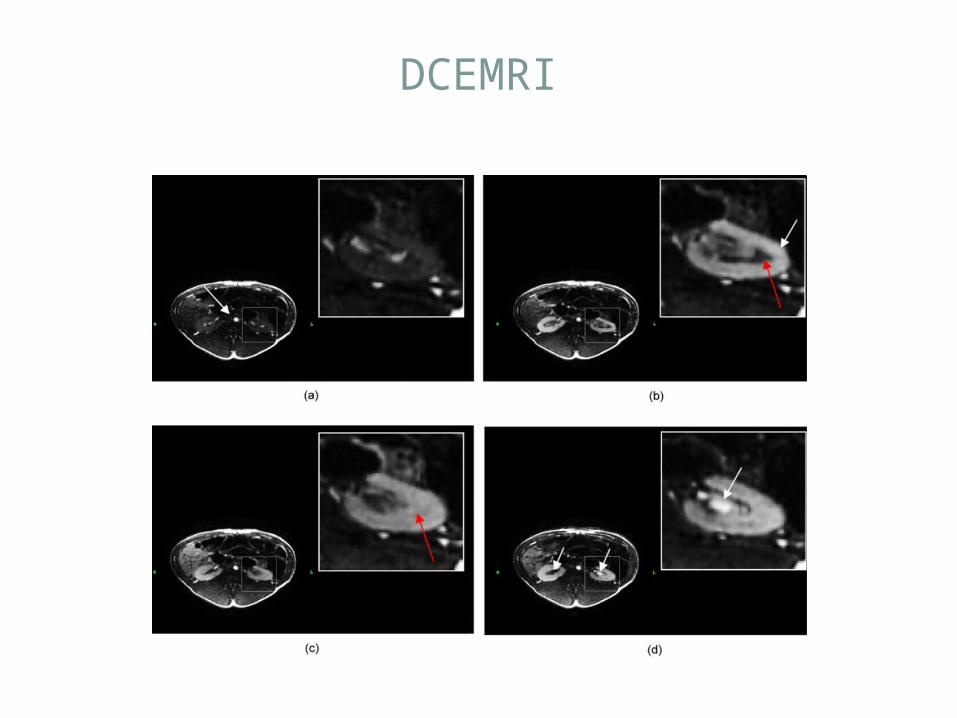
DCEMRI

DCEMRI
THANK YOU