mri knee trauma
TRANSCRIPT
MRI KNEE TRAUMA
DR MOHIT GOEL21/05/2013
There are three general mechanisms of ACL failure:
•External rotation and abduction with hyperextension •Direct forward displacement of the tibia •Internal rotation with the knee in full extension.
With varus or valgus stress, the ACL is injured after collateral ligament failure.
Forced valgus in external rotation is the most common mechanism of injury and causes disruption of the MCL and medial supporting structures.
ACL Tears

Primary Signs
•Abnormal ligament course (abnormal Blumensaat angle)
•Abnormal ligament signal intensity (coronal images should be used in conjunction with sagittal images to compensate for segmental visualization in the sagittal plane)
•Ligament discontinuity
(MRI findings)

Secondary Signs
•Lateral compartment ossesous contusions (posterolateral tibial plateau is most specific)
•Posteromedial tibial plateau contusion or fracture
•Anterior tibial displacement (assessed in the lateral aspect of the lateral compartment)
•Uncovered posterior horn lateral meniscus
•Posterior cruciate line and angle.
•Chronic ACL tears demonstrate resolution of the ossesous contusions, effusions, synovitis, and ligamentous hyperintensity that are characteristic of acute injuries unless seen in the setting of an acute injury.

The Blumensaat line courses parallel to the roof of the intercondylar notch (the posterior surface of the femur). The Blumensaat angle is formed by the Blumensaat line and a line along the margin (including the distal portion) of the ACL. A negative (normal) Blumensaat angle occurs when the apex of the angle is directed superiorly, and a positive (abnormal) Blumensaat angle occurs when the apex of the angle is directly inferior.
Blumensaat angle

The posterior cruciate line
A line was drawn tangent to the posterior margin of the distal posterior cruciate ligament and extended proximally. The posterior cruciate line was considered to be positive for ACL tear if the proximal extension of this line did not intersect the medullary cavity of the femur within 5 cm of its distal aspect.
This sign was considered to be negative if the proximal extension of the posterior cruciate ligament line intersected the medullary cavity within 5 cm of its distal aspect.

The posterior cruciate angle was defined as the point of intersection between lines drawn through the proximal and distal portions of the posterior cruciate ligament. A posterior cruciate angle measurement less than the mean value (114.8°) for all cases was used as the threshold for the diagnosis of ACL tear.

(A)Acute ACL rupture of proximal fibers. The slope of the ACL is decreased relative to the intercondylar roof (Blumensaat's line). Characteristic posterolateral tibial plateau contusion is demonstrated.
(B) Sagittal and(C) axial FS PD FSE images
of a grade 3 ACL tear. Complete loss of proximal ligament continuity with the lateral femoral condyle side wall is shown in both sagittal and axial planes. Acute findings of a joint effusion and posterior tibial plateau contusion are present.

ACL tears are classified into three grades:
Grade I ACL tears represent intraligamentous injury without a change in ligament length.
Grade II ACL tears represent intraligamentous injury and an increase in ligament length.
Grade III ACL tears represent complete ligamentous disruption.

Grade 1 to 2 ACL sprain.
(A)On PD FSE images the ligament demonstrates intermediate signal intensity.
(B) On corresponding FS PD FSE coronal images the ligament demonstrates continuity.
Loss of ligament hypointensity on a T1- or PD-weighted sequence is a sensitive sign of ligamentous strain or scarring. The FS PD sequence is specific for ligament continuity and can be used to differentiate a grade 2 from a grade 3 ACL sprain.

Grade 3 ACL tear- Disruption of the middle third of the ACL (FS PD FSE sagittal image)
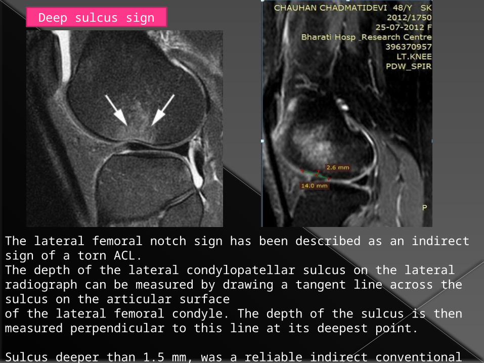
The lateral femoral notch sign has been described as an indirect sign of a torn ACL.The depth of the lateral condylopatellar sulcus on the lateral radiograph can be measured by drawing a tangent line across the sulcus on the articular surfaceof the lateral femoral condyle. The depth of the sulcus is then measured perpendicular to this line at its deepest point.
Sulcus deeper than 1.5 mm, was a reliable indirect conventional radiographic sign of a torn ACL.
Deep sulcus sign

Posterior Cruciate Ligament (PCL) tear
The PCL is twice as strong as the ACL, with a larger cross-sectional area and higher tensile strength. These features account for a lower incidence of rupture of the PCL.
Tears of the PCL are most common in the midportion (76%), followed by avulsions from the femur (36% to 55%) and the tibia (22% to 42%).
Rupture may be caused by excessive rotation, hyperextension, dislocation, or direct trauma while the knee is flexed.
Motor vehicle accidents (dashboard injuries) and injuries sustained in contact sports such as football are the most common causes of damage to the PCL .
Injuries to the PCL are usually associated with tears of the ACL, the meniscus, collateral ligaments, or postero-lateral structures .

(A)Dashboard injury caused by a posteriorly directed force applied to the proximal tibia with the knee in 90° of flexion.
This sagittal FS PD FSE image shows complete loss of PCL continuity secondary to a complex interstitial PCL tear.
(B) Axial FS PD FSE image showing an anterolateral fracture resulting from direct trauma by the dashboard during impact.


Normal Meniscal Anatomy
Medial meniscus
Both horns are triangular in shape and have very sharp points. The posterior horn is always larger than the anterior horn (figure).If this is not the case than the shape is abnormal, which can be a sign of a meniscal tear or a partial meniscectomy.

The posterior root is immediately anterior to the posterior cruciate ligament. If it is missing on the sagittal images, then there is a meniscal root tear (figure).

Lateral meniscus
On sagittal images the posterior horn is higher in position than the anterior horn.
Both horns are about the same size.

Meniscal tears
Criteria for tears
The two most important criteria for meniscal tears are an abnormal shape of the meniscus and high signal intensityon PD-images unequivocally contacting the surface .
Nomenclature of Meniscal Tears
Shapes. There are 3 basic shapes of meniscal tears: longitudinal, horizontal and radial .
Complex tears are a combination of these basic shapes.

Displaced TearsBucket-handle tear = displaced longitudinal tear.Flap tear = displaced horizontal tear.Parrot beak = displaced radial tear.

Longitudinal tears
Longitudinal tears parallel the long axis of the meniscus dividing the meniscus in an inner and outer part.
So the distance between the tear and the outer margin of the meniscus is always the same (figure).
The tear never touches the inner margin.

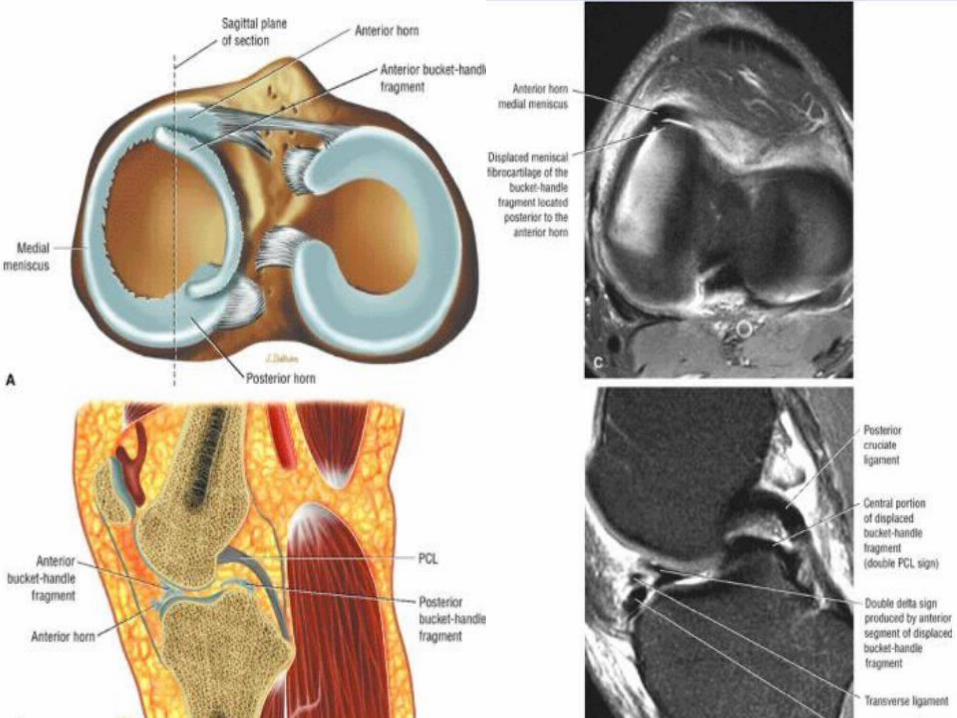
Bucket-Handle Tears
A displaced longitudinal tear of the meniscus, usually the medial meniscus, is called a bucket-handle tear because the separated central fragment resembles the handle of a bucket. The remaining larger peripheral section of the meniscus is the bucket.

Bucket-Handle Tears
• An unstable meniscal fragment locks into the intercondylar notch and involves at least two thirds of the meniscal circumference.
• Diagnosis of a bucket-handle tear requires identification of displaced meniscal tissue from posterior to a relative anterior coronal location.
• A double delta sign and/or a double PCL sign are sagittal MR findings of a displaced bucket-handle tear.
• Medial meniscus bucket-handle tears are three times more frequent than bucket-handle tears involving the lateral meniscus.
• A bucket-handle tear effectively reduces the width of the meniscus, and peripheral sagittal images fail to demonstrate the normal bowtie configuration of the body of the meniscus.

Double PCL sign
• The double PCL sign refers to visualization of the displaced meniscal fragment anterior to the PCL in the intercondylar notch.
Double delta sign
• The double delta sign refers to visualization of flipped inner meniscal fragments adjacent (posterior) to the anterior horn of the donor site.
• The double delta sign is produced by two triangular structures adjacent to each other anteriorly.
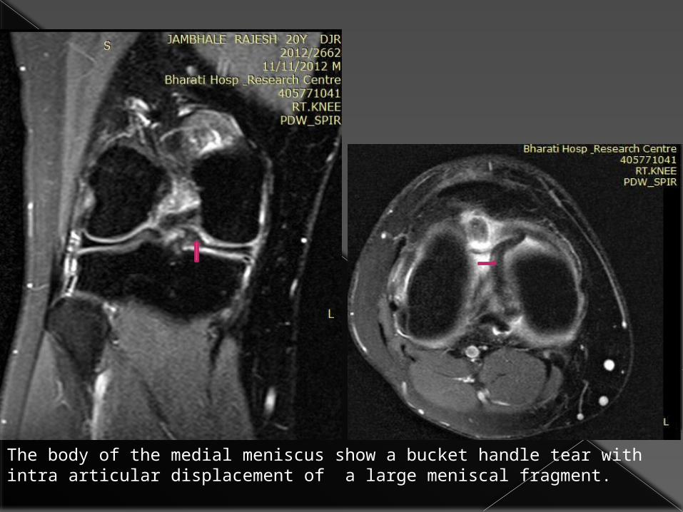
The body of the medial meniscus show a bucket handle tear with intra articular displacement of a large meniscal fragment.

Flipped meniscus
An enlarged bulky anterior horn is seen.
Posteriorly a very small posterior horn will be seen.

Horizontal tears
Horizontal tears divide the meniscus in a top and bottom part
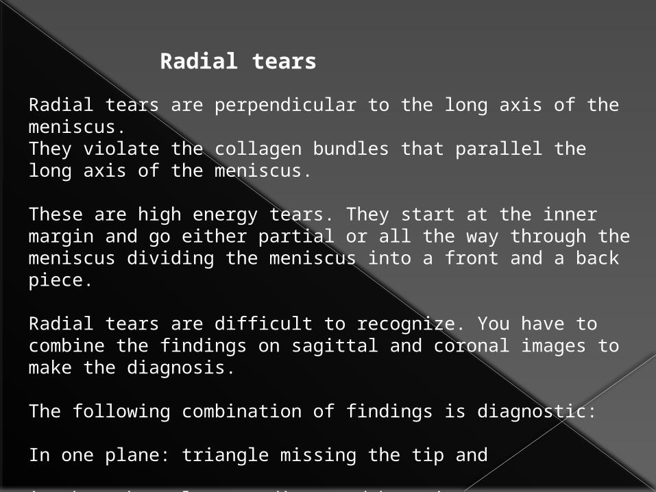
Radial tears
Radial tears are perpendicular to the long axis of the meniscus. They violate the collagen bundles that parallel the long axis of the meniscus.
These are high energy tears. They start at the inner margin and go either partial or all the way through the meniscus dividing the meniscus into a front and a back piece.
Radial tears are difficult to recognize. You have to combine the findings on sagittal and coronal images to make the diagnosis.
The following combination of findings is diagnostic:
In one plane: triangle missing the tip and
in the other plane: a disrupted bow tie.
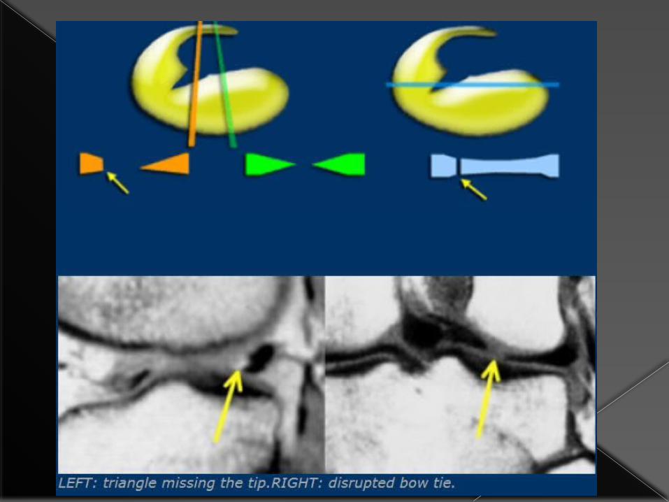

Small radial tears are difficult to diagnose. Sometimes the only sign is a disrupted bow tie.

Meniscal root tear
A meniscal root tear is a radial tear located at the meniscal root.
Normally when you image the posterior cruciate ligament on sagittal images you should see a considerable posterior horn of the meniscus on that image or the image adjacent to it.
If this is not the case it is an absent or empty meniscus-sign indicating a root tear.

Medial collateral ligament
The superficial medial collateral ligament (MCL) extends from the medial epicondyle to insert not just near the joint but 7 cm below the joint space.
At that point there are three landmarks: the inferomedial geniculate artery and paired veins (figure). The deep part of the MCL, even when it is normal, you may not be able to see.

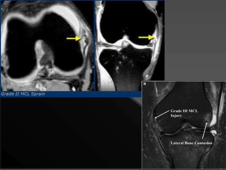
Injuries of the of the medial collateral ligament are graded into three groups on MRI, much in the same way as many other ligaments.
grade 1: (minor sprain) high signal is seen medial (superficial) to the ligament, which looks normal
grade 2 : (severe sprain or partial tear) high signal is seen medial to the ligament, with high signal or partial disruption of the ligament
grade 3 : complete disruption of the ligament

Chondromalacia patellae
Chondromalacia patellae refers to softening and degeneration of the articular hyaline cartilage of the patella, and is a frequent cause of anterior knee pain.
MRIT1
• poor sequence for cartilage and surface irregularity and subtly signal change may be inapparent
• areas of hypointensity may be seen in cartilage• sub-chondral reactive bone marrow oedema pattern (low signal)• secondary changes of osteoarthritis may be seen
T2 / PD
• best sequences for assessing cartilage• abnormal cartilage is usually of high signal compared to normal
cartilage• findings range from subtle increase in signal to complete loss of
cartilage
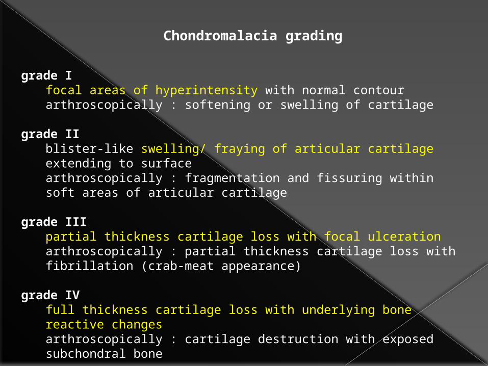
Chondromalacia grading
grade Ifocal areas of hyperintensity with normal contour arthroscopically : softening or swelling of cartilage
grade IIblister-like swelling/ fraying of articular cartilage extending to surfacearthroscopically : fragmentation and fissuring within soft areas of articular cartilage
grade IIIpartial thickness cartilage loss with focal ulcerationarthroscopically : partial thickness cartilage loss with fibrillation (crab-meat appearance)
grade IVfull thickness cartilage loss with underlying bone reactive changesarthroscopically : cartilage destruction with exposed subchondral bone

Grade 4 chondromalacia- full thickness cartilage loss with underlying bone reactive changes

Bone Contusion Patterns
The distribution of bone marrow edema is like a footprint left behind at injury, providing valuable clues to the associated soft-tissue injuries.
Five contusion patterns with associated soft-tissue injuries occur in the knee:
• pivot shift injury,
• dashboard injury,
• hyperextension injury,
• clip injury, and
• lateral patellar dislocation.

Pivot shift injury.
Drawing shows a skier with a right knee pivot shift injury (knee valgus, femur internally rotated).
Drawing shows that, with the foot planted, the combination of valgus stress on the knee and internal rotation of the femur results in disruption of the ACL.
After disruption of the ACL, the tibia is free to sublux anteriorly relative to the femur. This movement results in the impaction of the lateral femoral condyle against the posterolateral tibial plateau.

classic bone marrow edema pattern resulting from pivot shift injury of the knee. Extensive contusion is present in the posterolateral tibial plateau (straight arrow) and, to a lesser degree, the lateral femoral condyle (curved arrow)

Dashboard injury.
Drawing shows a woman striking her knee against the dashboard during an automobile accident. This is the most common mechanism of injury resulting in disruption of the PCL.
The tibia is forced posteriorly (open arrow) relative to the femur. The crosshatched region indicates the area of bone contusion on the anterior tibia caused by direct trauma.
The PCL is usually tight when the knee is in 90° of flexion and is, therefore, at risk for disruption (solid arrow).
The ACL, on the other hand, is normally lax while the knee is flexed and usually remains intact.

Sagittal T2-weighted fast SE image demonstrates edema in the anterior proximal tibia (white arrow) and complete disruption of the PCL (black arrow)

Hyperextension injury.
Drawing depicts a forceful kicking motion resulting in a hyperextension injury of the right knee.
Drawing shows how very severe hyperextension of the knee (arrow) can result in the impaction of the anterior aspect of the femoral condyle against the anterior aspect of the tibial plateau.
The crosshatched regions indicate the areas of bone contusion.
Depending on the amount of force applied during hyperextension, tears of the ACL, PCL, or both may occur.

Coronal T2-weighted fast SE MR image obtained with fat saturation reveals kissing bone marrow contusions (arrows) of the medial aspect of the anterior tibia and femur secondary to the hyperextension injury.
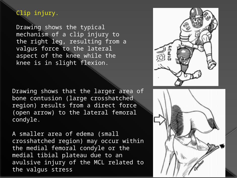
Clip injury.
Drawing shows the typical mechanism of a clip injury to the right leg, resulting from a valgus force to the lateral aspect of the knee while the knee is in slight flexion.
Drawing shows that the larger area of bone contusion (large crosshatched region) results from a direct force (open arrow) to the lateral femoral condyle.
A smaller area of edema (small crosshatched region) may occur within the medial femoral condyle or the medial tibial plateau due to an avulsive injury of the MCL related to the valgus stress
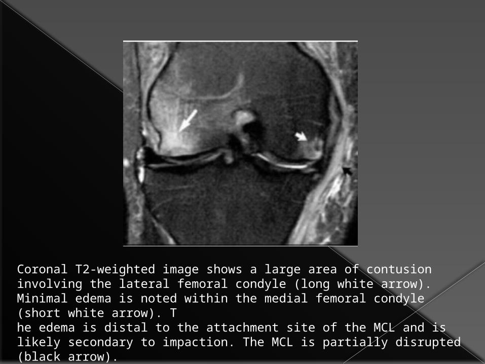
Coronal T2-weighted image shows a large area of contusion involving the lateral femoral condyle (long white arrow). Minimal edema is noted within the medial femoral condyle (short white arrow). The edema is distal to the attachment site of the MCL and is likely secondary to impaction. The MCL is partially disrupted (black arrow).
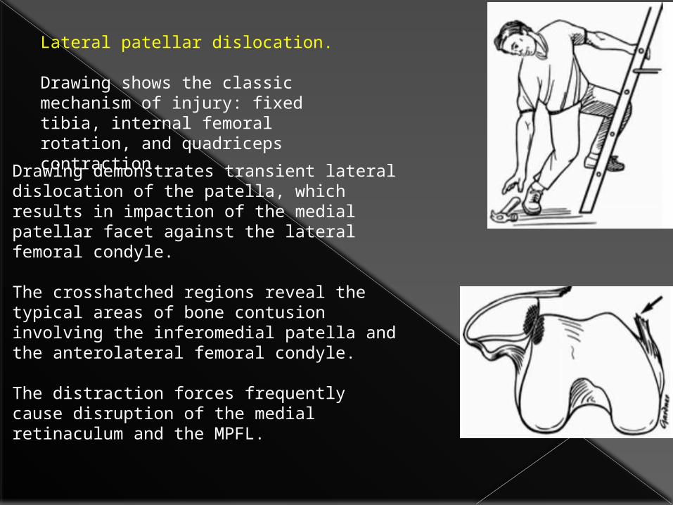
Lateral patellar dislocation.
Drawing shows the classic mechanism of injury: fixed tibia, internal femoral rotation, and quadriceps contraction.
Drawing demonstrates transient lateral dislocation of the patella, which results in impaction of the medial patellar facet against the lateral femoral condyle.
The crosshatched regions reveal the typical areas of bone contusion involving the inferomedial patella and the anterolateral femoral condyle.
The distraction forces frequently cause disruption of the medial retinaculum and the MPFL.
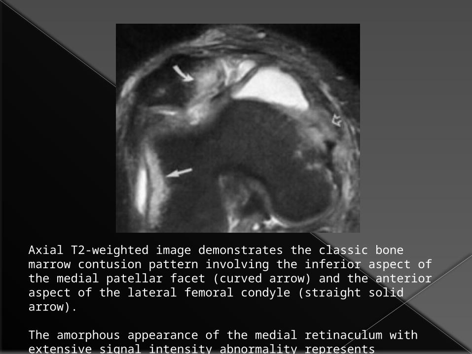
Axial T2-weighted image demonstrates the classic bone marrow contusion pattern involving the inferior aspect of the medial patellar facet (curved arrow) and the anterior aspect of the lateral femoral condyle (straight solid arrow).
The amorphous appearance of the medial retinaculum with extensive signal intensity abnormality represents disruption (open arrow).

THANK YOU