mri features of combined hepatocellular
TRANSCRIPT

RESEARCH ARTICLE Open Access
MRI features of combined hepatocellular-cholangiocarcinoma versus mass formingintrahepatic cholangiocarcinomaJennifer Sammon1, Sandra Fischer2, Ravi Menezes1, Hooman Hosseini-Nik1, Sara Lewis3, Bachir Taouli3
and Kartik Jhaveri1*
Abstract
Background: Combined hepatocellular-cholangiocarcinoma (cHCC-CC) is a rare primary liver tumor, which hasoverlapping imaging features with mass forming intra-hepatic cholangiocarcinoma (ICC) and hepatocellular carcinoma(HCC). Previous studies reported imaging features more closely resemble ICC and the aim of our study was to examinethe differential MRI features of cHCC-CC and ICC with emphasis on enhancement pattern observations of gadoliniumenhanced MRI.
Methods: Institutional review board approval with consent waiver was obtained for this retrospective bi-centric study.Thirty-three patients with pathologically proven cHCC-CC and thirty-eight patients with pathologically proven ICC,who had pre-operative MRI, were identified. MRI images were analyzed for tumor location and size, T1 and T2 signalcharacteristics, the presence/absence of: cirrhosis, intra-lesional fat, hemorrhage/hemosiderin, scar, capsular retraction,tumor thrombus, biliary dilatation, degree of arterial enhancement, enhancement pattern, pseudocapsule and washout.Associations between MRI features and tumor type were examined using the Fisher’s exact and chi-square tests.
Results: Strong arterial phase enhancement and the presence of: washout, washout and progression, intra-lesional fatand hemorrhage were all strongly associated with cHCC-CC (P < 0.001). While cHCC-CC had a varied enhancementpattern, the two most common enhancement patterns were peripheral persistent (n = 6) and heterogeneoushyperenhancement with washout (n = 6), compared to ICC where the most common enhancement patterns wereperipheral hypoenhancement with progression (n = 18) followed by heterogeneous hypoenhancement withprogression (n = 14) (P < 0.001).
Conclusion: The cHCC-CC enhancement pattern seems to more closely resemble HCC with the degree of arterialhyperenhancement and the presence of washout being valuable in differentiating cHCC-CC from ICC. However thepresence of washout and progression, in the same lesion or a predominantly peripheral /rim hyperenhancing masswere also seen as important features that should alert the radiologist to the possibility of a cHCC-CC.
Keywords: Combined hepatocellular-cholangiocarcinoma, Intrahepatic cholangiocarcinoma, Biphenotypic tumor,Liver MRI, Primary liver tumor
* Correspondence: [email protected] Joint Department of Medical Imaging, University Health Network,Sinai Health System and Women’s College Hospitals, University of Toronto,Toronto, CanadaFull list of author information is available at the end of the article
© The Author(s). 2018 Open Access This article is distributed under the terms of the Creative Commons Attribution 4.0International License (http://creativecommons.org/licenses/by/4.0/), which permits unrestricted use, distribution, andreproduction in any medium, provided you give appropriate credit to the original author(s) and the source, provide a link tothe Creative Commons license, and indicate if changes were made. The Creative Commons Public Domain Dedication waiver(http://creativecommons.org/publicdomain/zero/1.0/) applies to the data made available in this article, unless otherwise stated.
Sammon et al. Cancer Imaging (2018) 18:8 https://doi.org/10.1186/s40644-018-0142-z

BackgroundCombined hepatocellular-cholangiocarcinoma (cHCC-CC)is a rare primary liver tumor that expresses both biliaryand hepatocellular markers on immunohistochemistry.The WHO reclassified cHCC-CC in 2010 into two sub-groups: cHCC-CC classical type and cHCC-CC with stemcell features. These tumors must show unequivocal hepa-tocellular (HCC) and cholangiocarcinoma (ICC) compo-nents which have transition zones, thus differentiatingcHCC-CC from collision tumors [1].As cHCC-CC is a rare tumor, only a few studies have
looked at prognosis and management of this tumor, withcomplete tumor resection and lymph node clearancehaving the best prognosis. Survival rates post resectionappear to be worse than HCC and similar to ICC [2–7],with several studies reporting 5-year survival rates of16–41.1% for cHCC-CC post-transplant compared tonear 70% for HCC patients [8–11]. There are no ac-cepted transplant criteria for cHCC-CC to date, withprevious studies reporting poor outcome post livertransplant for patients with presumed HCC who werefound to have cHCC-CC on the explant pathology. Aspatients can proceed to transplant without histology,pre-operative diagnosis of cHCC-CC is important, butremains challenging, as there is both clinical and radio-logical overlap in these tumors. cHCC-CC can occur inpatients with risk factors for HCC and in patients withrisk factors for ICC and due to the heterogeneity of thetumor, cHCC-CC can have overlapping imaging featureswith HCC and ICC. Tumor markers cannot be reliedupon to differentiate, as only just over half of patients inone study had elevated Alpha-fetoprotein (AFP) and/orcarbohydrate antigen 19.9 (CA19.9) [1].Previous studies report imaging features of cHCC-CC
appear to more closely resemble ICC and metastasis ra-ther than HCC [12–18] and to the best of our know-ledge there are only a few studies that have attempted toinvestigate the MRI features of cHCC-CC [12–15]. Weperformed a step-wise systematic evaluation of MRI ex-aminations of pathologically proven cHCC-CC versusICC. The aim of our study was to examine the differen-tial MRI features of cHCC-CC and ICC with emphasison enhancement pattern observations of gadolinium en-hanced MRI.
MethodsPatientsInstitutional review board approval with consent waiverwas obtained for this retrospective bi-centric study.Pathology databases at both centers were searched forconsecutive cHCC-CC/biphenotypic tumors betweenJanuary 2005 and December 2014 and these results werecross-referenced with radiology databases, excluding anypatients who did not have preoperative MRI. Over the
same period the pathology and radiology databases weresearched for ICC cases.The patient demographics of the two groups are sum-
marized in Table 1. Thirty-three patients who hadpathologically proven cHCC-CC and MRI at baselinewere identified. Within this cohort, 25 of the patientswere male and 8 were female. The mean age was59.5 years with an age range of 36–82. Twenty-five pa-tients had chronic liver disease: 16 patients had hepatitisB, 9 patients had hepatitis C, 3 patients had a history ofalcohol abuse, 1 patient had hemochromatosis, 1 patienthad non-alcoholic steatohepatitis and 1 patient had pri-mary biliary cirrhosis. Two of the patients with historiesof alcohol excess were also hepatitis C positive and 1 pa-tient had both hepatitis B and hepatitis C positive ser-ology. Twenty-three (69.7%) of the patients had cirrhosison imaging, defined as lobar redistribution (hypertrophyof the caudate and left lateral segments, with atrophy ofthe right lobe and left medial segments) and/or nodularhepatic contour.AFP was recorded for 29 patients pre-treatment and 8
patients had an AFP > 100 ng/ml, with 5 patients in thecohort having an AFP > 400 ng/ml (range < 5–353,014).Only 7 patients had CA19.9 recorded pre-treatment and4 of those had elevated CA19.9 (> 37 U/ml), with onlyone greater than twice the normal limit at 125 U/ml.The remaining patients with a positive CA19.9 rangedfrom 38 to 49 U/ml.Forty consecutive patients with pathologically proven
ICC with MRI at baseline were identified. Two patientswere excluded; one as they did not have dynamic con-trast enhanced imaging and the other, as the quality ofthe study was deemed non-diagnostic. Within this co-hort there were a similar amount of male and female pa-tients with 20 males and 18 females. The mean age was61, with an age range of 32–86. Ten patients had riskfactors for liver disease, 7 had hepatitis B and 3 hadhepatitis C.AFP was recorded in 24 patients pre-treatment and no
patient had an elevated AFP. CA19.9 was recorded in 26patients pre-treatment and the median CA19.9 was
Table 1 Patient demographics
Parameter cHCC-CC Cholangiocarcinoma
Mean age(range)
59.5 (36–82) 61 (32–86)
Sex (M:F) 25:8 20:18
Median AFP(range)
23.5 ng/ml (< 5–353,014) 2 ng/ml (< 5–15)
Median Ca19.9(range)
25 U/ml (< 1–49) 16.5 U/ml (< 1–129,207)
Hepatitis B 16 7
Hepatitis C 9 3
Sammon et al. Cancer Imaging (2018) 18:8 Page 2 of 9

16.5 U/ml (range < 1–129,207). Seven patients had aCA19.9 > 37 U/ml.
Image acquisitionMRI examinations were performed at 1.5 T or 3 T (n = 63at 1.5 T and n = 8 at 3 T) using a phased array torso coil.MRI protocol included: T2 single shot turbo spin echowith TE 180, axial T2 turbo spin echo with TE 90, axialT1 volumetric interpolated breath-hold (VIBE) opposed-in phase sequences, axial diffusion weighted imaging andaxial T1 VIBE pre-contrast and dynamic post-contrast im-ages (Table 2). The majority of the patients (22 cHCC-CCand 38 ICC) received routine extracellular gadoliniumbased contrast agent gadobutrol (Gadovist, Bayer Health-care, Berlin, Germany) at a dose of 0.1 mmol/kg at 1 ml/s.Eleven cases in the cHCC-CC group and 3 cases in theICC group had imaging with hepatocyte specific contrastagent gadoxetic acid (Primovist, Bayer AG, Germany) at adose of 0.025 mmol/kg at 1 ml/s. At our institution, theprimary contrast agent for initial liver imaging is an extra-cellular based gadolinium contrast agent, and as this is aretrospective study, only the extracellular phases of con-trast imaging were analyzed.
Image analysisTwo abdominal radiologists (one abdominal imagingfellow and one faculty with 15 years subspecialty MRIexperience) retrospectively reviewed the studies in con-sensus. Images were reviewed on a picture archive com-munication system. The following characteristics wereevaluated: tumor location and size, T1 and T2 signalcharacteristics, the presence/absence of: cirrhosis on im-aging, intra-lesional fat, hemorrhage/hemosiderin, scar,capsular retraction, tumor thrombus, biliary dilatation,
degree of arterial enhancement, enhancement pattern onarterial portal-venous and delayed (5 min) phases,pseudocapsule and washout. T2 intermediate signal in-tensity was defined as the same signal intensity as thespleen and T2 hyperintense lesions were defined as be-ing of higher signal intensity than the spleen. Capsularretraction was recorded for peripheral tumors, which wedefined as being within 1 cm of the liver capsule. Thedegree of arterial enhancement was defined as beingstrong if any part of the lesion showed similar enhance-ment to the aorta, mild to moderate if the enhancementwas less than the aorta and absent if there was no arter-ial enhancement. For the overall enhancement pattern,lesions were characterized as being associated withwashout even if there was an area of progressive en-hancement in the same lesion as our main aim of thisstudy was comparing cHCC-CC to ICC. Lesions withboth washout and progression were captured separately.Lesions were defined as having peripheral enhancementpatterns, rather than heterogeneous enhancement pat-terns, if there was peripheral (< 1 cm depth) enhance-ment on the arterial or venous phase (in lesions thatwere hypoenhancing on arterial phase). If there was anycentral enhancement these lesions were characterized asa heterogeneous enhancement pattern. Evidence ofcirrhosis included a lobulated/nodular contour and/orvolume redistribution to the left lobe and caudate.
Statistical analysisDescriptive statistics (frequencies, percentage, mean)were used to summarize demographics, clinical historyand MRI features, by tumor type. Associations betweenMRI features and tumor type were examined using theFisher’s exact and chi-square tests. All tests were two
Table 2 MRI parameters
Image sequence TR (ms) TE (ms) NEX FOV (mm) ST (mm) Gap (mm) Matrix (phase x frequency)
Pre-contrast imaging:
Axial T2 HASTE SPAIR 1600 90 1 360 5 1 259 × 320
Axial T2 HASTE SPAIR 1600 180 1 360 5 1 259 × 320
Axial T1 VIBE opp/in 4.43 1.39–2.49 1 360 3 0 218 × 320
ep2d diff b100,600 7600 66 6 380 5 0 156 × 192
T1 VIBE axial SPAIR 4.19 1.47 1 300 3 0 195 × 320
Post-contrast imaging:
T1 VIBE axial SPAIR dynamic: arterial(care bolus trigger), venous (45–60 s)and interstitial phase (90–120 s)
4.19 1.47 1 300 3 0 195 × 320
T1 VIBE axial SPAIR 5-min delay 4.19 1.47 1 300 3 0 195 × 320aPost-contrast Primovist:
T1 VIBE axial SPAIR 20 min 4.37 1.47 1 300 4 0 195 × 320
T1 VIBE axial SPAIR 20 min 4.19 1.47 1 300 1.5 0 202 × 320aIf hepatocyte specific contrast agent (gadoxetic acid) used
Sammon et al. Cancer Imaging (2018) 18:8 Page 3 of 9
sided, and p < 0.05 was considered an indicator of a sta-tistically significant association. Statistical analyses wereperformed using SPSS software (version 20.0, IBM).
ResultsThe MRI features of cHCC-CC and ICC are summarizedin Table 3. On T1 the majority of the lesions werehomogenously hypointense in the cHCC-CC and ICCgroups, 23/33 and 30/38 respectively. On T2, the majorityof the cHCC-CC group (23/33), had a homogenous inter-mediate/hyperintense appearance. In the ICC group, 14/38had heterogeneous signal intensity on T2, 12/38 hadhomogenous intermediate/hyperintense appearance andperipheral hyperintensity with a central hypointense regionwas seen in 9/38.Two patients in the cHCC-CC group had intra-lesional
fat and 4 patients in the cHCC-CC group had intra-lesional hemorrhage. No patient in the ICC cohort hadintra-lesional fat. One patient in the ICC cohort had evi-dence of intra-lesional hemorrhage, however this patienthad a percutaneous biopsy three days prior to the MRI.Excluding the post biopsy patient in the ICC group, bothintra-lesional fat and intra-lesional hemorrhage are highlyspecific (100%) for cHCC-CC versus ICC, although theyhave poor sensitivities (6% {95% CI: -2 to 14%} and 12%{95% CI: 1–23%} respectively).In the cases of peripherally located tumors, 13/21 in
the ICC group showed capsular retraction compared to3/23 in the cHCC-CC group (P < 0.001).
The presence of biliary dilatation associated with themass was seen in 5 of the cHCC-CC group and 23 ofthe ICC group, P-value of less than 0.001. Portal veintumor thrombus was seen in 3 of the cHCC-CC groupcompared to 0 in the ICC group.The enhancement characteristics of cHCC-CC and
ICC are summarized in Table 4. Arterial enhancementwas seen in 90.9% (n = 30) of the cHCC-CC group com-pared to 57.9% (n = 22) of the ICC group. The degree ofarterial enhancement in 15 patients in the cHCC-CCgroup was similar to the degree of enhancement of theaorta (strong) and in the remaining 15 patients it wasless intense (mild to moderate) than the aorta, comparedto 1 and 22, respectively in the ICC group (P < 0.001;strong arterial enhancement). Peripheral rim enhance-ment on the arterial phase was seen in 14 cases in boththe cHCC-CC group and the ICC group.With regards to the overall enhancement characteris-
tics of the lesions, the most common enhancement pat-terns in the cHCC-CC group were peripheral persistent(n = 6) (Fig. 1) and heterogeneous hyperenhancementwith washout (n = 6). The most common enhancementpattern in the ICC group was peripheral hypoenhance-ment with progression (n = 18) followed by heteroge-neous hypoenhancement with progression (n = 14)(Fig. 2). Combining peripheral hypoenhancement withprogression, heterogeneous hypoenhancement with pro-gression and hypoenhancement versus the other sub-groups, there was a statistically significant differencebetween the ICC and cHCC-CC groups. 79% of the pa-tients who had either one of these three enhancementpatterns had ICC and 89% of the patients in the othercategory had cHCC-CC (P < 0.001).Progressive enhancement was seen in 13 of the cHCC-
CC group and 33 of the ICC group (P < 0.001). Washoutwas seen in 13 of the cHCC-CC group and in 0 of the ICCgroup (P < 0.001), with a sensitivity of 39% (95% CI: 23–56%) and specificity of 100% in differentiating cHCC-CCfrom ICC. Both washout and progression were seen in thesame tumor in 3 cases in the cHCC-CC group.
Table 3 MRI characteristics of cHCC-CC and ICC
Parameter cHCC-CC Cholangiocarcinoma P-value
T1 WI
Hypointense 23 30 0.37
Heterogeneous 9 5 0.136
Isointense/not seen 1 3 0.375
T2 WI
Homogenouslyintermediate/hyperintense
23 12 0.001
Peripheral hyperintensityand central hypointensity
3 9 0.102
Heterogeneous 7 14 0.15
Isointense/not seen 0 3 0.99
Intralesional fat 2 0 0.124
Intralesional hemorrhage 4 1a 0.119
Capsular retractionb 3/23 (13%) 13/21 (62%) < 0.001
Cirrhosis on imaging 23 0 < 0.001
Biliary dilatation 5 23 < 0.001
Tumor thrombus 3 0 0.058aThis patient had recently had a percutaneous biopsybRecorded for lesions within 1 cm of the liver capsule
Table 4 Enhancement characteristics of cHCC-CC and ICC
Parameter CombinedHCC/CC
Cholangiocarcinoma P value
Degree of arterialenhancement
Strong: 15/33 Strong: 1/38 < 0.001
Mild: 15/33 Mild: 22/38 0.295
Hypo: 3/33 Hypo: 15/38 0.003
Peripheral rim arterialenhancement
14 (42%) 14 (37%) 0.631
Progression 13 (39%) 33 (87%) < 0.001
Washout 13 (39%) 0 < 0.001
Washout and Progression 3 (9%) 0 0.058
Sammon et al. Cancer Imaging (2018) 18:8 Page 4 of 9

Three patients in the cHCC-CC cohort also had a sep-arate mass characteristic of HCC on their MRI. In twoof these cases, the cHCC-CC tumors had similar im-aging characteristics to the foci of HCC within the sameliver, in that they demonstrated arterial hyperenhance-ment and washout. In one case, the cHCC-CC and HCCwere both over 4 cm in diameter and in this case thecHCC-CC was relatively hypovascular compared to theHCC and it did not contain fat, unlike the HCC. TheHCC demonstrated washout, but the cHCC-CC did not(Fig. 3). In two other cases, separate 1–2 cm foci ofHCC were identified on the explanted liver, but notdetected on pre-operative imaging.
DiscussionFew studies have been published evaluating the imagingfeatures of cHCC-CC, with most of the earlier studiesusing the Allen and Lisa or Goodman classifications,which include collision tumors. As mentioned previouslymost studies report similar imaging characteristics toICC [12–19]. However, the enhancement characteristicsof cHCC-CC in our study appear to more closely resem-ble HCC rather than ICC, with 13/33 patients in thecHCC-CC cohort having a typical HCC enhancement
pattern (arterial enhancement and washout). This maybe partly explained by the demographics of our popula-tion. In our study the prevalence of cirrhosis (69.7%) andpositive hepatitis serology (hepatitis B: 48% and hepatitisC: 27%) in the cHCC-CC cohort is greater than previ-ously reported North American studies [14, 20, 21],where patient demographics and the presence of chronicliver disease risk factors resembled those of ICC ratherthan HCC. However, some of the earlier studies ofcHCC-CC are in Asian populations and these studies re-port demographics, risk factors and survival similar toHCC [1–3, 22–24]. One European study suggests thatthe risk factors of the cHCC-CC population lie in be-tween the HCC and ICC groups, but continued to reporta male predominance [25]. The differences in our groupcompared to previously published North American stud-ies could be explained by the increasing Asian popula-tion in Canada, higher prevalence of chronic liverdisease and increasing incidence of liver cancer [26].There were also other features associated with HCC in
the cHCC-CC group: n = 2 had intra-lesional fat and n = 4had intra-lesional hemorrhage. While these features arehighly specific in differentiating cHCC-CC from ICC, thelow sensitivity does not help in differentiating cHCC-CC
Fig. 1 Pathologically proven cHCC-CC with peripheral persistent enhancement: There is a T2 hyperintense (a) lesion in segment 8/4A of the liver,which demonstrates peripheral arterial hyperenhancement (b). The enhancement pattern remains peripheral on both the portal venous and de-layed phases (c & d). This enhancement pattern (peripheral persistent) was one of the most common enhancement patterns of cHCC-CC seen inour cohort
Sammon et al. Cancer Imaging (2018) 18:8 Page 5 of 9
from ICC. In 3 cases of cHCC-CC there was both washoutand progression in the same lesion (Fig. 4), which doesdifferentiate cHCC-CC from ICC, as washout is not seenin ICC. These features can be seen in scirrhous HCC,but this should alert the radiologist to the possibilityof a cHCC-CC tumor and consideration for biopsy asthe potential treatment options for these two tumorsvary [27, 28].Previous studies have reported that tumor markers
can be helpful in raising the possibility of a cHCC-CCtumor, where both CA19.9 and AFP can be elevated[4, 14, 29, 30]. In our cHCC-CC cohort, AFP was re-corded for 29 patients pre-treatment with 8 patients hav-ing an AFP > 100 ng/ml and 7 patients had CA19.9recorded pre-treatment, with 4 of those patients having anelevated CA19.9. While the midrange elevation of AFPhelps differentiate these tumors from ICC, there was onlyone patient who had a CA19.9 above twice the upper limitof normal. This could partially be due to limited samplingin these patients, as the presumptive diagnosis was HCCin the setting of cirrhosis and cHCC-CC was a postresection/explant or post biopsy diagnosis. With previousstudies reporting low to mid-level elevation of CA19.9, itdoes raise an argument for routine CA19.9 testing inpatients who have a liver mass as this would alert
the radiologist to the possible presence of a cHCC-CC tumor and prompt biopsy, to aid in a pre-treatment diagnosis.Our study has several limitations, including that this is
a retrospective study and the readers were aware the co-hort comprised of cHCC-CC and ICC, even though spe-cific pathological diagnosis was not known at the time ofimage review. Our study group is also small, with only33 patients in the cHCC-CC group, however this is at-tributable to the rare nature of this tumor. Despite this,our population for MRI is larger than most other pub-lished studies. Another limitation is the absence of histo-logical quantification of HCC and ICC components inthe cHCC-CC tumors as not all cases went to resection.
ConclusionPre-operative imaging diagnosis of cHCC-CC tumors re-mains a challenge. In our study, cHCC-CC tumors dis-played predominant arterial hyperenhancement patternand the presence of washout, similar to HCC, perhapsdue to a population with a high prevalence of HCC riskfactors. We found that the presence of washout; washoutand progression in the same lesion; intra-lesional fat andintra-lesional hemorrhage help differentiate cHCC-CCfrom ICC.
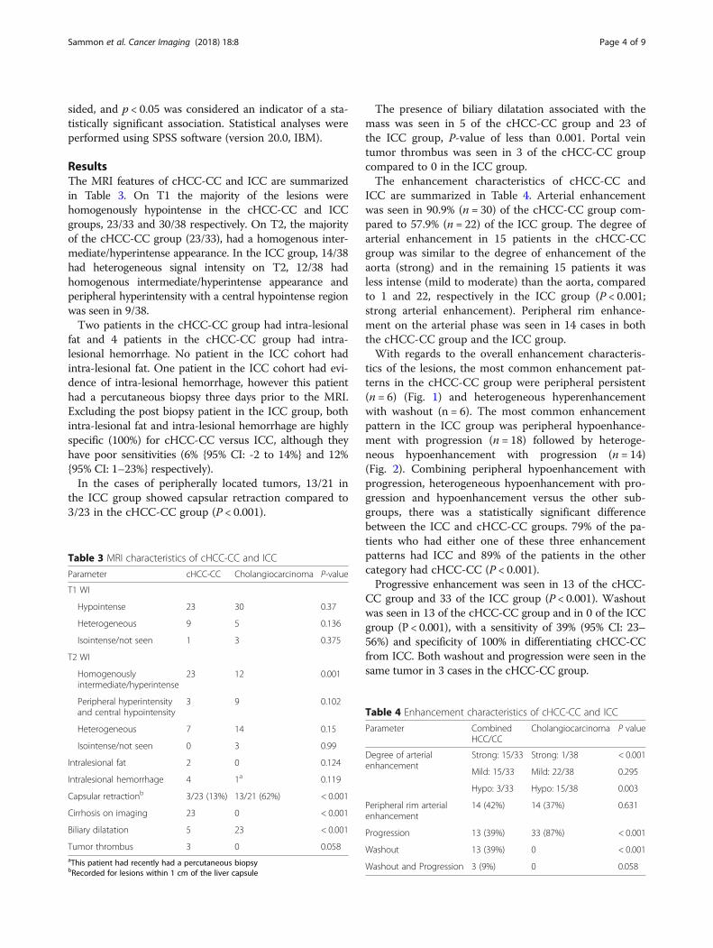
Fig. 2 Pathologically proven cHCC-CC with peripheral progressive enhancement: There is a large mass, which is predominantly intermediate sig-nal on T2-WI (a) in segment 8 of the liver. This demonstrates peripheral arterial hyperenhancement (b), but then shows progressive enhancementon the portal venous and delayed phases (c & d) demonstrating enhancement pattern similar to that seen in mass forming ICC
Sammon et al. Cancer Imaging (2018) 18:8 Page 6 of 9
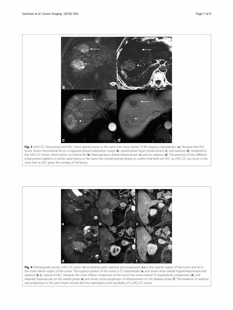
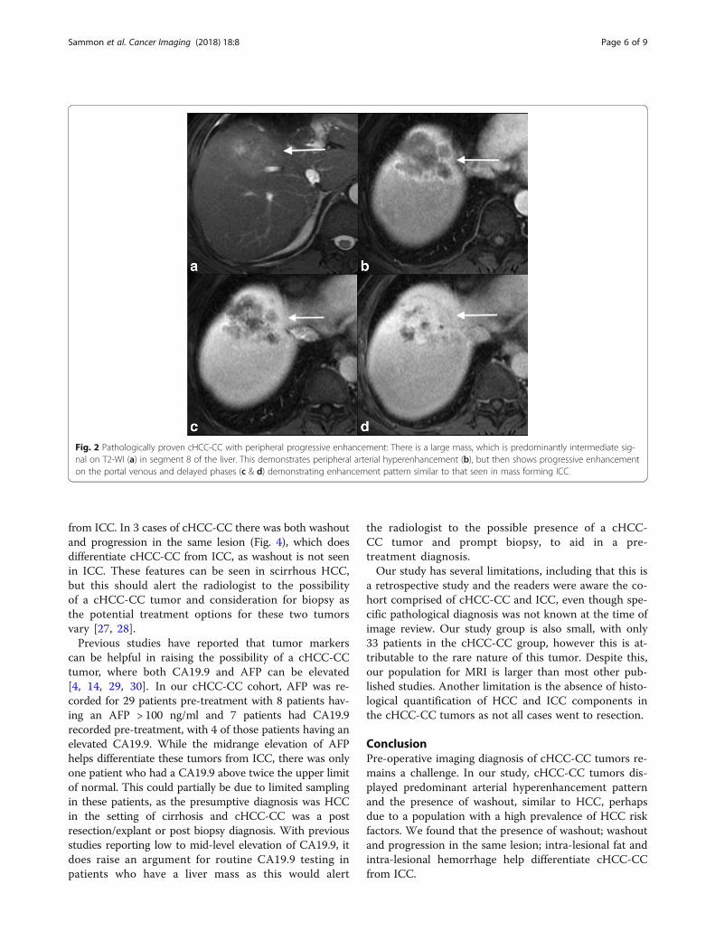
Fig. 3 cHCC-CC (long arrow) and HCC (short dashed arrow) in the same liver; show similar T2-WI imaging characteristics (a). However the HCCtumor shows intra-lesional fat on in-opposed phased subtraction image (b), arterial phase hyper enhancement (c) and washout (d) compared tothe cHCC-CC tumor, which shows no internal fat (b) heterogeneous arterial enhancement (c) and no washout (d). The presence of two differentenhancement patterns in similar sized lesions in the same liver should prompt biopsy to confirm that both are HCC as cHCC-CC can occur in thesame liver as HCC given the overlap of risk factors
Fig. 4 Pathologically proven cHCC-CC tumor demonstrating both washout and progression: a-c is the superior aspect of the tumor and d-f isthe more inferior aspect of the tumor. The superior portion of the tumor is T2 intermediate (a) and shows show arterial hyperenhancement andwashout (b, c), typical of HCC. However the more inferior component of the tumor has some internal T2 hypointense components (d), andrelatively hypovascular on the arterial phase (e) and shows some progression of enhancement on the delayed phase (f). The presence of washoutand progression in the same lesion should alert the radiologist to the possibility of a cHCC-CC tumor
Sammon et al. Cancer Imaging (2018) 18:8 Page 7 of 9

AbbreviationsAFP: Alpha-fetoprotein; CA19.9: Carbohydrate antigen 19.9; cHCC-CC: Combinedhepatocellular-cholangiocarcinoma; HCC: Hepatocellular carcinoma; ICC: Intra-hepatic cholangiocarcinoma; MRI: Magnetic resonance imaging
AcknowledgementsN/A
FundingNo funding was provided for this study.
Availability of data and materialsThe datasets used and/or analyzed during the current study are availablefrom the corresponding author on reasonable request.
Authors’ contributionsJS and KJ analyzed and interpreted the images. JS is the primary author. KJ andBT critically reviewed the paper and revised it. HH and SL performed thedatabase search and literary review. HH and SL also contributed to the primarydraft of the manuscript. SF did the pathology review and analysis. RM performedthe statistical analysis. All authors read and approved the final manuscript.
Ethics approval and consent to participateInstitutional review board approval with consent waiver was obtained forthis retrospective bi-centric study.
Consent for publicationImages are entirely unidentifiable and there are no details on individualsreported within the manuscript.
Competing interestsThe authors declare that they have no competing interests.
Publisher’s NoteSpringer Nature remains neutral with regard to jurisdictional claims inpublished maps and institutional affiliations.
Author details1Toronto Joint Department of Medical Imaging, University Health Network,Sinai Health System and Women’s College Hospitals, University of Toronto,Toronto, Canada. 2Department of Pathology, University Health Network,University of Toronto, Toronto, Canada. 3Department of Radiology, MountSinai New York, New York, USA.
Received: 25 September 2017 Accepted: 15 February 2018
References1. Yin X, Zhang B-H, Qiu S-J, Ren Z-G, Zhou J, Chen X-H, et al. Combined
hepatocellular carcinoma and cholangiocarcinoma: clinical features, treatmentmodalities, and prognosis. Ann Surg Oncol. 2012;19(9):2869–76. http://www.springerlink.com/index/10.1245/s10434-012-2328-0.
2. Koh KC, Lee H, Choi MS, Lee JH, Paik SW, Yoo BC, et al. Clinicopathologicfeatures and prognosis of combined hepatocellular cholangiocarcinoma.Am J Surg. 2005;189(1):120–5. http://linkinghub.elsevier.com/retrieve/pii/S0002961004004799.
3. Lee WS, Lee KW, Heo JS, Kim SJ, Choi SH, Kim YI, et al. Comparison of combinedhepatocellular and cholangiocarcinoma with hepatocellular carcinoma andintrahepatic cholangiocarcinoma. Surg Today. 2006;36(10):892–7.
4. Kassahun WT, Hauss J. Management of combined hepatocellular andcholangiocarcinoma. Int J Clin Pract. 2008;62(8):1271–8. http://onlinelibrary.wiley.com/doi/10.1111/j.1742-1241.2007.01694.x/abstract.
5. Chi M, Mikhitarian K, Shi C, Goff LW. Management of combinedhepatocellular-cholangiocarcinoma: a case report and literature review.Gastrointest Cancer Res. 2012;5(6):199–202. http://www.pubmedcentral.nih.gov/articlerender.fcgi?artid=3533848&tool=pmcentrez&rendertype=abstract.
6. Zuo H-Q, Yan L-N, Zeng Y, Yang J-Y, Luo H-Z, Liu J-W, et al. Clinicopathologicalcharacteristics of 15 patients with combined hepatocellular carcinomaand cholangiocarcinoma. Hepatobiliary Pancreat Dis Int. 2007;6(2):161–5.http://www.ncbi.nlm.nih.gov/pubmed/17374575.
7. Lee J-H, Chung GE, Yu SJ, Hwang SY, Kim JS, Kim HY, et al. Long-termprognosis of combined hepatocellular and cholangiocarcinoma aftercurative resection comparison with hepatocellular carcinoma andcholangiocarcinoma. J Clin Gastroenterol. 2011;45(1):69–75. http://www.ncbi.nlm.nih.gov/pubmed/20142755.
8. Garancini M, Goffredo P, Pagni F, Romano F, Roman S, Sosa JA, et al.Combined hepatocellular-cholangiocarcinoma: a population-level analysisof an uncommon primary liver tumor. Liver Transpl. 2014;20(8):952–9.http://www.ncbi.nlm.nih.gov/pubmed/24777610.
9. Sapisochin G, Fidelman N, Roberts JP, Yao FY. Mixed hepatocellularcholangiocarcinoma and intrahepatic cholangiocarcinoma in patientsundergoing transplantation for hepatocellular carcinoma. Liver Transpl.2011;17(8):934–42. http://www.ncbi.nlm.nih.gov/pubmed/21438129.
10. Panjala C, Senecal DL, Bridges MD, Kim GP, Nakhleh RE, Nguyen JHH, et al.The diagnostic conundrum and liver transplantation outcome for combinedhepatocellular-cholangiocarcinoma. Am J Transplant. 2010;10(5):1263–7.http://doi.wiley.com/10.1111/j.1600-6143.2010.03062.x.
11. Sapisochin G, de Lope CR, Gastaca M, de Urbina JO, López-Andujar R,Palacios F, et al. Intrahepatic cholangiocarcinoma or mixed hepatocellular-cholangiocarcinoma in patients undergoing liver transplantation: aSpanish matched cohort multicenter study. Ann Surg. 2014;259(5):944–52.http://www.ncbi.nlm.nih.gov/pubmed/24441817.
12. de Campos ROP, Semelka RC, Azevedo RM, Ramalho M, Heredia V, Armao DM,et al. Combined hepatocellular carcinoma-cholangiocarcinoma: report of MRappearance in eleven patients. J Magn Reson Imaging. 2012;36(5):1139–47.http://doi.wiley.com/10.1002/jmri.23754.
13. Hwang J, Kim YK, Park MJ, Lee MH, Kim SH, Lee WJ, et al. Differentiatingcombined hepatocellular and cholangiocarcinoma from mass-formingintrahepatic cholangiocarcinoma using gadoxetic acid-enhanced MRI.J Magn Reson Imaging. 2012;36(4):881–9.
14. Fowler KJ, Sheybani A, Parke R a., Doherty S, Brunt EM, Chapman WC, et al.Combined hepatocellular and cholangiocarcinoma (biphenotypic) tumors:imaging features and diagnostic accuracy of contrast-enhanced CT and MRI.Am J Roentgenol 2013;201(2):332–339.
15. Potretzke TA, Tan BR, Doyle MB, Brunt EM, Heiken JP, Fowler KJ. Imagingfeatures of biphenotypic primary liver carcinoma(hepatocholangiocarcinoma) and the potential to mimic hepatocellularcarcinoma: LI-RADS analysis of CT and MRI features in 61 cases. AJR Am JRoentgenol. 2016;207:1–7.
16. Akoi K, Takayasu K, Kawano T, Muramatsu Y, Moriyama N, Wakao F, et al.Combined hepatocellular carcinoma and cholangiocarcinoma: clinicalfeatures and computed tomographic findings. Hepatology. 1993;18(5):1090–5.
17. Jeon T, Kim S, Lee W, Lim H. The value of gadobenate dimeglumine-enhancedhepatobiliary-phase MR imaging for the differentiation of scirrhous hepatocellularcarcinoma and cholangiocarcinoma with or without hepatocellular carcinoma.Abdom Imaging. 2010;35(3):337–45. http://resolver.scholarsportal.info/resolve/09428925/v35i0003/337_tvogdhcwowhc.xml.
18. Wells ML, Venkatesh SK, Chandan VS, Fidler JL, Fletcher JG, Johnson GB, et al.Biphenotypic hepatic tumors : imaging findings and review of literature. AbdomImaging. 2015;40(7):2293–305. https://doi.org/10.1007/s00261-015-0433-9.
19. Allen RA, Lisa JR. Combined liver cell and bile duct carcinoma. Am J Pathol.1949;25:647–55.
20. Jarnagin WR, Weber S, Tickoo SK, Koea JB, Obiekwe S, Fong Y, et al.Combined hepatocellular and cholangiocarcinoma. Cancer. 2002;94(7):2040–6. http://doi.wiley.com/10.1002/cncr.10392.
21. Bhagat V, Javle M, Yu J, Agrawal A, Gibbs JF, Kuvshinoff B, et al. Combinedhepatocholangiocarcinoma: case-series and review of literature. Int JGastrointestinal Cancer. 2006;37(1):27–34.
22. Yano Y, Yamamoto J, Kosuge T, Sakamoto Y, Yamasaki S, Shimada K, et al.Combined hepatocellular and cholangiocarcinoma : a clinicopathologicstudy of 26 resected cases. Jpn J Clin Oncol. 2003;33(6):283–7.
23. Park HS, Bae JS, Jang KY, Lee JH, Yu HC, Jung JH, et al. Clinicopathologicstudy on combined hepatocellular carcinoma and cholangiocarcinoma:with emphasis on the intermediate cell morphology. J Korean Med Sci.2011;26(8):1023. http://synapse.koreamed.org/DOIx.php?id=10.3346/jkms.2011.26.8.1023.
24. Lee SD, Park S-J, Han S-S, Kim SH, Kim Y-K, Lee S-A, et al. Clinicopathologicalfeatures and prognosis of combined hepatocellular carcinoma andcholangiocarcinoma after surgery. Hepatobiliary Pancreat Dis Int.2014;13(6):594–601. http://www.hbpdint.com/CN/abstract/abstract4238.shtml.
Sammon et al. Cancer Imaging (2018) 18:8 Page 8 of 9

25. Cazals-hatem D, Rebouissou S, Bioulac-sage P, Bluteau O, Franco D, BelghitiJ, et al. Clinical and molecular analysis of combined hepatocellular-cholangiocarcinomas. J Hepatol. 2004;41:292–8.
26. Jiang X, Pan SY, De Groh M, Liu S, Morrison H. Increasing incidence in livercancer in Canada, 1972–2006: age-period-cohort analysis. J GastrointestOncol. 2011;2(4):223–31.
27. Kim SH, Lim HK, Lee WJ, Choi D, Park CK. Scirrhous hepatocellularcarcinoma: comparison with usual hepatocellular carcinoma based onCT-pathologic features and long-term results after curative resection.Eur J Radiol. 2009;69(1):123–30. http://www.ejradiology.com/article/S0720048X07004676/fulltext.
28. Chung YE, Park M-S, Park YN, Lee H-J, Seok JY, Yu J-S, et al. Hepatocellularcarcinoma variants: radiologic-pathologic correlation. AJR Am J Roentgenol.2009;193(1):W7–13. http://www.ajronline.org/doi/full/10.2214/AJR.07.3947.
29. Maximin S, Ganeshan DM, Shanbhogue AK, Dighe MK, Yeh MM, KolokythasO, et al. Current update on combined hepatocellular-cholangiocarcinoma.Eur J Radiol Open. 2014;1:40–8. http://linkinghub.elsevier.com/retrieve/pii/S2352047714000021.
30. O’Connor K, Walsh JC, Schaeffer DF. Combined hepatocellular-cholangiocarcinoma(cHCC-CC): a distinct entity. Ann Hepatol. 2014;13(3):317–22.
• We accept pre-submission inquiries
• Our selector tool helps you to find the most relevant journal
• We provide round the clock customer support
• Convenient online submission
• Thorough peer review
• Inclusion in PubMed and all major indexing services
• Maximum visibility for your research
Submit your manuscript atwww.biomedcentral.com/submit
Submit your next manuscript to BioMed Central and we will help you at every step:
Sammon et al. Cancer Imaging (2018) 18:8 Page 9 of 9