mouse chemerin quantikine - resources.rndsystems.com · mouse chemerin immunoassay ... chemerin,...
TRANSCRIPT

Mouse Chemerin Immunoassay
Quantikine® ELISA
This package insert must be read in its entirety before using this product. For research use only. Not for use in diagnostic procedures.
Catalog Number MCHM00
For the quantitative determination of mouse Chemerin concentrations in cell culture supernates, serum, and plasma.
This product and/or its use is the subject of European Patent 1 405 083 B1, US Patents 7,332,291; 7,419,658 and 7,842,453 as well as foreign equivalents licensed to R&D Systems® Inc. The purchase of this product is intended for research purposes only, not including the screening of compounds for the development of therapeutic and/or diagnostic products. Buyers may require a separate license to the patent rights for applications beyond such research purposes. For information on licensing, please contact Euroscreen SA rue Adrienne Bolland n°47 B-6041 Gosselies Belgium. Phone: +32-71-348500, Fax: +32-71-348519, e-mail: [email protected]. Attention: Dr. Vincent Lannoy.

TABLE OF CONTENTS
SECTION PAGE
INTRODUCTION .....................................................................................................................................................................1PRINCIPLE OF THE ASSAY ...................................................................................................................................................2LIMITATIONS OF THE PROCEDURE .................................................................................................................................2TECHNICAL HINTS .................................................................................................................................................................2MATERIALS PROVIDED & STORAGE CONDITIONS ...................................................................................................3OTHER SUPPLIES REQUIRED .............................................................................................................................................3PRECAUTIONS .........................................................................................................................................................................4SAMPLE COLLECTION & STORAGE .................................................................................................................................4SAMPLE PREPARATION........................................................................................................................................................4REAGENT PREPARATION .....................................................................................................................................................5ASSAY PROCEDURE .............................................................................................................................................................6CALCULATION OF RESULTS ...............................................................................................................................................7TYPICAL DATA .........................................................................................................................................................................7PRECISION ................................................................................................................................................................................8RECOVERY.................................................................................................................................................................................8LINEARITY .................................................................................................................................................................................8SENSITIVITY .............................................................................................................................................................................9CALIBRATION ..........................................................................................................................................................................9SAMPLE VALUES .....................................................................................................................................................................9SPECIFICITY ........................................................................................................................................................................... 10REFERENCES ......................................................................................................................................................................... 10
Manufactured and Distributed by:
USA R&D Systems, Inc. 614 McKinley Place NE, Minneapolis, MN 55413TEL: 800 343 7475 612 379 2956FAX: 612 656 4400E-MAIL: [email protected]
Distributed by:
Europe | Middle East | Africa Bio-Techne Ltd.19 Barton Lane, Abingdon Science ParkAbingdon OX14 3NB, UKTEL: +44 (0)1235 529449FAX: +44 (0)1235 533420E-MAIL: [email protected]
China Bio-Techne China Co., Ltd.Unit 1901, Tower 3, Raffles City Changning Office,1193 Changning Road, Shanghai PRC 200051TEL: +86 (21) 52380373 (400) 821-3475FAX: +86 (21) 52371001E-MAIL: [email protected]

www.RnDSystems.com 1
INTRODUCTIONChemerin, also known as TIG2 and RARRES2, is a distant member of the cystatin/cathelicidin superfamily of proteins (1-3). Members of this superfamily contain at least two intrachain disulfide bonds and an α-helical structure over a distance of about 100 amino acids (aa) (2, 3). Mouse Chemerin is synthesized as a 162 aa precursor that contains a probable 19 aa N-terminal signal sequence, an intervening 137-140 aa cystatin-fold containing domain, and a variable length C-terminal pro-segment (4-7). The cystatin-fold domain contains three intrachain disulfide bonds that contribute to the molecule’s characteristic fold (4, 8). Following secretion, the 18 kDa pro-form undergoes proteolytic processing at the C-terminus. This generates the bioactive molecule. Multiple serine proteases are involved, including plasmin, cathepsin G, elastase, and tryptase (5, 8-10). In mice, this creates multiple isoforms that end with Gln152, Ala154, and Ser155. The activity seems to be concentrated in the 9 amino acids preceding the pro-segment (aa 147-155) (10). Retention of the pro-segment blocks activity (4, 7, 9). One potential splice variant exists that shows a 6 aa substitution for aa 61-162. Chemerin is potentially produced by multiple cell types, including adipocytes, endothelial cells, fibroblasts, hepatocytes, and keratinocytes (1, 11, 12). It circulates at high ng/mL concentrations and is suggested to form dimers or higher order multimers (7, 10-12). Due to plasmin activation, only chemerin in serum (not plasma) shows bioactivity (7). Mature mouse Chemerin shares 68%, 85%, and 82% aa sequence identity to human, rat, and hamster Chemerin, respectively (6).
In mice, mature Chemerin binds to the G-protein coupled receptor termed GPCR-DEZ (ChemR23 in humans) (5, 13, 14). The receptor has limited expression, being found on endothelial cells, mature adipocytes, macrophages, early dendritic cells (DC), and natural killer (NK) cells (12, 15, 16). Although human plasmacytoid DC are reported to express ChemR23, this does not appear to be the case in mice (16).
Functionally, Chemerin has two principal activities. The first is chemoattraction, which is comparable to that of known chemokine family members. It is posited that high concentrations of tissue pro-chemerin become activated by serine proteases released by early-responding inflammatory cells. Both elastase and cathepsin G are components of neutrophil granules, and their release in response to microbial insult is proposed to activate chemerin and induce macrophage/monocyte infiltration (9, 10). The second activity involves adipocytes. Mature white adipocytes are known to secrete, activate, and respond to Chemerin. Activation of the adipocyte Chemerin receptor increases lipolysis and the efficiency of insulin-induced glucose uptake, thus materially impacting fat metabolism (11-13, 17).
The Quantikine® Mouse Chemerin Immunoassay is a 4.5 hour solid-phase ELISA designed to measure mouse Chemerin in cell culture supernates, serum, and plasma. It contains E. coli-expressed recombinant mouse mature Chemerin and antibodies raised against the recombinant factor. This immunoassay has been shown to accurately quantitate the recombinant mouse Chemerin. Results obtained using natural mouse Chemerin showed dose response curves that were parallel to the standard curves obtained using the Quantikine® kit standards. These results indicate that this kit can be used to determine relative mass values for natural mouse Chemerin.
For research use only. Not for use in diagnostic procedures.2
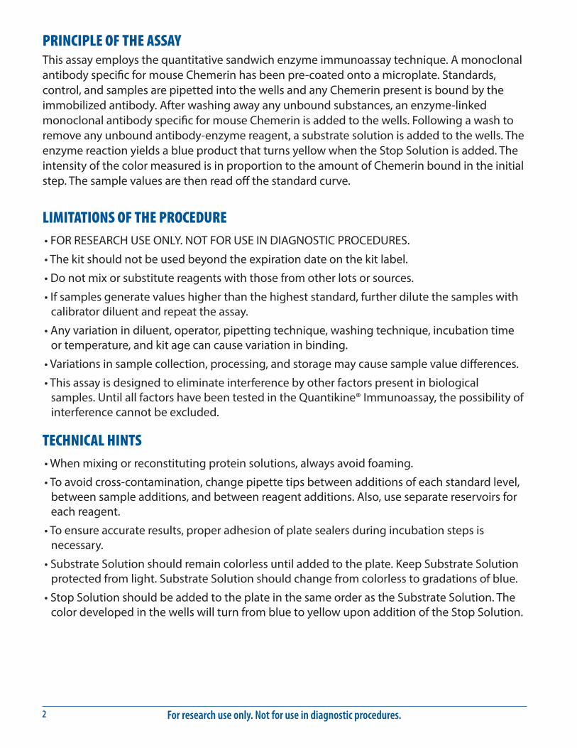
PRINCIPLE OF THE ASSAYThis assay employs the quantitative sandwich enzyme immunoassay technique. A monoclonal antibody specific for mouse Chemerin has been pre-coated onto a microplate. Standards, control, and samples are pipetted into the wells and any Chemerin present is bound by the immobilized antibody. After washing away any unbound substances, an enzyme-linked monoclonal antibody specific for mouse Chemerin is added to the wells. Following a wash to remove any unbound antibody-enzyme reagent, a substrate solution is added to the wells. The enzyme reaction yields a blue product that turns yellow when the Stop Solution is added. The intensity of the color measured is in proportion to the amount of Chemerin bound in the initial step. The sample values are then read off the standard curve.
LIMITATIONS OF THE PROCEDURE• FOR RESEARCH USE ONLY. NOT FOR USE IN DIAGNOSTIC PROCEDURES.
• The kit should not be used beyond the expiration date on the kit label.
• Do not mix or substitute reagents with those from other lots or sources.
• If samples generate values higher than the highest standard, further dilute the samples with calibrator diluent and repeat the assay.
• Any variation in diluent, operator, pipetting technique, washing technique, incubation time or temperature, and kit age can cause variation in binding.
• Variations in sample collection, processing, and storage may cause sample value differences.
• This assay is designed to eliminate interference by other factors present in biological samples. Until all factors have been tested in the Quantikine® Immunoassay, the possibility of interference cannot be excluded.
TECHNICAL HINTS• When mixing or reconstituting protein solutions, always avoid foaming.
• To avoid cross-contamination, change pipette tips between additions of each standard level, between sample additions, and between reagent additions. Also, use separate reservoirs for each reagent.
• To ensure accurate results, proper adhesion of plate sealers during incubation steps is necessary.
• Substrate Solution should remain colorless until added to the plate. Keep Substrate Solution protected from light. Substrate Solution should change from colorless to gradations of blue.
• Stop Solution should be added to the plate in the same order as the Substrate Solution. The color developed in the wells will turn from blue to yellow upon addition of the Stop Solution.
www.RnDSystems.com 3
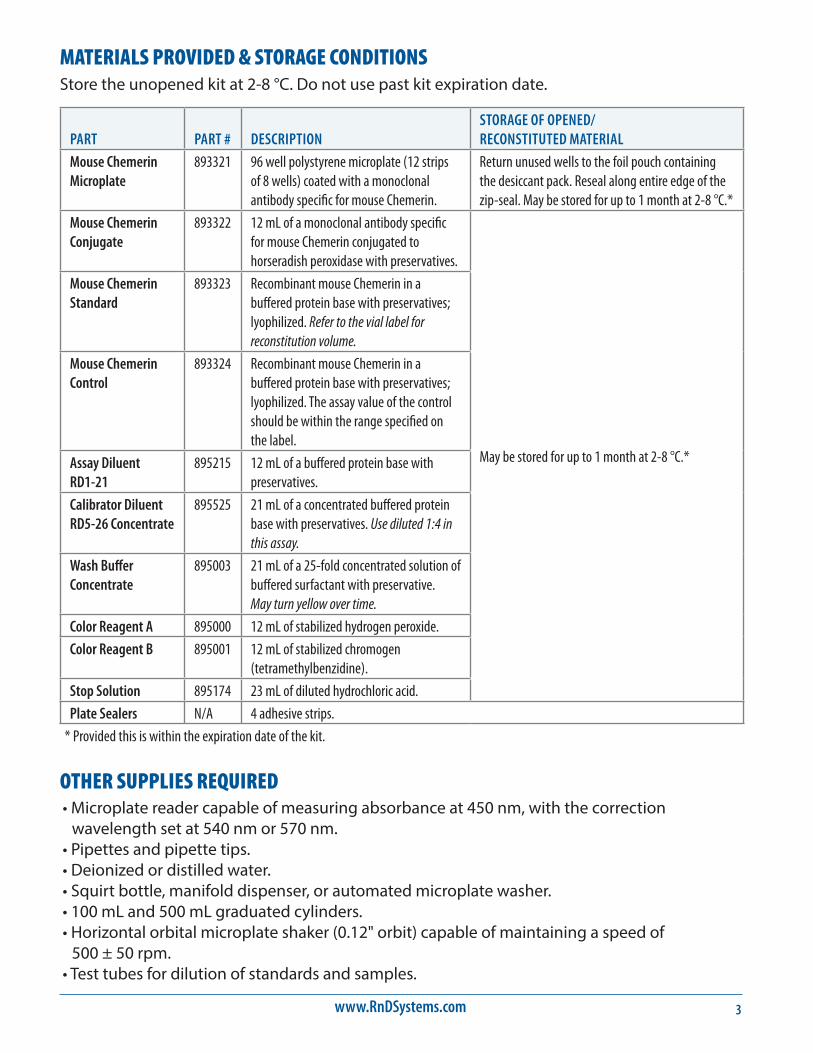
MATERIALS PROVIDED & STORAGE CONDITIONSStore the unopened kit at 2-8 °C. Do not use past kit expiration date.
PART PART # DESCRIPTIONSTORAGE OF OPENED/ RECONSTITUTED MATERIAL
Mouse Chemerin Microplate
893321 96 well polystyrene microplate (12 strips of 8 wells) coated with a monoclonal antibody specific for mouse Chemerin.
Return unused wells to the foil pouch containing the desiccant pack. Reseal along entire edge of the zip-seal. May be stored for up to 1 month at 2-8 °C.*
Mouse Chemerin Conjugate
893322 12 mL of a monoclonal antibody specific for mouse Chemerin conjugated to horseradish peroxidase with preservatives.
May be stored for up to 1 month at 2-8 °C.*
Mouse Chemerin Standard
893323 Recombinant mouse Chemerin in a buffered protein base with preservatives; lyophilized. Refer to the vial label for reconstitution volume.
Mouse Chemerin Control
893324 Recombinant mouse Chemerin in a buffered protein base with preservatives; lyophilized. The assay value of the control should be within the range specified on the label.
Assay Diluent RD1-21
895215 12 mL of a buffered protein base with preservatives.
Calibrator Diluent RD5-26 Concentrate
895525 21 mL of a concentrated buffered protein base with preservatives. Use diluted 1:4 in this assay.
Wash Buffer Concentrate
895003 21 mL of a 25-fold concentrated solution of buffered surfactant with preservative. May turn yellow over time.
Color Reagent A 895000 12 mL of stabilized hydrogen peroxide.
Color Reagent B 895001 12 mL of stabilized chromogen (tetramethylbenzidine).
Stop Solution 895174 23 mL of diluted hydrochloric acid.
Plate Sealers N/A 4 adhesive strips.
* Provided this is within the expiration date of the kit.
OTHER SUPPLIES REQUIRED• Microplate reader capable of measuring absorbance at 450 nm, with the correction
wavelength set at 540 nm or 570 nm.• Pipettes and pipette tips.• Deionized or distilled water.• Squirt bottle, manifold dispenser, or automated microplate washer.• 100 mL and 500 mL graduated cylinders.• Horizontal orbital microplate shaker (0.12" orbit) capable of maintaining a speed of
500 ± 50 rpm.• Test tubes for dilution of standards and samples.
For research use only. Not for use in diagnostic procedures.4
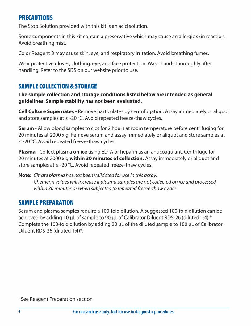
PRECAUTIONSThe Stop Solution provided with this kit is an acid solution.
Some components in this kit contain a preservative which may cause an allergic skin reaction. Avoid breathing mist.
Color Reagent B may cause skin, eye, and respiratory irritation. Avoid breathing fumes.
Wear protective gloves, clothing, eye, and face protection. Wash hands thoroughly after handling. Refer to the SDS on our website prior to use.
SAMPLE COLLECTION & STORAGEThe sample collection and storage conditions listed below are intended as general guidelines. Sample stability has not been evaluated.
Cell Culture Supernates - Remove particulates by centrifugation. Assay immediately or aliquot and store samples at ≤ -20 °C. Avoid repeated freeze-thaw cycles.
Serum - Allow blood samples to clot for 2 hours at room temperature before centrifuging for 20 minutes at 2000 x g. Remove serum and assay immediately or aliquot and store samples at ≤ -20 °C. Avoid repeated freeze-thaw cycles.
Plasma - Collect plasma on ice using EDTA or heparin as an anticoagulant. Centrifuge for 20 minutes at 2000 x g within 30 minutes of collection. Assay immediately or aliquot and store samples at ≤ -20 °C. Avoid repeated freeze-thaw cycles.
Note: Citrate plasma has not been validated for use in this assay. Chemerin values will increase if plasma samples are not collected on ice and processed within 30 minutes or when subjected to repeated freeze-thaw cycles.
SAMPLE PREPARATIONSerum and plasma samples require a 100-fold dilution. A suggested 100-fold dilution can be achieved by adding 10 μL of sample to 90 μL of Calibrator Diluent RD5-26 (diluted 1:4).* Complete the 100-fold dilution by adding 20 μL of the diluted sample to 180 μL of Calibrator Diluent RD5-26 (diluted 1:4)*.
*See Reagent Preparation section

www.RnDSystems.com 5
REAGENT PREPARATIONBring all reagents to room temperature before use.
Mouse Chemerin Control - Reconstitute the control with 1.0 mL of deionized or distilled water. Mix thoroughly. Assay the control undiluted.
Wash Buff er - If crystals have formed in the concentrate, warm to room temperature and mix gently until the crystals have completely dissolved. Add 20 mL of Wash Buff er Concentrate to 480 mL of deionized or distilled water to prepare 500 mL of Wash Buff er.
Substrate Solution - Color Reagents A and B should be mixed together in equal volumes within 15 minutes of use. Protect from light. 100 μL of the resultant mixture is required per well.
Calibrator Diluent RD5-26 (diluted 1:4) - Add 20 mL of Calibrator Diluent RD5-26 Concentrate to 60 mL of deionized or distilled water to prepare 80 mL of Calibrator Diluent RD5-26 (diluted 1:4).
Mouse Chemerin Standard - Refer to the vial label for reconstitution volume. Reconstitute the Mouse Chemerin Standard with Calibrator Diluent RD5-26 (diluted 1:4). Do not substitute other diluents. This reconstitution produces a stock solution of 3000 pg/mL. Allow the stock solution to sit for a minimum of 5 minutes with gentle mixing prior to making dilutions.
Pipette 200 μL of Calibrator Diluent RD5-26 (diluted 1:4) into each tube. Use the stock solution to produce a dilution series (below). Mix each tube thoroughly before the next transfer. The undiluted Mouse Chemerin Standard (3000 pg/mL) serves as the high standard. Calibrator Diluent RD5-26 (diluted 1:4) serves as the zero standard (0 pg/mL).
200 µL Std.
3000 pg/mL 1500 pg/mL 750 pg/mL 375 pg/mL 188 pg/mL 93.8 pg/mL 46.9 pg/mL
200 µL 200 µL 200 µL 200 µL 200 µL

For research use only. Not for use in diagnostic procedures.6
ASSAY PROCEDURE Bring all reagents and samples to room temperature before use. It is recommended that all standards, control, and samples be assayed in duplicate.
1. Prepare all reagents, standard dilutions, control, and samples as directed in the previous sections.
2. Remove excess microplate strips from the plate frame, return them to the foil pouch containing the desiccant pack, and reseal.
3. Add 50 μL of Assay Diluent RD1-21 to each well.
4. Add 50 μL of standard, control, or sample* per well. Cover with the adhesive strip provided. Incubate for 2 hours at room temperature on a horizontal orbital microplate shaker (0.12" orbit) set at 500 ± 50 rpm.
5. Aspirate each well and wash, repeating the process four times for a total of five washes. Wash by filling each well with Wash Buffer (400 μL) using a squirt bottle, manifold dispenser, or autowasher. Complete removal of liquid at each step is essential to good performance. After the last wash, remove any remaining Wash Buffer by aspirating or decanting. Invert the plate and blot it against clean paper towels.
6. Add 100 μL of Mouse Chemerin Conjugate to each well. Cover with a new adhesive strip. Incubate for 2 hours at room temperature on the shaker.
7. Repeat the aspiration/wash as in step 5.
8. Add 100 μL of Substrate Solution to each well. Incubate for 30 minutes at room temperature on the benchtop. Protect from light.
9. Add 100 μL of Stop Solution to each well. Gently tap the plate to ensure thorough mixing.
10. Determine the optical density of each well within 30 minutes, using a microplate reader set to 450 nm. If wavelength correction is available, set to 540 nm or 570 nm. If wavelength correction is not available, subtract readings at 540 nm or 570 nm from the readings at 450 nm. This subtraction will correct for optical imperfections in the plate. Readings made directly at 450 nm without correction may be higher and less accurate.
*Samples may require dilution. See the Sample Preparation section.

www.RnDSystems.com 7
CALCULATION OF RESULTSAverage the duplicate readings for each standard, control, and sample and subtract the average zero standard optical density (O.D.).
Create a standard curve by reducing the data using computer software capable of generating a four parameter logistic (4-PL) curve-fit. As an alternative, construct a standard curve by plotting the mean absorbance for each standard on the y-axis against the concentration on the x-axis and draw a best fit curve through the points on the graph. The data may be linearized by plotting the log of the mouse Chemerin concentrations versus the log of the O.D. and the best fit line can be determined by regression analysis. This procedure will produce an adequate but less precise fit of the data.
If samples have been diluted, the concentration read from the standard curve must be multiplied by the dilution factor.
TYPICAL DATAThis standard curve is provided for demonstration only. A standard curve should be generated for each set of samples assayed.
(pg/mL) O.D. Average Corrected0 0.012 0.013 —
0.01346.9 0.063 0.066 0.053
0.06993.8 0.123 0.124 0.111
0.124188 0.235 0.240 0.227
0.245375 0.460 0.468 0.455
0.475750 0.890 0.907 0.894
0.9231500 1.643 1.698 1.685
1.7523000 2.810 2.871 2.858
2.932
For research use only. Not for use in diagnostic procedures.8
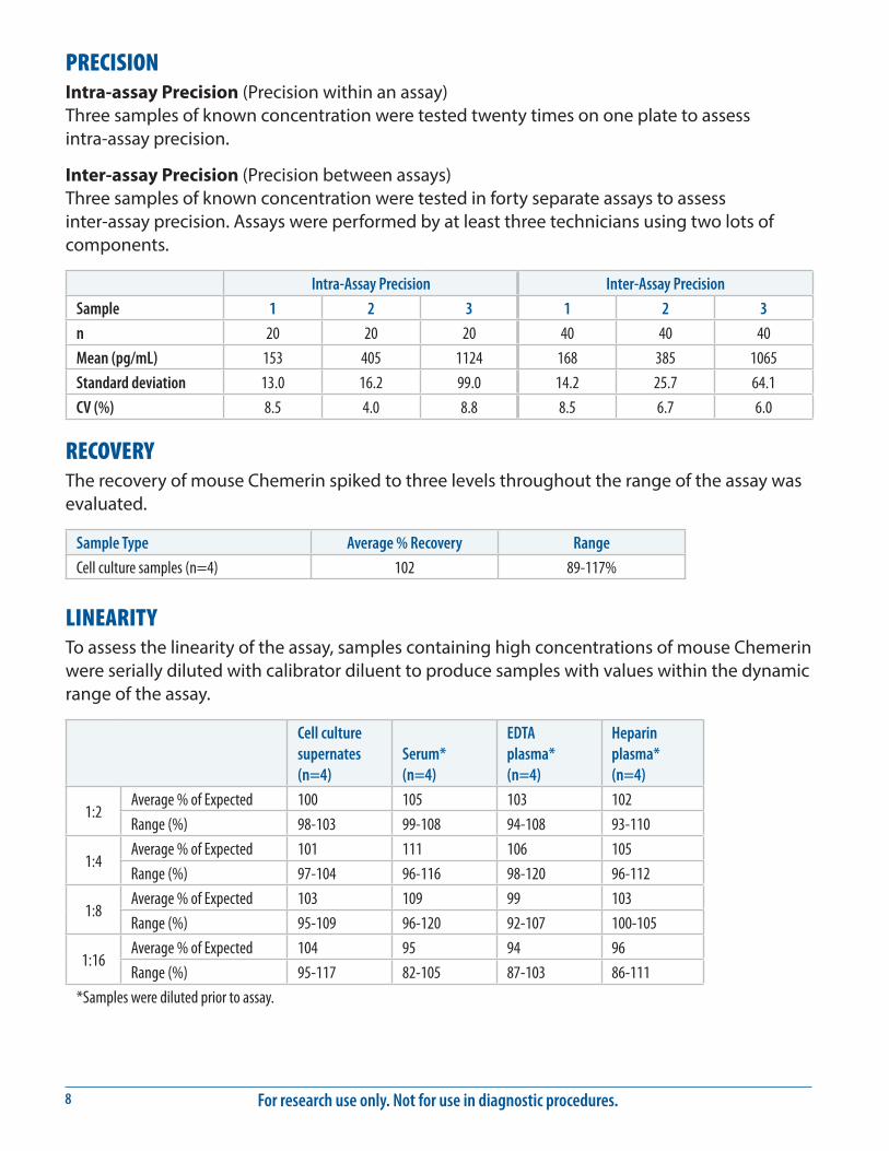
PRECISIONIntra-assay Precision (Precision within an assay) Three samples of known concentration were tested twenty times on one plate to assess intra-assay precision.
Inter-assay Precision (Precision between assays) Three samples of known concentration were tested in forty separate assays to assess inter-assay precision. Assays were performed by at least three technicians using two lots of components.
Intra-Assay Precision Inter-Assay Precision
Sample 1 2 3 1 2 3
n 20 20 20 40 40 40
Mean (pg/mL) 153 405 1124 168 385 1065
Standard deviation 13.0 16.2 99.0 14.2 25.7 64.1
CV (%) 8.5 4.0 8.8 8.5 6.7 6.0
RECOVERYThe recovery of mouse Chemerin spiked to three levels throughout the range of the assay was evaluated.
Sample Type Average % Recovery Range
Cell culture samples (n=4) 102 89-117%
LINEARITYTo assess the linearity of the assay, samples containing high concentrations of mouse Chemerin were serially diluted with calibrator diluent to produce samples with values within the dynamic range of the assay.
Cell culture supernates (n=4)
Serum* (n=4)
EDTA plasma* (n=4)
Heparin plasma* (n=4)
1:2Average % of Expected 100 105 103 102
Range (%) 98-103 99-108 94-108 93-110
1:4Average % of Expected 101 111 106 105
Range (%) 97-104 96-116 98-120 96-112
1:8Average % of Expected 103 109 99 103
Range (%) 95-109 96-120 92-107 100-105
1:16Average % of Expected 104 95 94 96
Range (%) 95-117 82-105 87-103 86-111
*Samples were diluted prior to assay.

www.RnDSystems.com 9
SENSITIVITYForty-four assays were evaluated and the minimum detectable dose (MDD) of mouse Chemerin ranged from 1.08-3.47 pg/mL. The mean MDD was 1.88 pg/mL.
The MDD was determined by adding two standard deviations to the mean O.D. value of twenty zero standard replicates and calculating the corresponding concentration.
CALIBRATIONThis immunoassay is calibrated against a highly purified E. coli-expressed recombinant mouse Chemerin produced at R&D Systems®.
SAMPLE VALUESSerum/Plasma - Samples were evaluated for the presence of mouse Chemerin in this assay.
Sample Type Mean (ng/mL) Range (ng/mL) Standard Deviation (ng/mL)
Serum (n=20) 163 99.1-247 45.6
EDTA plasma (n=20) 58.3 39.1-87.0 12.8
Heparin plasma (n=20) 62.7 49.1-83.0 12.0
Cell Culture Supernates - Tissues from two mice were homogenized and seeded into 100 mL of RPMI containing 10% fetal bovine serum, 2 mM L-glutamine, 100 U/mL penicillin, and 100 μg/mL streptomycin sulfate for 18 hours. Aliquots of the cell culture supernates were removed and assayed for levels of mouse Chemerin.
Tissue Type (pg/mL)
Lung 5014
Kidney 1567
Brain 194
For research use only. Not for use in diagnostic procedures.10
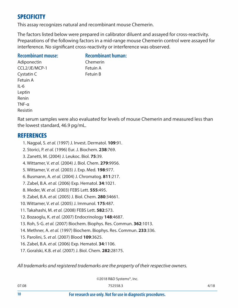
SPECIFICITYThis assay recognizes natural and recombinant mouse Chemerin.
The factors listed below were prepared in calibrator diluent and assayed for cross-reactivity. Preparations of the following factors in a mid-range mouse Chemerin control were assayed for interference. No significant cross-reactivity or interference was observed.
Recombinant mouse:AdiponectinCCL2/JE/MCP-1Cystatin CFetuin AIL-6LeptinReninTNF-αResistin
Recombinant human:ChemerinFetuin AFetuin B
REFERENCES1. Nagpal, S. et al. (1997) J. Invest. Dermatol. 109:91.2. Storici, P. et al. (1996) Eur. J. Biochem. 238:769.3. Zanetti, M. (2004) J. Leukoc. Biol. 75:39.4. Wittamer, V. et al. (2004) J. Biol. Chem. 279:9956.5. Wittamer, V. et al. (2003) J. Exp. Med. 198:977.6. Busmann, A. et al. (2004) J. Chromatog. 811:217.7. Zabel, B.A. et al. (2006) Exp. Hematol. 34:1021.8. Meder, W. et al. (2003) FEBS Lett. 555:495.9. Zabel, B.A. et al. (2005) J. Biol. Chem. 280:34661.
10. Wittamer, V. et al. (2005) J. Immunol. 175:487.11. Takahashi, M. et al. (2008) FEBS Lett. 582:573.12. Bozaoglu, K. et al. (2007) Endocrinology 148:4687.13. Roh, S-G. et al. (2007) Biochem. Biophys. Res. Commun. 362:1013.14. Methner, A. et al. (1997) Biochem. Biophys. Res. Commun. 233:336.15. Parolini, S. et al. (2007) Blood 109:3625.16. Zabel, B.A. et al. (2006) Exp. Hematol. 34:1106.17. Goralski, K.B. et al. (2007) J. Biol. Chem. 282:28175.
07.08 752558.3 4/18
©2018 R&D Systems®, Inc.
Rat serum samples were also evaluated for levels of mouse Chemerin and measured less than the lowest standard, 46.9 pg/mL.
All trademarks and registered trademarks are the property of their respective owners.