motor system pathways for students
TRANSCRIPT
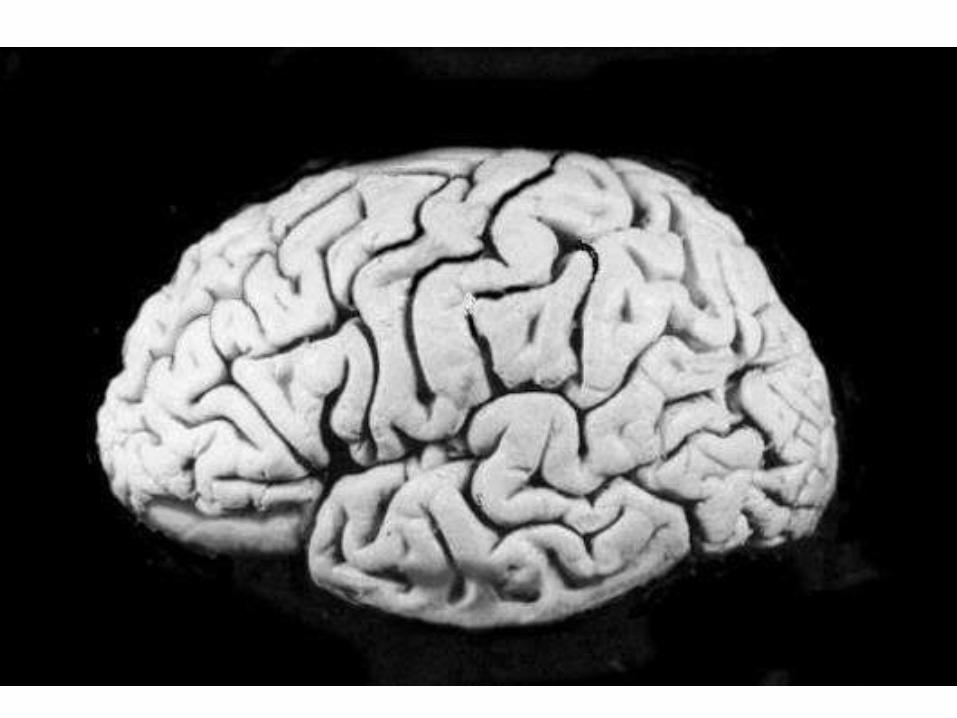
Motor System
• Starts at the motor cortex
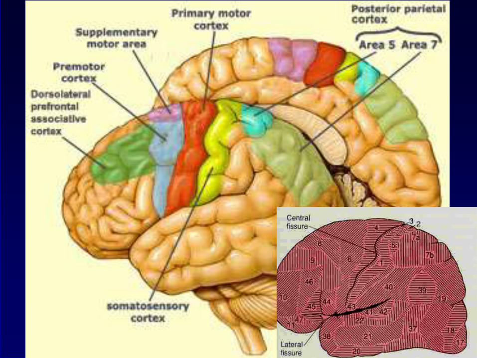
• Motor cortex is located at the frontal lobe
– precentral cortex


Motor homunculus
First discovered
by
Penfield

Brodmann areas Primary motor cortex
Area 4

Motor cortex
• different areas of the body are
represented in different cortical areas in
the motor cortex
• Motor homunculus
– somatotopic representation
– not proportionate to structures but
proportionate to function
– distorted map
– upside down map

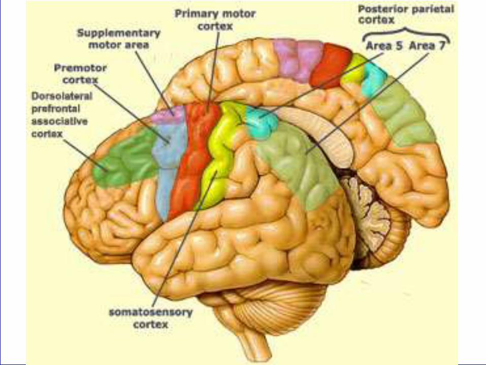
Motor cortical areas
• primary motor cortex (MI)
– precentral gyrus
• Movements are executed
• secondary motor cortex (MII)
– premotor cortex
– supplementary motor area (SMA)
• Movements are planned together with cerebellum, basal
ganglia and other cortical areas
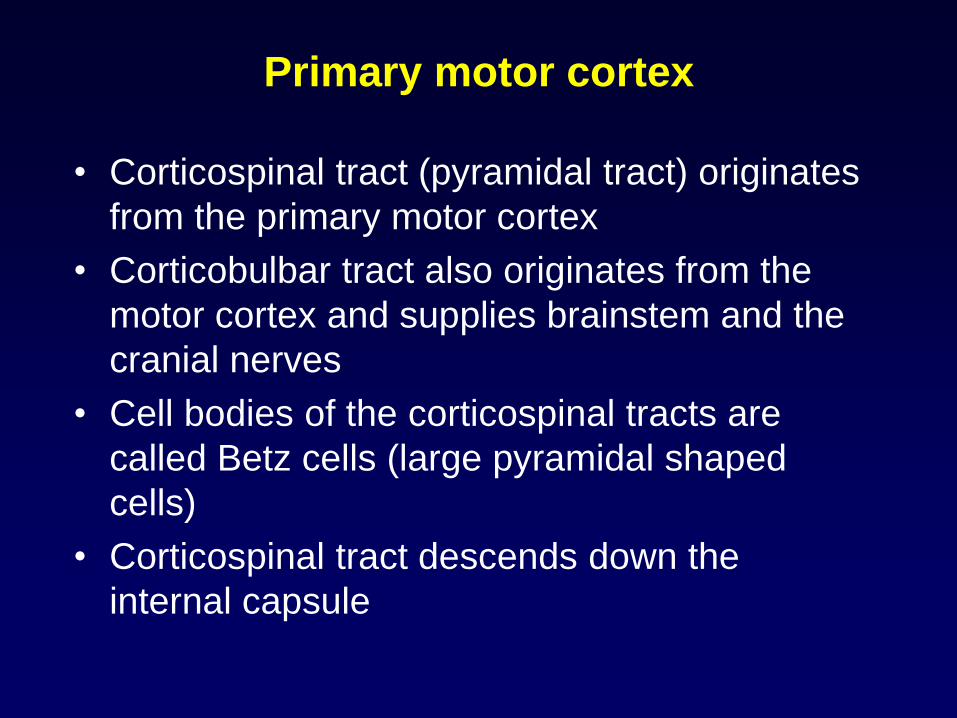
Primary motor cortex
• Corticospinal tract (pyramidal tract) originates
from the primary motor cortex
• Corticobulbar tract also originates from the
motor cortex and supplies brainstem and the
cranial nerves
• Cell bodies of the corticospinal tracts are
called Betz cells (large pyramidal shaped
cells)
• Corticospinal tract descends down the
internal capsule

Course of the corticospinal tract
• Descends through
– internal capsule
– at the medulla • cross over to the other side
• uncrossed tracts
– descends down as the corticospinal tract
– ends in each anterior horn cell
– synapse at the anterior horn cell (directly or through interneurons)

Medulla
internal capsule
Upper
motor
neuron
Lower
motor
neuron
anterior horn cell

Primary and secondary cortical
areas• Primary areas are primarily connected with the
peripheral organs/structures
– Primary motor cortex (area 4)
• Secondary areas are inter-connected to each
other by cortico-cortical pathways and perform
complex processing
– Premotor cortex (area 6)
– Supplementary motor area (superomedial part of
area 6)
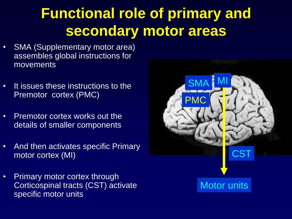
Functional role of primary and
secondary motor areas• SMA (Supplementary motor area)
assembles global instructions for movements
• It issues these instructions to the Premotor cortex (PMC)
• Premotor cortex works out the details of smaller components
• And then activates specific Primary motor cortex (MI)
• Primary motor cortex through Corticospinal tracts (CST) activate specific motor units
SMA
PMC
MI
CST
Motor units
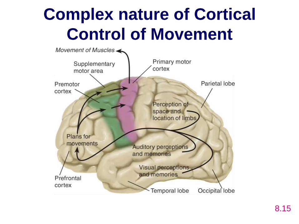
Complex nature of Cortical
Control of Movement
8.15
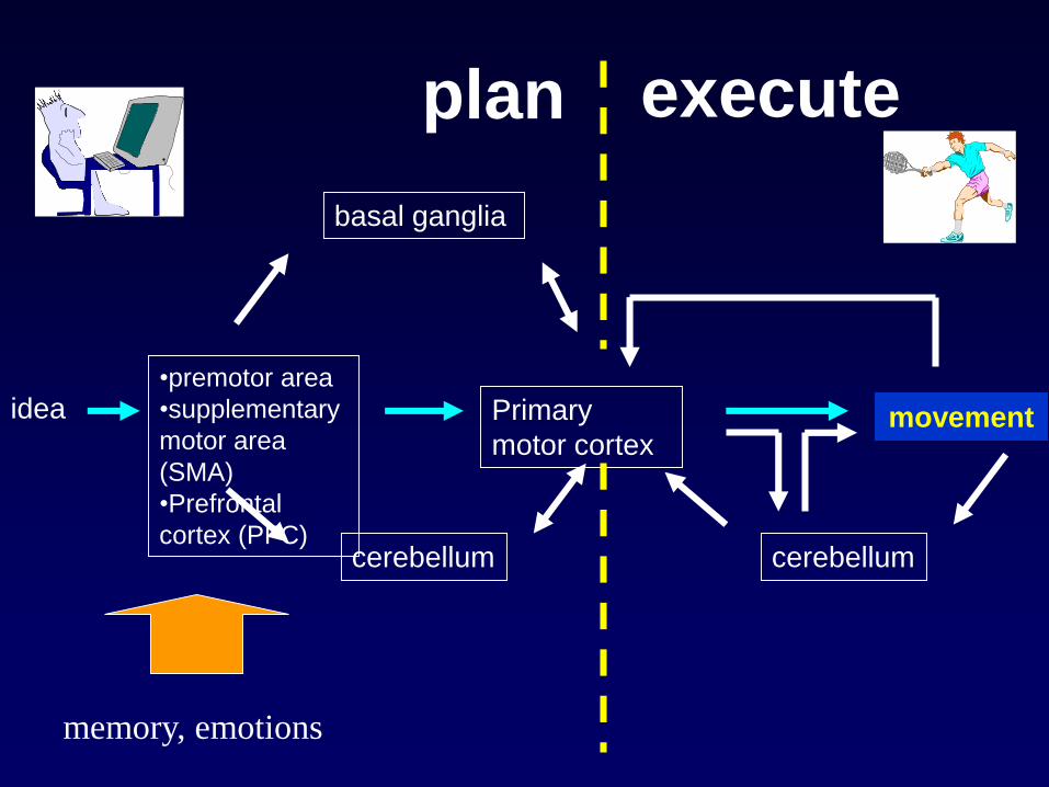
idea•premotor area
•supplementary
motor area
(SMA)
•Prefrontal
cortex (PFC)
Primary
motor cortexmovement
basal ganglia
cerebellum cerebellum
plan execute
memory, emotions

Motor system
• Consists of
– Upper motor neuron
– Lower motor neuron
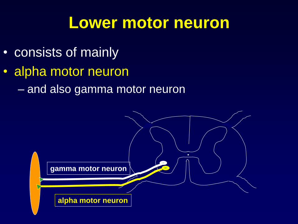
Lower motor neuron
• consists of mainly
• alpha motor neuron
– and also gamma motor neuron
alpha motor neuron
gamma motor neuron

alpha motor neuron
gamma motor neuron
corticospinal tract
Arrangement at the
anterior horn cell

alpha motor neuron
• this is also called the final common pathway
• Contraction of the muscle occurs through this
whether
– voluntary contraction through corticospinal tract
or
– involuntary contraction through gamma motor
neuron - stretch reflex - Ia afferent
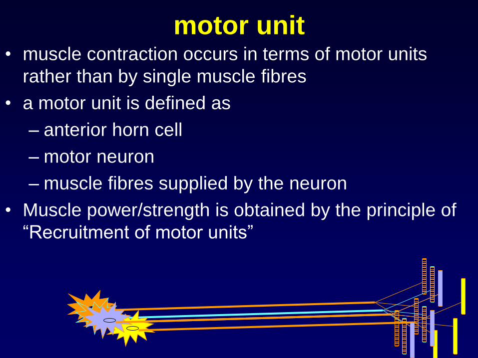
motor unit• muscle contraction occurs in terms of motor units
rather than by single muscle fibres
• a motor unit is defined as
– anterior horn cell
– motor neuron
– muscle fibres supplied by the neuron
• Muscle power/strength is obtained by the principle of
“Recruitment of motor units”

motor unit
• Innervation ratio
– motor neuron:number of muscle
fibres
• in eye muscles
– 1:23 offers a fine degree of
control
• in calf muscles
– 1:1000 more strength

Upper motor neuron
• Consists of – Corticospinal tract (pyramidal tract)
– Extrapyramidal tracts• Start from the brainstem
• Ipsilateral/contralateral
• Cortical pathways can excite/inhibit these tracts
• Modify the movement that is initiated by the CST
• Influence (+/-) gamma motor neuron, stretch reflex, muscle tone
• Important for postural control
• Cerebellar and basal ganglia influence on the lower motor neuron will be through extrapyramidal tracts
Extrapyramidal tracts
• starts at the brain stem
• descends down either ipsilaterally or
contralaterally
• ends at the anterior horn cell
• modifies the motor functions
Extrapyramidal tracts
• there are 4 tracts
– reticulospinal tracts
– vestibulospinal tracts
– rubrospinal tracts
– tectospinal tracts
reticulospinal tract
• relay station for descending motor impulses
except pyramidal tracts
• receives & modifies motor commands to the
proximal & axial muscles
• maintain normal postural tone
• excitatory to alpha & gamma motorneurons
• end on interneurons too
• this effect is inhibited by cerebral influence
• mainly ipsilateral

midbrain
pons
medulla
spinal cord
reticulospinal tract

• pontine reticular formation
– medial reticulospinal tracts
• controls proximal muscles (axial), excitatory to flexor
• medullary reticular formation
– lateral reticulospinal tracts (also medial)
• excitatory or inhibitory to axial muscles
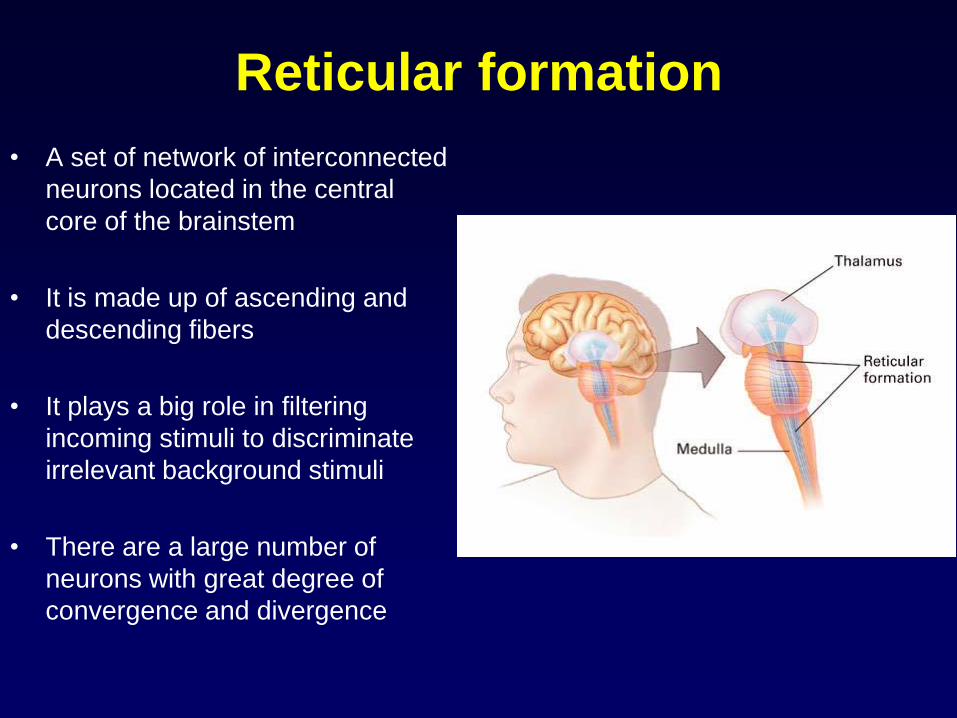
Reticular formation
• A set of network of interconnected
neurons located in the central
core of the brainstem
• It is made up of ascending and
descending fibers
• It plays a big role in filtering
incoming stimuli to discriminate
irrelevant background stimuli
• There are a large number of
neurons with great degree of
convergence and divergence
Functions
• Maintain consciousness, sleep and arousal
• Motor functions (postural and muscle tone
control)
– Reticulospinal pathways are part of the
extrapyramidal tracts
• Pain modulation (inhibition)
– Several nuclei (PAG, NRM) are part of the
descending pain modulatory (inhibitory) pathway

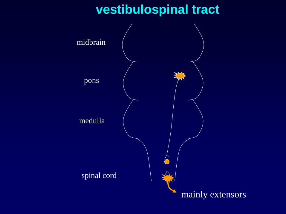
vestibular nuclei & tracts
• responsible for maintaining tone in antigravity
muscles & for coordinating the postural
adjustments in limbs & eyes
• connections with vestibular receptors (otolith
organs) & cerebellum
• mainly ipsilateral
• supplies extensors
midbrain
pons
medulla
spinal cord
vestibulospinal tract
mainly extensors
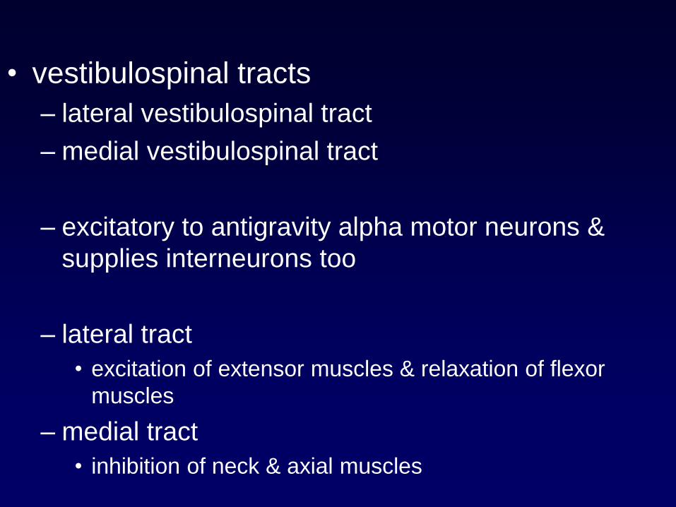
• vestibulospinal tracts
– lateral vestibulospinal tract
– medial vestibulospinal tract
– excitatory to antigravity alpha motor neurons &
supplies interneurons too
– lateral tract
• excitation of extensor muscles & relaxation of flexor
muscles
– medial tract
• inhibition of neck & axial muscles
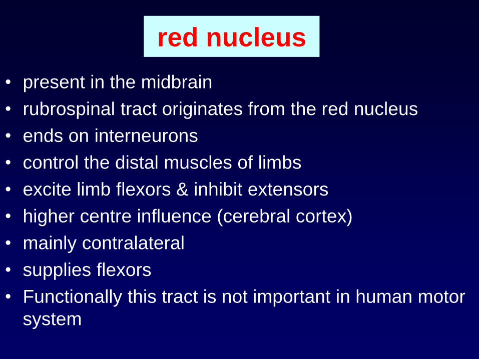
red nucleus
• present in the midbrain
• rubrospinal tract originates from the red nucleus
• ends on interneurons
• control the distal muscles of limbs
• excite limb flexors & inhibit extensors
• higher centre influence (cerebral cortex)
• mainly contralateral
• supplies flexors
• Functionally this tract is not important in human motor
system
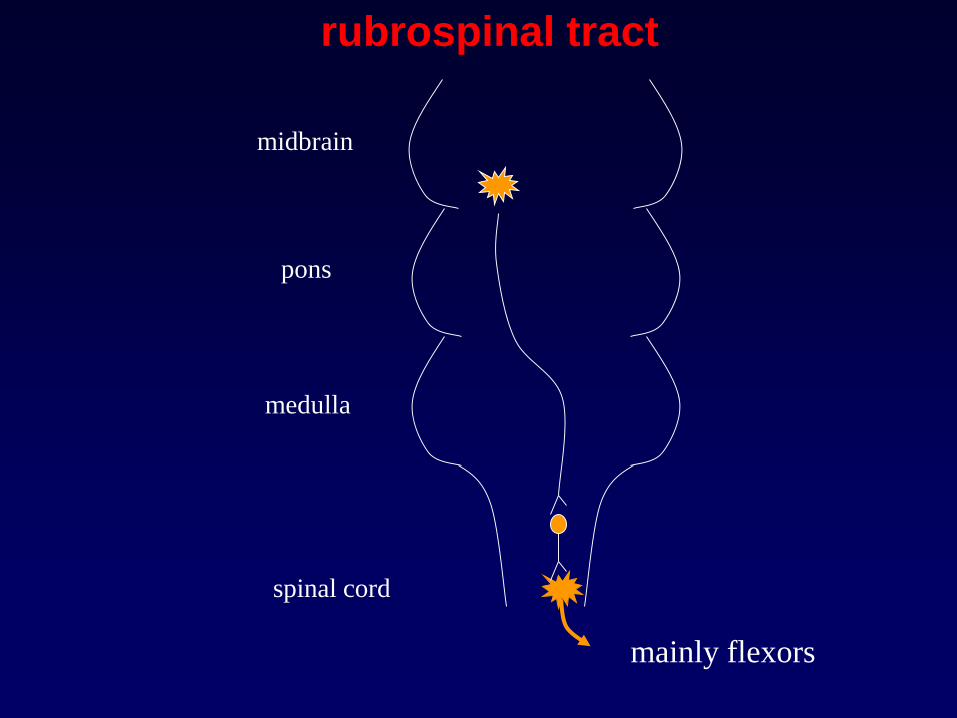
midbrain
pons
medulla
spinal cord
rubrospinal tract
mainly flexors
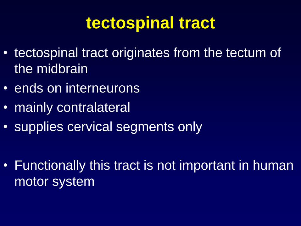
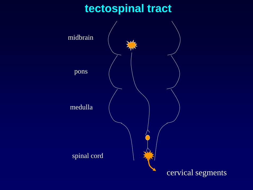
tectospinal tract
• tectospinal tract originates from the tectum of
the midbrain
• ends on interneurons
• mainly contralateral
• supplies cervical segments only
• Functionally this tract is not important in human
motor system
midbrain
pons
medulla
spinal cord
tectospinal tract
cervical segments

inferior olivary nucleus
• present in the medulla
• function:
– motor coordination
• via projections to the cerebellum
• sole source of climbing fibres to the cerebellum
– motor learning
– Functionally this nucleus is not important in human
motor system

Upper
motor
neuron
Lower
motor
neuron
extrapyramidal tracts
pyramidal tracts
alpha motor neurone
gamma motor neurone
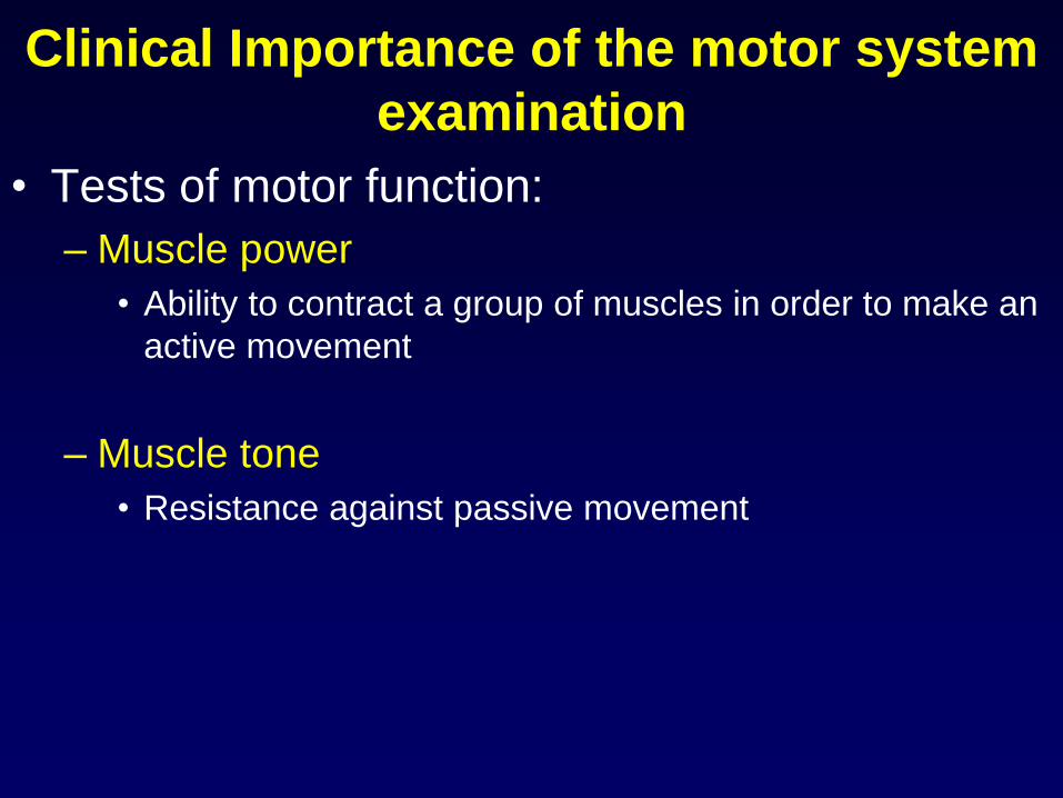
Clinical Importance of the motor system
examination
• Tests of motor function:
– Muscle power
• Ability to contract a group of muscles in order to make an
active movement
– Muscle tone
• Resistance against passive movement
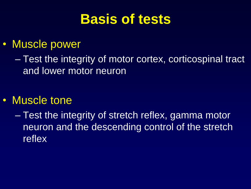
Basis of tests
• Muscle power
– Test the integrity of motor cortex, corticospinal tract
and lower motor neuron
• Muscle tone
– Test the integrity of stretch reflex, gamma motor
neuron and the descending control of the stretch
reflex

Muscle tone
• Resistance against passive movement
– Gamma motor neuron activate the spindles
– Stretching the muscle will activate the stretch reflex
– Muscle will contract involuntarily
– Gamma activity is under higher centre inhibition
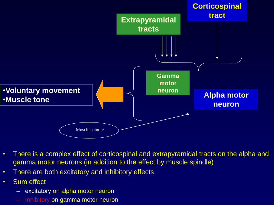
• There is a complex effect of corticospinal and extrapyramidal tracts on the alpha and
gamma motor neurons (in addition to the effect by muscle spindle)
• There are both excitatory and inhibitory effects
• Sum effect
– excitatory on alpha motor neuron
– Inhibitory on gamma motor neuron
Corticospinal
tractExtrapyramidal
tracts
Alpha motor
neuron
Gamma
motor
neuron•Voluntary movement
•Muscle tone
Muscle spindle
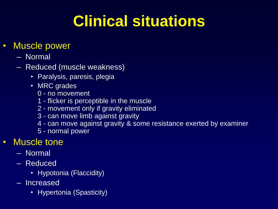
Clinical situations
• Muscle power– Normal
– Reduced (muscle weakness)
• Paralysis, paresis, plegia
• MRC grades 0 - no movement1 - flicker is perceptible in the muscle2 - movement only if gravity eliminated3 - can move limb against gravity4 - can move against gravity & some resistance exerted by examiner5 - normal power
• Muscle tone – Normal
– Reduced
• Hypotonia (Flaccidity)
– Increased
• Hypertonia (Spasticity)

Main abnormalities
• Muscle Weakness / paralysis
– Reduced muscle power
• Flaccidity
– Reduced muscle tone
• Spasticity
– Increased muscle tone

• Lower motor neuron lesion causes
– flaccid paralysis (flaccid weakness)
• Upper motor neuron lesion causes
– spastic paralysis (spastic weakness)

Lower motor neuron lesion
• muscle weakness
• flaccid paralysis
• muscle wasting (disuse atrophy)
• reduced muscle tone (hypotonia)
• reflexes: reduced or absent (hyporeflexia or areflexia)
• spontaneous muscle contractions (fasciculations)
• plantar reflex: flexor
• superficial abdominal reflexes: present
• eg. Brachial plexus damage
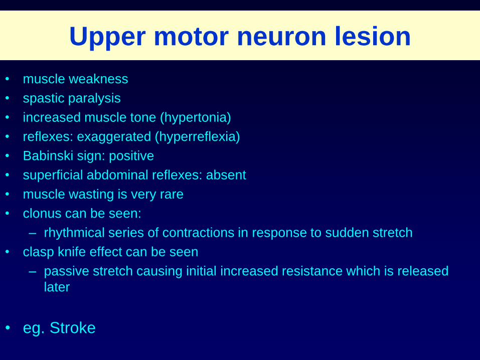
Upper motor neuron lesion
• muscle weakness
• spastic paralysis
• increased muscle tone (hypertonia)
• reflexes: exaggerated (hyperreflexia)
• Babinski sign: positive
• superficial abdominal reflexes: absent
• muscle wasting is very rare
• clonus can be seen:
– rhythmical series of contractions in response to sudden stretch
• clasp knife effect can be seen
– passive stretch causing initial increased resistance which is released
later
• eg. Stroke

Babinski sign• when outer border of the sole of the foot is
scratched
• upward movement of big toe
• fanning out of other toes
• feature of upper motor neuron lesion
• extensor plantar reflex
• seen in infants during 1st year of life (because
of immature corticospinal tract)
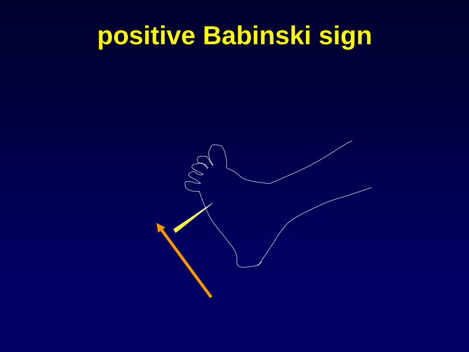
positive Babinski sign

• Observation
• When the spinal cord is suddenly transected, essentially all
cord functions, including spinal cord reflexes, immediately
become depressed
• This is called “spinal shock”
• Period of spinal shock is about 2 weeks in humans
• It may vary depending on the level spinal cord injury
• Higher the animal in evolution greater is the spinal shock
period
Spinal cord transection and spinal shock
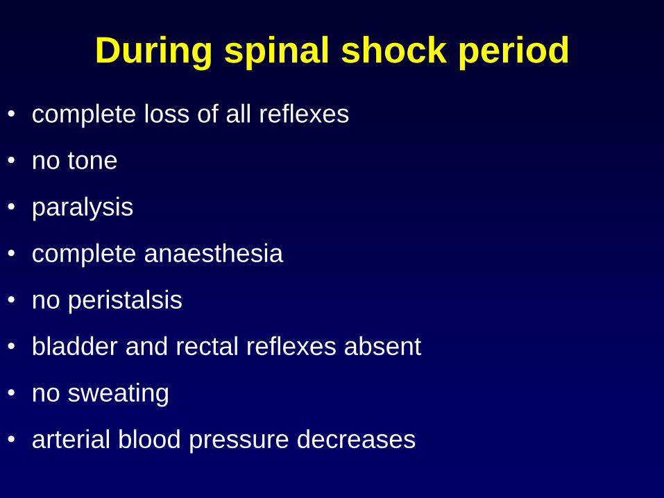
During spinal shock period
• complete loss of all reflexes
• no tone
• paralysis
• complete anaesthesia
• no peristalsis
• bladder and rectal reflexes absent
• no sweating
• arterial blood pressure decreases

Possible mechanism of spinal shock
• Normal activity of the spinal cord reflexes depends to a great
extent on continual tonic excitation from higher centers
(pyramidal and extrapyramidal tracts)
• Spinal shock may be due to the sudden cessation of tonic
bombardment of spinal cord interneuron pool by descending
influences
• During recovery from spinal shock, the excitability of spinal cord
reflexes increase due to the lack of descending inhibition and
possible denervation hypersensitivity
• After the spinal shock period typical upper motor neuron
features appear

after the spinal shock
• reflexes will reappear, mostly exaggerated
• bladder become reflex
• mass reflex will appear
– afferent stimuli irradiate to several reflex centres
– noxious stimulus causes: withdrawal response,
evacuation of bladder, rectum, sweating, pallor
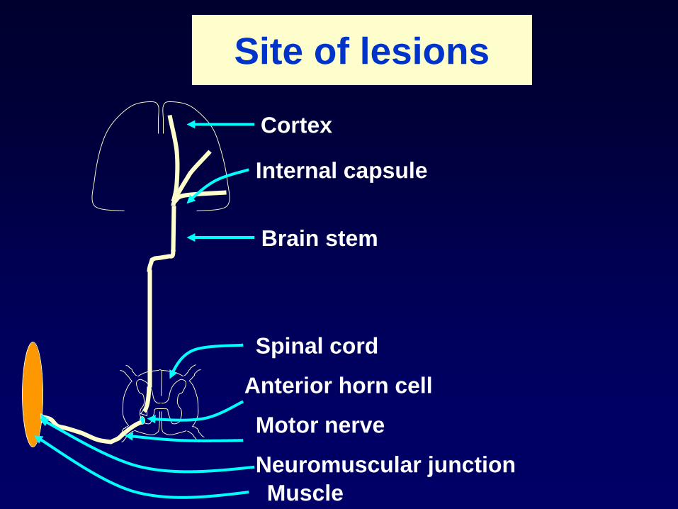
Site of lesions
Cortex
Internal capsule
Brain stem
Spinal cord
Anterior horn cell
Motor nerve
Neuromuscular junction
Muscle
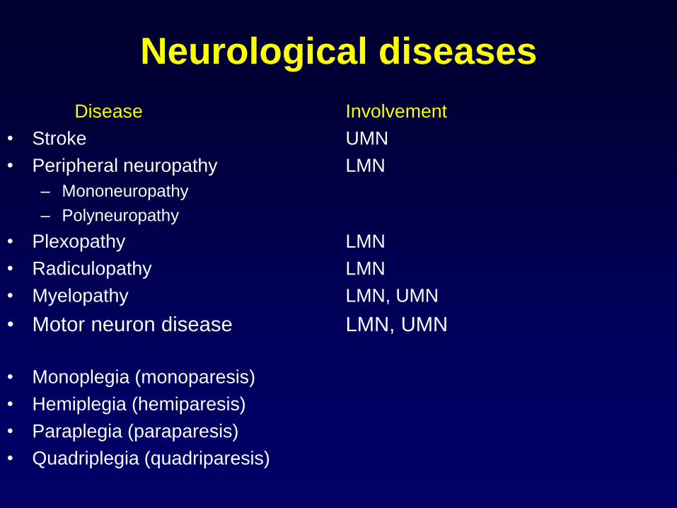
Neurological diseases
Disease Involvement
• Stroke UMN
• Peripheral neuropathy LMN
– Mononeuropathy
– Polyneuropathy
• Plexopathy LMN
• Radiculopathy LMN
• Myelopathy LMN, UMN
• Motor neuron disease LMN, UMN
• Monoplegia (monoparesis)
• Hemiplegia (hemiparesis)
• Paraplegia (paraparesis)
• Quadriplegia (quadriparesis)

Site of lesions
monoplegia
only 1 limb is affected either UL or LL,
lower motor neuron lesion
hemiplegia
one half of the body including
UL and LL
lesion in the Internal capsuleparaplegia
both lower limbs
thoracic cord lesion
quadriplegia (tetraplegia)
all 4 limbs are affected
cervical cord or brain stem lesion

Some common neurological
diseases

Stroke
• Cerebrovascular accident (CVA)
• A serious neurological disease
• Large number of deaths per year
• Cerebrovascular ischaemia causing
infarction or haemorrhage
• Sudden onset hemiplegia
• Hypertension, diabetes, obesity are
risk factors

Peripheral neuropathies
• Mononeuropathies
– Carpal tunnel syndrome (CTS)
– Ulnar neuropathy - claw hand
– Saturday night palsy (radial nerve lesion) – wrist drop
– Common peroneal nerve lesion – foot drop
– Posterior tibial nerve lesion – tarsal tunnel syndrome
– Sciatic nerve lesion
– Facial nerve lesion – Bell’s palsy
• Polyneuropathies
– Diabetic, vitamin deficiency, toxic

Median nerve compression
(Carpal tunnel syndrome)

Ulnar nerve lesion
(Ulnar tunnel syndrome)
Clawing of the hand
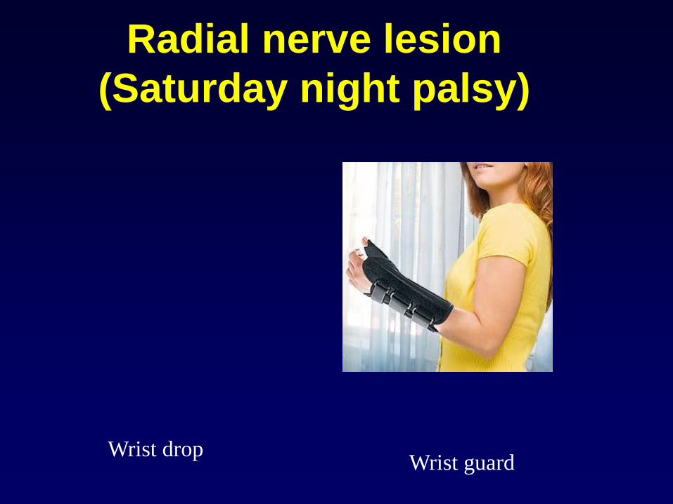
Radial nerve lesion
(Saturday night palsy)
Wrist drop Wrist guard
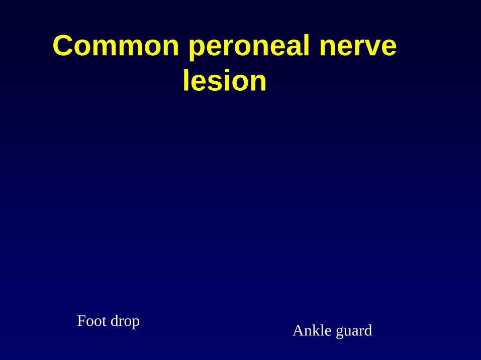
Common peroneal nerve
lesion
Foot drop Ankle guard
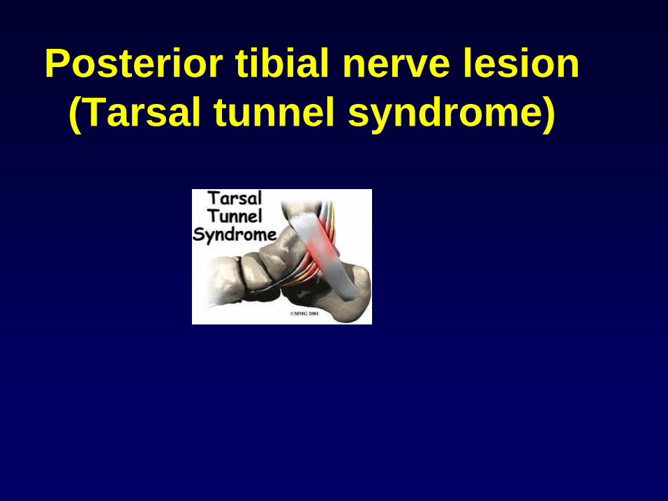
Posterior tibial nerve lesion
(Tarsal tunnel syndrome)
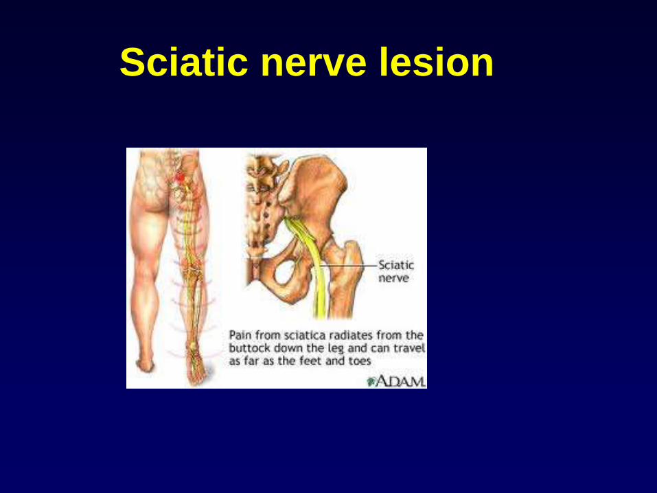
Sciatic nerve lesion
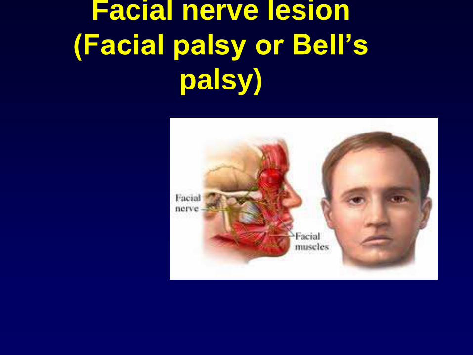
Facial nerve lesion
(Facial palsy or Bell’s
palsy)
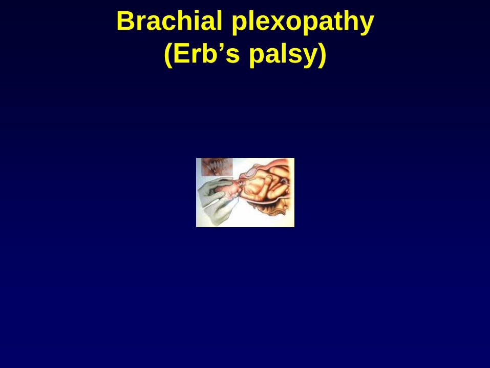
Brachial plexopathy
(Erb’s palsy)
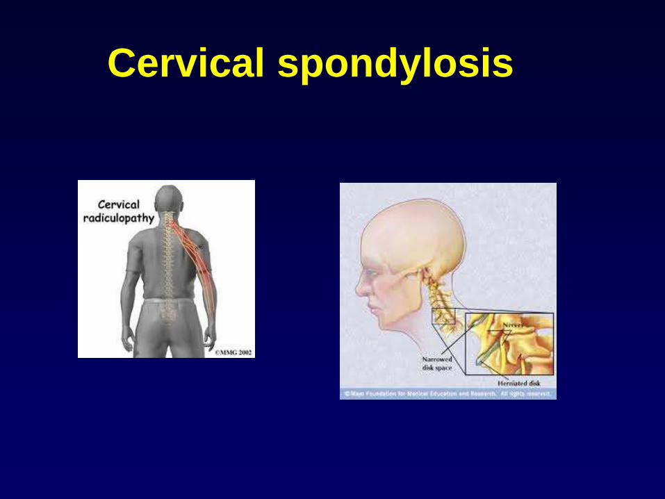
Cervical spondylosis
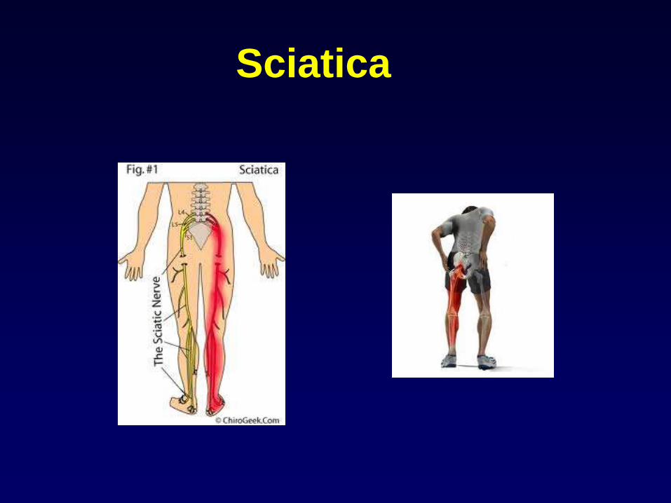
Sciatica

Cervical or thoracic
myelopathy
Paraplegia
Quadriplegia

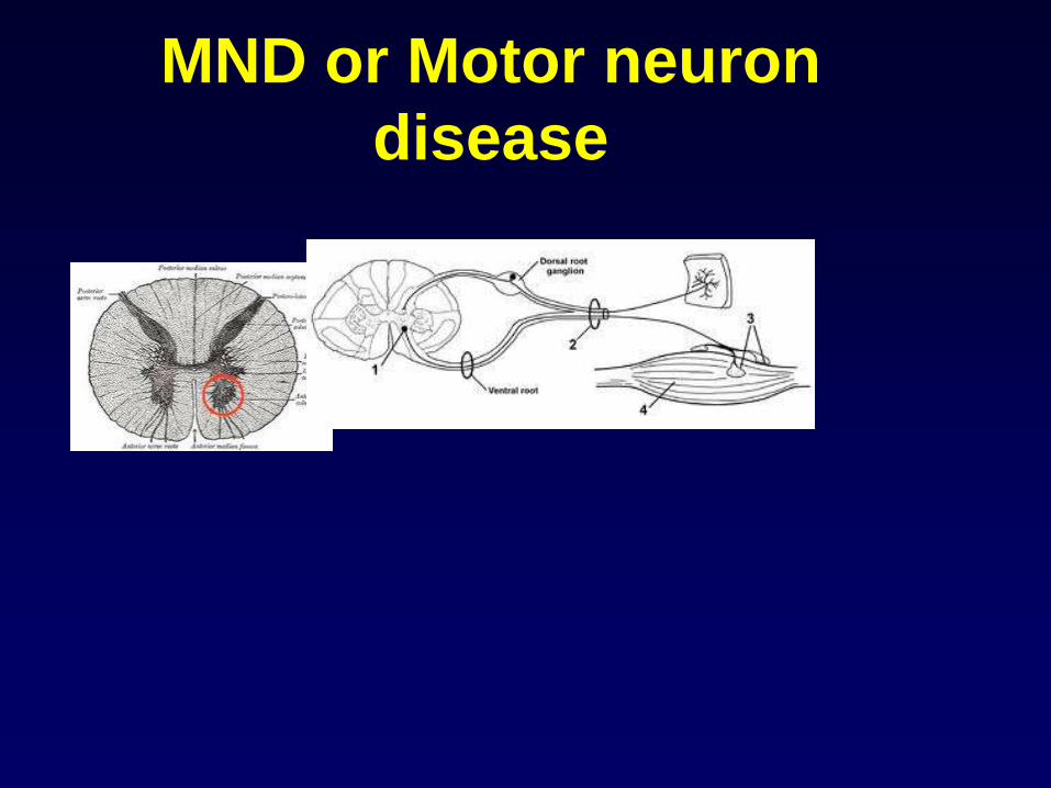
MND or Motor neuron
disease
• Anterior horn cell disease
• MND: motor neuron disease
• ALS: Amyotrophic lateral sclerosis
• Weakness of lower limbs, upper limbs
• Speech defect: dysarthria
• Difficulty in swallowing: dysphagia
MND or Motor neuron
disease