morphology and taxonomy of the japanese rhodymeniales (1 ... · were stained with 1 erythrosin or...
TRANSCRIPT
—11—
Lomentaria lubrica (Yendo) Yamada, anendemic species distributed in northernHonshu, Japan, was first described by Yendo(1920) as Chylocladia lubrica from Oma,Aomori Prefecture. Yamada (1932) latertransferred it from Chylocladia toLomentaria on noting the absence of adiaphragm (transverse septum) andtetrasporangia protruding into the cavity.Kawashima (1960) observed the thallusstructure and tetrasporangia formation. Themale gametophytes and carposporophyte for-mations are still unknown. Recently, we col-lected L. lubrica including male and female
gametophytes and tetrasporophytes from Sai,also in Aomori Prefecture, and describe herethe detailed morphological features. Thefindings allow further examination of thetaxonomic position of L. lubrica.
Materials and MethodsUsed specimens were collected at Sai,
Shimokita Co., Aomori Prefecture (41°27’N,141°52’E) on 4 August 2005 (Fig. 1).Specimens were preserved in 10� formalin/seawater. Voucher herbarium specimens aredeposited at TNS. The specimens were sec-tioned by a freezing microtome, and sections
植物研究雑誌J. Jpn. Bot.83: 11–19 (2008)
Morphology and Taxonomy of the Japanese Rhodymeniales (1)Thallus Structure and Reproductive Organs of Lomentaria lubrica
(Lomentariaceae, Rhodophyta)
Masahiro SUZUKI and Makoto YOSHIZAKI
The Graduate School of Science, Toho University,2–2–1, Miyama, Funabashi, 274-8510 JAPAN
E-mail: [email protected]
(Received on July 24, 2007)
Detailed morphological studies have been carried out on the vegetative thallus andreproductive organs of Lomentaria lubrica collected from Sai, Aomori Prefecture, Japan.This is the first report of male gametophytes and carposporophyte formation in the spe-cies. The thallus structure is multiaxial, hollow, and is composed of a one-layered cortexand a one or two-layered medulla surrounding the central cavity. The transverse septumoccurs only at the base of the branch or branchlet. The cortical cells cut off small cellsaround them. The medullary cells longitudinally border the cortical cells and solely bearsecretory cells. The procarp is composed of a three-celled carpogonial branch and a two-celled auxiliary cell branch. After fertilization, the cells of the carpogonial branch arefused. The fused carpogonial branch directly touches the auxiliary cell. The primarygonimoblast is cut off transversely from the auxiliary cell. Gonimoblasts develop out-wards from large fusion cells. Most gonimoblasts transform into carposporangia. Thecarposporophyte is covered with pericarp. The mature cystocarp is urceolate and has anostiole. Spermatangia are cut off from mother cells produced on the cortical cells.Tetrasporangia are terminally produced on the cortical cells, tetrahedrally divided, andformed in the depressed sori.
Key words: Carposporophyte formation, Lomentaria lubrica, morphology, Rhodophyta,Rhodymeniales.

were stained with 1�erythrosin or 0.5�cot-ton blue, and mounted in 50� Karo syrup.Drawings were made with a camera lucida.
ResultsLomentaria lubrica (Yendo) Yamada in
J. Fac. Sci., Hokkaido Imper. Univ., Ser. V(Bot.) 1 (3): 121 (1932).
Japanese name: Ito-taoyagisô.Type locality: Oma, Shimokita Co.,
Aomori Prefecture, Japan.Distribution: Mutsu Bay (Yamada 1928);
Tsugaru Strait (Takamatsu 1938); Sai,Aomori Prefecture (Kawashima 1960).
Specimen examined: Sai (41°27’N, 141°52’E),Shimokita Co., Aomori Prefecture, Japan. (4 August
植物研究雑誌 第83巻 第1号 平成20年2月12
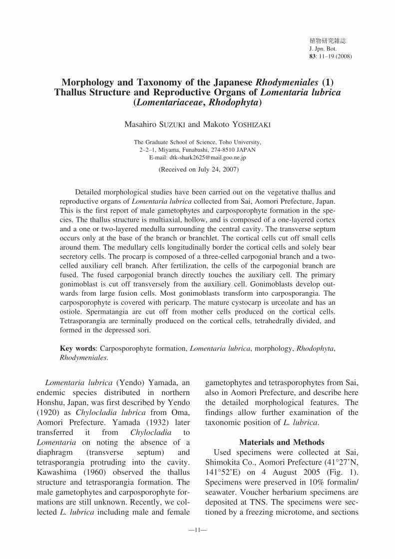
Fig. 1. Voucher specimen of Lomentaria lubrica. (TNS-AL-161634, August4, 2005, Sai, Shimokita Co., Aomori Pref.). Scale = 10 cm.

2005, M. Suzuki, cystocarpic TNS-AL-161633,spermatangial TNS-AL-161635, tetrasporangial TNS-AL-161634).
External morphology (Figs. 1–3)The plants grow on rocks or on the thallus
of Sargassum spp. or Laurencia spp. at-tached by small discoid holdfast, 0.5 to 1mm in diameter. Usually, two or three plantsgrow caespitose, forming tufts. The thallus iserect, flaccid, filiform, mucilaginous, light topale red or greenish red, to 20 cm in height,much branched dichotomously, with or with-out percurrent axes. The short branchletsalternately or oppositely produced at theupper parts giving an irregularly branching
appearance. The branches are cylindrical,slender, unconstricted, 1.0 to 1.5 mm in di-ameter. The branchlets terete, rarely con-stricted at the base, 2.0 to 6.0 mm in length,0.2 to 0.5 mm in diameter. The specimenfirmly adhered to paper on drying.
Thallus structure (Figs. 4–7)Thallus structure multiaxial with a cluster
of apical cells (Fig. 4), hollow, composed ofone-layered cortex and one or two-layeredmedulla surrounding the central cavity (Figs.5, 6). The transverse septum occurs only atthe base of the branch or branchlet. At theupper parts of the branches or branchlets, thecortical cells are rectangular or polygonal,
Journal of Japanese Botany Vol. 83 No. 1February 2008 13
Fig. 2–3. Habit of Lomentaria lubrica. Fig. 2. Cystocarpic plant bearing cystocarps (arrows). Fig. 3.Tetrasporangial plant bearing tetrasporangial sori (arrows). Scale = 5 mm.

植物研究雑誌 第83巻 第1号 平成20年2月14
Fig. 4–7. Thallus structure of Lomentaria lubrica. Fig. 4. Longitudinal section of apical partof thallus. Fig. 5. Longitudinal section of middle part of thallus. Fig. 6. Transverse sec-tion of middle part of thallus. Fig. 7. Surface view of middle part of thallus. The smallcells (arrows) around cortical cells forming rosettes like appearance. Scale = 50 �m. a:Apical cell. h: Hair cell. s: Secretory cell.

uniform and regularly arranged, 5.5 to 10 �min length, 2.5 to 6.0 �m in width, 10 to 20 �m in thickness, whereas at the middle tolower parts of the branches, they are round topolygonal, irregularly arranged, 15 to 33 �min length, 10 to 28 �m in width, 15 to 27 �min thickness, and cut off the small cellsaround them. The small cells are round, 5.0to 11 �m in diameter, and often cut off aunicellular hair (Fig. 6). In surface view, thecortical cells are irregularly and loosely ar-ranged, underlying the medullary cells werevisible through the cortex; the small cellsaround cortical cells form rosette-like ap-pearance (Fig. 7). The medullary cells areelongate rectangular, longitudinally borderthe cortical cells, 17 to 57 �m in length, 8 to30 �m in width, 4 to 18 �m in thickness,solely bear secretory cells (gland cells). Thesecretory cells are round to pyriform, 10 to13 �m in length, 8 to 11 �m in diameter.
Life historyThe gametophytes are dioecious: The
tetrasporophyte and gametophyte are uni-form. Therefore, the life history is isomor-phic(Polysiphonia type).
Female gametophyte (Figs. 2, 8–13)The cystocarps protrude from the surface
of the thallus, sessile, developed solely, scat-tered on the upper part of branches orbranchlets of the female gametophyte (Fig.2).
The procarp is composed of a three-celledcarpogonial branch and a two-celled auxil-iary cell branch (Fig. 8). The supporting celluniform with a small cell cuts off fromcortical cell, 10 to 11 �m in length, 7.5 to 13�m in diameter. The first and second cell ofcarpogonial branch are cubic, 5.0 to 6.0 �min length, 4.0 to 7.0 �m in diameter. Thecarpogonium is conical, 6.0 to 7.5 �m inlength, 2.0 to 2.5 �m in diameter at thebase, with a trichogyne. The trichogynefilamentous, protruded from the terminal of
carpogonia. The auxiliary mother cell is ob-long, 9.5 �m in length, 6.0 �m in diameter.The auxiliary cell is hemispherical, 4.0 �min length, 7.0 �m in diameter.
After fertilization, the supporting cell,auxiliary mother cell and auxiliary cell areenlarged, and the cells of the carpogonialbranch are fused. The fused carpogonialbranch directly touches the auxiliary cell(Fig. 9). The primary gonimoblast cuts offtransversely from the auxiliary cell. After thediploid nucleus migrates into the auxiliarycell, the carpogonial branch is fused with thesupporting cell, and forms a column-likefusion cell (Fig. 10). The auxiliary mothercell begins to fuse with the auxiliary cell andsome vegetative cells, and form a largetrunk-like fusion cell. The column-likefusion cell reduces in its contents, while thetrunk-like fusion cell becomes enlarged(Figs. 11, 12).
The gonimoblasts develop outwards fromthe trunk-like fusion cell, and form a globosecarposporophyte (Figs. 11, 12). As thecarposporophyte grows, the gonimoblasts areenlarged. Most gonimoblasts transform intocarposporangia (Fig. 13). The maturecarposporangium is round to polygonal, 46to 65 �m in length, 38 to 54 �m in diameter.
The carposporophyte is covered withpericarp. After fertilization, the cortical cellsaround the procarp begin to divide, and elon-gate outwards. The mature pericarp is com-posed of an outer layer and an inner layer.The inner layer is composed of one to twocells that are narrow elliptical or rectangular,not form stellate cells (Fig. 13).
The mature cystocarp is urceolate, and hasan ostiole, 0.6 to 1.0 mm in length, 0.5 to 0.6mm in diameter (Fig. 13).
Male gametophyte (Fig. 14)Spermatangia develop in sori over the
surface of upper parts of branches andbranchlets of male gametophyte. The corticalcells abcise two to four spermatangial
Journal of Japanese Botany Vol. 83 No. 1February 2008 15

植物研究雑誌 第83巻 第1号 平成20年2月16
Fig. 8–13. Carposporophyte formation of Lomentaria lubrica. Fig. 8. Procarp. Fig. 9. After fertiliza-tion. The fused carpogonial branch contacts directly with the auxiliary cell (arrowhead). Fig. 10.Auxiliary cell cut off a primary gonimoblast. Fig. 11, 12. Development of carposporophyte. Fig. 13.Mature cystocarp. Scale = 10 �m (Figs. 8–11); 50 �m (Figs. 12, 13). am: Auxiliary mother cell. au:Auxiliary cell. c: Cells of a carpogonial branch. ca: Carpogonium. cf: Column-like fusion cell. cs:Carposporangium. fc: Fused carpogonial branch. g: Gonimoblast. o: Ostiole. p: Pericarp. pg:Primary gonimoblast. sp: Spermatium. su: Supporting cell. t: Trichogyne. tf: Trunk-like fusion cell.

Journal of Japanese Botany Vol. 83 No. 1February 2008 17
Fig. 14–15. Transverse section of male gametophyte and tetrasporophyte of Lomentaria lubrica. Fig.14. Transverse section of spermatangial sori. Scale = 10 �m. s: Secretory cell. sm: Spermatangialmother cell. spt: Spermatangium. Fig. 15. Transverse section of tetrasporangial sori. Scale = 50�m. tp: Tetrasporangium.

mother cells around their upper corners.These mother cells cut off laterally oneto two secondary mother cells. Thespermatangia develop terminally on thesemother cells by centripetal constriction.After the liberation of primary sperma-tangium, the mother cell give rise tosecondary spermatangium. These secondaryspermatangia are often produced while theprimary ones still remain. The primarymother cells rarely have plastids, and areelongate rectangular or lanceolate with around apex, often slackly constricted at thecenter, 15 to 24 �m in length, 1.5 to 4.0 �min diameter. The secondary mother cell issmaller than the primary one, 7.5 to 16 �m inlength, 2.0 to 3.5 �m in diameter. Themature spermatangium and spermatium haveno plastids, and is elliptical or orbicular, 3.0to 5.0 �m in length, 3.0 to 4.0 �m in diame-ter.
Tetrasporophyte (Figs. 3, 15)Tetrasporangial sori are produced on the
upper part of branches and branchlets (Fig.3). The tetrasporangia are terminally pro-duced on cortical cells, tetrahedrally divided,formed in the depressed sori. The maturetetrasporangium is obovate, 50 to 55 �m inlength, 43 to 50 �m in diameter (Fig. 15).
DiscussionThe external morphology of L. lubrica
accords with Yendo (1920), Yamada (1928)and Kawashima (1960). The thallus structureand tetrasporangia formation accord withKawashima (1960). The terete, irregularlybranched and unconstricted thallus is similarto L. clavellosa (Irvine and Guiry 1983). Thethallus structure is similar to L. clavellosaand L. orcadensis (Irvine and Guiry 1983)and L. pinnata (Suzuki unpublished). Theyform the transverse septa only at the base ofbranch and branchlet. L. lubrica has smallcells cut off from cortical cells. In surfaceview, these small cells form a rosette-like
appearance. The formation of rosettes issimilar to L. australis (Womersley 1996) andL. pinnata. The one-layered cortex and oneto two-layered medulla are thinner than theother species. The development of sperma-tangium of L. lubrica accords with L.hakodatensis (Tazawa 1975, Lee 1978). Thestructure of procarp and carposporophyteformation accord with L. articulata (Bliding1928), L. catenata and L. hakodatensis (Lee1978), although the structure of the pericarpis thinner than the other species. The forma-tion of tetrasporangium accords withLomentaria species (cf. Bliding 1928, Lee1978, Irvine and Guiry 1983, Womersley1996).
The genus Lomentaria is characterizedby a hollow thallus, three-celled carpogonialbranch, mature cystocarp protruding on thethallus, tetrahedrally divided tetrasporangiaand tetrasporangia formed in depressed sori(Irvine and Guiry 1983, Womersley 1996and Yoshida 1998). Lomentaria lubrica sat-isfies the above characteristics. However, L.articulata the type species of the genusLomentaria and some other species such asL. catenata and L. hakodatensis have trans-verse septa constricting the medullary cavityat regular intervals. Others, such as L.clavellosa, L. orcadensis and L. pinnata havesepta occurring only at the base of branchesand branchlets, where the thallus has anunconstricted appearance. Guiry in Irvineand Guiry (1983) suggested removal of thislatter series of species to a separate genus,Chondrothamnion Kützing (1843). In accor-dance with Guiry’s opinion, L. lubricashould also be transferred to genusChondrothamnion. Further study is requiredand should include L. articulata and L.clavellosa, which is the type species of genusChondrothamnion.
We wish to express our gratitude to Mr.Masakazu Hirai and Ms. Hisako Hirai fortheir help in sampling at Institute of Hirai
植物研究雑誌 第83巻 第1号 平成20年2月18

Plant and Education. We are grateful to Ms.D. R. Olson for improving the English of themanuscript.
References
Bliding C. 1928. Die Florideenordnung Rhody-meniales. Lunds Universitets Årsskrift. N. F. Avd.2. 24: 1–74.
Irvine L. M. and Guiry M. D. 1983. Rhodymeniales.In: Irvine L. M. (ed.), Seaweeds of the British Isles.Vol. 1. Rhodophyta Part IIA–Cryptonemiales(sensu strict), Palmariales, Rhodymeniales. 115 pp.British Museum (Natural History), London.
Kawashima S. 1960. Notes on some marine algae fromthe Northeastern Honshu, Japan (4). Bull. Jpn. Soc.Phycol. 8: 100–107 (in Japanese with English sum-mary).
Lee I. K. 1978. Studies on Rhodymeniales fromHokkaido. J. Fac. Sci., Hokkaido Univ. Ser. (Bot.).11: 1–194, pls. 1–5.
Takamatsu M. 1938. Marine algae from Tsugaru Strait,northeastern Honshu, Japan. Res. Bull. Saito Ho-on
kai Mus. 14: 1–75, pls. 1–9.Tazawa N. 1975. A study of the male reproduction
organ of the Florideae from Japan and its vicinity.Sci. Pap. Inst. Algol. Res., Hokkaido Univ. 6: 95–179, pls. 1–10.
Womersley H. B. S. 1996. The Marine Benthic Floraof Southern Australia–Part IIIB–Gracilariales,Rhodymeniales, Corallinales and Bonnemaiso-niales. Vol. 5. 392 pp. Australian BiologicalResources Study, Canberra.
Yamada Y. 1928. Report of the biological survey ofMutsu Bay. 9. Marine algae of Mutsu Bay and ad-jacent waters. II. Sci. Rep. Tohoku Imp. Univ., ser.4, Biol. 3: 497–533.
Yamada Y. 1932. Notes on some Japanese algae III. J.Fac. Sci., Hokkaido Imper. Univ., Ser. V (Bot.) 1:109–123, pls. 21–25.
Yendo K. 1920. Novae algae Japoniae. Decas I–III.Bot. Mag. (Tokyo) 34: 1–12.
Yoshida T. 1998. Marine Algae of Japan. 1222 pp.Uchida Rokakuho, Tokyo.
Journal of Japanese Botany Vol. 83 No. 1February 2008 19
鈴木雅大, 吉� 誠:マサゴシバリ目の分類学的研究 ( 1 ) イトタオヤギソウ (フシツナギ科, 紅藻) の体構造と生殖器官青森県佐井村で採集したイトタオヤギソウについて, 体構造, 生殖器官の詳細な形態観察を行った. 本種の雄性配偶体と果胞子体形成過程の観察は, 初めての報告である. 体は多軸型構造で中空,1層の皮層と 2 , 3層の内層が内腔を囲む. 皮層細胞は周りに小細胞を生じる. 内層細胞は皮層細胞を縦方向に裏打ちし, 球形の腺細胞を単独に生じる. プロカルプは, 3細胞からなる造果枝と,2細胞からなる 1本の助細胞枝で構成される. 受
精後, 造果枝を構成する細胞は融合し, 造果枝融合細胞は助細胞と直接接触する. 助細胞はゴニモブラストを生じ, ほとんどのゴニモブラストが果胞子となる. 果胞子体は果皮に覆われる. 成熟した嚢果は壺形で, 果孔を持つ. 精子嚢は, 皮層細胞の上に形成された母細胞から切り出される. 四分胞子嚢は, 皮層細胞に頂生し, 三角錐状に分裂し, 体表面に作られたくぼみの中に集まって形成される.
(東邦大学大学院理学研究科)