morphological characteristics of particulate material...
TRANSCRIPT

Ali'A TL-TR- 78-117
c~8 FINAL REPORT FOR PERIDO OCT68ER 1971-0CTOllER 1918
UJ-J-Li-
Morphological Characteristics ofParticulate Material ForDled froDiHigh Velocity IDlpact of Depleted
;; Uranium Projectiles with Armor~ Targetsi'-~
O EIVIIOIICS OFFICE
<• NOVEIBER 1978
,I
I
•
~ Appraved far public release; distribution unlimited .
~

f--·uNCLASSI F....;.IE==D=-- _
SECU~TY CLASSIFICATION 01' fHIS PAGE (1f'hM D.,. «nl.,ed)
l.\,
UNCLASSIFIED
IS••-DE'CL ASSI l'.CATIONI OOWhGJt "DIN(,;SCHEDULE
ReAD INSTRUCTIONSBEFORE·COMPLF.TINO FOttM
2. GOIIT ACCUSION NO.3. RECIPIENT'S.CA ALOO MUIo4.e:A,/
REPORT DOCUMENTATION PACE
..
A85TRACT (C"tll'nu. ori '.".r•••Id. Il n'c••••ry ."d Id.nllly by block numb.,)
AFATL-TR-78~li7
JSORPHOLOGICAL !:HARACTERISTICS OF l'ARTICULATE i' 1!~==:~~~:::lr~7~8~~TERI~LORME~. FROM.JiIIGH~LOCIt't PACT OF o_-_-~...~DEPLETED RANIUM ROJECTIL~S WITH R GETS ..'-"t ... ".:.'}'1I~":" .' 4~' '" ... - "~<"5" .~,.~..-.....__~'I,,, _,
Available in ODe J
19. K EY WORDS (Contlnu. on ,.v.,•••id. /I n.c•••.;;.-..~d:-:,-:d~-n~tI~fy-b:-y-:b~/o-c~lr-n-um-:-b.-,):----~-------.--.------
Scanning Electron Microscc,py Armor.. Piercing MunitioTiParticles X-Ray Energy SpectruscopyPtmetrat01' Depleted tlraniumGALJ-8 Respirable Hazard
'6. 0151 RIBUTION STATEMENT (01 Ihi. R.po'l)
App~oved for public release; distribution unlimited
1&. SUPP~L-::!~:l='EN:-:-:T::-:,-=.R~Y~N~OT::-:E::-::S------------------------.-----
Scanning electron microscopy was used to investigate the particles fornedwhen depleteduranium projectiles (99.25 percent depleted uranium, 0.75 percenttitanium) impact a~'l!Ior targets, Spedal enlphasis was placed on the iOorphological ~haracteristics of the particles in relationship to ,ize, crystallinestructure, and inherent stability, Airbon·te particles, which had becn col··lected on cellophane tape, were observed directly following sputter coating.These particlcs were primarBy spherical in shape, indicating formation fre 111 --7 ,. A
7. AUTHOA e l. CONTAAC:T 01' GI'V.lfT MUMBER(.1
Capt, USAF, Ph.D ••D.
go oPER;ORMING ORGAMI 1ATION N·AME AND ADDR!!;SS
Environics Oftice /Air Force Armament LaboratJryEglin Air Force Base,-!lor~iduaiS3_2_5_4_2 ~~~~~;n~~iW-';;::~
Il. CONTROLLING OFFICe: NAME ANO ADDREss ii Ai. ~Air Force A1'11lament Labol'atory N~v! ,.m ;.,;"8 IArrnament Development and Test Center '''I:-MUMft'''''01'ASiiJ
Eglin Air Force Base, Florida 32542 28~ MONITORING AGFNCY NAME a ;50D~R~E~s~s~~~~~~~~~~~~~1~5~o1siEEcrru';~I~T~Y~C~LJA~siL~(~oTI;'h~I.~,;~p;n;,ij~~~~
:;; DISTRIBUTION S"ATEMENT (..I Ihe lib.".c' en/.,.d In B/oclr 20, Il dlll.,enl Irorn R.po"J
."
..._loIl.lII.,...I........__·~·~,.._=__... ...(...··... ..........._-----~~ .........--,....~.--,--...... ...-.....-_..-..-..----- _._-
UNCLASS IF11m'. SECURITY~CLASSH·Ir.ATIONOF nils PA:Ù~-(~;Y-rhl" tEnl."Hl)
~~-t'l() Y'.;:," C::l -/' .J{{}'--'-
[Dl'rlON 01' 1 NOV 8lI 15 08S0LE'rE1473
, :, .
DD....._~--------------------------- -'O~Hil1 JA!" ?:t

"
,
J:J
l
l•• 1
;1
l~'J'1
,,
l:
-'
-~;~". --.-.•, - '.,.....J"-;~..............~~ ... ,-, .•
'.
r~ ......- ,. , . . ..- --- J
Item 20: (Conc:J.uded) molten materia). Crystallìne stroctul'e was highlyvariable and was dependent upon the degree of alloying with target materiai asdetermined by X-ray energy spe..:troscopy. TI!e surfaces of these particleswere covered to varying extents by Iarge numbers of coal~sced, ultrafine«~.l~ particulates. Soil particles, which were sieved and densit~
, ,~ated from debris prior to microscopic examination~ consisted of spheresand irregularly shaped fragments. X-ray analysis confirmed alloying andfusion with clay and smldp~rticulatas.
~.-----_o
~j
ll'l!
'.'
;1I
,I
I:,'.
·1
-'I' ..
.,',1
..,.·1
," ,
lì
t"
•
PREFACE
This report is the result of research conducted by the Air ForceArmament Laboratory. Arn.ament Development and Test Center. Eglin AirForce Base. Florida. from October 1977 to October 1978 under Air ForceExploratory Development Project 06COOlOl.
Reference to specific manufacturers or suppliers of scientificequipment used in this study is for the sole purpose of identlficationand does not consti tute endorsement of the products by the United StatesAir FOi·ce.
This report has been reviewed by the Information Office (01) andis releasable to the National Techni\.al Information Servic~ (NTIS).At NTIS. it will be available to the generaI public. including foreignnations.
This technical report has been reviewed and is approved forpublication.
FOR THE COl~DER
d:~~'~OEA. FARMERChief. Environics Office
i(The reverse af this page is blank)
" - b .:'...,' __........... ~ • '_'__ ~--~~-_.-

TABLE OF CONTENTS
Titie
. . .. ....2
Page
1........ . . . .MATERI~LS ANO METHOOS.
INTROOUCTION .I
II
Section
,t;
.,1
:1
!I
III
I;1I IVi
II
li'i
"~ i
ti[,!,Ifl
~Il
RESULTS. . . . . . .
Airborne Pa.'ticiesParticles Isolated from Soil Samples . .Particle Composition
TECHNICAL SUMMARY .
REFERENCES . . . .
. . . . .
3
61313
18
20
-n /1.1 ~I t I~ .. ~.
iii
04

.- -'I
III
!'I
A'I
!
r Figure~. ;
f l
2
3,~ ~
.- 4
i'I 5
6
Overall View of Ail'borne Particles Collected on Double-Stick Tape at Sample Sìte 3; 8ar Represents 55.5 ~. . . 7
Dual Magnification (3X) of Airborne Partic1e ShowingConvoluted Surface Patte~n and Fusion of Concave SurfacePiates; Smaii Bar Represents 5 ~m at Lower Magnification 7
Duai M~gnification (5X) of Airborne Particle DepictingSurface Fracturing; Small Bar Represents 5 ~ at LowerMagnification. . . . . . . . . . .. ..... . .. 9
Demonstration of Particie Breakup as a Result of InherentFragility; Smali Bar Represents 5 prn • • • • • • ~
Uuai Magnification (5X) of Polycrystalline ParticieConsisting PrimariIy of Iron; Smaii Bar Represents 5 ~m
at Lower Magnification • . . . . . . . . . . . . . • lO
Hollow Particie Demonstrating Variable Waii Thickness;Bar Represents S~. . .. lO
~erforated, Hollow Sphere; Bar at Right Represents 5 ~m. Il
Several Airborne Particles Demonstrating Surface Coverageby U1trafine Particulates; Bar at Right Represents 5 prn.. Il
Dual Magnification (lOX) of a Depleted Uranium and IronParticle; Bar kepresents 55.5 ~ at Lower Magniticatiun 12
Flaring of an Ultrafine Aggregate from the Surface of anAirborne Particle Shown in Dual Magnification (3X); Barat Right Represents 5 ~ Lower Magnification . . . . . . 12
Ultrafine Particulates on the Surface of Depleted UraniumParticles Showing Interconnecting Thread; Bar at RightRepresents 5 ~m. . . . . . . . . . . . . . . . . . . 14
Concentric Pattern of Free Ultrafine Particulates onDouble-Stick Tape; Bar Represents 55.5~. . . . . . . . . . 14
High Magnification Showing Aggregation of UltrafineParticles; Gap Between Bars Represents 0.5 ~. . . . . . .. 15
I
i~
7
8
9
lO
11
12
13
14
lS
16
LIST OF FIG'JRES
Title
Diagram of Firing Site: Ford Farm Firing Range,Aberdeen Proving Ground, MD.
9verhead Diagr~ of Target .
Typica1 X-Ray Energy Spectrum Resulting from theBombardment of a Depleted Uranium and Iron Particle ~ith a25 kV Electron Beam. . . . . : . . . . . . . . . . . . .
Page
3
4
5
iv
~------~--_. d~~' . ._~_' ~ _
,~ I,
LI5T OF FIGURES (CONCLUDED) .~
"j
I.',
Figure
17
18
19
20
Title
Soi1 Partic1as of Density Greater Than 4.3 Which PassedThrough a 12 pm p01ycarbonate Nuc1epore Membrane; BarRepresents 55.5 }.Ull • • • • • • • • • • • • • • • • • •
Dual Magnification (10X) of Soi1 Partic1es Revea1ingBoth Spherical Partic1es and Fragments; Bar Represents55.5 ~ at Lower Magnification ..••........
Knobby 50i1 Partic1e; Gap Between Bars Repres~nts 0.5 ~
Dual Magnification (5X) of Partic1e Breakup Due to15-Second Exposure ~o Ultrasound; Bar Represents 55.5 ~at Lower Magnification . • . • . . . . . • • . . . . . •
Page
15
16
16
17
j,1l
ì~
j j~
1- !
1lj
i
v(The reverse of this page ìs blank)
'~".
. t_l--_.........._-----

SECTION 1
INTRODUCTION
tr
Development of high-density, annor··piereing munitions has led to widespread use of metallie depleted uranium by the armed services. Notableweapons employing depleted uranium include the Air Force GAll-8, the ArmyXM 774 and M735EI, and the PHALANX gun system5.
Armor-piereing munìtions are specifieally designed to defeat armoredtargets through primary impaet of a high-density, n~11explosive core orpenetrator. Using depl~ted uranium as the penetrator material, fire isrealized ~s a secondary damage-meehanism dee to the pyrophoric* nature ofthe depleted uranium projeetile which bursts into burning fragments uponi.mpact with armor. It was the flbjective of this \io1"k to study the nbtureand formation of these fragments and to d~scrihe the particulatcs which 0regenerated as a result of the physical breakup and the vigorous oxidation ofdepleted uranium. Scanning electron microscope techniques coupled withenergy dispersive X-ray spectroscopy were used to determine the morphologicalcharaeteristics of the particulate n:aterid. Emphasis was placed on determining the size range, crystalline structure, and stability of tI" resultingpaT.tìcles. Infortn~tion obtained from this study wlll be useful in futureassessments concerning ~he impact of depleted uranium munitions on theenvironment. The data wil1 a150 be valuable in understanding and providingfor protection of personnel aS50ciat~d with testing and operational use ofthis type of weaponry. It is anticipated that the results of these Aberdeentests will provide insight in understanding the events which occur during30 rom testing at Eglin Air Force Base.
*Popular usage of the term pyrophoric has led to its aceeptancp for ,'~
sr.ribing this high-den~ity metal (deplete~ uranium) since. at high \ClOClty.it spontaneously ignites and burns upon impact with armor.
1
~ il,l
~
1
j
11J
l1l1
. j
j;.,)" ~
'.1j
i
[rIt '
l "-----------_._--

1\,
:I ìA
iJ:1
~
SECTION Il
MATERIALS AND METHODS
Depleted uranium particulate samplag studicd in this report were obtai.ned following test firings of the Army 105 mm XM 774 antitank round at theFo~" Faro Firing Range, Aberdeen Proving Ground. MD during October 1977.The rounds were test fired against spaced armor targets from a distance ofapproximately 200 meters. Penetrators consisted of 3.5 kg alloyed depleteduranium containing 0.75 percent titanium by weight. A diagram of the firingsite and an overhead view of the target area are shown in Figures l and 2(courtesy of Battelle Pacific Northwest Laboratories). respectively.
Aìr sample collectors were positioned at three locations adjacent tothe target butti site 1 was direct1y east and approximately 2 meters aboveground level; site 2 was 4 meters south and 2 meters above ground level;and site 3 was 1.5 meters west and :2 meters above gl'ound level. Soìl samples were collected directly beneath and behind the target plates.
Airborne particles were collected or. double-stick cellophane tape.Cut portions of the tape were placed on carbon coated aluminum stubs (15 romin diameter) and coated with 100 percent gold in an International ScìentificInstrument PS-2 Sputter Coater.
AlI soil samples were sieved through a 400 mesh (37 ~ openìng) screento eliminate large particulate material. Only that fraction which passedthrough the 400 mesh screen was used for subsequent ar.alyses. Selectedsamples were passed through a 12 ~ pore diameter, hydrophobically treatedNucleopore Membrane, to concentrate particles in the respirable range.Following the sieving operations. samples were centrifuged at 500 rpm inan aqueous solution of thallium formate-thallium malonate (density 4.3).Particles with a density greater than that of the fluid medium were collectedat the bottom of the tube. This high density particulate material was thentrapped on 0.4 ~ pore diameter Nucleopore membr&nes. After repeatedwashings with disti11ed water, the membranes ware air dried. placed oncarbon coated aluminum stubs, and coated with 100 percent ~old as previous1ydescribed.
Particulate materials were examined 1n an International ScientificInstrument Supcr III A Scanning Electron Microscope at an acceleratingvoltage of 25 kV. Elemental composition of the particles (to 0.5 ~ indiameter) was identified with either a Kevex 5500 or Kevex 5100 X-RayEnergy S~)ectr()meteT with :l detector resolution of 146 eV. Specimen tU tangles vari0d from a 0- to 45-degree tilt. Uranium was identified by its~b and ~f~ X-ray peaks at 3.17 keV and 3.33 keV, respectively. A t}~ical
X-ray spectrwn 1s i llustrateù in Figure 3. The MLK marker (white bar) identi ...-ies the Ma peak for uranium. Additional peaks represented in tLe sp~ctrUJD
.ire a1uminum (Ka 1.4~ keV). gold (Ma 2.12 keVL titani'Jm ()Ca 4.51 keV), andiron (Ka 6.40 keV. K~ 7.06 keV).
2
I,ii"
\11'jt
1j
11
l.,.--..-----

"~·~l~;.:::._.~'.:~~· .. *~-::.:_._
~
Figur~ l.
ht tr~:t!: Il: " 'O,.'~" .....,~"- .._-,._-~--~._._."•.• _'~". •.•.• _'*"•. - .......
~'.-":."!,''''''_l!~'''j_ ""OL!IPIf~!!!:,--,!:: ·_~-'~"""~""""'~"~-'"'-'H "."", >... ,",,""""'" ii"" .""",,' "i ;.:= _,.. ;ai ;;;"':.'M.AlCiJ 1# Ae,..; .'*$#6;i~.".
BombproofBu: 1 ding
S W
E~~
105 ~,f Gun200 H to Target Area.'."....,
~Diagram of Firing Site: F')rd Fam Firing Range, Aberdeen Proving Ground, MD
" ,> "_.,,,_~'~.·i""'''',i,,''",",',,,.·, ••,,...., '••. "";'''''.---'••111*1'"•. "M"tl fll.UlJllllllllllli

Bombproof Building6 Feet High
Soil Samples
Bombproof BUilding/6 Feet High
.,.J
8 Feet HighBombproofBuilding
Sitc 2
Roofcd CatcherlO Feet High
Sìte l
TargetPlates
TareSamples
r
~
Iti...•.!.1'1.1
"t
8 Feet High, 24 Feet LongLi S:ld
_:----~
figure) ()vFrheaù Diagram of Target Ar~a
4
!,l .
~---

t,r
Figure ,). Typic:11 \-I~ay Llwr;.:y SpVl'tnllll I~('sltlting
fl'l)lll tlw HombardilL'llt l)f :1 Stah:lllll\' and [1'll/1
l'articlc' hitl. :1 ..;~) ]...,\. J:1l'l'tl'lll1 Ikam
r"

~~--------'--'._--- ......_---~-
SECTION I I I
RESULTS
AIRBORNE PARTI CLES
As sho\\'1l in Figure 4, illlpactioT of 105 mm depleted uranium penetratorsagainst multiple armor plate target ~~s~lted ir. the formation of largenumbers of airborne particulates. At this relatively low magnification, itwas apparent that particulate material was generated over an extremely broadsize range, i.e., From macro fragments at diameters great0r than SO ~m tosubmicron particulatc aerosols. Othcr than sheared or hroken fragments,most of the rarti~lùs observcd w~rc eithcr spherical or cllipsoidal in shapo,indicating sufficient heat geaeration following impaction to cause the formaticn vf mol ten l!l'aniutl and ùrani'lm uxi.:.ie:5. The extrcme temperat.u:'" rcquired for meltin~, in exccss ~f 1133°C (Refercnce l), undoubtedly rcsultcdfrom the combined effects of frictional forces at impact and oxidction ofthe pyrophoric penetrator naterial. During the process of penetration ofmultiple armor targets, depleted ùranium penetrators undergo severe fragmentation. With the type of targets utilized in the study, the onIyrecognizable remaining portion of the originaI penetrator is normally asmall sec tion near the base. Fragments pI'oduced are ignitetl spontaneous lyhy a combination of shock and friction heating at impact. Combustion offragments in air is ~xothermic and self-sustaining. Flash temperaturesrcached during iml'act of depleted uranium penetrators with armor plate ha'v'ebeen shown to fall in the range of 5500 to 5~~0°F (Reference 2). Testresults show further a nearly constant (3037 to 3093°C) impact flash temperature over the entire range of impact velocites from 4010 to 5560 feetper seco~d. Such temperatures, especially in view of the severe penetratorbreakup demonstrated by the XM 774, are sufficient to initiate combustionof thp. numerJus partic1es produced.
Measuring fine particles is generally a cumbersome, if not complcx,·.:ask. lIowever, due to the nearly spherical shapo of most of thc particlcsencountered in this study, direct diamctcr mcasurcmcnts werc relativclysimple to porform.
Examination of numerous particlos rcvealed several tlistinct morphologies.The grcat majority of airhornc particlcs exhibited a ~ugose or convolutedstructure which frequently appeared, at surfacE level, to consist of large,interconnecting concave plates (Figure 5). Each plate, formed independentlyfrom the solidific~tion of molten material, was observed to radiate from acommon origin or focai point upon the surface. At or near the junction ofadjacent plates considerable overlapping and fusion occurred, resulting indistinct b~' irregularly delineated boundary edges. Higher magnificationrevealed num. rous imperfections in the overall c.vystalline structure alongthe surface, thereby serving to fl!rther subdivide each major plate.
!I!
6
':':;j~
I\
l

f,
-.--: .. ~' ,....~.............. i.::..-:
Figure Il. Overall ViCI'; 01' fI,j l'borne Part icles Collectcùon DOl.lblc-Stick 'l'are at Samplc ~)it~ 3;
Bar l~cprc~('lIts :;S.:; 11m ,
Figure 5. lIlla] :.lagnific;ltillll l:';Xl or ;\ir!Jonll' l'al'ticlc ShowingConvolutcd Surfacc 1';lttCl'rl :llhJ FusjOI1 or COllcave SUl'face Plates;
Small Bar Rcpl'l'sCJlts ;; !;1Il at LOII'c'r ~'1agnification
..
II1,I'/
!',1c,j

As illustrated in Figure 6, some rugose particles exhibited a moreuniform surface morphology, devoid of major plate divisions. The convolutedsurface, although similar to that previously described, consisted of deeper,more numerous folds and extersive dimpling. Fissures and pore-like invag-inations were frequently observed and progressively developed into deepfractures, presumably as a consequence of thermal expansion or throughcollisions with other objects. These fractures, which traversed irregular
--' courses along the convoluted folds, were the eventual cause of extensiveparticle breakup (Figure 7).
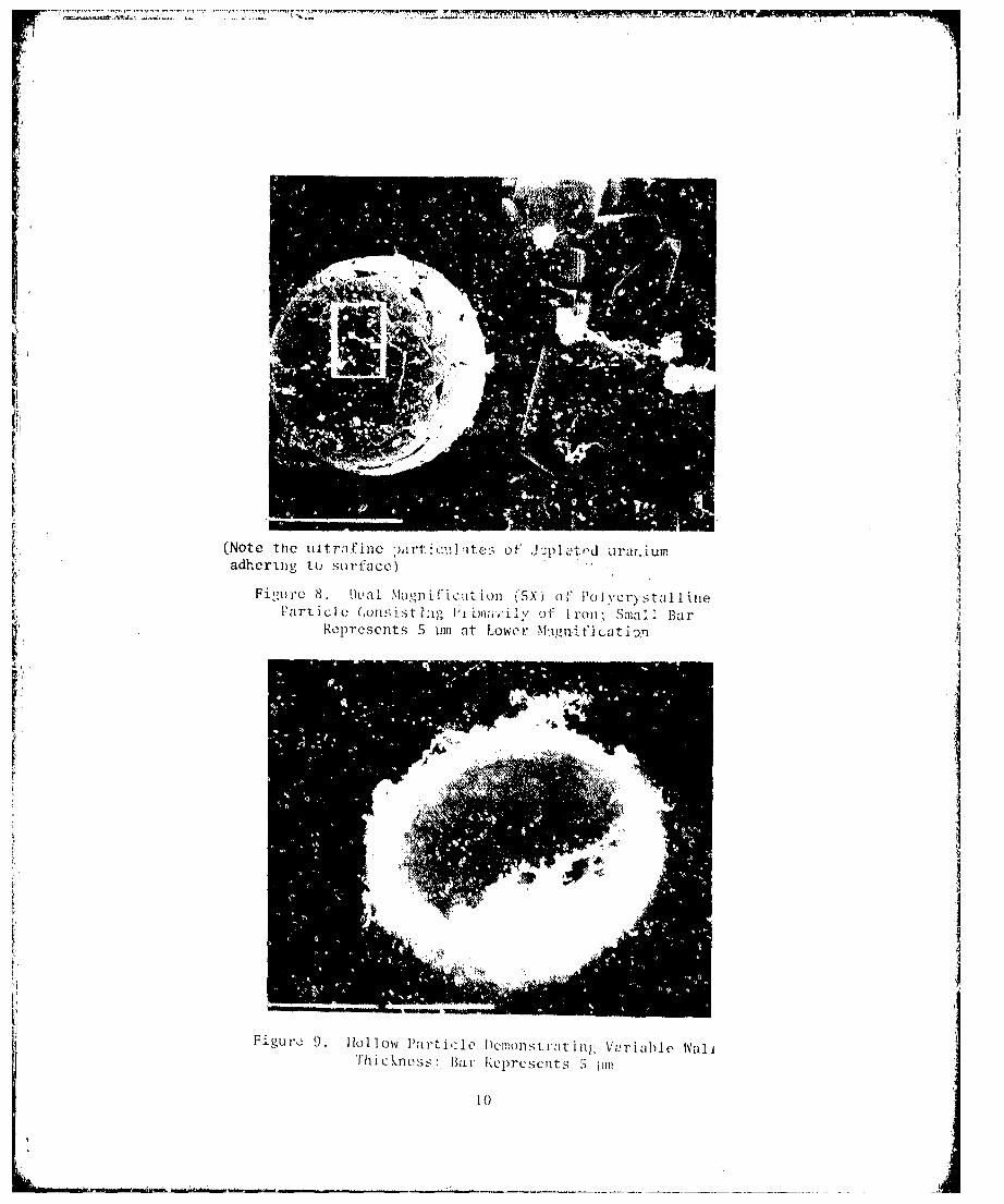
A unique class of particulates, best described as polycrystalline, wasfrequently found (Figure 8). Only observed at diameters greater than 40 pm,these particles consisted of a tight but irregular mass of nearly cuboidalcrystals. Although these particles lacked the orderly and systematic intra-crystalline alignment often associated with polycrystalline formations, thenet result was the creation ofsnearly perfect spheres exhibiting relativelynsmooth bult scohtinuousc.surofa ry. It should be noted that, when fracturedthese particles revealed a similar crystalline structure throughou theirsolid interiors. X-ray data indicated that these particles contained anextremely high content of iron with relatively small quantities of uraniumpresent.
Internal morphology was frequently revealed by examination of fracturedparticles, particularly those in which large portions had become detached.Both solid and hollow particles were observed, the latter clearly demon-strated by the presence of hemispherical fragments (Figure 9). Althoughwall thickness varied greatly, the inner surface morphology of hollow
particles consistently resembled that of the outer surface.
Perforated depleted uranium particles, similar in many respects to thosereported for fly ash and oil soot (Reference 3), are shown in Figure 10.Some of the particles observed were so highly perforated and thin-walledthat impact with the collecting tape resulted in distortion, collapse orcomplete breakýip. The unusual nature of these particles is consistent withthe oxidative processes and violent outgassing known to occur during forma-tion of such particles.
Prior to weathering, the surfaces of most airborne particles werecovered to a varyirg extent by immense numbers of nearly spherical, ultra-,fine particulates less titpn 0.1 Pm in diameter (Figure 11). Identity ofthese parti:ulates as pure or alloyed uranium was confirmed by X-ray analysis.
An example of the uniform dispersal of ultrafine particuiates on thesurface of a depleted uranium/iron particle is demonstrated in Figure 12.Any further accumulation, however, generally resulted in extensive coag-ulation and hence the formation of large billowing aggregates (Figure 13).
8

i,I
.;'
Figure (l. Dual magnification (5X) ci- /\irhorne PalticleDoplct ing Surface Fr1.!cturing; Sm,lll l3ar Represents
5 jJJn at 1..0\';C1' MagnificacioJI
F:i.gul'C 7. D(;lIlOn~;tratlon of Particle Brc,lkup as a ResultoE In::2rent j':ragi l ity; Small Bar l~eprescnts S pm
iii'I"
.,
",I
,),"
~
f
l' ,r~ :l'lit:I:I
"
(Note tllc uitrafinc :),lrt:c',lhtes of ,hpL"tf'd iJrur.iumadr,erlng tu su rface)
Fi;;Ul'C 8. ~ltlal ~Iugnifi\.~<l~inn (5X) of PolycrystalJ\1l0Farticlc (,on~;istbg Jilil!l;"ily é)f lrol1; Sma:: Bar
R~prcsC'nts 5 pm at LOI"'cl.' ~!:lgnifjcati'J'1
Figur~ 9, lIo11ow l'artj':1l~ /lcl1lonsrr:ltill); V<!riahle 1\'a11ThicblCS:::: Bar l\cprl'scnts :; ~1Il1
lO
q
',l,l;.
-.._-.....,----~-_., . . _........_-------_.__......._-

.... -- .
.... ---..--.,.-.<--_ .. - --. .:;;. •••..--; •.-~-~_.- -..~.;•. -f~.:._.·~c:.~::-:.~O"'."..;...&&... ....
.1
Figure J l. SC\'l"':I]:\ i rb,ll'llc' 1':1 rt i,: I ,'~ !ll'j;J()llstratinl.; SurfaceC()VCl'a)~l' ji)" Illtrafill<' 1';lrti,'lILI\l",;; l';il' :11' l{i~ht
":C'prl'svnt s ~ 111li '
(High iron contcn1" or tili,; P;ll't :çj.: i~ in..licltcd hy crystalforma t ion. P;l l't i eli ];1('1)" ;11 ()Il:, i llli" r Il':11 J ~;lll'fal.: l')
Fig'.1rc IO.
,,
I j

f
Il•
I~ti
(Note ultl'afinc partù:1I1atcs :11'0 cvcnly JistrihutcJupon the surface)
Figure 12. 1l1l:Il ~1ii,L~rlification llOX) or <l Stah .. lloy and Iranl'art iclc; Bar I~l'pl'csl'nts S:>. 5 I1lll at Lt)Wcr Magni fication
Figure B. Flaring ur :IIl lJlt l'afine Aggregate frolll thc Surfaccof an Airbornc PnrUclc SI\Oll'll in Dual t>lagnì fication (3X);
Bar at Ilight I~eprescnts S ,11ll Lowcr ~1agnificatjon
12
ìJì
lj

. I
ii!
j1
f
l'bis property or aJhcsion or coalcsccncc is partieularly evic.1ent in Fig-ure 14 which shows two ùcpletcù uranium particlcs intcrconncctec.1 by u singlc,almost invisible, stranù ol' material compriseJ cntirc1y 01' u1trafineparticulates.
Although generally found in association with larger partic1es, theultrafine pal'ticulates were also dctected in th~ free st~te directly uponthe surface of the collective tape. At low magnification these particulatesappeal'ed as 1arge concentric masses often reaching several hundl'ed micrometel's in diameter (Figure 15). At gl'eatly incl'eased ~agnification (Figul'e16), these masses were l'evealed as consisting of vast n~Imbers of smallaggregates and long angular chains. The ultrafine sphericp.l 'Jarticles comprising these a~gregates measured approximately 0.01 to 0.1 ~ in diameter.
PARTICLES ISOLATED FROM SOIL SAMPLES
Sampies C'ollected frt)ffi the soi l. n~ar thc tar~et si te wcre sepu.-atcc.1according to size and del by a multistep preparative procedure. l'hìstechnique proved beneficib~ in that it resulted in the exclusion of lowdensity materia1s such as sand and clay as we1l as particles larger thanthose l'equired for this stUQy.
An over~ll view of particles isolated from soil is shown in Figure 17.The use of porous, po1ycarbonate membranes as a supporting material resultedin we11 dispersed samples. The degl'ee of dispel'sion was readily control1edand was dependent onl~' upon initial partic1e concentration.
Morphology of particles removed from soi! sa.'nples was quite un1ike thatof their airbone counterparts. Far greater numbers of irregularly shapedfragments were present, presumably thc result of interaction and fusionwith sand and other materials within the soil (Figure 18). Spherical particles, although quitc numerous, generally lacked ~he convoluted surfacemorphology so apparent in airbcrne samples. Their surfaces were consistentlysmoother and frequently speck1ed with knobby b1ebs (Figure 19). Some of theseb1ebs were clear1y continuous with the sul'rounding surface whereas othersappeared neal'ly detachcd.
The relative fragility of these uranium particles was clearly cvidentfollowing brief exposure in the 1aboratory to ultrasounù (Figure 20). Al thoughsonification lasted no longer than IS seconds, particlcs showed cxtensivefl'acturing and in many inftances complete disintegration.
PARTICLE COMPOSITION
The elemental composition of individuaI particles was qualitativelydetermined by energy dispersive X-ray spectroscopy. Depleted uranium particlesfrequent1y contaìned iron, aluminum, silicon, calcium, magnesium, potassium,titanium, and tungsten aS a result of contamination during impaction andsettling.
13
,i.1
i1
, ì~
k...~_,__-_._._-t_O~_n~'}_*_::_·_......_'~.._......._..._I'li...·....' _._.........~~ •••• ,-.
'--_._~~ ,.-'

· '- .. ., .. -' ~.~~~-""--"",#_",,,.~; '~--"'-~"'.~_"'R~~",I'I'!T.'''''''''-'''''__'''l\I\lI!l';_.''
I!
Ultrafinc Particulates on thc Surface ofParticles Showing Interconnecting Thread;
Bar at Right Repl'escnts 5 pmf
IFigure 14.Staballoy
Figure 15. Conccntric Puttern of Free Ultrafine Particulateson Double-Stlck rape; Bar Rcpresents 55.5 pm
14
,
j
lj1
I
.._-------------_._---~.~_ ......•-..._...----_.. - .....

(Sam~ mate r i:11 ~;hOlm i 'l F i gu re 15)
Figure 16. lIigh ~bl;nificatjon Shc)\dng i\ggrcg:ttion of Ultraf.inc ]';'l'ticlc-;; G;'jJ B.:twcen Bars Represents 0.5 pm
Figure lì. Suil l'artidl:s 01' [l(:l1Sity Grcatcr Thall IL3 WhichPasseLi Throl1)',h ;, 12 j;m Pulycarbonate Nuclepo'Lc ~.lembrane;
Bar Reprcsents ·~S. 5 pm
tS
it il I1,Il'.> I~ ,, .

----.-.-.-'~~-~~~-~~----~.~#~-~~--~~----~_-,.--.-~-~._.~.~-~~~.~_2_n~_~5i~Q~.;4~_.~~.t~E~~~Q_'
I,.!,
~
Figure 18. \lu~11 ~iai~lljficatil'~1 (lC)X) ùf Suil P.trticles Revealing80th Spherk,il j):ll'til::lcs and Fragmcnts; Bar Represents
SS. S 11m at LOI"('1' ~(agni fication
Figure J9. f\i1obhy Soi.! Partide; (~ap Bctwccn BarsRl'lH'l'sC'nts 0.5 j.1I11
1{)

Ii1tf
I!I
Figure 20. Dual Magnification (SX) af Particle Breakup Due to15 Secand Exposure ta Ultrasaund; Bar Represents 55.5 ~m
at T"ower Magni ficat.iorl
17
,
Il
l
=- -..__ ,~-_'.~---- .. -- --~~. ",~ ........~. -<1< ., . - ....•. --..._--

,SI:t:T1 ON I V
:\lthough a nUllIhl'r ot he.I'Y l1lcLds ha\e 1ìl'l'1l fahricated il' high density jH'djcl.'tiles in iln attl' lII l' , te incr('ase the cffcctlveness t trmor piercingmuniti"n~ in rec<.>nt years, Jel,lc-tcù ur,ll\ium has heen uscd most '(tcnsively.Selccrlon of deplcted uranilllT' as thc 1.IO~è J0s:.ahle of the candidate materials was base~ primarily llpon it~ (l) high dcnsity, (2) pyrophorieity,(31 metallurgical propertics, (4) aval!a~i]ity, and (5) relatively low cost.
The medicaI and environmental impIlcatlons of depleted uranium haveheen widely studiett (References 4 and 5). Biotie hazards, gellerally con~idercd to be low and resulting onIy after prolonged exposu re , nl~ bclicve~
to be due primarily to its chemical rather than radiological properties.But even with the vast amount of phy~:ologieal data already aequireJ, additional research is necessary to better de fine the pnysieal and chemicalnature of fragmentary depleted uranium generated as a consequence of its usein military weaponry. Foremost attention should be focused on its potentialfar dissemination within the environment and entry into biological systems,particularly that of mano
In a recent report, Hanson et al (Reference 6), demonstrated the forma-t ion of particulate aerosols following penetration of depleted uranillm pro-.ieeti les into armor plato targets. This study was especially vduable in thatthe overall aerodynamic characteristics and size distribution of uranium pa:--ticles were defined.
The prcsent study was undertaken to examine the gross morphologicalcharacteristics of these particulates. Such spccific knowledge is requiredfor determination of potential safety hazards associateci with the respirationand deposition of uranium aerosols within the lungs. Particles in the 0.1to 0.5-~m size range are of greatest concern because of their high efficiencyfor deposition in thc Iungs. This range hus bccn dcfincd appropriatcly asthe respirable size range.
For this study, 105 mm rounds, each containing upproximately 3.5 kgdepleted uranium, were fired at multiple steel plate targets. Airborneparticulates were collected on doublc-stick tape through a combination ofsettling, impaetion, thcrmul precipitation, and diffusion. However, due tothe py~ophoric nature or daplated llranium and the high projectile velocityattained, the predominant mechanism appcared to be impaction. A relativelyhigh collcction cfficiency wa5 anticipated far alI but the larger fragmentsdue to the proximity of salllpling sitcs to the target.
Scanning electron microscopy revcaled that ail'horne particlllatcs wereprimarily spherical, the surfaces of ,,,h i ,'}. wcre n~ghly convolutecl. rarticleswore at times compriscd of rartially overlapping, concave plates, formed asa resul t of polyfoca 1 sol idi fication. Fxtensivc fracturing, particularly

--
~~.;.-....-~.. --- .- ...."7~--,-~---~_ ........~_........~-.~_-_----...--"lI!\I!""'--.....-'!iP.!"'."!5EPlX._P"'.!!!lWIl'.""A'!\!IM!!!!5 ....------..,
tt!
along the convoluted folds and piate Iin~s, account ed fcr the apparentfragility assoçiateù with airborne particles of the rugose type. Undernormai weathering conditions such particies could be expected to break uprapidIy, thereby contributing to an increase in the total numb~r of respirable-size particles.
Particle disintcgration would be further acceleratcd by the hollownature of many of the spheres. Hollow particles, which are frequently thinwailed or perforated, are extremel)' vuInerable to weathering and thus ~ub
ject to rapid deterioration.
The appal'ent fragi! i ty of th.:; rugose, ~lollow. and perforate particleswa3 further substantiated through observation of soil particulates. Thesesamples contained material which had accumulated over many mont~s of testingand therefore represented aged particulatc debris. The relatively lowincidence of these types of particies in suil Inust be attributed to theirinherent instabili ty and rcsults in rapiJ wtlathering and subsequent formation of smaller particulates.
An unexpected phenomenon was tho formation of ultrafine particles lessthan O.l ~ in diarneter. These particulates. generally observed adheringto the surfaces of larger particles. presumably were :orrned as a result ofthe extreme temperatures achieved and the highly reactive nature of pyrophoricdepleted uraniurn. TI ese ultrafine particles exhibited an extreme tendencytO coalesce. p~obably due to spontaneous diffusion charging. This coalescing tendency of particles. which were originally below the respirablesize-range. is especially significant sinc~ it resulted in the formationof abundant agglomerates that fell within the r0spirable range.
The elemental composition of individuaI particles was qualitativelydetermined by non-destructive X-ray spectroscopy. Airb~rne particles werecomprised primarily of alloyed uranìum and iron. Althol'gh the r.".tio of thebIO metals varied considerably arnong particles. the fact that alloy:i.ng didoccur is consistent with the violent interaction between penetrator andtarget at impact.
Particles isolated from soil samples near the target area. in additìonto uranium and iron. freqùent1y contai.ned appreciable amounts of silicon.Rluminum. and/or tungsten. Fusion with both silicon and aluminum had beenanticipated as a resul t of interaction with sand and clays within the soiI.The pl'esence of tungsten was dUE> to contarnination of the target site fromprevious test firings of high density penetrators employiilg that material.
This study has shown that scanning electron microscope techniques areideaI for examinati..on of particles formed from the impact of depletedurailium projectiles against armar plates. Results show that appreciablcquantities of respirable-size paricles ~re released during use of theseproj ectiles. Al though panicles are ini tially forrned over a:t extrerndybroad size rango, eventual weathering of large particies together withcoalescence ~f ultrafine particles combine to increase the potential totalnumber of particulates within the rcspirable range.
19
j1
ll
• __.Jn ... aa.:t'il--.--'_·",....,...., '_._ ..~ .. ···-
REFERENCES
1. Leibowitz, L., L. Baker, J.C. Schnizlein, L.W. Mishler, and J.P.Bingle, "Burning Velocities of Uranium and Zirconium in Air" in NuclearScience and Engineering, 15:395-403, 1963.
2. Physics of High Speed Impact, Final Summary Report, PATEC-TR-157-70,July 1970.
3. McCrone, W.C., and J.G. Delly, The Particle Atlas, Volume 3, AnnArbor Science Publishers, Inc., Ann Arbor, Michigan, 1973, pp. 773-779.
4. Medical and Environmental Evaluation of Depleted Uranium, Volume I, 4
JTCG/ME Ad Hoc Working Group for Depleted Uranium, 1974.
5. Hodge, H.C., J.N. Stannard, J.B. Hursh, Uranium, Plutonium,Transplutonic Elements, Springer-Verlag Publishers, New York, 1973.
6. Hanson, W.C., J.C. Elder, H.J. Ettinger, L.W. Hantel, and J.W. Owens,Particle Size Distribution of Fragments from Depleted Uranium PenetratorsFired Against Armor Plate Targets, LA-5654 Los Alamos Scientific Laboratory,1974.
2
1
:1I
20
__ 44

INITIAL OISTRIBUTION
ASD/ENFEA lHq USAF/SAMI 1OO-ALe/MMWMP 2AFIS/INT 1
';1 Hq TAC/ORA 1TAe/INAT 1
I ASD/ENESH 1'I USA TRADOe Sys Ana1y Act/ATAA-SL 1f Hq USAFE/DOQ ll
i Hq PACAF/DOOFQ 1.]
;1 ASD/XRP lCOMIPAe/I -.232 lAFATL/DLODL 2
,~DDR&E (Tech Lib) lUSAFA/DFCBS 1
,1 Dugway Prov Gd/Tech Lib l'II AFLC/MMNO l,.
·1 SAAMA/SFQT l
l AFSC/SDW l:I Hq USAF/RDP l~I AFSC/DEV l~ DDR&E/Env &Life Sciences l
I DRDAR-CW-L lUSAF (Env f.ea1th Lab) lr.;wc Env Eng lAMD/RD l
,.. . AMRL/THE l','
AMRL/THT 1ADTC/CSV lADTC/SGPE 1Det l (CEEDO/EC) lAUL/LSE 71-249 1AFLC/DS lADTC/DEN lUS Army Natick Lab lADTC/DLV 20Las Alamos Sci Lab, H-12 lDDC/DDA 2Deseret Test etr/Tech Lib lNWC/Tech Lib 1AEDC/DEE 1AFCEC/EO lARRADCOM/DRDAR-SCM-P 1Batte11e Pacific Northwest Lab 1
i 21(The :t'ever.s~' O;ìf this pa~e, is b1ank)