morphological and molecular identification of ...parasitol.kr/upload/pdf/kjp-57-3-257.pdf · there...
TRANSCRIPT

257
INTRODUCTION
Stellantchasmus is a genus of minute intestinal flukes from the family Heterophyidae. This genus includes only 4 known species: S. falcatus Onji and Nishio, 1924, S. aspinosus Pearson, 1964, S. gallinae Oshmarin, 1970, and S. batillans Pearson, 1964, all of which have been clearly defined by their morpho-logical characters [1]. The number and arrangement of testes and the presence of an armed or unarmed ventral sucker are used as the main characters for species identification [1]. Stel-
lantchasmus has been found in both experimental and natural hosts: S. falcatus in natural and experimental cats; S. aspinosus
in the wild water rat and the experimental cat, rat, and chick-en; S. gallinae in the domestic chicken; and S. batillans in the water rat and domestic duck [1,2].
S. falcatus was first reported in 1916 by Onji and Nishio [3]. Human infection by this parasite has been reported in Japan, the Philippines, the Republic of Korea (=Korea), Hawaii, and Thailand [4-7], and human infections also occur at a high rate in northern parts of Thailand [8]. So far only 1 species, S. falca-
tus, has been recorded in Thailand (and in some other coun-tries) [4-8]. During infection, eggs can pass through the blood vessels to the brain or heart of the patient [9]. Pathology in humans includes mild to colicky pain, leading to death in se-vere cases (as seen in the Philippines) [7,9].
S. falcatus is widespread and can infect various types of hosts [3,10]. The first intermediate host of this parasite, reported in Thailand, is freshwater snails such as Thiara scabra and Tarebia
granifera [11]. The metacercarial stage of S. falcatus, which in-fects humans, is found in several types of brackish water and
ISSN (Print) 0023-4001ISSN (Online) 1738-0006
Korean J Parasitol Vol. 57, No. 3: 257-264, June 2019https://doi.org/10.3347/kjp.2019.57.3.257▣ ORIGINAL ARTICLE
•Received 22 February 2019, revised 10 May 2019, accepted 10 May 2019.*Corresponding author ([email protected])
© 2019, Korean Society for Parasitology and Tropical MedicineThis is an Open Access article distributed under the terms of the Creative Commons Attribution Non-Commercial License (http://creativecommons.org/licenses/by-nc/4.0) which permits unrestricted non-commercial use, distribution, and reproduction in any medium, provided the original work is properly cited.
Morphological and Molecular Identification of Stellantchasmus dermogenysi n. sp. (Digenea:
Heterophyidae) in Thailand
Chalobol Wongsawad1,2,*, Nattawadee Nantarat1, Pheravut Wongsawad1,3, Preeyaporn Butboonchoo1,2, Jong-Yil Chai4,5
1Department of Biology, Faculty of Science, Chiang Mai University, Chiang Mai 50202, Thailand; 2Environmental Science Research Center (ESRC), Faculty of Science, Chiang Mai University, Chiang Mai 50202, Thailand; 3Economic Plant Genome Service Centre, Faculty of Science, Chiang Mai
University, Chiang Mai 50202, Thailand; 4Institute of Parasitic Diseases, Korea Association of Health Promotion (KAHP), Seoul 07649, Korea; 5Department of Tropical Medicine and Parasitology, Seoul National University College of Medicine, Seoul 03080, Korea
Abstract: We tried a series of morphological and molecular approaches to identify a new species of Stellantchasmus (Di-genea: Heterophyidae) originating from the wrestling half-beaked fish, Dermogenys pusillus of Thailand. Adult worm sam-ples of the new species were recovered from hamsters experimentally infected with the metacercariae from D. pusillus in Thailand. Two isolates (Thai and Korean) of Stellantchasmus falcatus were used as comparative control groups. Worm samples of 3 Stellantchasmus groups were morphologically observed and molecularly analyzed with the mitochondrial cytochrome c oxidase 1 gene. The morphological characteristics of S. dermogenysi n. sp. are similar to S. falcatus origi-nating from brackish water fish, but minor difference was noted including the absence of the prepharynx, position of the ovary near the ceca end, smaller body size, and shorter esophageal length. A phylogenetic tree derived from neighbor-joining and maximum-likelihood methods suggests that S. dermogenysi n. sp. is separated from S. falcatus supported by high bootstrap values. The relative divergences persist between these host-specific trematodes, which we suggest should be recognized as 2 distinct species. Comparisons of S. dermogenysi n. sp. with S. falcatus isolated from mullets in Thailand and Korea indicate a genetic divergence of mitochondrial DNA of 19.4% and 21.7%, respectively. By the present study, a new species, Stellantchasmus dermogenysi n. sp. (Digenea: Heterophyidae), is proposed in Thailand based on molecular evidences, in addition to minor morphological differences between S. falcatus and the new species.
Key words: Stellantchasmus dermogenysi, Stellantchasmus falcatus, heterophyid fluke, phylogeny, Thailand

258 Korean J Parasitol Vol. 57, No. 3: 257-264, June 2019
freshwater fish, the second intermediate host [10,12]. There are only 2 species of fish, Dermogenys pusillus (half-beaked fish, freshwater species) and Planiliza subviridis (a species of mullet, brackish water species), that were reported to be second inter-mediate hosts of S. falcatus in Thailand [12]. Traditional iden-tification of Stellantchasmus species has been based solely on morphological characteristics; however, numerous morpho-logical similarities can result in misidentification.
Over the last decade, cryptic speciation has been discovered in other digenean parasitic species such as Echinostoma, Phyllo-distomum, and Clinostomum [13-17]. Mechanisms of speciation in parasites may be unswervingly connected to their hosts and their complex life cycles [13,18]. The presence of cryptic spe-cies arises through morphological similarities and appears to be widespread among parasites [18,19]. Speciation almost al-ways occurs in parasite species that form a large diverse group of organisms, infecting various kinds of hosts [13,17,20]. There have been reports of morphological characteristics of S.
falcatus from the fish D. pusillus that seems to be different from S. falcatus from the fish P. subviridis [7,9,10,12,21]. In this case, no exception exists in the ordinary cryptic speciation that may occur in Stellantchasmus species.
Molecular approaches are the most efficient tools currently used to understand phylogenetic relationships and to deter-mine genetic variations in heterophyid flukes [22-25]. The conventional PCR and high annealing temperature-based ran-dom amplified polymorphic DNA (HAT-RAPD) methods have been used to discriminate Stellantchasmus species isolated from various hosts [22]. It has been suggested that different DNA fingerprints of Stellantchasmus worms acquired from dif-ferent hosts may be due to distinct species [22]. To clarify this point, HAT-RAPD can be used to develop specific markers to detect S. falcatus infections [25]. Moreover, many genes, such as nuclear genes (including internal transcribed spacers 1 and 2; ITS1 and ITS2) and mitochondrial genes (including mito-chondrial cytochrome c oxidase 1; CO1), have been used for identification and phylogenic studies of various parasites [13,23,24]. ITS2 and CO1 regions have been useful for investi-gation and identification of Stellantchasmus sp. [23,24].
The aim of this study was to investigate and identify a new species of Stellantchasmus (Digenea: Heterophyidae) obtained from the wrestling half-beaked fish, D. pusillus, in Thailand based on both morphological and molecular approaches us-ing CO1 gene.
MATERIALS AND METHODS
Ethical statementsAll experimental hosts were managed according to the
guidelines approved by the Animal Ethics Committee of the Faculty of Science, Chiang Mai University, Thailand, and the relevant document (no. RE 001/13) was approved by the com-mittee. The guidelines for animal care were used according to the International Guiding Principles of Biomedical Research Involving Animals of Council for International Organizations of Medical Sciences (CIOMS).
Morphological investigationThe metacercariae of Stellantchasmus spp. were collected
from different sites in Thailand. The hosts of this parasite were P. subviridis and D. pusillus, acquired from Chiang Mai and Chonburi Provinces in Thailand. Fish specimens were minced and digested using 1% pepsin solution for 2 hr at 37˚C and rinsed with 0.85% NaCl. A hundred metacercariae of Stellant-chasmus spp. were checked with light microscopy, and ham-sters (Phodopus campbelli) were force-fed with the metacercariae samples. After 2 days, the adult-stage worms were collected from the hamsters. Total 57 specimens of Stellantchasmus were used for identification and morphological studies. The worms were processed for permanent slides. Briefly, they were flat-tened under a cover slip pressure and fixed in 4% formalin, stained with hematoxylin and/or Borax’s carmine, dehydrated in alcohol series, and finally mounted in Permount. The speci-mens were checked with the descriptions of Onji and Nishio [3], Pearson [2], Kliks and Tantachamrun [5], Pearson and Ow-Yang [1], and Pubua and Wongsawad [12]. The body and organs of the worms were measured and analyzed. The holo-type and paratypes of the new species are kept in the Natural History Museum, London (NHMUK). The worms were frozen at -20˚C for DNA extraction.
DNA extractionGenomic DNA from each Stellantchasmus species and related
groups were collected from adult worms. S. falcatus from the laboratory of Prof. Jong-Yil Chai, Seoul National University College of Medicine and Korean Association of Health Promo-tion, Seoul, Korea, was used for molecular studies with the 2 Thai samples. Approximately, 2 mg of each trematode tissue sample was used for DNA extraction. The DNA extraction and purification steps were performed using 150 ml of 5% Chelex
Wongsawad et al.: Stellantchasmus dermogenysi n. sp. in Thailand 259
(Fluka) solution containing 10 µl of 20 mg/ml proteinase K (Sigma-Aldrich, St. Louis, Missouri, USA): the mixture con-taining the trematode tissue was washed and incubated at 55˚C for 1 hr, followed by heating at 95˚C for 30 min, then gently mixed. The mixture was centrifuged for 10 sec at 13,000 rpm, after which the supernatant was removed and stored at -20˚C until use.
Amplification and sequencing of the CO1 geneThe partial CO1 gene was amplified using the primers as
follows: JB3 (5′-TTTTTTGGGCATCCTGACGTTTAT-3′) as a for-ward primer and JB4.5 (5′–TAAAGAAAGAACATAATGAAAATG-3′) as a reverse primer. The PCR amplifications were carried out as described above. The amplification procedure included a 3 min initial denaturation at 95˚C, then 40 cycles of 1 min de-naturation at 95˚C, 1 min annealing at 50˚C, 1 min extension at 72˚C, and a 7 min final extension at 72˚C. The PCR prod-ucts were visualized on 1.0% agarose gels, purified using the Cleanup PCR Kit (Sigma-Aldrich), and directly sequenced in both directions by Ward Medic, Ltd. (Bangkok, Thailand).
Phylogenetic analysesThe molecular data in this study including the sequences
from Stellantchasmus spp. and outgroup (Metagonimus spp.) were aligned using ClustalW, a subprogram of MEGA version 6.0 [26]. The sequences were checked for ambiguous nucleo-tide sites before being subjected to a phylogenetic analysis. The jModel test 2 [27] was used to find the most appropriate sub-
stitution model for the CO1 datasets. Phylogenetic trees of all taxa were constructed using neighbor-joining, maximum like-lihood, and Bayesian inference. GTR+G model was applied for all analyses. The NJ analysis and the likelihood scores of datas-et was carried out using PAUP* version 4.0b10 [28] with 1,000 bootstrap replicates. The ML analysis was undertaken using PHYML version 2.4.5 [29]. The tree searching system used for this method involved the heuristic procedure with tree-bisec-tion-reconnection branch swapping. The bootstrap resampling [30,31] was done with 1,000 replicates that were assigned to support the particular branches. BI analysis was performed us-ing MrBayes version 3.2.5 [32], where the Bayesian analysis was run for 5 million generations (heating parameter=0.15), sampling was done for every 100 generations, and the first 25% of trees were discarded with burn-in procedure. The final consensus tree was built using the last 15,002 trees. Support for nodes was defined as posterior probabilities (P).
RESULTS
Morphological dataThe samples of S. dermogenysi were identified compared
with the original description and holotype picture of S. falca-
tus. In the morphology of Stellantchasmus spp. from different hosts [2-4], the differences in the presence or absence of the prepharynx, position of the ovary, body size, and esophagus length were distinguishing factors. The prepharynx was very short or absent in S. dermogenysi n. sp. but it was present in S.
Table 1. Helminth species used in this study and GenBank accession numbers for their corresponding CO1 sequence
Species Host Country GenBank accession no.
Stellantchasmus dermogenysi n.sp.a Experimental hamster Chiang Mai, Thailand -Stellantchasmus dermogenysi n.sp. Experimental hamster Chiang Mai, Thailand KU753571Stellantchasmus dermogenysi n.sp. Experimental hamster Chiang Mai, Thailand KU753572Stellantchasmus dermogenysi n.sp. Experimental hamster Chiang Mai, Thailand KU753573Stellantchasmus dermogenysi n.sp. Experimental hamster Chiang Mai, Thailand KU753574Stellantchasmus dermogenysi n.sp. Experimental hamster Chiang Mai, Thailand KU753575Stellantchasmus dermogenysi n.sp. Experimental hamster Chiang Mai, Thailand KU753576Stellantchasmus falcatusa Experimental hamster Chon Buri, Thailand AY382320Stellantchasmus falcatusa Korean human Korea -Stellantchasmus falcatus Experimental hamster Chon Buri, Thailand KU753568Stellantchasmus falcatus Experimental hamster Chon Buri, Thailand KU753569Stellantchasmus falcatus Experimental hamster Chon Buri, Thailand KU753570Metagonimus yokogawib Experimental hamster Korea AB470519Metagonimus takahashiib Experimental hamster Korea AF096231
aQuery sequence generated. bUsed as the outgroup.
260 Korean J Parasitol Vol. 57, No. 3: 257-264, June 2019
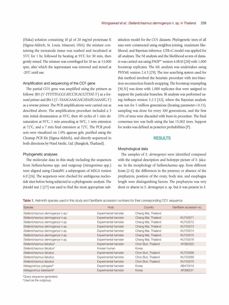
falcatus. The ovary of S. dermogenysi n. sp. is located near the ceca end, whereas in S. falcatus it is located in the middle of the ceca length. Of the new species, the body, esophagus, and ventral sucker were clearly smaller than those of S. falcatus (Ta-ble 2; Fig. 1). Similarity in the body shape, organ size, and their positions were observed, in addition to the ratios of the
body size (Table 2; Fig. 1). S. falcatus from P. subviridis revealed smaller values. Although the ratio of the body size of S. falcatus from P. subviridis to the new species was 1.33:1.39, ratios of the organs were found to be different, especially with regard to the esophagus length, ovary width, and the right testis width (Ta-ble 2).
Fig. 1. (A) Stellantchasmus falcatus adult fluke originating from the brackish water fish P. subviridis. Ventral view. (B) Stellantchasmus dermogenysi n. sp. originating from the freshwater fish D. pusillus.
A B
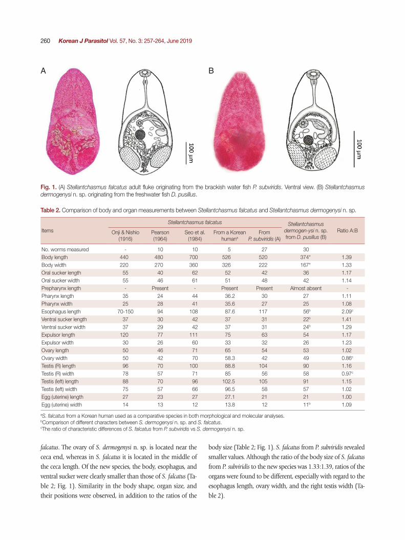
Table 2. Comparison of body and organ measurements between Stellantchasmus falcatus and Stellantchasmus dermogenysi n. sp.
Items
Stellantchasmus falcatus Stellantchasmus dermogen-ysi n. sp. from D. pusillus (B)
Ratio A:BOnji & Nishio (1916)
Pearson (1964)
Seo et al. (1984)
From a Korean humana
From P. subviridis (A)
No. worms measured - 10 10 5 27 30Body length 440 480 700 526 520 374* 1.39Body width 220 270 360 326 222 167* 1.33Oral sucker length 55 40 62 52 42 36 1.17Oral sucker width 55 46 61 51 48 42 1.14Prepharynx length - Present - Present Present Almost absent -Pharynx length 35 24 44 36.2 30 27 1.11Pharynx width 25 28 41 35.6 27 25 1.08Esophagus length 70-150 94 108 87.6 117 56b 2.09c
Ventral sucker length 37 30 42 37 31 22b 1.41Ventral sucker width 37 29 42 37 31 24b 1.29Expulsor length 120 77 111 75 63 54 1.17Expulsor width 30 26 60 33 32 26 1.23Ovary length 50 46 71 65 54 53 1.02Ovary width 50 42 70 58.3 42 49 0.86c
Testis (R) length 96 70 100 88.8 104 90 1.16Testis (R) width 78 57 71 85 56 58 0.97c
Testis (left) length 88 70 96 102.5 105 91 1.15Testis (left) width 75 57 66 96.5 58 57 1.02Egg (uterine) length 27 23 27 27.1 21 21 1.00Egg (uterine) width 14 13 12 13.8 12 11b 1.09
aS. falcatus from a Korean human used as a comparative species in both morphological and molecular analyses. bComparison of different characters between S. dermogenysi n. sp. and S. falcatus.cThe ratio of characteristic differences of S. falcatus from P. subviridis vs S. dermogenysi n. sp.

Wongsawad et al.: Stellantchasmus dermogenysi n. sp. in Thailand 261
Stellantchasmus dermogenysi n. sp. Wongsawad et al., 2019
Descriptions: Adult specimens were obtained from experi-mentally infected hamsters (P. campbelli). All measurements below are given in micrometers (µm). Body pyriform, with a length of 374 (256-480), and forebody narrower than the hindbody, with a maximum width of 167 (136-220) across the testes. Almost the entire surface covered with scale-like spines extending to half of the posterior body. Oral sucker subterminal and 36 (26-46) long by 42 (32-54) wide; preph-arynx 4 (2-10) long; pharynx 27 (22-40) long by 25 (20-34) wide; esophagus 56 (30-102) long, with bifurcation extending to the anterior border of testis. Ventral sucker 22 (20-30) long by 24 (20-30) wide. Testes paired, ovoid, opposite, and in the posterior part of the body, and measured 90 (68-116) long by 58 (40-80) wide (right). Left testis 90 (68-112) long by 57 (40-82) wide. Seminal vesicles thin-walled, located to the left of ovary, and connected to the expulsor lying dorsal and left of
cecum. Expulsor with muscle fibers 54 (26-80) long by 26 (20-36) wide, opened into the ventrogenital sac dorsally on the left side and anterior to ovary. Ovary slightly ovoid and positioned slightly right and anterior to testes, 53 (30-70) long by 49 (40-76). Uterus coiled in the posterior of the body and filled with operculate eggs. Vitelline follicles distributed at the inner intestine, with bifurcation extending to the posterior of the body. Eggs 21.6 (17.5-23.0) long by 11.5 (7.5-15.0) wide, thin-walled, possessing a smooth shell with a small opercu-lum at anterior end.
Type materials: Type specimens have been deposited in the Natural History Museum, London, UK and the Applied Parasi-tology Research Laboratory, Faculty of Science, Chiang Mai University, Chiang Mai, Thailand. Holotype NHMUK 2015.1.7.1 (Fig. 1), paratypes NHMUK 2015.1.7.2 (1 individ-ual) and CMUZ 01.1-01.28 (29 individuals).
Host: The metacercariae were collected from the freshwater fish Dermogenys pusillus and the adult stage in the experimental
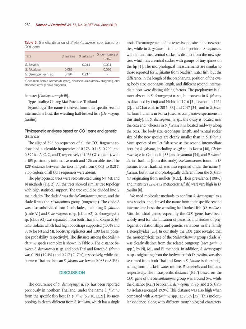
Fig. 2. A phylogenetic tree of Stellantchasmus spp. and related group based on CO1 gene. Statistics can be referred to in Materials and Methods.
262 Korean J Parasitol Vol. 57, No. 3: 257-264, June 2019
hamster (Phodopus campbelli). Type locality: Chiang Mai Province, Thailand Etymology: The name is derived from their specific second
intermediate host, the wrestling half-beaked fish (Dermogenys
pusillus).
Phylogenetic analyses based on CO1 gene and genetic distance
The aligned 396 bp sequences of all the CO1 fragment co-dons had nucleotide frequencies of 0.173, 0.145, 0.290, and 0.392 for A, C, G, and T, respectively (41.1% GC content), with a 105 parsimony informative result and 126 variable sites. The K2P-distance between the taxa ranged from 0.005 to 0.217. Stop codons of all CO1 sequences were absent.
The phylogenetic trees were reconstructed using NJ, ML and BI methods (Fig. 2). All the trees showed similar tree topology with high statistical support. The tree could be divided into 2 main clades. The clade A was the Stellantchasmus group, and the clade B was the Metagonimus group (outgroup). The clade A was also subdivided into 2 subclades, including S. falcatus (clade A1) and S. dermogenysi n. sp. (clade A2). S. dermogenysi n. sp. (clade A2) was separated from both Thai and Korean S. fal-
catus isolates which had high bootstraps supported (100% and 99% for NJ and ML bootstrap replicates and 1.00 for BI poste-rior probability, respectively). The distance among the Stellant-
chasmus species complex is shown in Table 3. The distance be-tween S. dermogenysi n. sp. and both Thai and Korean S. falcatus was 0.194 (19.4%) and 0.217 (21.7%), respectively, while that between Thai and Korean S. falcatus was lower (0.083 or 8.3%).
DISCUSSION
The occurrence of S. dermogenysi n. sp. has been reported previously in northern Thailand, under the name S. falcatus from the specific fish host D. pusillus [5,7,10,12,21]. Its mor-phology is clearly different from S. batillans, which has a single
testis. The arrangement of the testes is opposite in the new spe-cies, while in S. gallinae it is in tandem position. S. aspinosus, with an unarmed ventral sucker, is distinct from the new spe-cies, which has a ventral sucker with groups of tiny spines on the lip [1]. The morphological measurements are similar to those reported for S. falcatus from brackish water fish, but the difference in the length of the prepharynx, position of the ova-ry, body size, esophagus length, and different second interme-diate host were distinguishing factors. The prepharynx is al-most absent in S. dermogenysi n. sp., but present in S. falcatus, as described by Onji and Nishio in 1916 [3], Pearson in 1964 [2], and Chai et al. in 2016 [33] and 2017 [34], and in S. falca-tus from humans in Korea (used as comparative specimens in this study). In S. dermogenysi n. sp., the ovary is located near the ceca end, whereas in S. falcatus it is located mid-way along the ceca. The body size, esophagus length, and ventral sucker size of the new species are clearly smaller than in S. falcatus. Most species of mullet fish serve as the second intermediate host for S. falcatus, including Mugil sp. in Korea [10], Chelon macrolepis in Cambodia [33] and Myanmar [34], and P. subviri-dis in Thailand (from this study). Stellantchasmus found in D.
pusillus, from Thailand, was also reported under the name S. falcatus, but it was morphologically different from the S. falca-
tus originating from mullets [8,22]. Their prevalence (100%) and intensity (22-2,492 metacercaria/fish) were very high in D. pusillus [8].
We used molecular methods to confirm S. dermogenysi as a new species, and derived the name from their specific second intermediate host, the wrestling half-beaked fish (D. pusillus). Mitochondrial genes, especially the CO1 gene, have been widely used for identification of parasites and studies of phy-logenetic relationships and genetic variations in the family Heterophyidae [23]. In our study, the CO1 gene revealed that the monophyletic tree of the Stellantchasmus group (clade A) was clearly distinct from the related outgroup (Metagonimus
spp.), by NJ, ML, and BI methods. In addition, S. dermogenysi n. sp., originating from the freshwater fish D. pusillus, was also separated from both Thai and Korean S. falcatus isolates origi-nating from brackish water mullets P. subviridis and humans, respectively. The intraspecific distance (K2P) based on the CO1 gene of the Stellantchasmus group was around 5%, while the distance (K2P) between S. dermogenysi n. sp. and 2 S. falca-
tus isolates averaged 19.9%. This distance was also high when compared with Metagonimus spp., at 7.5% [35]. This molecu-lar evidence, along with different morphological characters,
Table 3. Genetic distance of Stellantchasmus spp. based on CO1 gene
Taxa S. falcatus S. falcatusa S. dermogenysi n. sp.
S. falcatus - 0.014 0.024S. falcatusa 0.083 - 0.026S. dermogenysi n. sp. 0.194 0.217 -
aSpecimen from a Korean (human), distance value (below diagonal), and standard error (above diagonal).

Wongsawad et al.: Stellantchasmus dermogenysi n. sp. in Thailand 263
clearly supports the distinction of the Stellantchasmus species. The present findings agree with Wongsawad and Wongsawad [22], who reported that differences in the morphology, habi-tat, DNA fingerprint, and phylogenetic relationships (based on HAT-RAPD) strongly suggested that Stellantchasmus from freshwater and brackish water habitats are different species.
The morphological and molecular characteristics obtained in this study allows us to confidently identify the parasite iso-late originating from the freshwater fish D. pusillus, in Thai-land, as a new species, S. dermogenysi n. sp. Although S. der-mogenysi shares many morphological similarities with S. falca-
tus, the molecular evidence supports the distinction. The pres-ent findings will help a definitive diagnosis in humans, may affect treatment, prevention measures, and research into anti-parasitic agents.
ACKNOWLEDGMENTS
This research work was partially supported by Chiang Mai University, Chiang Mai, Thailand. We thank the Applied Tech-nology for Biodiversity, Institute for Science and Technology; Applied Parasitology, Department of Biology, Faculty of Sci-ence; Economic Plant Genome Service Centre, Faculty of Sci-ence, Chiang Mai University, Thailand for the provision of in-struments, facilities, and access to laboratories. This work was supported by National Research Council of Thailand to CW (grant no. 2559A10402051, 2016) and The Thailand Research Fund to NN (grant no. TRG5880053). Special thanks are ex-tended to the Natural History Museum, London, and Senior Curator of Parasites and Vectors Division.
REFERENCES
1. Pearson JC, Ow-Yang CK. New species of Haplorchis from South-east Asia, together with keys to the Haplorchis-group of hetero-phyid trematodes of the region. Southeast Asian J Trop Med Pub-lic Health 1982; 13: 35-60.
2. Pearson JC. A revision of the subfamily Haplorchinae Looss, 1899 (Trematoda: Heterophyidae): I. The Haplorchis group. Para-sitology 1964; 54: 601-676.
3. Onji, Y, Nishio T. On intestinal distomes. Iji Shimbun 1916; 949: 589-593.
4. Seo BS, Hong, ST, Chai JY, Lee SH. Studies on intestinal trema-todes in Korea. VIII. A human case of Echinostoma hortense infec-tion. Korean J Parasitol 1983; 21: 219-223.
5. Kliks M, Tantachamrun T. Heterophyid (trematoda) parasites of cats in North Thailand, with notes on a human case found at
necropsy. Southeast Asian J Trop Med Public Health 1974; 5: 547-555.
6. Sohn WM, Chai JY, Lee SH. Two cases of natural human infec-tion by Heterophyes nocens and the infection status of heterophy-id metacercariae in mullets from Samcheonpo, Kyongnam Province. Inje Med J 1989; 10: 443-452.
7. Tantachamrun T, Kliks M. Heterophyid infection in human ile-um: report of three cases. Southeast Asian J Trop Med Public Health 1978; 9: 228-231.
8. Sripalwit P, Wongsawad C, Chai JY, Anuntalabhochai S, Ro-janapaibul A. Investigation of Stellantchasmus falcatus metacer-cariae in half-beaked fish, Dermogenus pusillus from four districts of Chiang Mai Province, Thailand. Southeast Asian J Trop Med Public Health 2003; 34: 281-285.
9. Africa CM, de Leon W, Garcia EY. Visceral complications in in-testinal heterophyidiasis of man. Acta Med Phil (Monographic series) 1940; 1: 1-32.
10. Chai JY, Sohn WM. Identification of Stellantchasmus falcatus metacercariae encysted in mullets in Korea. Korean J Parasitol 1988; 26: 65-68.
11. Chuboon S, Wongsawad C. Molecular identification of larval trematodes in intermediate host from Chiang Mai, Thailand. Southeast Asian J Trop Med Public Health 2009; 40: 1216-1220.
12. Pubua J, Wongsawad C. Redescription of the trematode meta-cercariae from the mullet (Liza subviridis) and half-beak (Der-mogenys pusillus). Southeast Asian J Trop Med Public Health 2007; 38: 106-109.
13. Noikong W, Wongsawad C, Chai JY, Saenphet S, Trudgett A. Mo-lecular analysis of echinostome metacercariae from their second intermediate host found in a localised geographic region reveals genetic heterogeneity and possible cryptic speciation. PLoS Negl Trop Dis 2014; 8: e2778.
14. Georgieva S, Selbach C, Faltýnková A, Soldánová M, Sures B, Skírnisson K, Kostadinova A. New cryptic species of the “revolu-tum” group of Echinostoma (Digenea: Echinostomatidae) re-vealed by molecular and morphological data. Parasit Vectors 2013; 6: 64.
15. Ho HW, Bray RA, Cutmore SC, Ward S, Cribb TH. Two new spe-cies of Phyllodistomum Braun, 1899 (Trematoda: Gorgoderidae Looss, 1899) from Great Barrier Reef fishes. Zootaxa 2014; 3779: 551-562.
16. Sereno-Uribe AL, Pinacho-Pinacho CD, García-Varela M, de León GPP. Using mitochondrial and ribosomal DNA sequences to test the taxonomic validity of Clinostomum complanatum Ru-dolphi, 1814 in fish-eating birds and freshwater fishes in Mexi-co, with the description of a new species. Parasitol Res 2013; 112: 2855-2870.
17. Nadler SA, DE León GP. Integrating molecular and morphologi-cal approaches for characterizing parasite cryptic species: impli-cations for parasitology. Parasitology 2011; 138: 1688-1709.
18. Cable J, van Oosterhout C. The impact of parasites on the life history evolution of guppies (Poecilia reticulata): the effects of host size on parasite virulence. Int J Parasitol 2007; 37: 1449-

264 Korean J Parasitol Vol. 57, No. 3: 257-264, June 2019
1458.19. Huyse T, Volckaert FA. Identification of a host-associated species
complex using molecular and morphometric analyses, with the description of Gyrodactylus rugiensoides n. sp. (Gyrodactylidae, Monogenea). Int J Parasitol 2002; 32: 907-919.
20. Dyer NA, Ravel S, Choi KS, Darby AC, Causse S, Kapitano B, Hall MJ, Steen K, Lutumba P, Madinga J, Torr SJ, Okedi LM, Le-hane MJ, Donnelly MJ. Cryptic diversity within the major try-panosomiasis vector Glossina fuscipes revealed by molecular markers. PLoS Negl Trop Dis 2011; 5: e1266.
21. Wongsawad C, Rojanapaibul A, Vanittanakom P. Surface ultra-structure of encysted metacercariae and of adult Stellantchasmus sp. (Trematoda: Heterophyidae). J Electron Microscop Soc Thai-land 1997; 11: 19-26.
22. Wongsawad C, Wongsawad P. Molecular markers for identifica-tion of Stellantchasmus falcatus and a phylogenic study using the HAT-RAPD method. Korean J Parasitol 2010; 48: 303-307.
23. Chontananarth T, Wongsawad C, Chomdej S, Krailas D, Chai JY. Molecular phylogeny of trematodes in Family Heterophyidae based on mitochondrial cytochrome c oxidase subunit I (mCOI). Asian Pac J Trop Med 2014; 7: 446-450.
24. Sripalwit P, Wongsawad C, Chontananarth T, Anuntalabhochai S, Wongsawad P, Chai JY. Developmental and phylogenetic charac-teristics of Stellantchasmus falcatus (Trematoda: Heterophyidae) from Thailand. Korean J Parasitol 2015; 53: 201-207.
25. Wongsawad C. Development of HAT-RAPD marker for detec-tion of Stellantchasmus falcatus infection. Southeast Asian J Trop Med Public Health 2011; 42: 46-52.
26. Tamura K, Stecher G, Peterson D, Filipski A, Kumar S. MEGA6: Molecular evolutionary genetics analysis version 6.0. Mol Biol Evol 2013; 30: 2725-2729.
27. Darriba D, Taboada GL, Doallo R, Posada D. jModelTest 2:
more models, new heuristics and parallel computing. Nat Meth-ods 2012; 9: 772.
28. Swofford DL. PAUP*. Phylogenetic Analysis Using Parsimony (* and Other Methods). Version 4. Sunderland, USA. Sinauer Asso-ciates. 2003.
29. Guindon S, Gascuel O. A simple, fast, and accurate algorithm to estimate large phylogenies by maximum likelihood. Syst Biol 2003; 52: 696-704.
30. Felsenstein J. Bootstraps and testing trees. [Internet]; Availabe from: http://evolution.gs.washington.edu/sisg/2016/2016_SISG_19_7.pdf
31. Felsenstein J. Confidence limits on phylogenies: an approach using the bootstrap. Evolution 2010; 39: 783-791.
32. Ronquist F, Teslenko M, van der Mark P, Ayres DL, Darling A, Höhna S, Larget B, Liu L, Suchard MA, Huelsenbeck JP. MrBayes 3.2: efficient Bayesian phylogenetic inference and model choice across a large model space. Syst Biol 2012; 61: 539-542.
33. Chai JY, Sohn WM, Na BK, Jeoung HG, Sinuon M, Socheat D. Stellantchasmus falcatus (Digenea: Heterophyidae) in Cambodia: discovery of metacercariae in mullets and recovery of adult flukes in an experimental hamster. Korean J Parasitol 2016; 54: 537-541.
34. Chai JY, Sohn WM, Na BK, Park JB, Jeoung HG, Hoang EH, Htoon TT, Tin HH. Zoonotic trematode metacercariae in fish from Yangon, Myanmar and their adults recovered from experi-mental animals. Korean J Parasitol 2017; 55: 631-641.
35. Pornruseetairatn S, Kino H, Shimazu T, Nawa Y, Scholz T, Ru-angsittichai J, Saralamba NT, Thaenkham U. A molecular phy-logeny of Asian species of the genus Metagonimus (Digenea)--small intestinal flukes--based on representative Japanese popula-tions. Parasitol Res 2016; 115: 1123-1130.