morphogenesis and mechanisms of penetration by plant
TRANSCRIPT

July 12, 1996 11:3 Annual Reviews MENDTEXT.DUN AR14-19
Annu. Rev. Phytopathol. 1996. 34:367–86Copyright c© 1996 by Annual Reviews Inc. All rights reserved
MORPHOGENESIS ANDMECHANISMS OF PENETRATION BYPLANT PATHOGENIC FUNGI
K. Mendgen, M. Hahn, and H. DeisingUniversitat Konstanz, Fakult¨at fur Biologie, Lehrstuhl f¨ur Phytopathologie,Universitatsstrasse 10, D-78434 Konstanz, Germany
KEY WORDS: penetration, fungi, cell wall-degrading enzymes, infection structures
ABSTRACT
Infection structures of phytopathogenic fungi are modified hyphae specializedfor the invasion of plant tissues. Initial events are adhesion to the cuticle anddirected growth of the germ tube on the plant surface. At the site of penetration,appressoria are often formed that may have melanized walls and develop highturgor pressure to support the penetration process. The penetration hypha accu-mulates components of the cytoskeleton in the tip and secretes a variety of cellwall–degrading enzymes in a highly regulated fashion in order to penetrate thecuticle and the plant cell wall. This article reviews recent papers on the cytology,physiology, and molecular biology of the penetration process.
INTRODUCTION
Plant parasitic fungi have conquered the living plant as a copious source ofnutrients. For successful parasitism, a crucial step is penetration. In order toovercome the various barriers present in leaves, stems, or roots, fungi haveevolved astonishingly diverse invasion strategies. For this purpose, infectionstructures are produced that enable the fungus to penetrate different types ofplant cell walls. The morphogenetic events leading to formation of the infectionstructure often depend on specific signals provided by the plant surface and areprerequisites for a particular mode of penetration. Physiological changes suchas targeted secretion of enzymes or an increase of pressure within the infectionstructure support the penetration process. We believe that infection structureshave evolved from nonpathogenic hyphae that are able to penetrate into theirsubstrate.
3670066-4286/96/0901-0367$8.00
Ann
u. R
ev. P
hyto
path
ol. 1
996.
34:3
67-3
86. D
ownl
oade
d fr
om a
rjou
rnal
s.an
nual
revi
ews.
org
by D
euts
che
Fors
chun
gsge
mei
nsch
aft o
n 07
/25/
07. F
or p
erso
nal u
se o
nly.

July 12, 1996 11:3 Annual Reviews MENDTEXT.DUN AR14-19
368 MENDGEN, HAHN, & DEISING
To understand the mechanisms of penetration, the peculiar properties of plant-penetrating hyphae must be distinguished from those shared with saprophytichyphae growing through nonliving organic substrata. This distinction is notyet possible for many fundamental aspects, e.g. the role of cell wall–degradingenzymes, in the penetration process. Pathogenic fungi exhibit various degreesof specialization. Hyphae ofCladosporium fulvumincrease in diameter im-mediately after growth through the open stomata and theavr9 gene product isonly produced during this advanced stage of fungal development (110a). Rootpathogens such asFusarium oxysporumor Rhizoctonia solaniaccumulate hy-phae that may form infection cushions before individual hyphae penetrate withminor modifications of their morphology. More elaborate structures are differ-entiated by leaf pathogens such asColletotrichumspp. orMagnaporthe grisea.These pathogens produce germ tubes that differentiate melanized appressoriafrom which penetration hyphae develop. The most sophisticated differentia-tion is observed in biotrophic rust fungi in the dikaryophase; these invade theintercellular space of the leaf with a complex series of infection structures untilhaustoria are produced within plant cells.
New techniques have contributed to a better understanding of these processes.Cryofixation has improved the preservation of cell organelles and membranesand has revealed new wall layers at the penetration site. Molecular componentsof the plant parasite interface have been recognized with immuno-electronmicroscopy. Using molecular genetic techniques, genes thought to be involvedin the penetration process have been cloned and tested for their importance bytargeted gene disruption.
This article focuses on important processes during penetration by pathogenicfungi into their host plants. Recent work dealing with interactions involvingmycorrhizal fungi has been reviewed by Bonfante & Perotto (7) and Cairney& Bruke (12) and is not discussed here. We have selected from the vast bodyof literature just those recent citations that have significantly contributed to ourunderstanding of the penetration process.
APICAL GROWTH OF HYPHAE
Germ tubes and hyphae elongate by apical deposition of wall glycoproteins andpolysaccharides such as chitin and glucans (115, 116). During extension of thefungal apex, these components are assembled into microfibrils as a result of hy-drogen bonding and crosslinking of adjacent polysaccharide chains. Anotherminor component of some hyphae is melanin (26). Chitin andβ-1,3-glucans aresynthesized by transmembrane enzymes. The wall proteins or glycoproteins areprocessed in the Golgi apparatus or Golgi-equivalent (52, 67). Vesicles leavingthe Golgi migrate to the hyphal tip and interact in the middle of the apex with an
Ann
u. R
ev. P
hyto
path
ol. 1
996.
34:3
67-3
86. D
ownl
oade
d fr
om a
rjou
rnal
s.an
nual
revi
ews.
org
by D
euts
che
Fors
chun
gsge
mei
nsch
aft o
n 07
/25/
07. F
or p
erso
nal u
se o
nly.

July 12, 1996 11:3 Annual Reviews MENDTEXT.DUN AR14-19
FUNGAL PENETRATION 369
accumulation of vesicles, called the Spitzenk¨orper (36). This structure is veryoften visible by light microscopy as a refractory body. The Spitzenk¨orper seemsto function as a center of microtubule nucleation and organization (93). In ad-dition, it is a source of directional mass transport of vesicles toward the hyphalapex (41, 65). The Spitzenk¨orper may also control vesicle-mediated secretionof enzymes required for host cell wall degradation. For example, inPhytoph-thora infestans, a subpopulation of apical vesicles was shown to contain pectinmethylesterase (32). In plant pathogens, the Spitzenk¨orper is eccentric and iscloser to the substrate. Thus, the parasite appears to grow “nose down,” whichmay help to recognize topographical features of the plant surface (18, 62, 89).
An important feature of the hypha is the mechanism of forward movementat the apex. Hyphae ofSaprolegnia feraxgrown at full turgor (0.44 MPa) areable to penetrate into the agar medium. When the turgor of the fungus wasdecreased by increasing concentrations of osmolytes in the medium, the abilityof the hyphae to penetrate agar was reduced concomitantly. However, reduc-tion of turgor pressure to less than 0.02 MPa did not affect the ability of thehyphae to grow forward on the agar surface (71). Thus, turgor pressure con-stitutes an essential component of fungal penetration ability, whereas forwardmovement and the shape of the hyphal tip appear to be mediated mainly bycytoskeletal elements such as actin filaments (41). Hyphal growth is directedin such a way as to explore the environment (39). Thigmotropic responses di-rect germ tubes on leaf surfaces (44). This mode of growth enables some plantpathogens to recognize an array of anticlinal walls or the stomatal opening (45,89). Mechano-sensitive ion channels in the fungal plasma membrane may beinvolved in recognition (122), but the signal transduction pathway leading fromsignal perception to directional growth is not yet understood.
With the ability of the hyphae to sense the surface, to secrete cell wall–degrading enzymes, and to exert pressure to the substratum beneath (63), thefungus has to modulate these tools in order to penetrate the host wall.
HYPHAL ADHESION AND PREPARATION OF THEINFECTION COURT
Hydrophobic interaction between spore and cuticle has been observed in manyfoliar pathogens (19, 27). This initial, nonmetabolic (passive) adhesion isfollowed by a second stage involving secretion of a film ensheathing the germtube and parts of the cuticle in the vicinity of the hypha (10, 14, 51, 70, 73).These fungal sheaths, which are associated with germ tubes ofBotrytis cinerea(28, 92), rusts such asU. viciae-fabae(19) andPuccinia sorghi(16), and manyother fungi, are assumed to mediate adhesion.
Ann
u. R
ev. P
hyto
path
ol. 1
996.
34:3
67-3
86. D
ownl
oade
d fr
om a
rjou
rnal
s.an
nual
revi
ews.
org
by D
euts
che
Fors
chun
gsge
mei
nsch
aft o
n 07
/25/
07. F
or p
erso
nal u
se o
nly.

July 12, 1996 11:3 Annual Reviews MENDTEXT.DUN AR14-19
370 MENDGEN, HAHN, & DEISING
Germlings ofB. cinereaadhering to artifical surfaces were resistant to re-moval when treated with hydrolytic enzymes including protease, acids, or boil-ing (28). In contrast, protease treatment of rust germlings resulted in detachment(16, 29). Thus, in some fungi, proteins or glycoproteins present in the extra-cellular matrix support adhesion and enable hyphae to sense the surface andto differentiate infection structures (14, 29, 119), whereas in others, carbohy-drates seem to be involved in adhesion (74). At the macroconidial tip ofNectriahaematococca, the mucilage and a 90-kDa glycoprotein are specifically associ-ated with adhesion (63a). With monoclonal antibodies, stage-specific glycopro-teins have been identified as cell surface components from spores and infectionstructures ofColletotrichum lindemuthianumandBotrytis cinerea(80a, 19b).Experimental proof for the involvement of these molecules in adhesion has notyet been obtained by directed mutagenesis.
In Colletotrichum graminicola, the extracellular matrix embedding conidiacontains several enzymes (74), some of which were identified as cutinases(83). Likewise, conidia of the powdery mildew fungusErysiphe graminisf. sp.hordeirelease a liquid containing cutinase after landing on any hard surface (76,84). Enzymes in the liquid are thought to erode the cuticle (60). Cutinase andesterase activities in the matrix surrounding rust uredospores have been demon-strated to contribute to spore adhesion to the leaf cuticle (24). Interestingly,appressoria of powdery mildew of barley are preferentially formed after contactwith hydrophilic surfaces, and it has been demonstrated that the area coveredby the conidial liquid is converted from a hydrophobic to a hydrophilic state,possibly by cutinases that hydrolize the cutin polymer (75). Thus, enzymesthat are either present in the extracellular matrix covering germ tubes or spores,or released from these structures, may contribute to adhesion and preparationof the infection court.
Terhune & Hoch (110) found that the degree of adhesion of germlings ofU. appendiculatusto artificial surfaces correlated closely with substratum hy-drophobicity. A class of abundant cell wall proteins called hydrophobins me-diates adhesion by forming an amphipathic protein layer between hyphal wallpolysaccharides and the hydrophobic plant cuticle (115, 118). Mutants ofM. griseawith a disruption in the hydrophobin-encodingMPG1gene showedstrongly impaired ability to form appressoria and to cause lesion developmenton rice leaves (107).
PENETRATION WITH MINOR MODIFICATIONSOF HYPHAE
Several plant pathogenic fungi penetrate their host without differentiation ofa fully developed (= septate) appressorium. The root pathogenFusarium
Ann
u. R
ev. P
hyto
path
ol. 1
996.
34:3
67-3
86. D
ownl
oade
d fr
om a
rjou
rnal
s.an
nual
revi
ews.
org
by D
euts
che
Fors
chun
gsge
mei
nsch
aft o
n 07
/25/
07. F
or p
erso
nal u
se o
nly.
July 12, 1996 11:3 Annual Reviews MENDTEXT.DUN AR14-19
FUNGAL PENETRATION 371
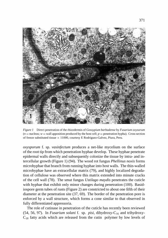
Figure 1 Direct penetration of the rhizodermis ofGossypium barbadenseby Fusarium oxysorum(n= nucleus;w=wall apposition produced by the host cell;p= penetration hypha). Cross sectionof freeze substituted tissue× 11000, courtesy E Rodriguez-Galvez, Piura, Peru.
oxysporumf. sp. vasinfectumproduces a net-like mycelium on the surfaceof the root tip from which penetration hyphae develop. These hyphae penetrateepidermal walls directly and subsequently colonize the tissue by intra- and in-tercellular growth (Figure 1) (94). The wood rot fungusPhellinus noxisformsmicrohyphae that branch from running hyphae into host walls. The thin-walledmicrohyphae have an extracellular matrix (79), and highly localized degrada-tion of cellulose was observed where this matrix extended into minute cracksof the cell wall (78). The smut fungusUstilago maydispenetrates the cuticlewith hyphae that exhibit only minor changes during penetration (100). Basid-iospore germ tubes of rusts (Figure 2) are constricted to about one fifth of theirdiameter at the penetration site (37, 69). The border of the penetration pore isenforced by a wall structure, which forms a cone similar to that observed infully differentiated appressoria.
The role of cutinase in penetration of the cuticle has recently been reviewed(54, 56, 97). InFusarium solanif. sp. pisi, dihydroxy-C16 and trihydroxy-C18 fatty acids which are released from the cutin polymer by low levels of
Ann
u. R
ev. P
hyto
path
ol. 1
996.
34:3
67-3
86. D
ownl
oade
d fr
om a
rjou
rnal
s.an
nual
revi
ews.
org
by D
euts
che
Fors
chun
gsge
mei
nsch
aft o
n 07
/25/
07. F
or p
erso
nal u
se o
nly.

July 12, 1996 11:3 Annual Reviews MENDTEXT.DUN AR14-19
372 MENDGEN, HAHN, & DEISING
constitutively expressed cutinase, together with soluble nuclear protein factorswere shown to activate cutinase gene transcription. Cyclic AMP and phospho-rylation of transcription factors appear to be important in regulating cutinasegene expression (55). Initially, a large body of data indicated that cutinaseof Fusariumplays an important role in penetration. Antibodies raised againstcutinase were used in microscopical studies to substantiate that secretion occursaround the infection site, and both chemical cutinase inhibitors and antibodiesdecreased the rate of infection when present in the infection droplet. Fur-thermore, virulence correlated with levels of cutinase expression in differentisolates. The cutinase gene ofFusarium solanif. sp. pisi was cloned andinserted into a cutinase-deficient isolate of the papaya wound pathogenMy-cosphaerellaspp. Transformants expressing cutinase were able to infect pa-paya fruits through the intact cuticle. Antibodies againstFusarium solanif. sp.pisicutinase prevented infection. These data were taken as strong evidence thatcutinase is a determinant of virulence inFusarium solanif. sp. pisi (54, 55).
The role of cutinase in penetration was questioned by Sch¨afer and co-workers,analyzing cutinase-deficient mutants ofFusarium solanif. sp. pisi generated
Figure 2 Direct penetration of the leaf cuticle by the basidiospore germling ofUromyces ap-pendiculatus( p = penetration site). Scanning electron micrograph× 3100, courtesy RE Gold,Limburgerhof, Germany.
Ann
u. R
ev. P
hyto
path
ol. 1
996.
34:3
67-3
86. D
ownl
oade
d fr
om a
rjou
rnal
s.an
nual
revi
ews.
org
by D
euts
che
Fors
chun
gsge
mei
nsch
aft o
n 07
/25/
07. F
or p
erso
nal u
se o
nly.
July 12, 1996 11:3 Annual Reviews MENDTEXT.DUN AR14-19
FUNGAL PENETRATION 373
by transformation-mediated gene disruption (102). Detailed quantitative andmicroscopic examination of infection and the colonization process did not revealany differences between wild-type and cutinase-deficient mutants and thereforegave no indication that cutinase is a virulence determinant in theF. solani f.sp. pisi-pea interaction (103). Kolattukudy and co-workers used the mutantproduced by Stahl & Sch¨afer (102) and inoculated pea seedlings with a rangeof different spore concentrations (95). These authors, however, reported thatvirulence of the mutant was strongly decreased. One explanation for theseconflicting data may be derived from the observation by K¨oller et al (56, 112a)that distinct cutinases are expressed during saprophytic and pathogenic stages ofAlternaria brassicicola. All Fusariumcutinases can be assumed to be inhibitedby chemical inhibitors, and possibly also by the polyclonal antisera used in peastem infection assays. However, targeted gene-disruption experiments mayhave inactivated only the cutinase gene active during saprophytic growth, butnot the cutinase gene(s) required for pathogenicity. Further studies are requiredto clarify the different roles played by cutinases.
Directly penetrating fungi that do not differentiate appressoria clearly needcell wall–degrading enzymes for penetration. Localized degradation of plantcell wall material, about 0.2µm deep, was observed along the penetrationhypha ofU. vignae. In this area, the density of pectin and xyloglucan epi-topes was reduced by about 50% ( H Xu & K Mendgen, unpublished result).Culture fluids of isolates ofFusarium solanif. sp. pisi differing in virulenceshowed similar polygalacturonase activities, but polygalacturonate lyase ac-tivities correlated with virulence (53); a comparable situation was found inFusarium oxysporumf. sp. lycopersiciandFusarium oxysporumf. sp.ciceri(30, 85). Antibodies against the lyase ofFusarium solanif. sp. pisi, but notthose against the polygalacturonases, protected pea tissue against infection (38).Studies using UV- or chemically induced mutants ofFusarium, Verticillium,Sclerotinia,or Alternaria did not indicate a critical role played by pectic en-zymes in pathogenesis (112). Also, after targeted disruption of aPenicilliumolsoniigene encodingendo-polygalacturonase, mutants infected the host,Ara-bidopsis thaliana, at rates comparable to the wild type (61). However, since notall of the polygalacturonases had been inactivated in the mutants, no conclusionconcerning their role can be drawn.
Elucidation of the importance of cell wall–degrading enzymes is hamperedby their redundancy and variable regulation. Often, several isoforms of a partic-ular enzyme occur that may be encoded by a single gene (13). In culture mediacontaining purified plant cell walls or polymers such as pectin, polygalacturonicacid, cellulose, xylans, and others, many fungi synthesize an array of enzymesrequired to degrade these carbon sources. The presence of a single polymersuch as polygalacturonic acid is often sufficient to induce a number of differ-ent enzymes, e.g. polygalacturonases, pectin and pectate (polygalacturonate)
Ann
u. R
ev. P
hyto
path
ol. 1
996.
34:3
67-3
86. D
ownl
oade
d fr
om a
rjou
rnal
s.an
nual
revi
ews.
org
by D
euts
che
Fors
chun
gsge
mei
nsch
aft o
n 07
/25/
07. F
or p
erso
nal u
se o
nly.

July 12, 1996 11:3 Annual Reviews MENDTEXT.DUN AR14-19
374 MENDGEN, HAHN, & DEISING
lyases, and pectin methylesterases. In plants, conserved promoter elementsof defense-related genes responsive to pectic fragments have been identified(15). A similar situation may exist in fungi, such that plant cell wall fragmentscould activate fungal transcription factors capable of simultaneous induction ofa number of genes encoding cell wall–degrading enzymes.
Whether enzymes produced in culture media are required for penetration ofplant cell walls or only for saprophytic growth of the fungus is unresolved. Fora better understanding of the role played by cell wall–degrading enzymes, thefocus of research needs to be shifted toward those enzymes produced duringpathogenesis, i.e. during penetration and growthin planta.
In the broad host range pathogenB. cinerea, pectin not only induces pecticenzymes (50), but also serves as a second inducer of laccase synthesis andsecretion (66). Laccases are thought to contribute to degradation of lignin,a polymer incorporated into plant cell walls in response to pathogen attack(111). Since pectin can be esterified to phenolic compounds like ferulic acid andcovalently linked with lignin (35), laccases may also be required for penetration(77). Importantly, specific inhibition of laccase by tetracyclic triterpenoidscalled cucurbitacins protected both cucumber fruits and cabbage leaves againstinfection byBotrytis cinerea(3).
PENETRATION FROM FULLY DEVELOPEDAPPRESSORIA
Many fungi such asColletotrichum, Magnaporthe, Cochliobolus, or the di-karyon of rusts (Figure 3) form well-differentiated appressoria. Chemical sig-nals such as potassium and calcium ions, simple sugars, acrolein, and pH gra-dients, as well as temperature shifts induce appressorium formation (44), buttheir significance is unclear. The major signals provided by the host are hy-drophobicity (49, 64), hardness (120), components of the plant surface (86), andtopographical properties (44).Colletotrichum gloeosporioidesandC. musae,which cause rot in climacteric fruits such as tomato, avocado, and banana, usethe fruit-ripening hormone ethylene as a signal for germination and differenti-ation (31). Conidia of these fungi germinate and form multiple appressoria onglass surfaces after exposure to micromolar concentrations of ethylene gas. Thatethylene emanating from ripening fruit is the actual signal was elegantly shownby using transgenic tomato fruits incapable of ethylene production. In contrastto normal tomato fruits, these transgenic fruits did not support fungal germina-tion and appressorium differentiation unless exogenous ethylene was applied.In C. gloeosporioides(86), host surface wax components have also been shownto induce germination and appressorium differentiation. Recently, genes ex-pressed during induction of appressorium formation inC. gloeosporioidesby
Ann
u. R
ev. P
hyto
path
ol. 1
996.
34:3
67-3
86. D
ownl
oade
d fr
om a
rjou
rnal
s.an
nual
revi
ews.
org
by D
euts
che
Fors
chun
gsge
mei
nsch
aft o
n 07
/25/
07. F
or p
erso
nal u
se o
nly.

July 12, 1996 11:3 Annual Reviews MENDTEXT.DUN AR14-19
FUNGAL PENETRATION 375
Figure 3 Penetration of the stomatal pore from an appressorium by the uredospore germling ofU. appendiculatus(s= stoma,a= appressorium). Low temperature scanning electron micrograph× 1000.
the host surface wax have been cloned. Disruption of one of these genes,cap20,caused failure of the mutants to penetrate into the host tissue (48).
During different stages of its life cycle, the same fungus may respond todifferent signals provided by the same plant surface. Whereas the monokaryonof U. appendiculatus(Figure 2) differentiates infection structures in responseto the hardness of the substrate (33), the dikaryon (Figure 3) differentiatesappressoria in response to the correct dimensions of a ridge formed by thestomatal lips of the guard cell (44, 104). The signal for differentiation can beperceived inU. appendiculatusby the first 10µm of the hyphal tip (20). Within4 min after signal perception, the cytoskeleton and the vesicles in the apex ofthe hypha are reorganized along the wall (62, 63). After the appressorial wallstructure is completed, a new polarity emerges: Glycoproteins unique for theplasmalemma in the appressorium ofC. lindemuthianumwere detected onlyoutside of the area of contact between the appressorium and the host cuticle,where the initials of the penetration hypha arise (82).
Fungi penetrating from fully developed appressoria show some variabilitywith respect to melanization of the appressorial wall. For example,M. grisea
Ann
u. R
ev. P
hyto
path
ol. 1
996.
34:3
67-3
86. D
ownl
oade
d fr
om a
rjou
rnal
s.an
nual
revi
ews.
org
by D
euts
che
Fors
chun
gsge
mei
nsch
aft o
n 07
/25/
07. F
or p
erso
nal u
se o
nly.
July 12, 1996 11:3 Annual Reviews MENDTEXT.DUN AR14-19
376 MENDGEN, HAHN, & DEISING
andColletotrichumspp. both incorporate a distinct melanin layer into theirappressorial walls. Since only water but not osmotically active compounds canpass the plasma membrane and appressorial wall, a substantial pressure can begenerated. A turgor pressure of 8.0 MPa (80 bar) was measured in appressoriaof M. grisea(47). This tugor pressure seems to be due to molar concentra-tions of glycerol in the appressoria ofM. grisea(BJ McCormack & NJ Talbot,unpublished result). Thus, even though these fungi are also capable of synthe-sizing cutinases and cell wall–degrading enzymes (11, 43, 106, 117), the datapresented by Howard et al (47) suggest that fungi with melanized appressoriasuch asMagnaportheand possiblyColletotrichummay be able to penetrate thehost cuticle and cell wall mainly by means of turgor pressure. The essentialrole of melanin for penetration is supported by the observation that chemi-cally induced melanin-deficient mutants are both unable to form melanizedappressoria and are apathogenic (17, 57). Addition of appropriate precursorsof melanin biosynthesis restored melanin incorporation into the appressoriumand pathogenicity of the mutants. Correspondingly, after transformation of amutant ofC. lagenariumwith wild-type DNA, transformants were obtainedwith melanized appressoria and normal virulence (58, 84a). Microscopical in-vestigations of melanin-deficient mutants ofMagnaportheandColletotrichumrevealed differences in the attempts to penetrate the host. While differentiationstopped in the mutant ofM. grisea, the mutant ofC. lagenariumformed lateralappressorial germ tubes and secondary appressoria (57). Lateral developmentmay indicate that appressoria ofColletotrichumdo not adhere as tightly to theirsubstratum as those ofMagnaporthe. The tight adhesion ofMagnaportheap-pressoria could be mediated by a ring of glue-like substances visible below thebase of appressoria around the penetration pore (46). These substances maybe similar to those found at conidial tips (40), or to extracellular glycoproteinsthought to be involved in cellular differentiation (119).
THE PENETRATION HYPHA
Extensively studied examples for penetration hypha development areC. linde-muthianum(80), M. grisea(46), and rusts (68). The penetration hypha startsto grow from a pore in the middle of the appressorial base. The pore wall isvery thin but often has reinforced borders. This reinforcement can turn into athick wall structure, called the appressorial cone. Thus, pressure, generated byturgor and possibly also by the cytoskeleton, is exerted over the restricted areaof the pore by the growing penetration hypha.
A pressure of 8 MPa within the appressorium ofM. griseapushes the pen-etration hypha into artificial nonbiodegradable substrata such as Teflon (47).Assuming that the wall of the fungus offers no resistance to extension, the max-imum force produced at the hyphal tip would be in the order of 8× 10⁻⁶ N (71).
Ann
u. R
ev. P
hyto
path
ol. 1
996.
34:3
67-3
86. D
ownl
oade
d fr
om a
rjou
rnal
s.an
nual
revi
ews.
org
by D
euts
che
Fors
chun
gsge
mei
nsch
aft o
n 07
/25/
07. F
or p
erso
nal u
se o
nly.

July 12, 1996 11:3 Annual Reviews MENDTEXT.DUN AR14-19
FUNGAL PENETRATION 377
In contrast, appressoria ofU. appendiculatushave a turgor pressure of only 0.35MPa. Nevertheless, the base of this appressorium can distort polystyrene ridges,and the emerging penetration hypha can curl the stomatal lip inward (109).
The contribution of the cytoskeleton to these forces is unclear. Forces prob-ably produced by actin and exerted on microneedles at the front of migratingkeratocytes only reached a value of 6× 10⁻⁸ N (81), corresponding to a pres-sure of less than 0.002 MPa. These figures suggest that the force generated bythe cytoskeleton is of some importance only if the turgor pressure is very low,as suggested for the water moldSaprolegniawhen grown under osmotic stress(72). On the other hand, the high concentration of cytoskeletal elements ob-served in the penetration hyphae ofM. grisea(8) andU. appendiculatus(121)may be necessary to determine the shape of this hypha. Even on the surface ofscratched membranes, where no host cell wall restricts development of the fun-gus, penetration hyphae ofUromycesstill keep their typical shape, indicatingthat it is controlled morphogenetically (23, 68). A stabilization of the tip of thepenetration hypha would also be needed in fungi likeM. griseato compensatefor the dramatic differences in osmotic pressure between appressorial and hostcell protoplasts during the initial contact with the infected cell.
Compared to the tip of the germ tube, the wall of the penetration hypha hasnew features. InM. grisea(46),C. lindemuthianum(80), and several rusts (22,34), the wall has strongly reduced affinity to wheat germ agglutinin, suggestinga reduced chitin content or a modification of chitin. InC. lagenariumandU. viciae-fabae, an increase in chitin deacetylase activity was observed afterappressorium development (25, 99a, Figure 4). This enzyme activity probablymodifies the chitin molecule on the surface of the penetration hypha. Sincedecreasing degrees of acetylation result in less efficient cleavage of chitin byendochitinases (91), the penetration hypha could be protected by enzymaticdeacetylation. Furthermore, the amount of chitin degradation products, whichmay serve as elicitors of host defense reactions, would be reduced (22).
Penetration in the area of the appressorial pore is likely to be supported byenzymes that soften the host cell wall. At the beginning of development ofthe rust penetration hypha, apical vesicles start to accumulate over the pen-etration pore (105). InU. viciae-fabae, differentiation of appressorium andpenetration hypha is accompanied by secretion of a complex pattern of lyticenzymes (Figure 4). With thigmo-inductive polyethylene membranes (23), en-zyme formation during infection structure differentiation was analyzed in theabsence of the plant. Thus, enzymes synthesized and secreted by the funguscan clearly be distiguished from cell wall polymer-degrading or -modifying en-zymes formed by the plant at different stages of development (30a, 35a). Duringappressorium formation, several extracellular proteases appear that show speci-ficity for fibrous hydroxyproline-rich proteins (88), reminiscent of structural
Ann
u. R
ev. P
hyto
path
ol. 1
996.
34:3
67-3
86. D
ownl
oade
d fr
om a
rjou
rnal
s.an
nual
revi
ews.
org
by D
euts
che
Fors
chun
gsge
mei
nsch
aft o
n 07
/25/
07. F
or p
erso
nal u
se o
nly.
July 12, 1996 11:3 Annual Reviews MENDTEXT.DUN AR14-19
378 MENDGEN, HAHN, & DEISING
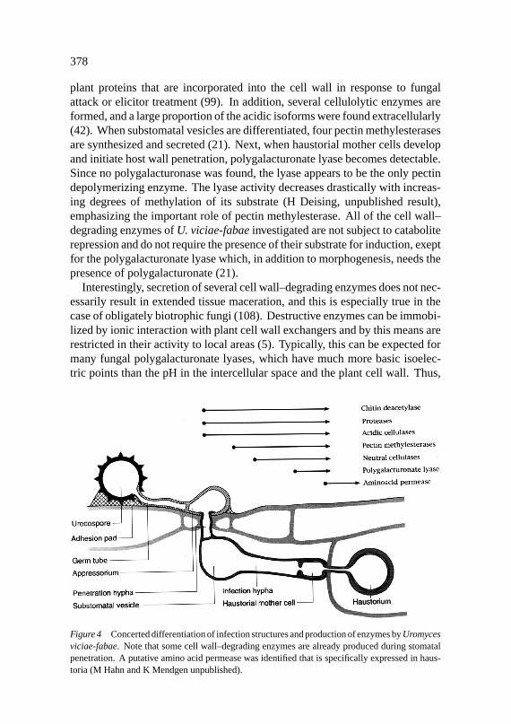
plant proteins that are incorporated into the cell wall in response to fungalattack or elicitor treatment (99). In addition, several cellulolytic enzymes areformed, and a large proportion of the acidic isoforms were found extracellularly(42). When substomatal vesicles are differentiated, four pectin methylesterasesare synthesized and secreted (21). Next, when haustorial mother cells developand initiate host wall penetration, polygalacturonate lyase becomes detectable.Since no polygalacturonase was found, the lyase appears to be the only pectindepolymerizing enzyme. The lyase activity decreases drastically with increas-ing degrees of methylation of its substrate (H Deising, unpublished result),emphasizing the important role of pectin methylesterase. All of the cell wall–degrading enzymes ofU. viciae-fabaeinvestigated are not subject to cataboliterepression and do not require the presence of their substrate for induction, exeptfor the polygalacturonate lyase which, in addition to morphogenesis, needs thepresence of polygalacturonate (21).
Interestingly, secretion of several cell wall–degrading enzymes does not nec-essarily result in extended tissue maceration, and this is especially true in thecase of obligately biotrophic fungi (108). Destructive enzymes can be immobi-lized by ionic interaction with plant cell wall exchangers and by this means arerestricted in their activity to local areas (5). Typically, this can be expected formany fungal polygalacturonate lyases, which have much more basic isoelec-tric points than the pH in the intercellular space and the plant cell wall. Thus,
Figure 4 Concerted differentiation of infection structures and production of enzymes byUromycesviciae-fabae. Note that some cell wall–degrading enzymes are already produced during stomatalpenetration. A putative amino acid permease was identified that is specifically expressed in haus-toria (M Hahn and K Mendgen unpublished).
Ann
u. R
ev. P
hyto
path
ol. 1
996.
34:3
67-3
86. D
ownl
oade
d fr
om a
rjou
rnal
s.an
nual
revi
ews.
org
by D
euts
che
Fors
chun
gsge
mei
nsch
aft o
n 07
/25/
07. F
or p
erso
nal u
se o
nly.
July 12, 1996 11:3 Annual Reviews MENDTEXT.DUN AR14-19
FUNGAL PENETRATION 379
physicochemical properties of these enzymes are likely to determine the degreeof cell or tissue damage (21).
In C. gloeosporioides, evidence for the role of cell wall–degrading enzymesduring the penetration process has been obtained by Prusky and coworkers(114). Both pectin-degrading enzymes and penetration of fruit tissue are inhi-bited by epicatechin, a phenolic compound present in the peel of unripe avocadofruits. During ripening, levels of epicatechin decrease to a threshold not in-hibitory to the pectic enzymes, and the fungus gains the ability to spread withinthe fruit.
Cochliobolusspp. form only weakly melanized appressoria (113), andmelanin-deficient mutants of the rice pathogenC. miyabeanusremain infec-tious (59). On the other hand,Cochliobolusspecies produce a large number ofcell wall–degrading enzymes that might play an important role in the penetra-tion process. To test this hypothesis, Walton and co-workers performed targetedinactivation of genes ofC. carbonumencoding cell wall–degrading enzymessuch asendo-polygalacturonase, xylanase,β-1,3-glucanase, and cellulase (2,96, 98, 101). Likewise, in the apple fruit pathogenGlomerella cingulata, a genecoding for a pectin lyase was inactivated (9). In neither case was the virulenceof the resulting mutants affected. However, all mutants retained various degreesof residual enzyme activity, probably due to genes encoding isoforms of therespective enzyme, which were not affected by mutagenesis. Thus, based onthe data currently available, no general conclusion can be drawn concerning therole played by fungal cell wall–degrading enzymes in the penetration process.
CONCLUDING REMARKS
Research over the past few years has significantly increased our knowledgeof penetration by plant pathogenic fungi, unfolding a process of unexpectedcomplexity. The diversity of fungal penetration strategies as well as the manytools needed by a particular pathogen to successfully enter its host make gen-eralizations difficult.
The ability to construct defined single-gene disruption mutants in a numberof phytopathogenic fungi has so far given clear-cut answers only in a few cases,such as the role of melanin in the formation of functional appressoria. Theparticular role of cutinase and of cell wall–degrading enzymes is still unre-solved, due to the redundancy of the encoding genes. Functional redundancyis often indicative of processes with vital importance to an organism. Numer-ous enzymes, sometimes with overlapping activities, are needed to degradethe complex web of carbohydrates, glycoproteins, and phenolic compounds ofthe plant cell wall. This might explain the difficulty in detecting the effect of asingle enzyme in the combination of activities involved in cell wall degradation.
Ann
u. R
ev. P
hyto
path
ol. 1
996.
34:3
67-3
86. D
ownl
oade
d fr
om a
rjou
rnal
s.an
nual
revi
ews.
org
by D
euts
che
Fors
chun
gsge
mei
nsch
aft o
n 07
/25/
07. F
or p
erso
nal u
se o
nly.
July 12, 1996 11:3 Annual Reviews MENDTEXT.DUN AR14-19
380 MENDGEN, HAHN, & DEISING
There is an interesting parallel in the bacterial soft rottingErwinia spp., inwhich the cell wall–degrading enzymes are encoded by several gene families(4). Disruption of single genes never completely abolished pathogenicity, andin many cases no effect on virulence could be observed. Even the deletion ofall known pectate lyase genes did not eliminate macerating ability because ofthe presence of newly detected pectate lyase genes that were only expressed inplanta (1, 4). These observations are similar to those obtained by K¨oller et al(56) with cutinases fromAlternariaand suggest similar molecular strategies ofbacterial and fungal plant pathogens.
Transformation and targeted gene disruption mutagenesis remains a laboriousand time-consuming task for most of the phytopathogenic fungi, and there is stillno effective way to stably transform the biotrophic fungi. Therefore, we cannotexpect rapid progress in elucidating the mechanisms of cell wall penetration,except for some model systems such asC. carbonum(112).
Instead of altering the ability of a particular fungal pathogen to produce cellwall–degrading enzymes and to analyze the resulting phenotypes, work withgenetically altered host plants offers a promising alternative. For example, intransgenic tomatoes, high-level expression of pear polygalacturonase inhibitoryproteins (PGIPs) led to increased resistance of the fruits toB. cinerea, providingindirect evidence that polygalacturonase might be a virulence factor in thisfungus (87). Mutants defective in certain cell wall carbohydrates have beenobtained fromArabidopsis(90), and these mutants can be tested for an alteredsusceptibility toArabidopsispathogens.
Because of the limitations of approaches toward known enzymatic activi-ties, molecular genetic techniques that do not requirea priori knowledge ofthe pathosystem are increasingly used by plant pathologists. These techniquesinclude the search for genes specifically expressed during penetration and sub-sequent analysis of their mutant phenotypes (48), as well as random inser-tion mutagenesis (e.g. REMI) followed by screening for mutants defective inpathogenicity (6). The great promise of these “black box” approaches is thatthey will provide insight into the molecular mechanisms of penetration that arestill obscure.
With modern molecular techniques, it now seems possible to uncover thecascade of signaling events between and within the fungal parasite and its hostplant during penetration. Identification of plant-derived signals and parts ofthe signal transduction chains involved in cutinase induction inF. solaniandappressorium formation inC. gloeosporioidesandM. grisea illustrate thesedevelopments. On the other hand, our knowledge of molecular and cytologicalevents inside and surrounding the invading fungus is limited. We still do notunderstand the mechanism of highly specific, pathogen-induced responses ofhost plants, for example, the formation of a unique type of plasma membrane
Ann
u. R
ev. P
hyto
path
ol. 1
996.
34:3
67-3
86. D
ownl
oade
d fr
om a
rjou
rnal
s.an
nual
revi
ews.
org
by D
euts
che
Fors
chun
gsge
mei
nsch
aft o
n 07
/25/
07. F
or p
erso
nal u
se o
nly.

July 12, 1996 11:3 Annual Reviews MENDTEXT.DUN AR14-19
FUNGAL PENETRATION 381
(the extrahaustorial membrane) that surrounds haustoria of biotrophic fungiand apparently supports their nutrition. So far, cytological techniques havemainly been used for a descriptive view of fungal pathogenesis, but with thehelp of molecular biology, electron microscopy can be used to locate specificmolecules at the penetration site. Careful cytological investigations will alsobe indispensable for the analysis of gene disruption mutants that do not reveala phenotype by macroscopic evaluation. During penetration, the decision isoften made whether or not a pathogen will succeed in colonizing the plant. Weare still far from understanding all the factors determining the outcome of thisbattle, and therefore the initial phase of fungal infection remains a subject ofgreat scientific and practical interest.
ACKNOWLEDGMENTS
We thank RL Nicholson, HC Hoch, NP Money, and M Rauscher for helpfulcomments on the manuscript, and the Deutsche Forschungsgemeinschaft forfinancial support.
Literature Cited
1. Alghisi P, Favaron F. 1995. Pectin-degrading enzymes and plant-parasiteinteractions. Eur. J. Plant Pathol.101:365–75
2. Apel PC, Panaccione DG, Holden FR,Walton JD. 1993. Cloning and tar-geted gene disruption ofXYL1, aβ1,4-xylanase gene from the maize pathogenCochliobolus carbonum. Mol. Plant-Microbe Interact.6:467–73
3. Bar-Nun N, Mayer AM. 1990. Cucur-bitacins protect cucumber tissue againstinfection by Botrytis cinerea. Phyto-chemistry29:787–91
4. Barras F, von Gijsegem F, ChatterjeeAK. 1994. Extracellular enzymes andpathogenesis of soft-rotErwinia. Annu.Rev. Phytopathol.32:201–34
5. Benhamou N, Chamberland H, Pauz´eFJ. 1990. Implication of pectic com-ponents in cell surface interactions be-tween tomato root cells andFusar-ium oxysporumf. sp.radicis-lycopersici.Plant Physiol.92:995–1003
6. Bolker M, Bohnert HU, Braun KH,Gorl J, Kahmann R. 1995. Taggingpathogenicity genes inUstilago may-disby restriction enzyme-mediated inte-gration (REMI).Mol. Gen. Genet.248:547–52
7. Bonfante P, Perotto S. 1995. Strategiesof arbuscular mycorrhizal fungi when in-
fecting host plants.New Phytol.130:3–21
8. Bourett TM, Howard RJ. 1992. Actin inpenetration pegs of the fungal rice blastpathogen,Magnaporthe grisea. Proto-plasma168:20–26
9. Bowen JK, Templeton MD, SharrockKR, Crowhurst RN, Rikkerink EHA.1995. Gene inactivation in the plantpathogenGlomerella cingulata: threestrategies for the disruption of the pectinlyase genepnlA. Mol. Gen. Genet.246:196–205
10. Braun EJ, Howard RJ. 1994. Adhesionof Cochliobolus heterostrophusconidiaand germlings to leaves and artificial sur-faces.Exp. Mycol.18:211–20
11. Bucheli P, Doares SH, AlbersheimP, Darvill A. 1990. Host-pathogeninteractions XXXVI. Partial purifica-tion and characterization of heat-labilemolecules secreted by the rice blastpathogen that solubilize plant cell wallfragments that kill plant cells.Physiol.Mol. Plant Pathol.36:159–73
12. Cairney JWG, Bruke RM. 1994. Fun-gal enzymes degrading plant cell walls:their possible significance in the ec-tomycorrhizal symbiosis.Mycol. Res.98:1345–56
13. Caprari C, Bergmann C, Migheli Q,Salvi G, Albersheim P, Darvill A, Cer-
Ann
u. R
ev. P
hyto
path
ol. 1
996.
34:3
67-3
86. D
ownl
oade
d fr
om a
rjou
rnal
s.an
nual
revi
ews.
org
by D
euts
che
Fors
chun
gsge
mei
nsch
aft o
n 07
/25/
07. F
or p
erso
nal u
se o
nly.

July 12, 1996 11:3 Annual Reviews MENDTEXT.DUN AR14-19
382 MENDGEN, HAHN, & DEISING
vone F, De Lorenzo G. 1993.Fusar-ium moniliforme secretes four endo-polygalacturonases derived from a sin-gle gene product.Physiol. Mol. PlantPathol.43:453–62
14. Carver TLW, Ingerson-Morris SM,Thomas BJ, Zeyen RJ. 1995. Early in-teractions during powdery mildew infec-tion. Can. J. Bot.73:S632–39
15. Cervone F, De Lorenzo G, Caprari C,Clark AJ, Desiderio A, et al. 1993. Theinteraction between fungal endopoly-galacturonase and plant cell wall PGIP(polygalacturonase-inhibiting protein).In Mechanisms of Plant Defense Re-sponses, ed. B Fritig, M Legrand, pp.64–7. Dordrecht: Kluwer
16. Chaubal R, Wilmot VA, Wynn WK.1991. Visualization, adhesiveness andcytochemistry of the extracellular ma-trix produced by germ tubes ofPucciniasorghi. Can. J. Bot.69:2044–54
17. Chumley FG, Valent B. 1990. Ge-netic analysis of melanin-deficient, non-pathogenic mutants ofMagnaporthegrisea. Mol. Plant-Microbe Interact.3:135–43
18. Clay RP, Enkerli J, Fuller MS. 1994. In-duction and formation ofCochliobolussativusappressoria.Protoplasma178:34–47
19. Clement JA, Porter R, Butt TM, BeckettA. 1994. The role of hydrophobicity inattachment of urediniospores and sporel-ings of Uromyces viciae-fabae. Mycol.Res.98:1217–28
19a. Cole GT, Hoch HC, eds. 1991.TheFungal Spore and Disease Initiation inPlants and Animals. New York: Plenum.555 pp.
19b. Cole L, Dewey F M, Hawes C R.1996. Infection mechanisms ofBotry-tis species: pre-penetration and pre-infection processes of dry and wet coni-dia.Mycol. Res.100:277–86
20. Correa AJ, Hoch HC. 1995. Identifica-tion of thigmoresponsive loci for celldifferentiation inUromycesgermlings.Protoplasma186:34–40
21. Deising H, Frittrang AK, Kunz S, Mend-gen K. 1995. Regulation of pectinmethylesterase and polygalacturonatelyase activity during differentiation ofinfection structures inUromyces viciae-fabae. Microbiology141:561–71
22. Deising H, Heiler S, Rauscher M, Xu H,Mendgen K. 1996. Cellular aspects ofrust infection structure differentiation:spore adhesion and fungal morphogene-sis. See Ref. 79a, pp. 135–56
23. Deising H, Jungblut PR, Mendgen K.1991. Differentiation-related proteins ofthe broad bean rust fungusUromycesviciae-fabae, as revealed by high reso-lution two-dimensional polyacrylamidegel electrophoresis.Arch. Microbiol.155:191–98
24. Deising H, Nicholson RL, Haug M,Howard RJ, Mendgen K. 1992. Adhe-sion pad formation and the involvementof cutinase and esterases in the attach-ment of uredospores to the host cuticle.Plant Cell4:1101–11
25. Deising H, Siegrist J. 1995. Chitindeacetylase activity of the rustUromycesviciae-fabae is controlled by fungalmorphogenesis.FEMS Microbiol. Lett.127:207–12
26. Dixon DM, Szanislo PJ, Polak A. 1991.Dihydroxynaphthalene melanin and itsrelationship with virulence in the earlystages of Phaeohyphomycosis. See Ref.19a, pp. 297–318
27. Doss RP, Potter SW, Chastagner GA,Christian JK. 1993. Adhesion of non-germinatedBotrytis cinereaconidia toseveral substrata.Appl. Environ. Micro-biol. 59:1786–91
28. Doss RP, Potter SW, Soeldner AH,Christian JK, Fukunaga LE. 1995. Ad-hesion of germlings ofBotrytis cinerea.Appl. Environ. Microbiol.61:260–5
29. Epstein L, Laccetti LB, Staples RC,Hoch HC. 1987. Cell-substratum adhe-sive protein involved in surface contactresponses of the bean rust fungus.Phys-iol. Mol. Plant Pathol.30:373–88
30. Fernandez N, Pati˜no B, Vazquez C.1993. Pectin degrading enzymes se-creted by six isolates ofFusarium oxys-porum. Mycol. Res.97:461–66
30a. Fischer RL, Bennett AB. 1991. Role ofcell wall hydrolases in fruit ripening.Annu. Rev. Plant Physiol. Plant Mol.Biol. 42:675–703
31. Flaishman MA, Kolattukudy PE. 1994.Timing of fungal invasion using host’sripening hormone as a signal.Proc. Natl.Acad. Sci. USA91:6579–83
32. Forster H, Mendgen K. 1987. Im-munocytochemical localization ofpectinesterases in hyphae ofPhy-tophthora infestans. Can. J. Bot.65:2607–13
33. Freytag S, Bruscaglioni L, Gold RE,Mendgen K. 1988. Basidiospores of rustfungi (Uromycesspecies) differentiateinfection structuresin vitro. Exp. Mycol.12:275–83
34. Freytag S, Mendgen K. 1991. Carbohy-
Ann
u. R
ev. P
hyto
path
ol. 1
996.
34:3
67-3
86. D
ownl
oade
d fr
om a
rjou
rnal
s.an
nual
revi
ews.
org
by D
euts
che
Fors
chun
gsge
mei
nsch
aft o
n 07
/25/
07. F
or p
erso
nal u
se o
nly.

July 12, 1996 11:3 Annual Reviews MENDTEXT.DUN AR14-19
FUNGAL PENETRATION 383
drates on the surface of urediniospore-and basidiospore-derived infectionstructures of heteroecious and au-toecious rust fungi. New Phytol.119:527–34
35. Fry SC. 1986. Cross-linking of matrixpolymers in the growing cell walls ofangiosperms.Annu. Rev. Plant Physiol.37:165–86
35a. Fry SC. 1995. Polysaccharide-modify-ing enzymes in the plant cell wall.Annu.Rev. Plant Physiol. Plant Mol. Biol.46:497–520
36. Girbardt M. 1957. Der Spitzenk¨orpervon Polystictus versicolor. Planta50:47–59
37. Gold RE, Mendgen K. 1991. Rust basid-iospore germlings and disease initiation.See Ref. 19a, pp. 67–99
38. Gonzalez-Candelas L, Kolattukudy PE.1992. Isolation and analysis of anovel inducible pectate lyase gene fromthe phytopathogenic fungusFusariumsolani f. sp. pisi (Nectria haemato-cocca, mating population VI).J. Bac-teriol. 174:6343–49
39. Gow NAR. 1994. Growth and guid-ance of the fungal hypha.Microbiology140:3193–205
40. Hamer JE, Howard RJ, Chumley FG, Va-lent B. 1988. A mechanism for attach-ment in spores of a plant pathogenic fun-gus.Science239:288–90
41. Heath IB. 1994. The cytoskeleton in hy-phal growth, organelle movements, andmitosis. In The Mycota I, ed. J Wes-sels, F Meinhardt, pp. 43–65. Heidel-berg: Springer
42. Heiler S, Mendgen K, Deising H. 1993.Cellulolytic enzymes of the obligatelybiotrophic rust fungusUromyces viciae-fabae are regulated differentiation-specifically.Mycol. Res.97:77–85
43. Hirayama T, Sudo T, Nagayama H, Mat-suda K, Tamari K. 1976. Number andinterrelation of components of CX en-zyme from Pyricularia oryzae. Agric.Biol. Chem.40:2137–42
44. Hoch HC, Staples RC. 1991. Signal-ing for infection structure formation infungi. See Ref. 19a, pp. 25–46
45. Hoch HC, Staples RC. 1987. Struc-tural and chemical changes among therust fungi during appressorium develop-ment.Annu. Rev. Phytopathol.25:231–47
46. Howard RJ, Bourett TM, Ferrari MA.1991. Infection byMagnaporthe: an invitro analysis. See Ref. 68a, pp. 251–64
47. Howard RJ, Ferrari MA, Roach DH,
Money NP. 1991. Penetration of hardsubstrates by a fungus employing enor-mous turgor pressures.Proc. Natl. Acad.Sci. USA88:11281–84
48. Hwang C-S, Flaishman MA, Kolat-tukudy PE. 1995. Cloning of a gene ex-pressed during appressorium formationby Colletotrichum gloeosporioidesanda marked decrease in virulence by dis-ruption of this gene.Plant Cell 7:183–93
49. Jelitto TC, Page HA, Read ND. 1994.Role of external signals in regulating thepre-penetration phase of infection by therice blast fungus,Magnaporthe grisea.Planta194:471–77
50. Johnston DJ, Williamson B. 1992.Purification and characterization offour polygalacturonases fromBotrytiscinerea. Mycol. Res.96:343–49
51. Jones EBG. 1994. Fungal adhesion.My-col. Res.98:961–81
52. Klis FM. 1994. Protein secretion inyeast. InThe Mycota I, ed. J Wessels,F Meinhardt, pp. 25–41. Heidelberg:Springer
53. Kolattukudy PE, Crawford MS. 1987.The role of polymer degrading enzymesin fungal pathogenesis. InMolecularDeterminants of Plant Diseases, ed. SNishimura, VP Vance, N Doke, pp. 75–95. Heidelberg/Tokyo: Springer
54. Kolattukudy PE, K¨amper J, K¨amper U,Gonzalez-Candel´as L, Guo W. 1994.Fungus-induced degradation and rein-forcement of defensive barriers of plants.See Ref. 85a, pp. 67–79
55. Kolattukudy PE, Rogers LM, Li D,Hwang C-S, Flaishman MA. 1995. Sur-face signaling in pathogenesis.Proc.Natl. Acad. Sci. USA92:4080–87
56. Koller W, Yao C, Trail F, Parker DM.1995. Role of cutinase in the invasion ofplants.Can. J. Bot.73:S1109–18
57. Kubo Y, Furusawa I. 1991. Melaninbiosynthesis. Prerequisite for success-ful invasion of the host by appressoriaof ColletotrichumandPyricularia. SeeRef. 19a, pp. 205–18
58. Kubo Y, Nakamura H, Kobayashi K,Okuno T, Furusawa I. 1991. Cloningof a melanin biosynthetic gene es-sential for appressorial penetration ofColletotrichum lagenarium. Mol. Plant-Microbe Interact.4:440–45
59. Kubo Y, Tsuda M, Furusawa I,Shishiyama J. 1989. Genetic analysis ofgenes involved in melanin biosynthesisof Cochliobolus miyabeanus. Exp. My-col. 13:77–84
Ann
u. R
ev. P
hyto
path
ol. 1
996.
34:3
67-3
86. D
ownl
oade
d fr
om a
rjou
rnal
s.an
nual
revi
ews.
org
by D
euts
che
Fors
chun
gsge
mei
nsch
aft o
n 07
/25/
07. F
or p
erso
nal u
se o
nly.

July 12, 1996 11:3 Annual Reviews MENDTEXT.DUN AR14-19
384 MENDGEN, HAHN, & DEISING
60. Kunoh H, Nicholson RL, Yoshioka H,Yamaoka N, Kobayashi I. 1990. Prepa-ration of the infection court byErysiphegraminis: degradation of the host cuti-cle.Physiol. Mol. Plant Pathol.36:397–407
61. Kusserow H, Scha¨afer W. 1994. The roleof polygalacturonase in the interactionbetweenPenicillium olsonii and Ara-bidopsis thaliana. Abstr. 442. 7thInt.Symp. Mol. Plant Micobe Interact.,Ed-inburgh
62. Kwon YH, Hoch HC, Aist JR. 1991.Initiation of appressorium formation inUromyces appendiculatus: organizationof the apex, and the responses involvingmicrotubules and apical vesicles.Can. J.Bot.69:2560–73
63. Kwon YH, Hoch HC, Staples RC. 1991.Cytoskeletal organization inUromycesurediospore germlings apices duringappressorium formation.Protoplasma165:37–50
63a. Kwon YH, Epstein L. 1993. A 90 kDaglycoprotein associated with adhesion ofNectria haematococcamacroconidia tosubstrata.Mol. Plant Microbe Interact.6:481–87
64. Lee Y-H, Dean RA. 1993. cAMP regu-lates infection structure formation in theplant pathogenic fungusMagnaporthegrisea. Plant Cell5:693–700
65. Lopez-Franco R, Howard RJ, BrackerCE. 1995. Satellite Spitzenk¨orper ingrowing hyphal tips. Protoplasma188:85–103
66. Marbach I, Harel E, Mayer AM. 1985.Pectin, a second inducer for laccase pro-duction byBotrytis cinerea. Phytochem-istry 24:2559–61
67. Mendgen K, Bachem U, Stark-Urnau M,Xu H. 1995. Secretion and endocytosisat the interface of plants and fungi.Can.J. Bot.73:S640–48
68. Mendgen K, Deising H. 1993. Infectionstructures of fungal plant pathogens—a cytological and physiological evalua-tion. New Phytol.124:193–213
68a. Mendgen K, Lesemann D-E, eds.1991. Electron Microscopy of PlantPathogens. Berlin: Springer. 336 pp.
69. Mims CW. 1991. Using electron mi-croscopy to study plant pathogenicfungi. Mycologia83:1–19
70. Moloshok TD, Leinhos GME, StaplesRC, Hoch HC. 1993. The autogenicextracellular environment ofUromycesappendiculatusurediospore germlings.Mycologia85:392–400
71. Money NP. 1995. Turgor pressure and
the mechanics of fungal penetration.Can. J. Bot.73:S96–102
72. Money NP, Harold FM. 1993. Two wa-ter molds can grow without measurabletugor pressure.Planta190:426–30
73. Nicholson RL. 1996. Adhesion of fungalpropagules. See Ref. 79a, pp. 117–34
74. Nicholson RL, Epstein L. 1991. Adhe-sion of fungi to the plant surface: pre-requisite for pathogenesis. See Ref. 19a,pp. 3–23
75. Nicholson RL, Kunoh H, Shiraishi T,Yamada T. 1993. Initiation of the infec-tion process byErysiphe graminis: con-version of the conidial surface from hy-drophobicity to hydrophilicity and influ-ence of the conidial exudate on the hy-drophobicity of the barley leaf surface.Physiol. Mol. Plant Pathol.43:307–18
76. Nicholson RL, Yoshioka H, Yamaoka N,Kunoh H. 1988. Preparation of the infec-tion court byErysiphe graminis. II. Re-lease of esterase enzyme from conidiain response to a contact stimulus.Exp.Mycol.12:336–49
77. Nicole M, Chamberland H, Geiger JP,Lecours N, Valero J, et al. 1992. Im-munocytochemical localization of lac-case in wood decayed byRigido-porus lignosus. Appl. Environ. Micro-biol. 58:1727–39
78. Nicole M, Chamberland H, Rioux D,Lecours N, Rio B, et al. 1993. A cyto-chemical study of extracellular sheathsassociated withRigidiporus lignosusduring wood decay.Appl. Environ. Mi-crobiol. 59:2578–88
79. Nicole M, Chamberland H, Rioux D,Xixuan X, Blanchettte RA, et al. 1995.Wood degradation byPhellinus noxius:ultrastructure and cytochemistry.Can. J.Microbiol. 41:253–65
79a. Nicole M, Gianinazzi-Pearson V, eds.1996. Histology, Ultrastructure andMolecular Cytology of Plant-Micro-organism Interactions. Dordrecht:Kluwer. 261 pp.
80. O’Connell RJ, Bailey JA. 1991.Hemibiotrophy in Colletotrichumlindemuthianum. See ref. 68a, pp.211–22
80a. O’Connell RJ, Pain NA, HutchinsonKA, Jones GL, Green JR. 1996. Ul-trastructure and composition of thecell surfaces of infection structuresformed by the fungal plant pathogenColletotrichum lindemuthianum. J. Mi-croscopy181:204–12
81. Oliver T, Lee J, Jacobson K. 1994.
Ann
u. R
ev. P
hyto
path
ol. 1
996.
34:3
67-3
86. D
ownl
oade
d fr
om a
rjou
rnal
s.an
nual
revi
ews.
org
by D
euts
che
Fors
chun
gsge
mei
nsch
aft o
n 07
/25/
07. F
or p
erso
nal u
se o
nly.
July 12, 1996 11:3 Annual Reviews MENDTEXT.DUN AR14-19
FUNGAL PENETRATION 385
Forces exerted by locomoting cells.Semin. Cell Biol.5:139–47
82. Pain NA, O’Connell RJ, Green JR.1995. A plasma membrane-associatedprotein is a marker for differentiationand polarisation ofColletotrichum lin-demuthianumappressoria.Protoplasma127:233–42
83. Pascholati SF, Deising H, Leite B, An-derson D, Nicholson RL. 1993. Cutinaseand non-specific esterase activities inthe conidial mucilage ofColletotrichumgraminicola.Physiol. Mol. Plant Pathol.42:37–51
84. Pascholati SF, Yoshioka H, Kunoh H,Nicholson RL. 1992. Preparation of theinfection court byErysiphe graminisf.sp. hordei: cutinase is a component ofthe conidial exudate.Physiol. Mol. PlantPathol.41:53–59
84a. Perpetua NS, Kubo Y, Okuno T, Furu-sawa I. 1994. Restoration of pathogenic-ity of a penetration-deficient mutantof Colletotrichum lagenariumby DNAcomplementation.Curr. Genet.25:41–46
85. Perez-Artes E, Tena M. 1990. Purifica-tion and characterization of pectic en-zymes from two races ofFusarium oxys-porumf.sp.ciceri differing in virulenceto chickpea (Cicer arietinum L.). Phys-iol. Mol. Plant Pathol.37:107–24
85a. Petrini O, Ouellette GB, eds. 1994.HostWall Alterations by Parasitic Fungi. St.Paul: APS. 159 pp.
86. Podila GK, Rogers LM, KolattukudyPE. 1993. Chemical signals from av-ocado surface wax trigger germina-tion and appressorium formation inColletotrichum gloeosporioides. PlantPhysiol.103:267–72
87. Powell ALT, D’hallewin G, Hall BD,Stotz H, Labavitch JM, Bennett AB.1994. Glycoprotein inhibitors of fungalpolygalacturonases: expression of pearPGIP improves resistance in transgenictomatoes.Plant Physiol.105:159
88. Rauscher M, Mendgen K, Deising H.1995. Extracellular protease of the rustfungusUromyces viciae-fabae.Exp. My-col. 19:26–34
89. Read ND, Kellock LJ, Knight H, Tre-wavas AJ. 1992. Contact sensing duringinfection by fungal pathogens. InPer-spectives in Plant Cell Recognition, ed.JA Callow, JR Green, 48:137–72. Cam-bridge: Cambridge Univ. Press
90. Reiter W-D. 1994. The use ofArabidop-sisgenetics to analyze synthesis, struc-ture, and function of the plant cell wall.
In Plant Molecular Biology. MolecularGenetic Analysis of Plant Developmentand Metabolism, ed. G Coruzzi, P Puig-domenech, pp. 117–27. NATO ASI Ser.Berlin: Springer
91. Ride JP, Barber MS. 1990. Purificationand characterization of multiple forms ofendochitinase from wheat leaves.PlantSci.71:185–97
92. Rijkenberg FHJ, De Leeuw GTN, Ver-hoeff K. 1980. Light and electron mi-croscopy studies on the infection oftomato fruits byBotrytis cinerea. Can.J. Bot.58:1394–404
93. Roberson RW, Vargas MM. 1994. Thetubulin cytoskeleton and its sites of nu-cleation in hyphal tips ofAllomycesmacrogynus. Protoplasma182:19–31
94. Rodriguez-Galvez E, Mendgen K. 1995.The infection process ofFusarium oxys-porum in cotton root tips.Protoplasma189:61–72
95. Rogers LM, Flaishman MA, Kolat-tukudy PE. 1994. Cutinase gene disrup-tion in Fusarium solanif sp pisi de-creases its virulence on pea.Plant Cell6:935–45
96. Schaeffer HJ, Leykam J, Walton JD.1994. Cloning and targeted gene dis-ruption of EXG1, encoding exo-β-1,3-glucanase, in the phytopathogenic fun-gusCochliobolus carbonum. Appl. Env-iron. Microbiol. 60:594–98
97. Schafer W. 1994. Molecular mecha-nisms of fungal pathogenicity to plants.Annu. Rev. Phytopathol.32:461–77
98. Scott-Craig JS, Panaccione DG,Cervone F, Walton JD. 1990. En-dopolygalacturonase is not requiredfor pathogenicity ofCochliobolus car-bonumon maize.Plant Cell2:1191–200
99. Sheng J, Showalter AM. 1994. Plant cellwall structural proteins: regulated ex-pression and roles in fungal infection.See Ref. 85a, pp. 91–102
99a. Siegrist J, Kauss H. 1990. Chitindeacetylase in cucumber leaves infectedby Colletotrichum lagenarium. Physiol.Mol. Plant Pathol.36:267–75
100. Snetselaar KM, Mims CW. 1994. Lightand electron microscopy ofUstilagomaydis hyphae.Mycol. Res.98:347–55
101. Sposato P, Ahn J-H, Walton JD. 1995.Characterization and disruption of agene in the maize pathogenCochliobo-lus carbonumencoding a cellulase lack-ing a cellulose binding domain andhinge region.Mol. Plant-Microbe Inter-act.8:602–9
Ann
u. R
ev. P
hyto
path
ol. 1
996.
34:3
67-3
86. D
ownl
oade
d fr
om a
rjou
rnal
s.an
nual
revi
ews.
org
by D
euts
che
Fors
chun
gsge
mei
nsch
aft o
n 07
/25/
07. F
or p
erso
nal u
se o
nly.

July 12, 1996 11:3 Annual Reviews MENDTEXT.DUN AR14-19
386 MENDGEN, HAHN, & DEISING
102. Stahl DJ, Sch¨afer W. 1992. Cutinase isnot required for fungal pathogenicity onpea.Plant Cell4:621–29
103. Stahl DJ, Theuerkauf A, Heitefuss R,Schafer W. 1994. Cutinase ofNectriahaematococca(Fusarium solanif. sp.pisi) is not required for fungal virulenceor organ specificity on pea.Mol. Plant-Microbe Interact.7:713–25
104. Stark-Urnau M, Mendgen K. 1993.Differentiation of aecidiospore- anduredospore-derived infection structureson cowpea leaves and on artificial sur-faces byUromyces vignae. Can. J. Bot.71:1236–42
105. Swann EC, Mims CW. 1991. Ultra-structure of freeze-substituted appresso-ria produced by aeciospore germlingsof the rust fungusArthuriomyces peck-ianus. Can. J. Bot.69:1655–65
106. Sweigard JA, Chumley FG, Valent B.1992. Disruption of aMagnaporthegriseacutinase gene.Mol. Gen. Genet.232:183–90
107. Talbot NJ, Ebbole DJ, Hamer JE. 1993.Identification and characterisation ofMpg1,a gene involved in pathogenicityfrom the rice blast fungusMagnaporthegrisea. Plant Cell5:1575–90
108. Taylor J, Mims CW. 1991. Fungal de-velopment and host cell responses tothe rust fungusPuccinia substriatavar.indica in seedlings and mature leavesof susceptible and resistant pearl millet.Can. J. Bot.69:1207–19
109. Terhune BT, Bojko RJ, Hoch HC. 1993.Deformation of stomatal guard celllips and microfabricated artificial to-pographies during appressorium forma-tion by Uromyces. Exp. Mycol.17:70–78
110. Terhune BT, Hoch HC. 1993. Sub-strate hydrophobicity and adhesion ofUromycesurediospores and germlings.Exp. Mycol.17:241–52
110a. Van den Ackerveken GFJM, Dunn RM,Cozijnsen AJ, Vossen JPMJ, Van denBroek HWJ, De Wit PGM. 1994. Ni-trogen limitation induces expression ofthe avirulence geneavr9 in the tomatopathogenCladosporium fulvum. Mol.Gen. Genet.243:277–85
111. Viterbo A, Staples RC, Yagen B, MayerAM. 1994. Selective mode of action ofcucurbitacin in the inhibition of laccaseformation in Botrytis cinerea. Phyto-chemistry35:1137–42
112. Walton JD. 1994. Deconstructing thecell wall. Plant Physiol.104:1113–18
113. Walton JD, Bronson CR, Panaccione
DG, Braun EJ, Akimitsu K. 1995.Cochliobolus. In Pathogenesis andHost Specificity in Plant Diseases.Histopathological, Biochemical, Ge-netic and Molecular Bases. Eukaryotes,ed. K Kohmoto, US Singh, RP Singh,2:65–81. Oxford: Elsevier
114. Wattad C, Dinoor A, Prusky D. 1994.Purification of pectate lyase producedby Colletotrichum gloeosporioidesandits inhibition by epicatechin: a possiblefactor involved in the resistance of un-ripe avocado fruits to anthracnose.Mol.Plant-Microbe Interact.7:293–97
115. Wessels JGH. 1994. Developmental reg-ulation of fungal cell wall formation.Annu. Rev. Phytopathol.32:439–59
116. Wessels JGH. 1993. Wall growth, pro-tein excretion and morphogenesis infungi. New Phytol.123:397–413
117. Wijesundera RLC, Bailey JA, ByrdeRJW, Fielding AH. 1989. Cell wall de-grading enzymes ofColletotrichum lin-demuthianum: their role in the develop-ment of bean anthracnose.Physiol. Mol.Plant Pathol.34:403–13
118. Wosten HAB, Schuren FHJ, WesselsJGH. 1994. Interfacial self-assembly ofa hydrophobin into an amphipathic pro-tein membrane mediates fungal attach-ment to hydrophobic surfaces.EMBO J.13:5848–54
119. Xiao J-Z, Ohshima A, Kamakura T,Ishiyama T, Yamaguchi I. 1994. Extra-cellular glycoprotein(s) associated withcellular differentiation inMagnaporthegrisea. Mol. Plant-Microbe Interact.7:639–44
120. Xiao JZ, Ohshima A, Watanabe T, Ka-makura T, Yamaguchi I. 1994. Stud-ies on cellular differentiation ofMag-naporthe grisea. Physico-chemical as-pects of substratum surfaces in rela-tion to appressorium formation.Physiol.Mol. Plant Pathol.44:227–36
121. Xu H, Mendgen K. 1994. Endocytosisof 1,3-β-glucans by broad bean cells atthe penetration site of the cowpea rust(haploid stage).Planta195:282–90
121a. Yao C, K¨oller W. 1995. Diversity ofcutinases from plant pathogenic fungi:Different cutinases are expressed dur-ing saprophytic and pathogenic stagesof Alternaria brassicicola. Mol. Plant-Microbe Interact.8:122–30
122. Zhou XL, Stumpf MA, Hoch HC, KungC. 1991. A mechano-sensitive cationchannel in the plasma membrane of thetopography sensing fungusUromyces.Science253:1415–17
Ann
u. R
ev. P
hyto
path
ol. 1
996.
34:3
67-3
86. D
ownl
oade
d fr
om a
rjou
rnal
s.an
nual
revi
ews.
org
by D
euts
che
Fors
chun
gsge
mei
nsch
aft o
n 07
/25/
07. F
or p
erso
nal u
se o
nly.