morpho-histological characterisation of the alimentary ... · subjects aquaculture, fisheries and...
TRANSCRIPT
Submitted 22 March 2016Accepted 29 July 2016Published 24 August 2016
Corresponding authorsShubha Vij, [email protected]ászló Orbán, [email protected]
Academic editorVirginia Abdala
Additional Information andDeclarations can be found onpage 14
DOI 10.7717/peerj.2377
Copyright2016 Purushothaman et al.
Distributed underCreative Commons CC-BY 4.0
OPEN ACCESS
Morpho-histological characterisation ofthe alimentary canal of an importantfood fish, Asian seabass (Lates calcarifer)Kathiresan Purushothaman1, Doreen Lau1, Jolly M. Saju1, Syed Musthaq SK1,Declan Patrick Lunny2, Shubha Vij1 and László Orbán1,3,4
1Reproductive Genomics Group, Temasek Life Sciences Laboratory, Singapore, Singapore2 Institute of Medical Biology, Agency for Science, Research and Technology, Singapore3Centre for Comparative Genomics, Murdoch University, Murdoch, Australia4Department of Animal Sciences and Animal Husbandry, Georgikon Faculty, University of Pannonia,Keszthely, Hungary
ABSTRACTAsian seabass (Lates calcarifer) is a food fish of increasing aquaculture importance. Inorder to improve our understanding on the digestive system and feeding of this species,morphological and histological features of the gut were studied. Morphologically,the Asian seabass gut is defined by a short and muscular esophagus, well-developedstomach and comparatively short intestine. Mucous secreting goblet cells reactive toPAS (Periodic Acid Schiff) and AB (Alcian Blue) stain were present throughout theesophagus. The stomachwas sac-like and could be distinguished into the cardiac, fundicand pyloric regions. Gastric glands and mucus cells were predominately present inthe cardiac and fundic regions. Five finger-like pyloric caeca were present betweenthe stomach and intestine. The intestine was a short, tubular structure with nomorphological differences between the various regions. Histologically, the intestinalregions were similar, the main difference being in the number of goblet cells thatincreased from anterior to posterior intestine, with 114± 9, 153± 7 and 317± 21 gobletcells in the anterior, mid and posterior regions, respectively. The intestinal epitheliumstained positively for PAS, but the staining was stronger for acidic glycoproteins. Therectum was similar to intestine, except for increased goblet cell numbers (anteriorrectum: 529± 26; posterior rectum: 745± 29). Gut morpho-histology did not respondto salinity changes, however, there was a significant reduction of mucosal height, gobletcell numbers and muscularis thickness upon food deprivation.
Subjects Aquaculture, Fisheries and Fish Science, Marine Biology, HistologyKeywords Gut, Lates calcarifer , Morphohistology, Fish alimentary canal
INTRODUCTIONThe Asian seabass (Lates calcarifer ; Centropomidae), also known as barramundi or giantperch, is an important food fish in many parts of the world (Nelson, 2006; Vij et al., 2016;Vij et al., 2014). As such, there has been an increasing interest in its growth mechanismand nutritional needs in recent years, especially with a view to improve the production ofgood fillet of marketable quantity and quality (Alhazzaa et al., 2011; Glencross et al., 2008;
How to cite this article Purushothaman et al. (2016), Morpho-histological characterisation of the alimentary canal of an important foodfish, Asian seabass (Lates calcarifer). PeerJ 4:e2377; DOI 10.7717/peerj.2377

Katersky & Carter, 2009; Matthews et al., 1997; Ngoh et al., 2015; Srichanun et al., 2013; Tuet al., 2013; Xia et al., 2013).
The Asian seabass is an opportunistic predator that naturally feeds on live crustaceans,molluscs and pelagic bony fishes (Davis, 1985). In commercial aquaculture, however, thefishes are generally fed with frozen bait fish and pelleted feed composed of both plant andanimal contents (Glencross, Wade & Morton, 2013).They exhibit moderate cannibalism,and may prey on their smaller siblings (Davis, 1985).The alimentary canal is one of themajor organ systems of fishes that interacts with the environment. It plays a critical role ingrowth, nutrition, as well as survival of the fish under different conditions. A typical teleostgut is a tube-like structure beginning at themouth and ending in the anus. Themouth servesto capture and sometimes, pre-process the food before it enters the esophagus. The latter isa short and muscular, mucous-secreting connection leading to the stomach. The stomachserves the purpose of digesting food, a function which is completed in the intestine. Theintestine is the main organ for nutrient absorption, usually aided by the variable finger-likeappendages, the pyloric caeca, present between the stomach and intestine which serve toincrease the surface area of the intestine. The undigested food is expelled through the anus,aided by the many mucous secreting cells located in the posterior intestine and rectum.The alimentary canal shows distinct differences in terms of morphology and function inrelation to the type of food and feeding habits, as well as body weight and sex (Day, Tibbetts& Secor, 2014; Gu et al., 2014)
Over the past two decades, an increasing number of research projects and publicationshave been focusing on the Asian seabass (Kuznetsova et al., 2014; Ngoh et al., 2015).Selection projects utilized the tools of molecular genetics to improve seabass lines (Wanget al., 2011; Wang et al., 2006), transcriptomic studies have analyzed the genetic regulationof its sex change (Ravi et al., 2014) or responses to a vaccine and bacterial infection (Jianget al., 2014) and others have investigated the evolution of the species (Vij et al., 2016; Vijet al., 2014). One of the areas of primary focus was to study the effects of feeds onto thefish using molecular tools (Ngoh et al., 2015). A good understanding of the functionalmorphology of the Asian seabass alimentary canal is fundamental for learning more abouttheir feeding physiology and habits especially for feed formulation prior to productionage. Furthermore, studies related to the growth and nutritional needs of the species havebeen focussed on feed formulation trials (Alhazzaa et al., 2011; Boonyaratpalin & Williams,2001; Tu et al., 2013). Studies have also been performed to improve our understanding ofthe feeding dynamics of Asian seabass based on biochemical analysis of digestive enzymesin the larval/juvenile stages (Monjoyo, Tan-Fermin & Macaranas, 2003; Walford & Lam,1993) and stomach content analysis of the adult fish (Davis, 1985). However, there is limitedinformation available on the functional characterisation and morphological analysis of theAsian seabass alimentary canal and that information is essential for better understandingthe potential effects of feeds onto the digestive system of the fish.
According to our knowledge, this is the first report on themorphological and histologicalanalysis of digestive system of Asian seabass. We performed a detailed examination of thealimentary canal of this economically important food fish species and describe our findingsin relation to its feeding behaviour and environment. The structural features of the gut
Purushothaman et al. (2016), PeerJ, DOI 10.7717/peerj.2377 2/20

correlated well with the feeding habits of a carnivorous fish species. In order to identifythe components of mucus substances secreted from the mucus cells present in the variousregions of the gut, we used Periodic acid-Schiff (PAS) and Alcian Blue (AB) staining for thejoint detection of neutral and acid glycoproteins. The knowledge gained here will be utilizedfor setting up a reference/baseline for the healthy seabass gut, which would be useful forunderstanding changes to the digestive system under altered conditions such as diseaseor varied feed/feeding regime. This, in turn will help the development of an informedaquaculture program and further improve the management of Asian seabass stocks.
MATERIALS AND METHODSEthics statementFarmed Asian seabass were obtained from the Marine Aquaculture Centre (Singapore). Allexperiments were approved byAgri-food andVeterinary Authority (AVA) Institutional An-imal Care andUseCommittee (IACUC) (approval ID: AVA-MAC-2012-02) and performedaccording to guidelines set by the National Advisory Committee on Laboratory AnimalResearch (NACLAR) for the care and use of animals for scientific research in Singapore.
Sample collectionFormorpho-histological analyses, three-month-old Asian seabass fed with commercial feedwere used for studying the morpho-histology. Samples had a mean weight of 71.3 ± 20.7 g,standard length of 13.2± 1.2 cm and total length 16.3± 1.5 cm at the time of sacrifice. Theheight of mucosal folds from different regions of the Asian seabass gut was also measured.
For measuring the intestinal coefficient, we procured twenty three, three-month-oldAsian seabass from a commercial farm based in Singapore. These fishes had a meanstandard length (SL) of 7.7 ± 0.86 cm and a mean body weight (BW) of 15.3 ± 3.3 g.The fishes were dissected to obtain the gastrointestinal tract and from which the length ofintestine (IL; cm) was measured. The intestinal coefficient (IC) was calculated using theformula: IC = IL/SL as described earlier (Angelescu & Gneri, 1949).
For the starvation study, Asian seabass individuals fed with commercial feed until fourmonths of age were starved for one and three weeks respectively, while the control groupwas continued to be fed commercial feed (thrice per day). In each case, three individualsfrom three separate tanks (a total of nine for each group—control and starvation) werecollected for sampling both at the first as well as third week.
For the salinity stress experiment, ca. 300 Asian seabass individuals were reared inseawater (30–32 ppt) until 44 dph. Around 75 fishes were transferred to a separate tank at30–32 ppt to serve as a control while ⇠75 fishes were transferred to fresh water (0 ppt).The fishes in both control (30–32 ppt) and sample (0 ppt) tanks were kept for three weeks(i.e., till 65 dph). The remaining fishes from the control tank were allowed to grow foran additional 3 weeks under full seawater conditions, while the remaining fishes from thesample tank (0 ppt) were returned to seawater (30–32 ppt) at 65 dph and kept for 3 weeks(i.e., till 86 dph). From each group, the gut histology of at least six fishes was examined. Aflow chart describing the experimental setup is given in Fig. S1.
Purushothaman et al. (2016), PeerJ, DOI 10.7717/peerj.2377 3/20
In each case, the fishes were sacrificed by immersion in 2% tricaine. A ventral incisionwas performed to expose the coelomic cavity in order to remove organs of the alimentarycanal for subsequent treatment and analysis.
Histochemical preparationFor each individual, organ specimens from the alimentary canal were collected and fixedfor 48 h in 10% buffered formalin for subsequent histological analyses. The fixed specimenswere dehydrated using increasing concentrations of ethanol (50–100%) and embedded inhydroxyethyl methacrylate (Historesin, Leica). For each specimen, 10–20 cross sections(thickness: ⇠5 µm) were obtained and mounted onto slides for subsequent histologicalstaining by hematoxylin-eosin (HE). The sections were also stained for the identificationof glycoproteins (Bancroft & Gamble, 2002). The combined staining of periodic acid-Schiff (PAS) and Alcian Blue (AB) (Novaultra staining kit, IHC World) was used for thejoint detection of neutral and acid glycoproteins.
In addition, six histochemical procedures were used for the detection and visualizationof different classes of glycoproteins (Table S2). Sections were stained with: (1) ↵-amylase digestion before PAS: identification of GPs with oxidizable vicinal diols; (2)acetylation before PAS: to block the oxidation of the 1,2 glycol groups by the periodic acid;(3) acetylation—saponification—PAS: to restore the 1,2 glycol groups which reacts withthe periodic acid; (4) AB pH 2.5 (Alcian Blue 8GX, Sigma): to demonstrate glycoprotein’swith carboxyl groups (sialic acid or urinic acid) and/or with O-sulphate esters; (5) ABpH 1.0: to demonstrate glycoprotein’s with O-sulphate esters and (6) AB pH 0.5: todemonstrate strongly sulphated glycoproteins (Díaz et al., 2010). The specimens wereexamined using Zeiss Axioplan 2 mounted with a Nikon digital camera DXM 1200F andZeiss AxioImager Z.2 mounted with CoolCube1 camera (in each case, 3–6 out of the10–20 sections were evaluated in detail). Goblet cells were counted from same-sized villi(intestine/rectum: six villi per region) of five representative fishes and the results are shownas an average (±standard deviation). For both the starvation and salinity experiments,three cross-sections per specimen (six individuals per group) were quantified for mucosalheight and muscularis thickness.
Quantification and statistical analysesAll measurements were performed using Fiji software (Schindelin et al., 2012). The numberof goblet cells significantly different between the three different regions of intestine andtwo regions of rectum was determined by Student’s t -test and significant difference invalues is indicated by * (p< 0.05) (Table 1). Student’s t -test was also used to comparevalues between the control and test sample for both salinity and starvation experimentsand significant difference in mean is indicated by * (p< 0.05).
RESULTSThe Asian seabass showed an elongated and compressed body shape, with a large andslightly oblique mouth and upper jaw extended behind the eye (Fig. 1A). Three-month-oldAsian seabass had a mean body weight (BW) of 15.3 ± 3.3 g, mean standard length (SL)
Purushothaman et al. (2016), PeerJ, DOI 10.7717/peerj.2377 4/20

Table 1 Goblet cell counts from different regions of the Asian seabass gut showing increased numbersof goblet cells from the anterior intestine to posterior rectum (n= 9).
Organ Region Goblet cell number
Intestine Anterior 114.2 ± 9.1Mid 152.6 ± 7.1*
Posterior 317.4 ± 20.7*
Rectum Anterior 529.2 ± 26.3*
Posterior 744.6 ± 29.3*
Notes.*Indicates p-value (<0.05) calculated using Student’s t -test.
Figure 1 Gross morphology of the Asian seabass head. (A) Head portion of a three-month-old Asianseabass-mouth closed, (B) mouth opened to display the teeth (indicated by black arrows) and (C) thebranchial arch without and (D) with gill rakers.
of 7.7 ± 0.9 cm and mean intestinal length (IL) of 8.9 ± 0.9 cm. This yielded an intestinalcoefficient (IC) of 1.1 ± 0.04, calculated using the formula: IC = IL/SL (Table S1). Theteeth were villiform and were set in single rows on both sides of the jaw. No canine teethcould be seen (Fig. 1B). The gill chamber consisted of four gill arches, the first and secondadorned with thin and long gill rakers, while the third and fourth gill arches lacked them(Figs. 1C–1D).
Purushothaman et al. (2016), PeerJ, DOI 10.7717/peerj.2377 5/20

Figure 2 Gross morphology of the Asian seabass gut. (A) Esophagus, (B) cardiac stomach, (C) fundicstomach, (D) pyloric stomach, (E) pyloric caeca (F) anterior intestine, (G) mid intestine, (H) posterior in-testine, (I) anterior rectum and (J) posterior rectum.
The esophagus was a short and thick-walled tubular structure, located between the endof the pharynx and anterior region of the cardiac stomach (Fig. 2A). It was comprisedof four layers: mucosa, submucosa, muscularis and serosa (Figs. 3A–3C). The mucosaof the esophagus was a wrinkled mucosal epithelial fold, comprising of simple columnarepithelium with interspersed goblet cells and included the lamina propria, a layer packedwith collagen fibres which extended towards the submucosa, a structure made up ofconnective tissues. The muscularis was present between the submucosa and serosa. Itconsisted of interweaving striated muscle fibers that extended towards the stomach andcould be visualized as two distinct layers—the inner circular and outer longitudinal layers.Esophageal glands could also be seen in the mucosal region (Figs. 3A–3C). The majority ofthe goblet cells stained positively for both PAS and AB (Fig. 3D; Table S2) indicating thesecretion of mucous containing neutral and acidic glycoproteins, respectively. However,there were few goblet cells which contained either only neutral or acidic glycoproteins.
The stomach was sac-like with the whole surface lined with secretory simple columnarepithelium and could be divided into three parts based on histology: the cardiac, fundicand pyloric regions (Figs. 2 and 4). Gastric pits (crypts) could be observed on the thickstomach mucosa with glands opening at the bottom of the pits which were most distinctin the pyloric region (Fig. 4). Both gastric glands and surface mucus secreting cells werepredominately present in the cardiac and fundic regions of the stomach. The submucosawas seen as a loose connective tissue without the presence of glands. The surface mucuscells stained positive for PAS and only weakly for AB (pH 0.5/1.0/2.5), indicating thesecretion of mucous containing neutral and to a lesser extent, acidic glycoproteins (Figs.4G–4I; Table S2). The pyloric portion of the stomach (Fig. 4C) extended until the initial
Purushothaman et al. (2016), PeerJ, DOI 10.7717/peerj.2377 6/20
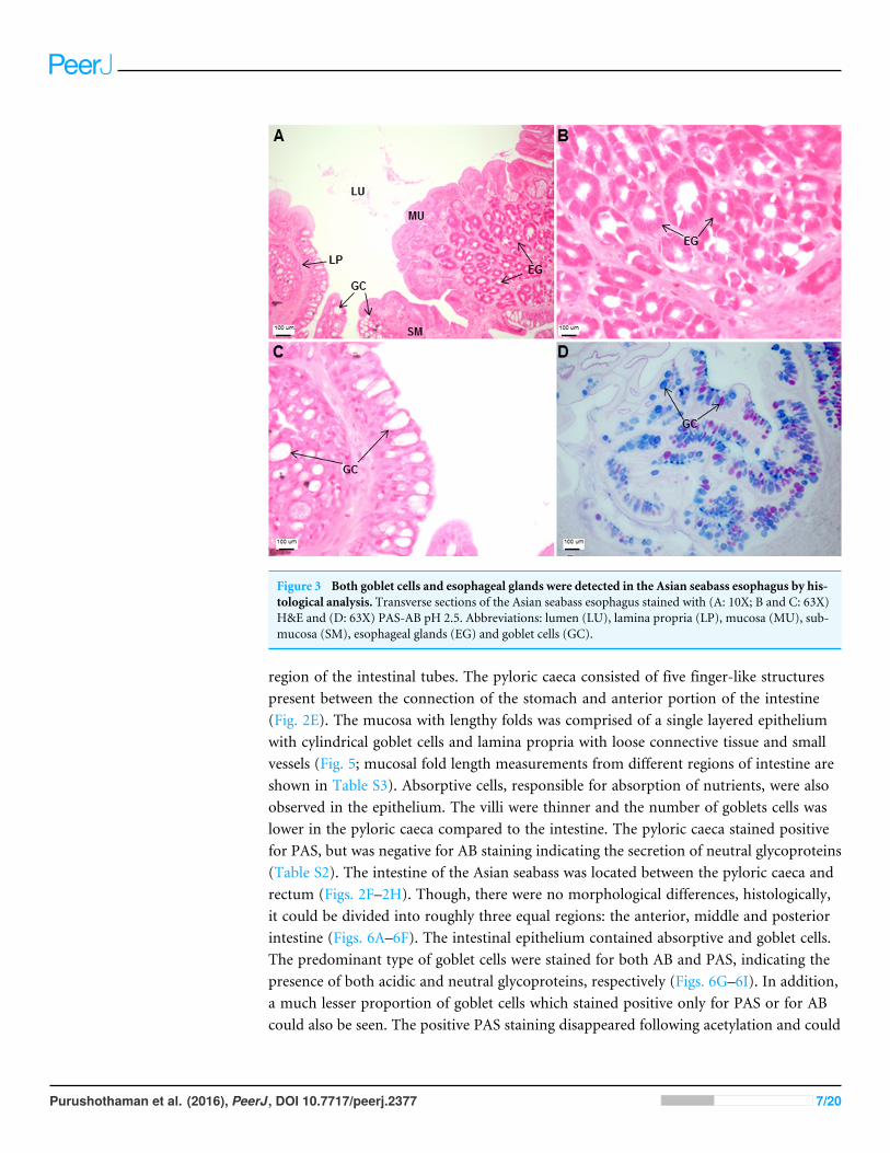
Figure 3 Both goblet cells and esophageal glands were detected in the Asian seabass esophagus by his-tological analysis. Transverse sections of the Asian seabass esophagus stained with (A: 10X; B and C: 63X)H&E and (D: 63X) PAS-AB pH 2.5. Abbreviations: lumen (LU), lamina propria (LP), mucosa (MU), sub-mucosa (SM), esophageal glands (EG) and goblet cells (GC).
region of the intestinal tubes. The pyloric caeca consisted of five finger-like structurespresent between the connection of the stomach and anterior portion of the intestine(Fig. 2E). The mucosa with lengthy folds was comprised of a single layered epitheliumwith cylindrical goblet cells and lamina propria with loose connective tissue and smallvessels (Fig. 5; mucosal fold length measurements from different regions of intestine areshown in Table S3). Absorptive cells, responsible for absorption of nutrients, were alsoobserved in the epithelium. The villi were thinner and the number of goblets cells waslower in the pyloric caeca compared to the intestine. The pyloric caeca stained positivefor PAS, but was negative for AB staining indicating the secretion of neutral glycoproteins(Table S2). The intestine of the Asian seabass was located between the pyloric caeca andrectum (Figs. 2F–2H). Though, there were no morphological differences, histologically,it could be divided into roughly three equal regions: the anterior, middle and posteriorintestine (Figs. 6A–6F). The intestinal epithelium contained absorptive and goblet cells.The predominant type of goblet cells were stained for both AB and PAS, indicating thepresence of both acidic and neutral glycoproteins, respectively (Figs. 6G–6I). In addition,a much lesser proportion of goblet cells which stained positive only for PAS or for ABcould also be seen. The positive PAS staining disappeared following acetylation and could
Purushothaman et al. (2016), PeerJ, DOI 10.7717/peerj.2377 7/20

Figure 4 The Asian seabass stomach is divided into the cardiac, fundic and pyloric regions and showsthe presence of glands andmucus cells. Transverse sections of the Asian seabass stomach stained with(A, B and C: 10X; D, E and F: 63X) H&E and (G, H and I: 63X) PAS-AB pH 2.5. The cardiac, fundic andpyloric regions are shown in (A, D, G)/(B, E, H) and (C, F, I), respectively. Abbreviations: Lumen (LU),gastric pits (GP), cardiac gland (CG), fundic gland (FG), lamina propria (LP), mucosa (MU), submucosa(SM), gland cells (GLC) and mucus secreting cells (MC).
be retrieved after saponification (as also seen for rest of the alimentary tract) (Table S2).Glands could not be seen in either the mucosa or submucosa. The rectum (Figs. 2I–2J)extended from the posterior part of the intestine and ended ventrally at the anus in thefacade of the anal fin. Similar to the intestine, the rectum contained mucosal folds withoutglands (Figs. 7A–7D). The number of goblet cells showed a progressive increase along thelength of the intestinal tract with the posterior part of rectum having over six times thenumber than the anterior intestine (see Table 1 for the cell counts for each region; andFigs. 7C–7D for representative examples).
The histology of the gut was also studied under the effect of two different stressors.The effect of starvation was analyzed at two different time points (one and three weekspost-starvation) (Fig. 8). It had a pronounced effect on gut histology, with a noticeabledecrease in mucosal height (>30% lesser) and goblet cell numbers (>40% reduction)observed within a week of starvation (Fig. 8), whereas, the muscularis thickness showeda nominal decrease (⇠4%) during this time period. The differences were much morepronounced following three weeks of starvation. The mucosal height of starved fishes was
Purushothaman et al. (2016), PeerJ, DOI 10.7717/peerj.2377 8/20
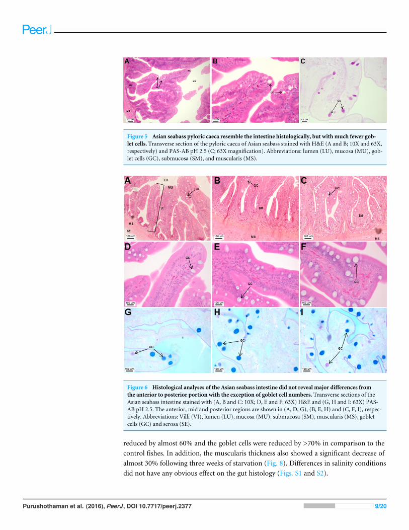
Figure 5 Asian seabass pyloric caeca resemble the intestine histologically, but with much fewer gob-let cells. Transverse section of the pyloric caeca of Asian seabass stained with H&E (A and B; 10X and 63X,respectively) and PAS-AB pH 2.5 (C; 63X magnification). Abbreviations: lumen (LU), mucosa (MU), gob-let cells (GC), submucosa (SM), and muscularis (MS).
Figure 6 Histological analyses of the Asian seabass intestine did not reveal major differences fromthe anterior to posterior portion with the exception of goblet cell numbers. Transverse sections of theAsian seabass intestine stained with (A, B and C: 10X; D, E and F: 63X) H&E and (G, H and I: 63X) PAS-AB pH 2.5. The anterior, mid and posterior regions are shown in (A, D, G), (B, E, H) and (C, F, I), respec-tively. Abbreviations: Villi (VI), lumen (LU), mucosa (MU), submucosa (SM), muscularis (MS), gobletcells (GC) and serosa (SE).
reduced by almost 60% and the goblet cells were reduced by >70% in comparison to thecontrol fishes. In addition, the muscularis thickness also showed a significant decrease ofalmost 30% following three weeks of starvation (Fig. 8). Differences in salinity conditionsdid not have any obvious effect on the gut histology (Figs. S1 and S2).
Purushothaman et al. (2016), PeerJ, DOI 10.7717/peerj.2377 9/20

Figure 7 An increased number of goblet cells in the rectum compared to the intestine. Transverse sec-tions of the Asian seabass rectum stained with (A and B: 10X; D and E: 63X) H&E and (E and F: 63X)PAS-AB pH 2.5. The anterior and posterior regions are shown in (A, C, E) and (B, D, F), respectively. Ab-breviations: Villi (VI), lumen (LU), goblet cells (GC), mucosa (MU) and muscularis (MS).
DISCUSSIONThe structural adaptation of the alimentary canal of each fish species is distinct and dependsnot only upon the body shape, weight and sex, but also upon its unique environment andthe feeding behaviour (Barlow et al., 1993; Davis, 1985). Knowledge on the functionalmorphology of the alimentary canal would be useful for establishing a feeding regime forAsian seabass, a fish of increasing aquaculture importance, especially in South-East Asia.The Asian seabass is a carnivorous species, this feeding behaviour is reflected in its large
Purushothaman et al. (2016), PeerJ, DOI 10.7717/peerj.2377 10/20

Figure 8 The intestinal mucosal height, muscularis thickness as well as goblet cell numbers showedsignificant decrease upon extended starvation. (A and B) Representative transverse section of H&Estained mid-intestine from control Asian seabass (A) and those starved for three weeks (B). Mucosalheight is indicated by the blue line and muscularis thickness is indicated by the green line. Scale bar =100 µm. (C–E) Comparison of mucosal height (C), muscularis thickness (D) and goblet cell numbers(E) between controls (green) and fish starved for one week (orange). (F–H) Comparison of mucosalheight (F), muscularis thickness (G) and goblet cell numbers (H) between controls (blue) and fish starvedfor three-weeks (red). Significantly different parameters are indicated by ⇤ (p < 0.05, Student’s t -test).Abbreviations: Serosa (SE), lumen (LU), mucosa (MU), muscularis (MS), goblet cells (GC).
mouth, oblique and wide upper jaw which enables the fish to swallow its prey as a whole(Pusey, Kennard & Arthington, 2004). Out of the four gill arches, the first two have thinand long rakers which are probably helpful in retaining smaller prey (Canan et al., 2012).
The Asian seabass generally feeds on prey such as molluscs, shrimps and fishes (Davis,1985), therefore, the short and thick esophageal muscularis seems to have a protective
Purushothaman et al. (2016), PeerJ, DOI 10.7717/peerj.2377 11/20

function useful for ingestion of solid material (Domeneghini, Straini & Veggetti, 1998). Theesophagus in the majority of teleost fishes is lined with stratified squamous epithelium. Theclosely-fitted multiple epithelial layers help to withstand various mechanical and chemicalabrasions as the food enters the alimentary canal (Machado et al., 2013; Raji & Norouzi,2010; Santos et al., 2015a;Vieira-Lopes et al., 2013). In contrast, the Asian seabass esophaguswas found to be lined with simple columnar epithelium only. Another interesting featurewas the presence of glands in the esophageal mucosa. Glands are not typically associatedwith the fish esophagus (Canan et al., 2012; Raji & Norouzi, 2010; Takiue & Akiyoshi, 2013;Xiong et al., 2011) but have been reported in a few fish species. The requirement for higherlevel of protection provided by the multi-layered epithelium in typical fishes could perhapsbe offset by the presence of numerous mucosal glands in the Asian seabass esophagus.This is since the secretion from these glands would be useful for protecting the mucosafrom mechanical wear and aid the passage of food into the stomach lining (Santos et al.,2015b; Vegas-Velez, 1972). Further, the mucous secreting cells interspersed in the simplecolumnar epithelium help in safeguarding the lining of the esophagus from chemicaland mechanical injury and also serve as a lubricant to aid in food passage (Machado etal., 2013). In fact, the esophageal mucous cells are a feature associated with most fishspecies (Díaz, García & Goldemberg, 2008; Domeneghini et al., 2005; Germano et al., 2014)and could likely be serving a function equivalent to the salivary glands in mammals (Díaz,García & Goldemberg, 2008; Pedini et al., 2004). The main function of the stomach whenpresent is the storage of food and production of hydrochloric acid to aid digestion (Gartneret al., 2002; Menin & Mimura, 1992; Stroband & Van Der Veen, 1981). The Asian seabassstomach is a straight, sac-like structure, comparable to those of other carnivorous fishes,such as pike, channel catfish and halibut (Evans & Claiborne, 2006). Though there is noclear distinction at the morphological level, histologically the stomach of the Asian seabasscan be divided into cardiac, fundic and pyloric regions. This is similar to other carnivorousfish species, such as Centropomus, where the stomach is well defined (Machado et al.,2013). PAS staining indicated the presence of neutral glycoproteins which serve to protectthe mucosal surface against microorganisms and high acidity of the stomach contents(Buddington & Doroshov, 1986; Díaz et al., 2003; Machado et al., 2013). The stomach hada large number of gastric glands, a feature typically associated with carnivorous fishspecies with a greater need to digest food high in protein content (Machado et al., 2013).In comparison to the other regions of the stomach, glands and surface mucus cells werescarce in the pyloric region of the stomach. This decrease is indicative of a ‘food retentive’more than a ‘food digestive’ function for this region. The storage of food in the pyloricregion (aided by the pyloric sphincter) before it enters the intestine would allow more timefor digestion and has been observed in other fishes such as the walking catfish (Clariasbatrachus) and red-bellied piranha (Serrasalmus nattereri; Raji & Norouzi, 2010).
Pyloric caeca are finger-like projections, highly variable in number (0–1,000 s). Theyare associated with the gut of the majority of fishes (⇠60% of identified species) with anincreased frequency seen amongst carnivorous fish species (Canan et al., 2012; Hossain &Dutta, 1996;Raji & Norouzi, 2010). Though a specific function has not been associated withthese finger-like projections, the most likely option seems to be an evolutionary adaptation
Purushothaman et al. (2016), PeerJ, DOI 10.7717/peerj.2377 12/20

to facilitate the digestive process by increasing the absorptive area (Canan et al., 2012).Also, in some cases, such as the pyloric caeca of the carnivorous red-bellied piranha, anabundance of goblet cells which stain positive for neutral glycoproteins, has been observedindicating an additional role for these accessory appendages (Raji & Norouzi, 2010). In thecase of Asian seabass, the five finger-like pyloric caeca have scarce neutral glycoproteinsecreting mucous cells indicative of a primary absorptive function. Although evidence ofendocrine activity has been shown in the pyloric caeca of some fishes (Anderson, 1986;Beorlegui, Martínez & Sesma, 1992), no secretory gland was observed in the pyloric caecaof the Asian seabass.
In teleost fishes, the length of the intestine provides a good gauge on their feedingbehaviour—longer in iliophagous, omnivorous and herbivorous species, and shorterin insectivorous and carnivorous species (Canan et al., 2012; Machado et al., 2013). Theintestinal coefficient of Asian seabass is relatively low (1.1) and falls within the rangeexpected for carnivorous fishes (0.2–2.5). On the other hand, the intestinal coefficientrange for omnivorous (0.6–8.0) and herbivorous (0.8–15.0) fishes are much higher(Al-Hussaini, 1949; Angelescu & Gneri, 1949; Bertin, 1958; Santos et al., 2015a; Santos et al.,2015b;Xiong et al., 2011). The Asian seabass intestine can be divided into three regions—theanterior, mid and posterior portions at the histological level, though no morphologicaldifferences existed as is the case for most fishes (Gargiulo et al., 1998; Scocco, Menghi &Ceccarelli, 1997). Numerous microvilli adorn the intestinal epithelium, which comprisesof many goblet and absorptive cells to serve lubricative function as well as aid digestionand absorption of food (Gartner et al., 2002). The intestinal epithelium, similarly to theesophagus and rectum, stained positive for both acidic and neutral glycoproteins. Inaddition, AB staining was positive at both pH 0.5 and pH 2.5 indicating the presence ofboth sulphated and carboxylated glycoproteins, respectively. Although the majority of thegoblet cells stained positive for both AB and PAS, some of them stained for only PAS orfor AB. The co-existence of goblet cells secreting neutral or acidic+neutral glycoproteinsprobably represents the sequential nature of mucous biosynthesis with cells producingneutral glycoprotein only (PAS-positive) representing an earlier stage in developmentcompared to the other cell types. On the other hand, mucins stain with AB pH 2.5 whenglycoproteins are carboxylated and with AB pH 0.5–1.0 when the sulphated groups areconjugated to glycoproteins (Sarasquete et al., 2001). Additionally, the number of gobletcells increased from the anterior to posterior portions in the intestine as well as rectum.The higher number of goblet cells in the rectum seems to be a universal feature in fishesand is probably useful for increased mucous production to safeguard the intestinal liningand aid faecal expulsion (Machado et al., 2013).
In addition, gut histology was also studied upon changing the feeding and salinityconditions. Food deprivation has been documented to have a major effect on the gas-trointestinal tract of several fish species such as white sturgeons (Acipenser transmontanus),neon damselfish (Pomacentrus coelestis), Red Sea surgeonfish (Acanthurus nigrofuscus),common carp (Cyprinus carpio L.) and rainbow trout (Oncorhynchus mykiss). The changesreported vary from reduction in mucosal surface area, mucosal thickness and intestinallength along with decreased mucous cell numbers (Domeneghini et al., 2002; Gas &
Purushothaman et al. (2016), PeerJ, DOI 10.7717/peerj.2377 13/20

Noailliac-Depeyre, 1976; Hall & Bellwood, 1995; MacLeod, 1978; Montgomery & Pollak,1988). A similar effect was observed on the Asian seabass gut with a pronounced decreasein all the parameters measured following three weeks of starvation. Further, the intestineof the species appears very sensitive to changes in food supply as even within a week ofstarvation, noticeable changes could be seen in the mucosal height as well as goblet cellnumbers. This is similar to the observation in damselfish wherein 13 days of starvationresulted in a reduction of mucosal surface area, thickness and intestinal length (Hall &Bellwood, 1995). On the other hand, repeated changes in salinity did not have any obviouseffect on the intestinal morpho-histology of the Asian seabass.
There are very few publications on the effect of stress on fish gut histology. In a studydone on damselfishes, no apparent effect on the intestinal mucosa could be observed uponstress treatment wherein fishes were placed in tubs containing rubble (Hall & Bellwood,1995). However, in another study, alkyl benzene sulphonate (an active ingredient foundin products such as detergents, shampoos and cosmetics) had a profound effect on the guthistology with villi losing their individuality and forming a mass in young giltheads. Inaddition, the lamina propria couldn’t be identified and the submucosa was hypertrophiedand the muscular layer was thickened in these fishes (Rosety et al., 2001). The lack ofeffect of salinity changes on the gut histology of Asian seabass could be attributed to theeuryhaline nature of Asian seabass due to which it can adapt well to a range of salinities(Moore, 1982; Vij et al., 2014).
In conclusion, our work is the first study describing the morphological as well ashistological features of the Asian seabass gut. The observations described in this publicationwill serve as a reference for future research aimed at studying the effect of environmentalvariables, stress conditions and diet on the digestive system of the species. In fact, in thisstudy, we have already made use of this information to analyze the effect of two differentstress conditions, namely changes in salinity and food deprivation, on the gut histology.Histological features of the gut were unaltered upon salinity stress, whereas starvationhad a major effect on the gut in terms of reduced mucosal height, muscularis thicknessand goblet cell numbers compared to control fishes. Future nutriphysiological (andpossibly even nutrigenomic) studies can potentially benefit from the ‘baseline information’described in the current publication.
ACKNOWLEDGEMENTSWe would like to thank Graham Wright and Sarah Zulkifli for help with microscopicphotography.
ADDITIONAL INFORMATION AND DECLARATIONS
FundingThis research is supported by the National Research Foundation, Prime Minister’s Office,Singapore under its Competitive Research Program (CRPAwardNo.NRF-CRP7-2010-01).The funders had no role in study design, data collection and analysis, decision to publish,or preparation of the manuscript.
Purushothaman et al. (2016), PeerJ, DOI 10.7717/peerj.2377 14/20

Grant DisclosuresThe following grant information was disclosed by the authors:National Research Foundation.Prime Minister’s Office, Singapore: NRF-CRP7-2010-01.
Competing InterestsThe authors declare there are no competing interests.
Author Contributions• Kathiresan Purushothaman conceived and designed the experiments, performed theexperiments, analyzed the data, wrote the paper, prepared figures and/or tables.
• Doreen Lau and Jolly M. Saju performed the experiments.• Syed Musthaq SK performed the experiments and prepared figures and/or tables.• Declan Patrick Lunny analyzed the data.• Shubha Vij and László Orbán conceived and designed the experiments, analyzed thedata, wrote the paper, reviewed drafts of the paper.
Animal EthicsThe following information was supplied relating to ethical approvals (i.e., approving bodyand any reference numbers):
All experiments were approved by Agri-food and Veterinary Authority (AVA)Institutional Animal Care and Use Committee (IACUC) (approval ID: AVA-MAC-2012-02) and performed according to guidelines set by the National Advisory Committeeon Laboratory Animal Research (NACLAR) for the care and use of animals for scientificresearch in Singapore.
Data AvailabilityThe following information was supplied regarding data availability:
The raw data has been supplied as Data S1.
Supplemental InformationSupplemental information for this article can be found online at http://dx.doi.org/10.7717/peerj.2377#supplemental-information.
REFERENCESAl-Hussaini A. 1949. On the functional morphology of the alimentary tract of some fish
in relation to differences in their feeding habits: anatomy and histology. QuarterlyJournal of Microscopical Science 90:109–139.
Alhazzaa R, Bridle AR, Nichols PD, Carter CG. 2011. Up-regulated desaturase andelongase gene expression promoted accumulation of polyunsaturated fatty acid(pufa) but not long-chain pufa in Lates calcarifer, a tropical euryhaline fish, fed astearidonic acid-and 0-linoleic acid-enriched diet. Journal of Agricultural and FoodChemistry 59:8423–8434 DOI 10.1021/jf201871w.
Purushothaman et al. (2016), PeerJ, DOI 10.7717/peerj.2377 15/20
Anderson TA. 1986.Histological and cytological structure of the gastrointestinal tract ofthe Luderick, Girella tricuspidata (Pisces, Kyphosidae), in relation to diet. Journal ofMorphology 190:109–119 DOI 10.1002/jmor.1051900110.
Angelescu V, Gneri FS. 1949. Adaptaciones Del Aparato Digestivo Al Régimen AlimenticioEn Algunos Peces Del Río Uruguay Y Del Río De La Plata: I. Tipo Omnívoro E IliófagoEn Representantes De Las Familias’’ Loricariidae’’ Y’’ Anostomidae’’: Casa Ed.’’ Coni’’.
Bancroft J, Gamble M. 2002. Theory and practice of histological techniques. Fifth edition.New York: Churchill Livingstone.
Barlow C, Rodgers L, Palmer P, Longhurst C. 1993. Feeding habits of hatchery-rearedbarramundi Lates calcarifer (Bloch) fry. Aquaculture 109:131–144DOI 10.1016/0044-8486(93)90210-P.
Beorlegui C, Martínez A, Sesma P. 1992. Endocrine cells and nerves in the pyloric cecaand the intestine of Oncorhynchus mykiss (Teleostei): an immunocytochemical study.General and Comparative Endocrinology 86:483–495DOI 10.1016/0016-6480(92)90073-S.
Bertin L. 1958. Appareil digestif. Traité de zoologie 13:1249–1301.Boonyaratpalin M,Williams K. 2001. Asian sea bass, Lates calcarifer . In: Webster
CD, Lim CE, eds. Nutrient requirements and feeding of finfish for aquaculture.Wallingford: CANI Publishing.
Buddington RK, Doroshov SI. 1986. Structural and functional relations of the whitesturgeon alimentary canal (Acipenser transmontanus). Journal of Morphology190:201–213 DOI 10.1002/jmor.1051900205.
Canan B, NascimentoWSD, Silva NBD, Chellappa S. 2012.Morphohistology of thedigestive tract of the damsel fish Stegastes Fuscus (Osteichthyes: Pomacentridae). TheScientific World Journal 2012:1–9 DOI 10.1100/2012/787316.
Davis T. 1985. The food of barramundi, Lates calcarifer (Bloch), in coastal and inlandwaters of van diemen gulf and the gulf of carpentaria, Australia. Journal of FishBiology 26:669–682 DOI 10.1111/j.1095-8649.1985.tb04307.x.
Day RD, Tibbetts IR, Secor SM. 2014. Physiological responses to short-term fastingamong herbivorous, omnivorous, and carnivorous fishes. Journal of ComparativePhysiology B 184:497–512 DOI 10.1007/s00360-014-0813-4.
Díaz A, Garcia A, Devincenti C, Goldemberg A. 2003.Morphological and histochemicalcharacterization of the mucosa of the digestive tract in Engraulis anchoita (Hubbsand Marini, 1935). Anatomia, Histologia, Embryologia 32:341–346DOI 10.1111/j.1439-0264.2003.00490.x.
Díaz A, García A, Escalante A, Goldemberg A. 2010. Glycoproteins histochemistry of thegills of Odontesthes bonariensis (Teleostei, Atherinopsidae). Journal of Fish Biology77:1665–1673 DOI 10.1111/j.1095-8649.2010.02803.x.
Díaz AO, García AM, Goldemberg AL. 2008. Glycoconjugates in the mucosa of thedigestive tract of Cynoscion guatucupa: a histochemical study. Acta Histochemica110:76–85 DOI 10.1016/j.acthis.2007.08.002.
Purushothaman et al. (2016), PeerJ, DOI 10.7717/peerj.2377 16/20

Domeneghini C, Arrighi S, Radaelli G, Bosi G, Veggetti A. 2005. Histochemical analysisof glycoconjugate secretion in the alimentary canal of Anguilla anguilla L. ActaHistochemica 106:477–487 DOI 10.1016/j.acthis.2004.07.007.
Domeneghini C, Radaelli G, Bosi G, Arrighi S, Giancamillo AD, Pazzaglia M, Mas-carello F. 2002.Morphological and histochemical differences in the structure ofthe alimentary canal in feeding and runt (feed deprived) white sturgeons (Acipensertransmontanus). Journal of Applied Ichthyology 18:341–346DOI 10.1046/j.1439-0426.2002.00384.x.
Domeneghini C, Straini RP, Veggetti A. 1998. Gut glycoconjugates in Sparus aurataL. (Pisces, Teleostei). A comparative histochemical study in larval and adult ages.Histology and Histopathology 13:359–372.
Evans D, Claiborne J. 2006. The physiology of fishes. Third edition. Boca Ration: CRCPress.
Gargiulo A, Ceccarelli P, Dall’Aglio C, Pedini V. 1998.Histology and ultrastructureof the gut of the tilapia (Tilapia Spp.), a hybrid teleost. Anatomia, Histologia,Embryologia 27:89–94 DOI 10.1111/j.1439-0264.1998.tb00162.x.
Gartner LP, Hiatt JL, De Souza LF, Sales MdGF. 2002. Atlas colorido de histologia. 3rdedition. Rio de Janeiro: Guanabara Koogan.
Gas N, Noailliac-Depeyre J. 1976. Studies on intestinal epithelium involution duringprolonged fasting. Journal of Ultrastructure Research 56:137–151DOI 10.1016/S0022-5320(76)80161-X.
Germano RM, Stabille SR, Mari RB, Pereira JNB, Faglioni JRS, Miranda NHM.2014.Morphological characteristics of the Pterodoras granulosus digestive tube(Valenciennes, 1821) (Osteichthyes, Doradidae). Acta Zoologica 95:166–175DOI 10.1111/azo.12016.
Glencross B, Michael R, Austen K, Hauler R. 2008. Productivity, carcass composition,waste output and sensory characteristics of large barramundi Lates calcarifer fedhigh-nutrient density diets. Aquaculture 284:167–173DOI 10.1016/j.aquaculture.2008.07.031.
Glencross B,Wade N, Morton K. 2013. Lates calcarifer nutrition and feeding practices.In: Biology and Culture of Asian Seabass Lates calcarifer. CRC Press, 178–228.
Gu J, Bakke AM, Valen EC, Lein I, Krogdahl Å. 2014. Bt-Maize (Mon810) and non-Gm soybean meal in diets for atlantic Salmon (Salmo salar L.) juveniles-impacton survival, growth performance, development, digestive function, and transcrip-tional expression of intestinal immune and stress responses. PLoS ONE 9:e99932DOI 10.1371/journal.pone.0099932.
Hall K, Bellwood DR. 1995.Histological effects of cyanide, stress and starvation on theintestinal mucosa of Pomacentrus coelestis, a marine aquarium fish species. Journal ofFish Biology 47:438–454 DOI 10.1111/j.1095-8649.1995.tb01913.x.
Hossain AM, Dutta HM. 1996. Phylogeny, ontogeny, structure and function of digestivetract appendages (caeca) in teleost fish. In: Fish morphology: horizons of new research.Brookfield: Balkema Pub, 59–76.
Purushothaman et al. (2016), PeerJ, DOI 10.7717/peerj.2377 17/20

Jiang J, Miyata M, Chan C, Ngoh SY, LiewWC, Saju JM, Ng KS,Wong FS, Lee YS,Chang SF, Orban L. 2014. Differential transcriptomic response in the spleen andhead kidney following vaccination and infection of Asian seabass with streptococcusiniae. PLoS ONE 9:e99128 DOI 10.1371/journal.pone.0099128.
Katersky RS, Carter CG. 2009. Growth and protein synthesis of barramundi, Latescalcarifer, fed lupin as a partial protein replacement. Comparative Biochemistry andPhysiology Part A: Molecular & Integrative Physiology 152:513–517DOI 10.1016/j.cbpa.2008.12.017.
Kuznetsova IS, ThevasagayamNM, Sridatta PS, Komissarov AS, Saju JM, Ngoh SY,Jiang J, Shen X, Orban L. 2014. Primary analysis of repeat elements of the Asianseabass (Lates calcarifer) transcriptome and genome. Frontiers in Genetics 5(223)DOI 10.3389/fgene.2014.00223.
MachadoMRF, De Oliveira Souza H, De Souza VL, De Azevedo A, Goitein R, NobreAD. 2013.Morphological and anatomical characterization of the digestive tract ofCentropomus parallelus and Centropomus undecimalis. Acta Scientiarum BiologicalSciences 35:467–474 DOI 10.4025/Actascibiolsci.V35i4.14352.
MacLeodM. 1978. Effects of salinity and starvation on the alimentary canal anatomy ofthe rainbow trout salmo gairdneri Richardson. Journal of Fish Biology 12:71–79DOI 10.1111/j.1095-8649.1978.tb04152.x.
Matthews S, Kinhult A, Hoeben P, Sara V, Anderson T. 1997. Nutritional regulation ofinsulin-like growth factor-I Mrna expression in barramundi, Lates calcarifer . Journalof Molecular Endocrinology 18:273–276 DOI 10.1677/jme.0.0180273.
Menin E, Mimura O. 1992. Comparative functional anatomy of three species of fish foodhabit of Teleostei omnivore. Revista Ceres 223:233–260.
Monjoyo H, Tan-Fermin J, Macaranas J. 2003. Localisation of enzymes in the digestivetract during the larval to early juvenile stages of sea bass (Lates calcarifer Bloch).Indonesian Fisheries Research Journal 9:46–53.
MontgomeryW, Pollak P. 1988. Epulopiscium fishelsoni Ng, N. Sp., a protist of uncertaintaxonomic affinities from the gut of an herbivorous reef fish1. The Journal ofProtozoology 35:565–569 DOI 10.1111/j.1550-7408.1988.tb04153.x.
Moore R. 1982. Spawning and early life history of burramundi, Lates calcarifer (Bloch),in Papua New Guinea.Marine and Freshwater Research 33:647–661DOI 10.1071/MF9820647.
Nelson JS. 2006. Fishes of the world. New York: John Wiley and Sons.Ngoh SY, Tan D, Shen X, Kathiresan P, Jiang J, LiewWC, ThevasagayamNM, Kwan
HY, Saju JM, Prakki SRS. 2015. Nutrigenomic and nutritional analyses reveal theeffects of pelleted feeds on Asian seabass (Lates calcarifer). PLoS ONE 10:e0145456DOI 10.1371/journal.pone.0145456.
Pedini V, Dall’Aglio C, Parillo F, Scocco P. 2004. A lectin histochemical study of theoesophagus of Shi Drum. Journal of Fish Biology 64:625–631DOI 10.1111/j.1095-8649.2004.00326.x.
Pusey B, KennardM, Arthington A. 2004. Freshwater fishes of North-Eastern Australia.Collingwood: CSIRO Publishing.
Purushothaman et al. (2016), PeerJ, DOI 10.7717/peerj.2377 18/20

Raji AR, Norouzi E. 2010.Histological and histochemical study on the alimentary canalin walking catfish (Claris batrachus) and Piranha (Serrasalmus nattereri). IranianJournal of Veterinary Research 11:255–261.
Ravi P, Jiang J, LiewWC, Orban L. 2014. Small-scale transcriptomics reveals differencesamong gonadal stages in Asian seabass (Lates calcarifer). Reproductive Biology andEndocrinology 12(5) DOI 10.1186/1477-7827-12-5.
Rosety M, Ordoñez F, Ribelles A, Rosety-Rodriguez M, Dominguez A, CarrascoC, Rosety J. 2001.Morpho-histochemical changes in the liver and intestine ofyoung giltheads (fish-nursery), Sparus aurata, L., induced by acute action of theanionic tensioactive Alkylbenzene Sulphonate. European Journal of Histochemistry45:259–265.
Santos MLD, Arantes FP, Pessali TC, Santos JED. 2015a.Morphological, histologicaland histochemical analysis of the digestive tract of Trachelyopterusstriatulus (Siluri-formes: Auchenipteridae). Zoologia (Curitiba) 32:296–305DOI 10.1590/S1984-46702015000400005.
Santos MLD, Arantes FP, Santiago KB, Santos JED. 2015b.Morphological characteris-tics of the digestive tract of Schizodon knerii (Steindachner, 1875), (Characiformes:Anostomidae): an anatomical, histological and histochemical study. Anais daAcademia Brasileira de Ciências 87:867–878 DOI 10.1590/0001-3765201520140230.
Sarasquete C, Gisbert E, Ribeiro L, Vieira L, Dinis M. 2001. Glyconjugates in epidermal,branchial and digestive mucous cells and gastric glands of gilthead sea bream,Sparus aurata, Senegal sole, Solea senegalensis and Siberian sturgeon, Acipenser baer idevelopment. European Journal of Histochemistry 45:267–278.
Schindelin J, Arganda-Carreras I, Frise E, Kaynig V, Longair M, Pietzsch T, Preibisch S,Rueden C, Saalfeld S, Schmid B. 2012. Fiji: an open-source platform for biological-image analysis. Nature Methods 9:676–682 DOI 10.1038/nmeth.2019.
Scocco P, Menghi G, Ceccarelli P. 1997.Histochemical differentiation of glycoconjugatesoccurring in the tilapine intestine. Journal of Fish Biology 51:848–857DOI 10.1111/j.1095-8649.1997.tb02005.x.
SrichanunM, Tantikitti C, Utarabhand P, Kortner TM. 2013. Gene expression andactivity of digestive enzymes during the larval development of Asian seabass (Latescalcarifer). Comparative Biochemistry and Physiology Part B: Biochemistry andMolecular Biology 165:1–9 DOI 10.1016/j.cbpb.2013.02.005.
Stroband HWJ, Van Der Veen FH. 1981. Localization of protein absorption duringtransport of food in the intestine of the grasscarp, Ctenopharyngodon idella (Val.).Journal of Experimental Zoology 218:149–156 DOI 10.1002/jez.1402180207.
Takiue S, Akiyoshi H. 2013.Histological and scanning electron microscopic examinationof the digestive tract in whitespotted conger, Conger myriaster (Anguilliformes).Journal of Phylogenetics & Evolutionary Biology 2(125)DOI 10.4172/2329-9002.1000125.
TuW-C, Mühlhäusler BS, JamesMJ, Stone DA, Gibson RA. 2013. Dietary alpha-linolenic acid does not enhance accumulation of omega-3 long-chain polyunsat-urated fatty acids in Barramundi (Lates calcarifer). Comparative Biochemistry and
Purushothaman et al. (2016), PeerJ, DOI 10.7717/peerj.2377 19/20
Physiology Part B: Biochemistry and Molecular Biology 164:29–37DOI 10.1016/j.cbpb.2012.10.001.
Vegas-Velez M. 1972. La Structure Histologique Typique Du Tube Digestif Des PoissonsTéleosteens. Téthys 4:163–174.
Vieira-Lopes DA, Pinheiro NL, Sales A, Ventura A, Araújo FG, Gomes ID, NascimentoAA. 2013. Immunohistochemical study of the digestive tract of Oligosarcus hepsetus.World Journal of Gastroenterology 19:1919–1929 DOI 10.3748/wjg.v19.i12.1919.
Vij S, Kuhl H, Kuznetsova IS, Komissarov A, Yurchenko AA, Heusden PV, Singh S,ThevasagayamNM, Prakki SRS, Purushothaman K, Saju JM, Jiang J, Mbandi SK,Jonas M, Tong AHY, Mwangi S, Lau D, Ngoh SY, LiewWC, Shen X, Hon LS, DrakeJP, BoitanoM, Hall R, Chin CS, Lachumanan R, Korlach J, Trifonov V, Kabilov M,Tupikin A, Green D, Moxon S, Garvin T, Sedlazeck FJ, Vurture GW, GopalapillaiG, Katneni VK, Noble TH, Scaria V, Sivasubbu S, Jerry DR, O’Brien SJ, Schatz MC,Dalmay T, Turner SW, Lok S, Christoffels A, Orban L. 2016. Chromosomal-levelassembly of the Asian seabass genome using long sequence reads and multi-layeredscaffolding. PLoS Genetics 12:e1005954.
Vij S, Purushothaman K, Gopikrishna G, lau D, Saju JM, Shamsudheen KV, Vinayu-mar K, Basheer VS, Gopalakrishnan A, Mohommad SH, Sridhar S, Vinod S, JenaJK, Ponniah AG, Orbán L. 2014. Barcoding of Asian seabass across its geographicrange provides evidence for its bifurcation into two distinct species. Frontiers inMarine Science 1: Art30 DOI 10.3389/fmars.2014.00030.
Walford J, Lam T. 1993. Development of digestive tract and proteolytic enzyme activityin seabass (Lates calcarifer) Larvae and Juveniles. Aquaculture 109:187–205DOI 10.1016/0044-8486(93)90215-K.
Wang CM, Bai ZY, He XP, Lin G, Xia JH, Sun F, Lo LC, Feng F, Zhu ZY, Yue GH.2011. A high-resolution linkage map for comparative genome analysis andqtl fine mapping in Asian seabass, Lates calcarifer . BMC Genomics 12:174DOI 10.1186/1471-2164-12-174.
Wang CM, Lo LC, Zhu ZY, Yue GH. 2006. A genome scan for quantitative trait lociaffecting growth-related traits in an F1 family of Asian seabass (Lates calcarifer).BMC Genomics 7:274 DOI 10.1186/1471-2164-7-274.
Xia JH, Liu P, Liu F, Lin G, Sun F, Tu R, Yue GH. 2013. Analysis of stress-responsivetranscriptome in the intestine of Asian seabass (Lates calcarifer) using Rna-Seq. DNAResearch 20:449–460 DOI 10.1093/dnares/dst022.
Xiong D, Zhang L, Yu H, Xie C, Kong Y, Zeng Y, Huo B, Liu Z. 2011. A study ofmorphology and histology of the alimentary tract of Glyptosternum maculatum(Sisoridae, Siluriformes). Acta Zoologica 92:161–169DOI 10.1111/j.1463-6395.2010.00458.x.
Purushothaman et al. (2016), PeerJ, DOI 10.7717/peerj.2377 20/20