monitoring respiratory disease severity in cystic fibrosis
TRANSCRIPT

Monitoring Respiratory Disease Severity in Cystic Fibrosis
Jane C Davies MB ChB MRCP MRCPCH MD and Eric WFW Alton MD FRCP FMedSci
IntroductionWhat Should Be the Goals of Monitoring?Current MonitoringPhysiology
Current Clinical PracticeHow Could We Do Better?
InfectionCurrent Clinical PracticeWhat Are the Problems?How Could We Do Better?
InflammationCurrent Clinical PracticeWhat Are the Problems?How Could We Do Better?
RadiologyCurrent Clinical PracticeWhat Are the Problems?How Could We Do Better?
Summary
Measurements of disease severity provide a guide for the physician to tailor therapies, for thepatient and family to gauge progress, and are required for clinical trials. For many respiratorydiseases, including cystic fibrosis, sensitive, noninvasive measurements are few, and some of thosethat are available are applicable only to certain subgroups of patients or lack sufficient sensitivity.We discuss currently available measurements in 4 groups: physiology, infection, inflammation, andradiology. For each group we highlight strengths and weaknesses, ask how we could improve uponthese, and provide details of alternative methods. Key words: cystic fibrosis, monitoring. [Respir Care2009;54(5):606–615. © 2009 Daedalus Enterprises]
Jane C Davies MB ChB MRCP MRCPCH MD is affiliated with theDepartment of Gene Therapy, Imperial College London, and the Depart-ment of Paediatric Respiratory Medicine, Royal Brompton Hospital, Lon-don, United Kingdom. Eric WFW Alton MD FRCP FMedSci is affiliatedwith the Department of Gene Therapy, Imperial College London, and theDepartment of Respiratory Medicine, Royal Brompton Hospital, London,United Kingdom.
The authors have disclosed no conflicts of interest.
Dr Davies presented a version of this paper at the 43rd RESPIRATORY
CARE Journal Conference, “Respiratory Care and Cystic Fibrosis,” heldSeptember 26-28, 2008, in Scottsdale, Arizona.
Correspondence: Jane C Davies MB ChB MRCP MRCPCH MD, De-partment of Gene Therapy, Imperial College, Emmanuel Kaye Building,Manresa Road, London SW3 6NP, United Kingdom. E-mail:[email protected].
606 RESPIRATORY CARE • MAY 2009 VOL 54 NO 5

Introduction
Every interaction between a person with cystic fibrosis(CF) and a health professional will involve some degree ofassessment of well-being and disease status. Consensusdocuments provide guidance as to which tools should beemployed, both routinely and for more detailed annualassessments.1,2 So, are the tools we have at our disposaland the methods with which we employ them up to thetask? Do they provide sufficient insight into an individu-al’s current status and allow us to gauge the direction andrate of progression of disease? Do they provide us with theoptimal pieces of information that allow us to tailor ther-apeutic interventions, assess their success, and provideprognostic information? In this paper we argue that theanswer to these questions is “no”. We discuss the inves-tigations in common use, with their limitations, some ofwhich are specific to disease stage or age group. Finally,we discuss newer techniques, many of which are onlyperformed in the research setting, which may offer a moresensitive and detailed insight into lower airway disease inthe future.
What Should Be the Goals of Monitoring?
It is our opinion that we should perform measurementsthat, either alone or in combination, are: sensitive; allowdetection of change both long-term and short-term; repeat-able and reproducible; minimally invasive or noninvasiveand well-tolerated; applicable across age groups and topatients of different illness severity; and complementary inthe information they provide. Such an approach wouldallow individually tailored management and provide long-term prognostic information to patients and professionals.
Current Monitoring
Techniques in current clinical use and listed in manage-ment guidelines fall broadly into 4 groups: physiology,infection, inflammation, and radiology. For each of thesecategories we outline strengths and limitations and makesome suggestions as to how we could do better in thefuture.
Physiology
Current Clinical Practice
Patients old enough to form a seal with their lips andperform prolonged forced expiratory maneuvers routinelyundergo spirometry at every clinic visit and at periods ofclinical instability or exacerbation. In addition, annual pleth-ysmographic and diffusion-capacity (gas-transfer) mea-surements are recommended in some guidelines.
Spirometry. Spirometry has long been the accepted stan-dard in disease monitoring. Forced expiratory maneuverssuch as forced expiratory volume in the first second (FEV1)and forced vital capacity are well understood, and almostuniversally FEV1 is used to define mild (� 60% or 70% ofpredicted), moderate, and severe (� 40% or 30%) disease.The predicted values have been generated with variousmodels, based on healthy persons and height, sex, and age.The different reference ranges should be borne in mindwhen comparing or extrapolating data sets, and absolutevalues should therefore also be obtained and recorded.
What Are the Problems With Spirometry? Althoughthe coefficient of variability for FEV1 in healthy people isreported to be around 2–3%,3 it is much higher in patientswith CF4 and for flows at lower lung volumes (eg, theforced expiratory flow during the middle half of the forcedexpiratory maneuver [FEF25%-75%]), which may reflectsmall airways, which is the site of interest. The measure-ments are highly technique-dependent and effort-depen-dent. Some patients find such maneuvers difficult, andthey are not routinely performed in young children � 5 yearsold. The measurements also lack sensitivity, particularly inmild, early stages of disease or when looking for smallchanges in response to an intervention, and there is cur-rently a very slow rate of decline (1–2% per year) in theCF population treated in modern centers.5 This means that,though patients who are deteriorating rapidly or over ashort time period can be easily identified, observing anyimprovement on this rate of decline in an individual pa-tient will be almost impossible. Finally, although FEV1
has historically been used in defining severity, there issome evidence to suggest it is not a very useful tool onwhich to base prognosis; FEV1 is no longer included in thelung allocation score6 as part of transplant-waiting-list as-sessment in the United States.
Plethysmography. Lung volumes and diffusion capacityare listed in some of the current consensus documents forannual assessment, and are performed in the majority oflarge centers, at least in Europe.
What Are the Problems With Plethysmography? Thesetechniques are expensive, technically challenging, and time-consuming for staff and patient. Similarly to spirometry, theyrequire cooperation and are, in general, not suitable for veryyoung patients. In addition, they are probably less well un-derstood by the clinical team, and in our experience the re-sults may not in fact be paid a great deal of attention. Finally,recent data suggest that lung-volume values add very little tospirometry for the majority of patients.7
So we appear to be using expensive resources, in termsof both equipment and skilled manpower, and asking pa-tients to spend substantial time, for a relatively small gain.
MONITORING RESPIRATORY DISEASE SEVERITY IN CYSTIC FIBROSIS
RESPIRATORY CARE • MAY 2009 VOL 54 NO 5 607

How Could We Do Better?
Several new approaches show substantial promise andmay be clinically useful in the near future.
Lung-Clearance Index. The lung-clearance index usesmultiple-breath wash-out of a nonabsorbable gas (originallynitrogen, but, more commonly now, sulfur hexafluoride) tomeasure ventilation inhomogeneity caused by airway nar-rowing from inflammation or partial mucus obstruction. Thesubject inhales a low concentration of sulfur hexafluoride, viaeither mask or mouthpiece, until the concentration in the lungis in equilibrium with the concentration administered (wash-inphase). The supply is then switched off and, during continuedtidal breathing, wash-out is monitored. Gas analyzers includeconventional mass spectrometer and, more recently, photo-acoustic and ultrasonic-based technologies. Wash-out is de-fined, for practical purposes, as the point when the sulfurhexafluoride reaches 1/40th of its original concentration. Pa-tients with more severe disease take longer to wash out, be-cause gas is trapped in narrowed airways; therefore, theyhave a higher lung-clearance index.
One advantage of the lung-clearance index is that ithas a relatively narrow range of normal values thatchanges very little with age, which obviates the require-ment for age/size-adjusted normal values (Fig. 1).8 Thetechnique is also: harmless; easy to perform (requiresonly tidal breathing, and no additional coordination,cooperation, or forced maneuvers); can be performed atall ages, including infancy and pre-school ages8-10; re-peatable, reproducible,11 and more sensitive at the early
stages of disease than is spirometry (Fig. 2).12 Finally,it is at least as sensitive as forced expiratory maneuversin infants10 and correlates better with structural changeson high-resolution computed tomography (CT) than doesFEV1 (Table 1).13
An important disadvantage of the lung-clearance indexis that completely obstructed lung regions do not contrib-ute to the overall measurement because the inhaled gasdoes not reach those regions. So in patients who havetotally obstructed lung regions, the lung-clearance indexcould underestimate disease severity. Also, the techniquemay be more burdensome for the most severely affectedpatients, who require much longer wash-in and wash-outtimes. Some of the more portable technologies, such as theInnocor, which relies on photoacoustic analysis of exhaledgas, may currently be less applicable with small children(who have faster respiratory rates), because of the some-what slower response time than a mass spectrometer. How-ever, mass spectrometers are expensive to set up and maybe challenging to maintain.
Lung Function Tests Applicable to Infants and Pre-School Children. The last decade has seen a massiveincrease in the number of studies that reported lung func-tion in infants and young children, many of which havefocused on CF. Consensus guidelines have been pub-lished.14 It is clear from the studies that: sensitive mea-
Fig. 1. There is a narrow range of lung-clearance index in normalsubjects (circles), compared to patients with cystic fibrosis (dots),and this is similar for older children and adults, which obviatesadjustment for age or size. (From Reference 9, with permission.) Fig. 2. In the early stages of cystic fibrosis lung disease, the lung-
clearance index (LCI) is more sensitive than standard spirometry.Of 274 children with a normal forced expiratory volume in the firstsecond (FEV1) Z score, 254 (93%) had an abnormality detected viaLCI. In contrast, the LCI failed to detect an abnormal FEV1 in only2% (5 of 213) of patients. SD-S � standard deviation score. (FromReference 12, with permission.)
MONITORING RESPIRATORY DISEASE SEVERITY IN CYSTIC FIBROSIS
608 RESPIRATORY CARE • MAY 2009 VOL 54 NO 5

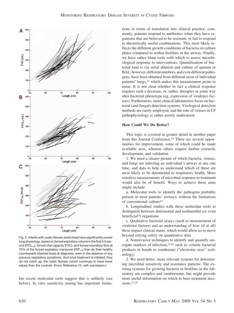
surements can detect abnormalities in pre-symptomatic ba-bies with CF10,15; these changes occur early, although theremay be a window of preserved lung function during thefirst few months of life in babies diagnosed via newbornscreening16; once physiologic changes are present, thesemay persist despite the initiation of standard management,so children with CF fail to catch up to their healthy peers(Fig. 3)15,17; and it has been difficult to explain these find-ings on the basis of infection and/or inflammation, fromthe limited number of studies that have included bron-choalveolar lavage (BAL) fluid.16 Further research isneeded.
Unfortunately, there are few specialized infant and pre-school child lung-function-testing laboratories. Most suchwork is done in the research setting. We hope that in thefuture some of these measurements will be routinely per-formed in the clinic on young children with CF, or thateasier measurement methods will be established, whichwould allow less specialized laboratories to participate.
Infection
Infection of the lower respiratory tract occurs early inCF, with what we had believed, until recently, to be arelatively well defined group of bacteria.18 In addition,viral infections are thought to play an important role, forexample, in infective exacerbations, although this role hasbeen less well studied and is not completely clear. Fungi,in particular Aspergillus fumigatus, cause problems withallergic sensitization, and there is also a substantial diseaseburden from nontuberculous mycobacteria, most notably,Mycobacterium abscessus.
Current Clinical Practice
The importance of bacterial infection has long been rec-ognized, and modern treatment is based on attempts toidentify organisms early, eradicate them if possible, andsuppress their numbers in the chronically infected state. Tothis end, guidelines recommend: culture and sensitivity atevery out-patient consultation and at the start and duringadmissions for intravenous antibiotic treatment; that non-expectorating patients (the majority of children) undergooropharyngeal, cough swab, or cough plate cultures; andthat CF subjects with their first isolation of Pseudomonasaeruginosa should undergo eradication therapy,19 which isusually a combination of nebulized and systemic (oral orintravenous) antibiotics. Cross-infection between patientsshould be limited by strict infection-control protocols thatprevent patient contact and emphasize the importance ofsimple measures such as staff handwashing.20 Patients withparticularly worrisome bacteria such as organisms of theBurkholderia cepacia complex, should attend separate clin-ics.
What Are the Problems?
It is our opinion that there are many opportunities forimprovement regarding both the ways we obtain samplesand the detection/sensitivity of the methods in conven-tional laboratories. It is clear from the literature that themethods of sampling from nonexpectorating patients lacksensitivity and specificity.21,22 Some patients have repeat-edly negative cultures despite substantial disease; it is un-known whether this truly reflects a sterile lower airway,
Table 1. Agreement Between Lung-Clearance Index, FEV1, and FEF75, and Structural Lung Changes Classified as Abnormal or Normal in aStudy of Patients With Cystic Fibrosis (n � 44)
Bronchiectasis HRCT Score Air-Trapping
Yes No � 5% � 5% � 30% � 30%
Lung-Clearance Index (n)*Abnormal 22 9 25 6 15 16Normal 4 9 2 11 1 12
(P � .03) (P � .001) (P � .03)FEV1 (n)
Abnormal 5 2 7 0 4 3Normal 21 16 20 17 12 25
(P � .76) (P � .06) (P � .41FEF75 (n)
Abnormal 16 3 17 2 12 7Normal 10 15 10 15 4 21
(P � .008) (P � .003) (P � .004)
* In a study of patients with cystic fibrosis, there was strong agreement between structural abnormalities identified via high-resolution computed tomography (HRCT) and lung-clearance index orforced expiratory flow at 75% of the forced expiratory maneuver (FEF75). In contrast, agreement with forced expiratory volume in the first second (FEV1) was poor. (Adapted from Reference 13.)
MONITORING RESPIRATORY DISEASE SEVERITY IN CYSTIC FIBROSIS
RESPIRATORY CARE • MAY 2009 VOL 54 NO 5 609
but recent molecular tools suggest that is unlikely (seebelow). In vitro sensitivity testing has important limita-
tions in terms of translation into clinical practice; com-monly, patients respond to antibiotics when they have or-ganisms that are believed to be resistant, or fail to respondto theoretically useful combinations. This most likely re-flects the different growth conditions of bacteria on cultureplates compared to within biofilms in the airway. Finally,we have rather blunt tools with which to assess microbi-ological response to interventions. Quantification of bac-terial load is via serial dilution and culture of sputum orBAL; however, different numbers, and even different patho-gens, have been obtained from different areas of individualpatients’ lungs,23 which makes this measurement prone tonoise. It is not clear whether in fact a clinical responserequires such a decrease, or, rather, therapies in some wayalter bacterial phenotype (eg, expression of virulence fac-tors). Furthermore, most clinical laboratories focus on bac-terial (and fungal) detection systems. Virological detectionmethods are rarely employed, and the role of viruses in CFpathophysiology is rather poorly understood.
How Could We Do Better?
This topic is covered in greater detail in another paperfrom this Journal Conference.18 There are several oppor-tunities for improvement, some of which could be madeavailable now, whereas others require further research,development, and validation.
1. We need a clearer picture of which bacteria, viruses,and fungi are infecting an individual’s airway at any onetime, and data to help us understand which of these aremost likely to be detrimental to respiratory health. Moresensitive measurements of microbial response to treatmentwould also be of benefit. Ways to achieve these aimsmight include:
a. Molecular tools to identify the pathogens probablypresent in most patients’ airways, without the limitationsof conventional culture24
b. Longitudinal studies with those molecular tools todistinguish between detrimental and nonharmful (or evenbeneficial?) organisms
c. Qualitative bacterial assays (such as measurement ofvirulence factors) and an understanding of how (if at all)these impact clinical status, which would allow us to movebeyond relying solely on quantitative data
d. Noninvasive techniques to identify and quantify sur-rogate markers of infection,25,26 such as volatile bacterialproducts in breath or condensate (“electronic nose” tech-nology)
2. We need better, more relevant systems for determin-ing microbial sensitivity and resistance patterns. The ex-isting systems for growing bacteria in biofilms in the lab-oratory are complex and cumbersome, but might providemore useful information on which to base treatment deci-sions.27,28
Fig. 3. Infants with cystic fibrosis (solid lines) have significantly poorerlung physiology, based on forced expiratory volume in the first 0.5 sec-ond (FEV0.5), forced vital capacity (FVC), and forced expiratory flow at75% of the forced expiratory maneuver (FEF75) than do their healthycounterparts (dashed lines) at diagnosis, even in the absence of anyprevious respiratory symptoms. And once treatment is initiated, theydo not catch up: the cystic fibrosis cohort continues to have lowervalues than the controls. (From Reference 15, with permission.)
MONITORING RESPIRATORY DISEASE SEVERITY IN CYSTIC FIBROSIS
610 RESPIRATORY CARE • MAY 2009 VOL 54 NO 5
Inflammation
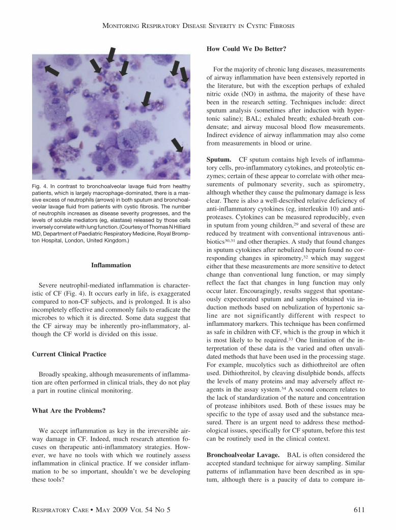
Severe neutrophil-mediated inflammation is character-istic of CF (Fig. 4). It occurs early in life, is exaggeratedcompared to non-CF subjects, and is prolonged. It is alsoincompletely effective and commonly fails to eradicate themicrobes to which it is directed. Some data suggest thatthe CF airway may be inherently pro-inflammatory, al-though the CF world is divided on this issue.
Current Clinical Practice
Broadly speaking, although measurements of inflamma-tion are often performed in clinical trials, they do not playa part in routine clinical monitoring.
What Are the Problems?
We accept inflammation as key in the irreversible air-way damage in CF. Indeed, much research attention fo-cuses on therapeutic anti-inflammatory strategies. How-ever, we have no tools with which we routinely assessinflammation in clinical practice. If we consider inflam-mation to be so important, shouldn’t we be developingthese tools?
How Could We Do Better?
For the majority of chronic lung diseases, measurementsof airway inflammation have been extensively reported inthe literature, but with the exception perhaps of exhalednitric oxide (NO) in asthma, the majority of these havebeen in the research setting. Techniques include: directsputum analysis (sometimes after induction with hyper-tonic saline); BAL; exhaled breath; exhaled-breath con-densate; and airway mucosal blood flow measurements.Indirect evidence of airway inflammation may also comefrom measurements in blood or urine.
Sputum. CF sputum contains high levels of inflamma-tory cells, pro-inflammatory cytokines, and proteolytic en-zymes; certain of these appear to correlate with other mea-surements of pulmonary severity, such as spirometry,although whether they cause the pulmonary damage is lessclear. There is also a well-described relative deficiency ofanti-inflammatory cytokines (eg, interleukin 10) and anti-proteases. Cytokines can be measured reproducibly, evenin sputum from young children,29 and several of these arereduced by treatment with conventional intravenous anti-biotics30,31 and other therapies. A study that found changesin sputum cytokines after nebulized heparin found no cor-responding changes in spirometry,32 which may suggesteither that these measurements are more sensitive to detectchange than conventional lung function, or may simplyreflect the fact that changes in lung function may onlyoccur later. Encouragingly, results suggest that spontane-ously expectorated sputum and samples obtained via in-duction methods based on nebulization of hypertonic sa-line are not significantly different with respect toinflammatory markers. This technique has been confirmedas safe in children with CF, which is the group in which itis most likely to be required.33 One limitation of the in-terpretation of these data is the varied and often unvali-dated methods that have been used in the processing stage.For example, mucolytics such as dithiothreitol are oftenused. Dithiothreitol, by cleaving disulphide bonds, affectsthe levels of many proteins and may adversely affect re-agents in the assay system.34 A second concern relates tothe lack of standardization of the nature and concentrationof protease inhibitors used. Both of these issues may bespecific to the type of assay used and the substance mea-sured. There is an urgent need to address these method-ological issues, specifically for CF sputum, before this testcan be routinely used in the clinical context.
Bronchoalveolar Lavage. BAL is often considered theaccepted standard technique for airway sampling. Similarpatterns of inflammation have been described as in spu-tum, although there is a paucity of data to compare in-
Fig. 4. In contrast to bronchoalveolar lavage fluid from healthypatients, which is largely macrophage-dominated, there is a mas-sive excess of neutrophils (arrows) in both sputum and bronchoal-veolar lavage fluid from patients with cystic fibrosis. The numberof neutrophils increases as disease severity progresses, and thelevels of soluble mediators (eg, elastase) released by those cellsinverselycorrelatewith lung function. (CourtesyofThomasNHilliardMD, Department of Paediatric Respiratory Medicine, Royal Bromp-ton Hospital, London, United Kingdom.)
MONITORING RESPIRATORY DISEASE SEVERITY IN CYSTIC FIBROSIS
RESPIRATORY CARE • MAY 2009 VOL 54 NO 5 611

flammation in those 2 types of sample. However, BAL,whether bronchoscopic or nonbronchoscopic, is highly in-vasive and not easily repeated in a short time period. Ad-verse effects such as fever have been reported, althoughthis is rarely important in our experience. Extensive safetydata in young children were recently reported by the Aus-tralasian study that addressed the utility of regular versussymptomatic BAL, further results of which are eagerlyawaited.35 Further limitations include the large and un-known dilution factor (markers for dilution are of limiteduse, and recent guidelines suggest they are not helpful36)and the fact that the technique samples only a small part ofthe airway, which may be problematic in a disease knownto be inhomogeneous.
Exhaled Breath. Both exhaled breath and breath con-densate are easy and noninvasive to obtain, and can beused reproducibly, even in young children, as long as at-tention is paid to methodological detail. Much interest hasfocused on the observation that the level of exhaled NO isreduced in CF. Given the anti-inflammatory and anti-in-fective properties of NO, some think it may play an im-portant primary role in CF pathophysiology—a hypothesissupported by the low level of NO synthase messengerribonucleic acid in relatively undamaged airways.37 Analternative view is that NO production is itself adverselyaffected by inflammation and that the low NO level issecondary to CF lung disease. The NO level is extremelylow in primary ciliary dyskinesia, a disease with a gener-ally much better outlook than CF. A recent clinical trial oforally administered L-arginine (an NO donor) with CFsubjects used exhaled NO as an outcome and reported asustained increase in NO production, although this was notmirrored by any significant effect on lung function.38 How-ever, no studies have addressed longitudinal change orcorrelations with other clinical variables, which would sup-port this measure as a useful monitoring tool in the clinic.
Exhaled-Breath Condensate. Condensate can be col-lected simply by asking a subject, even a quite small child,to exhale into a cold tube during tidal breathing. The con-densate contains a small (but variable and undetermined)volume of airway-lining fluid, the pH of which is abnor-mally low in CF (Fig. 5)39 and other inflammatory airwaydiseases.40 Thus, the condensate provides an “inflamma-mometer” to assess interventions. However, this techniqueappears to lack sufficient sensitivity for use with an indi-vidual patient. Attempts to measure other substances frompatients with CF have met with variable success, whichmight be partly related to methodological issues.41-43
Airway Mucosal Blood Flow. A universal downstreameffect of inflammation at any site in the body is an increasein blood flow. Measurements to detect this, based on the
rate of disappearance of an inhaled, absorbable gas fromthe airway, have shown some promise in asthma,44 in whichstudies have found a raised level, which is reduced byanti-inflammatory agents. There are, as yet, no publisheddata available from patients with CF, and current tech-niques require multiple, accurately-timed breath-hold ma-neuvers, which would probably restrict the technique toolder children and adults. However, this type of technique,unlike measurements of specific inflammatory markers, isnot dependent on a complete understanding of the com-plex inflammatory milieu within the airway, and may there-fore be more applicable as a generalized marker of inflam-mation.
Blood. Blood and serum markers, including inflamma-tory (total white blood cells and differential) cell countsand acute-phase reactants such as C-reactive proteinand immunoglobulin G, are recommended in many cur-rent clinical guidelines. Together with circulating cyto-kine levels, they have been used both as efficacy andsafety outcome measurements in CF clinical trials. Inmany studies in both those contexts they have proveduseful. However, although there are not the same meth-odological issues as exist with the airway-sampling tech-niques described above, blood/serum would probably beconsidered by most to be an adjunct to, rather than asubstitute for, such direct measurements. No studies havefound a tight correlation between such measurementsand direct airway inflammation.
Fig. 5. Exhaled-breath condensate from patients with cystic fibro-sis (CF) is significantly more acidic than that from healthy controls.Patients with and without CF pulmonary exacerbation also hadsignificant pH differences. This is unlikely to be directly related tothe cystic fibrosis transmembrane regulator defect, as it has alsobeen shown to occur in other inflammatory airway diseases, suchas asthma. (From Reference 39, with permission.)
MONITORING RESPIRATORY DISEASE SEVERITY IN CYSTIC FIBROSIS
612 RESPIRATORY CARE • MAY 2009 VOL 54 NO 5

Urine. Several groups have reported increased tissue-degradation products, such as desmosine and isodesmo-sine, in the urine of patients with chronic lung diseases,including CF,45 but the levels fluctuate rapidly, which mightlimit the applicability of these measurements in the clini-cal (or trial) context.
Radiology
Current Clinical Practice
Consensus guidelines recommend plain chest radio-graphs as part of the annual detailed assessment and atother periods of clinical concern.
What Are the Problems?
Plain chest radiographs lack sensitivity, particularly inthe early stages of disease. The scoring systems in clinicaluse also differ markedly, and although some researchershave reported confidence in the utility of these, othersview them as highly subjective and lacking in consistency.However, they carry a low radiation burden and are rela-tively cheap.
How Could We Do Better?
Computed Tomography. A very small number of cen-ters around the world advocate regular (every 1–2 years)CT, on the basis that CT is more sensitive than radiograph,and this was the topic of recent good reviews.46,47 Thechanges at various disease stages include air-trapping andbronchial wall thickening (Fig. 6), which appear to be atleast partially reversible, which renders them useful mark-ers of clinical progression and response to therapy. Severalscoring systems, of various complexities, have been de-
vised, and some authors strongly favor composite scoreswith, for example, spirometric indices.48 The routine useof CT in clinical practice is not proposed in the majority ofconsensus guidelines and has not been widely accepted bythe CF community, in particular by pediatricians, probablybecause the risk from radiation is thought to outweigh thebenefits of the knowledge gained from the CT. However,the radiation is substantially less with some of the moremodern CT scanners49 and could be further reduced witha CT protocol that takes fewer CT slices. The requirementthat the patient lie still and (as advocated by some) per-form respiratory maneuvers50 may limit the use of CT insome age groups.
Magnetic Resonance Imaging. Magnetic resonance im-aging was, until recently, widely regarded as lacking suf-ficient resolution for lung imaging, but in a small clinicaltrial the addition of hyperpolarized helium 3 improved thesensitivity so that significant differences were visible afterbronchodilator treatment.51 If further progress is made,this is potentially an attractive, radiation-free technique.
Positron Emission Tomography. This technique is rel-atively new in the context of lung disease. Labeled glucoseuptake indicates areas of inflammation. A recent studyfound greater uptake in patients with CF than in healthycontrols, and the difference was particularly marked insubjects with more impaired spirometry, and correlatedwith BAL neutrophilia.52 Research is ongoing on positronemission tomography in lung disease.
Summary
Patients with CF in developed countries are survivinglonger than ever before. In the United Kingdom, adultswith CF now outnumber children with CF, and almost50% of United States patients with CF are over 18 yearsold. Milder disease and a slower rate of decline makemonitoring more difficult, whereas the increasing numberof interventions available, and the fact that response totreatment is often unpredictable, make the requirement forgood monitoring interventions even more pressing. This isparticularly problematic in certain patient groups, such asinfants and young children. There are several opportunitiesfor improved monitoring with currently available tech-niques, and many new techniques are under investigation,fuelled largely by the requirement of more sensitive mea-surements for clinical trials. As an example, the UnitedKingdom CF Gene Therapy Consortium53 is assessingnewer techniques in both interventional and longitudinalclinical studies, to help determine which outcome mea-surements to use in our forthcoming multi-dose gene-ther-apy trial. We hope that certain of these techniques, andperhaps others as yet undeveloped, will show sufficient
Fig. 6. Typical changes on a computed tomogram of a patient withmoderately severe cystic fibrosis lung disease.
MONITORING RESPIRATORY DISEASE SEVERITY IN CYSTIC FIBROSIS
RESPIRATORY CARE • MAY 2009 VOL 54 NO 5 613
promise for routine clinical use and give us more insightinto our patients’ respiratory health.
REFERENCES
1. Cystic Fibrosis Trust. Standards for the clinical care of children andadults with cystic fibrosis in the UK 2001: a revised, expanded andreferenced version of the Cystic Fibrosis Trust’s 1996 guidelines. May2001. http://www.cftrust.org.uk/aboutcf/publications/consensusdoc/c_3000standards_of_care.pdf. Accessed March 18, 2009.
2. Kerem E, Conway S, Elborn S, Heijerman H; Consensus Committee.Standards of care for patients with cystic fibrosis: a European con-sensus. J Cyst Fibros 2005;4(1):7-26.
3. Cotes JE, Leathart GL Lung function, physiology, measurement andapplication in medicine, 6th edition. Blackwell Science; 1993.
4. Cooper PJ, Robertson CF, Hudson IL, Phelan PD. Variability ofpulmonary function tests in cystic fibrosis. Pediatr Pulmonol 1990;8(1):16-22.
5. Que C, Cullinan P, Geddes D. Improving rate of decline of FEV1 inyoung adults with cystic fibrosis. Thorax 2006;61(2):155-157.
6. Davis SQ, Garrity ER Jr. Organ allocation in lung transplant. Chest2007;132(5):1646-1651.
7. Rosenthal M. Annual assessment spirometry, plethysmography, andgas transfer in cystic fibrosis: do they predict death or transplanta-tion. Pediatr Pulmonol 2008;43(10):945-952.
8. Aurora P, Gustafsson P, Bush A, Lindblad A, Oliver C, Wallis CE,Stocks J. Multiple breath inert gas washout as a measure of venti-lation distribution in children with cystic fibrosis. Thorax 2004;59(12):1068-1073.
9. Aurora P, Bush A, Gustafsson P, Oliver C, Wallis C, Price J, et al;London Cystic Fibrosis Collaboration. Multiple-breath washout as amarker of lung disease in preschool children with cystic fibrosis.Am J Respir Crit Care Med 2005;171(3):249-256.
10. Lum S, Gustafsson P, Ljungberg H, Hulskamp G, Bush A, Carr SB,et al; London Cystic Fibrosis Collaboration. Early detection of cysticfibrosis lung disease: multiple-breath washout versus raised volumetests. Thorax 2007;62(4):341-347.
11. Horsley AR, Gustafsson PM, Macleod KA, Saunders C, GreeningAP, Porteous DJ, et al. Lung clearance index is a sensitive, repeat-able and practical measure of airways disease in adults with cysticfibrosis. Thorax 2008;63(2):135-140.
12. Kraemer R, Blum A, Schibler A, Ammann RA, Gallati S. Ventilationinhomogeneities in relation to standard lung function in patients withcystic fibrosis. Am J Respir Crit Care Med 2005;171(4):371-378.
13. Gustafsson PM, De Jong PA, Tiddens HA, Lindblad A. Multiple-breath inert gas washout and spirometry versus structural lung dis-ease in cystic fibrosis. Thorax 2008;63(2):129-134.
14. Beydon N, Davis SD, Lombardi E, Allen JL, Arets HG, Aurora P, etal. An official American Thoracic Society/European Respiratory So-ciety statement: pulmonary function testing in preschool children.Am J Respir Crit Care Med 2007;175(12):1304-1345.
15. Ranganathan SC, Stocks J, Dezateux C, Bush A, Wade A, Carr S, etal. The evolution of airway function in early childhood followingclinical diagnosis of cystic fibrosis. Am J Respir Crit Care Med2004;169(8):928-933.
16. Linnane BM, Hall GL, Nolan G, Brennan S, Stick SM, Sly PD, et al.;on behalf of the Australian Respiratory Early Surveillance Team forCystic Fibrosis (AREST-CF). Lung function in infants with cysticfibrosis diagnosed by newborn screening. Am J Respir Crit CareMed 2008;178(12):1238-1244.
17. Kozlowska WJ, Bush A, Wade A, Aurora P, Carr SB, Castle RA, etal.; London Cystic Fibrosis Collaboration. Lung function from in-fancy to the preschool years after clinical diagnosis of cystic fibrosis.Am J Respir Crit Care Med 2008;178(1):42-49.
18. Davies JC, Bilton D. Bugs, biofilms, and resistance in cystic fibrosis.Respir Care 2009;54(5):628-638; discussion 638-640.
19. Wood DM, Smyth AR. Antibiotic strategies for eradicating Pseudo-monas aeruginosa in people with cystic fibrosis. Cochrane DatabaseSyst Rev 2006;(1):CD004197.
20. Festini F, Buzzetti R, Bassi C, Braggion C, Salvatore D, Taccetti G,Mastella G. Isolation measures for prevention of infection with re-spiratory pathogens in cystic fibrosis: a systematic review. J HospInfect 2006;64(1):1-6.
21. Equi AC, Pike SE, Davies J, Bush A. Use of cough swabs in a cysticfibrosis clinic. Arch Dis Child 2001;85(5):438-439.
22. Rosenfeld M, Emerson J, Accurso F, Armstrong D, Castile R, Grim-wood K, et al. Diagnostic accuracy of oropharyngeal cultures ininfants and young children with cystic fibrosis. Pediatr Pulmonol1999;28(5):321-328.
23. Gutierrez JP, Grimwood K, Armstrong DS, Carlin JB, Carzino R,Olinsky A, et al. Interlobar differences in bronchoalveolar lavagefluid from children with cystic fibrosis. Eur Respir J 2001;17(2):281-286.
24. Rogers GB, Carroll MP, Serisier DJ, Hockey PM, Jones G, KehagiaV, et al. Use of 16S rRNA gene profiling by terminal restrictionfragment length polymorphism analysis to compare bacterial com-munities in sputum and mouthwash samples from patients with cys-tic fibrosis. J Clin Microbiol 2006;44(7):2601-2604.
25. Carroll W, Lenney W, Wang T, Spanel P, Alcock A, Smith D.Detection of volatile compounds emitted by Pseudomonas aerugi-nosa using selected ion flow tube mass spectrometry. Pediatr Pul-monol 2005;39(5):452-456.
26. Ryall B, Davies JC, Wilson R, Shoemark A, Williams HD. Pseudomo-nas aeruginosa, cyanide accumulation and lung function in CF andnon-CF bronchiectasis patients. Eur Respir J 2008;32(3):740-747.
27. Moskowitz SM, Foster JM, Emerson JC, Gibson RL, Burns JL. Useof Pseudomonas biofilm susceptibilities to assign simulated antibi-otic regimens for cystic fibrosis airway infection. J Antimicrob Che-mother 2005;56(5):879-886.
28. Caraher E, Reynolds G, Murphy P, McClean S, Callaghan M. Com-parison of antibiotic susceptibility of Burkholderia cepacia complexorganisms when grown planktonically or as biofilm in vitro. EurJ Clin Microbiol Infect Dis 2007;26(3):213-216.
29. Ordonez CL, Kartashov AI, Wohl ME. Variability of markers ofinflammation and infection in induced sputum in children with cysticfibrosis. J Pediatr 2004;145(5):689-692.
30. Colombo C, Costantini D, Rocchi A, Cariani L, Garlaschi ML, Tirelli S,et al. Cytokine levels in sputum of cystic fibrosis patients before andafter antibiotic therapy. Pediatr Pulmonol 2005;40(1):15-21.
31. Ordonez CL, Henig NR, Mayer-Hamblett N, Accurso FJ, Burns JL,Chmiel JF, et al. Inflammatory and microbiologic markers in in-duced sputum after intravenous antibiotics in cystic fibrosis. Am JRespir Crit Care Med 2003;168(12):1471-1475.
32. Ledson M, Gallagher M, Hart CA, Walshaw M. Nebulized heparinin Burkholderia cepacia colonized adult cystic fibrosis patients. EurRespir J 2001;17(1):36-38.
33. Suri R, Marshall LJ, Wallis C, Metcalfe C, Shute JK, Bush A. Safetyand use of sputum induction in children with cystic fibrosis. PediatrPulmonol 2003;35(4):309-313.
34. Kim JS, Hackley GH, Okamoto K, Rubin BK. Sputum processingfor evaluation of inflammatory mediators. Pediatr Pulmonol 2001;32(2):152-158.
35. Wainwright CE, Grimwood K, Carlin JB, Vidmar S, Cooper PJ, FrancisPW, et al. Safety of bronchoalveolar lavage in young children withcystic fibrosis. Pediatr Pulmonol 2008;43(10):965-972.
36. Haslam PL, Baughman RP. Report of ERS Task Force: guidelinesfor measurement of acellular components and standardization of BAL.Eur Respir J 1999;14(2):245-248.
MONITORING RESPIRATORY DISEASE SEVERITY IN CYSTIC FIBROSIS
614 RESPIRATORY CARE • MAY 2009 VOL 54 NO 5

37. Moeller A, Horak F Jr, Lane C, Knight D, Kicic A, Brennan S, et al.Inducible NO synthase expression is low in airway epithelium fromyoung children with cystic fibrosis. Thorax 2006;61(6):514-520.
38. Grasemann H, Grasemann C, Kurtz F, Tietze-Schillings G, Vester U,Ratjen F. Oral L-arginine supplementation in cystic fibrosis patients:a placebo-controlled study. Eur Respir J 2005;25(1):62-68.
39. Tate S, MacGregor G, Davis M, Innes JA, Greening AP. Airways incystic fibrosis are acidified: detection by exhaled breath condensate.Thorax 2002;57(11):926-929.
40. Carpagnano GE, Barnes PJ, Francis J, Wilson N, Bush A, Khari-tonov SA. Breath condensate pH in children with cystic fibrosis andasthma: a new noninvasive marker of airway inflammation? Chest2004;125(6):2005-2010.
41. Ojoo JC, Mulrennan SA, Kastelik JA, Morice AH, Redington AE.Exhaled breath condensate pH and exhaled nitric oxide in allergicasthma and in cystic fibrosis. Thorax 2005;60(1):22-26.
42. Carpagnano GE, Barnes PJ, Geddes DM, Hodson ME, KharitonovSA. Increased leukotriene B4 and interleukin-6 in exhaled breathcondensate in cystic fibrosis. Am J Respir Crit Care Med 2003;167(8):1109-1112.
43. Rosias PP, Dompeling E, Hendriks HJ, Heijnens JW, Donckerwol-cke RA, Jobsis Q. Exhaled breath condensate in children: pearls andpitfalls. Pediatr Allergy Immunol 2004;15(1):4-19.
44. Wanner A, Mendes ES, Atkins ND. A simplified noninvasive methodto measure airway blood flow in humans. J Appl Physiol 2006;100(5):1674-1678.
45. Bode DC, Pagani ED, Cumiskey WR, von Roemeling R, Hamel L,
Silver PJ. Comparison of urinary desmosine excretion in patientswith chronic obstructive pulmonary disease or cystic fibrosis. PulmPharmacol Ther 2000;13(4):175-180.
46. Robinson TE. High-resolution CT scanning: potential outcome mea-sure. Curr Opin Pulm Med 2004;10(6):537-541.
47. Brody AS. Scoring systems for CT in cystic fibrosis: who cares?Radiology 2004;231(2):296-298.
48. Robinson TE, Leung AN, Northway WH, Blankenberg FG, ChanFP, Bloch DA, et al. Composite spirometric-computed tomographyoutcome measure in early cystic fibrosis lung disease. Am J RespirCrit Care Med 2003;168(5):588-593.
49. Huda W. Radiation doses and risks in chest computed tomographyexaminations. Proc Am Thorac Soc 2007;4(4):316-320.
50. Long FR. High-resolution computed tomography of the lung in chil-dren with cystic fibrosis: technical factors. Proc Am Thorac Soc2007;4(4):306-309.
51. Mentore K, Froh DK, de Lange EE, Brookeman JR, Paget-BrownAO, Altes TA. Hyperpolarized HHe 3 MRI of the lung in cysticfibrosis: assessment at baseline and after bronchodilator and airwayclearance treatment. Acad Radiol 2005;12(11):1423-1429.
52. Chen DL, Ferkol TW, Mintun MA, Pittman JE, Rosenbluth DB,Schuster DP. Quantifying pulmonary inflammation in cystic fibrosiswith positron emission tomography. Am J Respir Crit Care Med2006;173(12):1363-1369.
53. UK Cystic Fibrosis Gene Therapy Consortium. Current research.http://www.cfgenetherapy.org.uk/consortiumresearch.htm. AccessedMarch 18, 2009.
Discussion
Geller: The lung-clearance indexseems like a nice, noninvasive, sensi-tive way of evaluating early disease,and we’re struggling with outcomemeasures in clinical trials where we’redealing with healthier and healthierkids with CF, in whom we don’t seelarge changes in variables such asFEV1, which is the standard outcomemeasure. I would think the lung-clear-ance index would be high on that listof new variables to consider, but inthe United States it’s not receiving alot of interest yet. How difficult and/orexpensive is it to set up for and collectthe data to calculate the lung-clear-ance index? Has it been commercial-ized or standardized?
Davies: It’s extremely easy, for thepatient and the technician, after a quiteshort training. It relies on tidal breath-ing and requires either a mask or thatthe patient be able to keep a lip-seal
on a mouthpiece. It requires patientcooperation, and we’ve found that pro-viding a cartoon or something to watchusually achieves that cooperation. Thesetup required used to be much moredifficult, because all the techniqueswere based on mass spectrometry, andthe machinery was constantly break-ing down, unreliable, or difficult orexpensive to maintain, but the ma-chines are now commercially avail-able, although they’re not beingheavily marketed.
We’re using an Innocor photoacous-tic machine, which is very much eas-ier to use. I think it costs about $30,000or $40,000. We usually do 3 tests,which takes up to a half an hour. Invery severely affected patients,wash-in and wash-out take muchlonger. Of the patient outcome vari-ables we have to choose from and areconsidering, I think the airway-clear-ance index currently tops the list. We’dnever use FEV1.
Ratjen: Though the Innocor lung-clearance-index technique may be finefor older individuals, it may not beideal for infants, with whom there havebeen some issues with this technol-ogy. For infants you probably still haveto use a mass-spectrometry-based sys-tem, which costs about $100,000 inNorth America, and those can be a bitdifficult to set up.
What we still don’t fully understandis how responsive the various mea-surements, such as the lung-clearanceindex, are in patients who have verymild disease, and we are very inter-ested in those patients because theyhave normal FEV1. We’re targetingthat population in interventional stud-ies with the lung-clearance index, hy-pertonic saline, and DNAse [recombi-nant human deoxyribonoclease], to seewhether we can pick up a signal inthis group. But using these data thatwe have from pulmonary exacerba-tions, where we use a population thatis not stable at baseline, may not be the
MONITORING RESPIRATORY DISEASE SEVERITY IN CYSTIC FIBROSIS
RESPIRATORY CARE • MAY 2009 VOL 54 NO 5 615
ideal comparative group to the kind ofintervention we’re ultimately looking at,because if we do interventional trialswe usually do the interventions in stablepatients.
Davies: I completely agree; that’s agood point. A lot of the data are skewedby picking a population that will re-gress to the mean anyway. One thingwe’re doing is a run in a study in which200 patients are being seen approxi-mately every 4 months, but only attimes of complete stability. We’remaking a basket of measurements, in-cluding lung-clearance index, at thosetime points, and we’re going to lookat the coefficient of variability, the re-producibility, et cetera, to see whetherthis would only be useful in the pa-tients who start out abnormal orwhether mild degrees of abnormalityare detectable.
It’s important, what you said aboutyoung children; the Innocor does havea slower detection time than the mass-spectrometry techniques, so with achild with a rapid respiratory rate thesemay not be completely applicable.We’ve been testing younger childrenwith both the Innocor and the massspectrometer and should have somecomparison data soon.
Ratjen: Oh good. If you do that,then we don’t have to.
Davies: Well, you could do it too,and then we could make sure we’re allgetting the same thing.
Rubin: Jane, that was great. I have2 queries. The first easy one is aboutresearch, and the second (harder) oneis practical. Have you any informa-tion on the use of hyperpolarized gasimaging to get a better idea of smallairways disease in CF? And for thepractical one, respiratory therapists atbedside are often asked to evaluatewhether a treatment that they’re pro-viding to somebody with CF, acutelyor chronically, is being beneficial orbeing harmful. They’re the ones at the
bedside; they’re the ones who get toobserve that patient. How would yourecommend that they determine if whatthey’re doing is helping the patient?
Davies: I have very limited knowl-edge about hyperpolarized helium andmagnetic resonance imaging. Groupsin the United States1 and the UnitedKingdom2 are working on this, andwe spoke to one of the United King-dom groups about whether it shouldbecome part of the armamentarium,but we haven’t taken it forward be-cause we haven’t yet been convincedof the degree of sensitivity it wouldgive us on top of the noninvasive mea-surements we’re already making. It’sa developing field, and worth keepingan eye on, but they haven’t yet con-vinced me that it’s worth overcomingthe technical issues.
With regard to patient assessmentand whether a treatment is doing harmor good, that’s a very difficult ques-tion. At our multidisciplinary meet-ings we quite often have such issues.For example, a physiotherapist (we callthem physiotherapists, and their jobdescription is not quite the same asthat of respiratory therapists) mightsay, “So-and-so really loves being onhypertonic saline, but we think it’s notdoing anything. They’re already non-adherent to the rest of their medica-tion. What should we do?” That’s adifficult situation.
It’s similar to a study Andy Bushand I did many years ago,3 in whichwe asked people how they felt aftertaking DNAse for several months. Andnearly everyone felt better, even pa-tients who’d had a 15% drop in FEV1,so I think there’s a mismatch betweenthe sorts of things we’re measuringand the patient-reported outcomes,which I deliberately didn’t stray into,because it’s a whole new minefield.
But, in general, I do think that theperson by the bedside is the best tojudge that acute response. And in gen-eral those clinicians will say, for in-stance, “This patient is feeling better;they are expectorating sputum better
with hypertonic saline than withDNAse.” And we don’t know whetherthat translates into medium or evenlong-term benefit. I don’t have the an-swer to that question, and I would loveto know if anyone else has. [Silence.]Obviously not.
REFERENCES
1. Altes TA, Eichinger M, Puderbach M. Mag-netic resonance imaging of the lung in cys-tic fibrosis. Proc Am Thorac Soc 2007;4(4):321-327.
2. van Beek EJ, Hill C, Woodhouse N, FicheleS, Fleming S, Howe B, et al. Assessmentof lung disease in children with cystic fi-brosis using hyperpolarized 3-helium MRI:comparison with Shwachman score,Chrispin-Norman score and spirometry. EurRadiol 2007;17(4):1018-1024.
3. Davies J, Trindade MT, Wallis C, RosenthalM, Crawford O, Bush A. Retrospective re-view of the effects of rhDNase in childrenwith cystic fibrosis. Pediatr Pulmonol 1997;23(4):243-248.
Marshall: What is the youngest pa-tient group with whom we can obtainthe lung-clearance index?
Davies: Janet Stocks’s group1 andothers2 have done it with newborns.It’s similar to getting the child to sleepand then putting on a mask with agood seal, and it’s no more diffi-cult—in fact, possibly easier—thansome of the raised-volume compres-sion techniques.
The group where I work doesn’t doinfant lung-function studies. We’vedone children down as low as age 7with the Innocor, although we are notcompletely certain how good thosedata are. But there’s probably a win-dow, somewhere between the ages of3 and 5 years, where they’re neither in-fants (who will go to sleep nicely) norcooperative children, depending on howcompliant your pediatric cohort is, butin general I would say it’s pretty muchacross the whole age range.
REFERENCES
1. Lum S, Gustafsson P, Ljungberg H, Hul-skamp G, Bush A, Carr SB, et al. Earlydetection of cystic fibrosis lung disease: mul-
MONITORING RESPIRATORY DISEASE SEVERITY IN CYSTIC FIBROSIS
616 RESPIRATORY CARE • MAY 2009 VOL 54 NO 5

tiple-breath wash-out versus raised volumetests. Thorax 2007;62(4):341-347.
2. Horsley A. Lung clearance index in the as-sessment of airways disease. Respir Med2009 Feb 24. [Epub ahead of print]
Marshall: You mentioned that lung-clearance index tops your list of pos-sible outcome measurements you’reconsidering at the gene-therapy con-
sortium. What other measurements areyou considering?
Davies: I don’t want to imply thatthe consortium has chosen the lung-clearance index as the primary endpoint. We haven’t looked at any ofthe data from the run-in, but I thinkit’s among the top contenders. Othermeasurements we’re looking at in-
clude: mucociliary clearance, for whichpatients from our center have to travelto Southampton, which is a couple ofhours away, and which requires a long24-hour scan that’s quite complicat-ed; computed tomography; varioussputum assays; exhaled breath assays;serum assays; normal spirometry; andquality-of-life questionnaires. That’severything you’d probably expect.
MONITORING RESPIRATORY DISEASE SEVERITY IN CYSTIC FIBROSIS
RESPIRATORY CARE • MAY 2009 VOL 54 NO 5 617