moleculardiversity of ca2+ channel a1 subunits fromthe ... ofca2+ channel a1 subunits fromthe marine...
TRANSCRIPT

Proc. Natl. Acad. Sci. USAVol. 90, pp. 3787-3791, May 1993Neurobiology
Molecular diversity of Ca2+ channel a1 subunits from the marineray Discopyge ommataW. A. HORNE*t, P. T. ELLINORt, I. INMANt, M. ZHOU*t, R. W. TSIENt, AND T. L. SCHWARZO§tDepartment of Molecular and Cellular Physiology, Stanford University Medical Center, Stanford, CA 94305; and *Department of Pharmacology, College ofVeterinary Medicine, Cornell University, Ithaca, NY 14850
Communicated by James A. Spudich, January 7, 1993
ABSTRACT In many neurons, transmitter release frompresynaptic terminals is triggered by Ca2+ entry via dihydro-pyridine-insensitive Ca2+ channels. We have looked for cDNAsfor such channels in the nervous system of the marine rayDiscopyge ommata. One cDNA (doe-2) is similar to dihydro-pyridine-sensitive L-type channels, and two cDNAs (doe-i anddoe-4) are more similar to the subfamily of dihydropyridine-insensitive non-L-type channels. doe-4, which encodes a pro-tein of 2326 aa, most closely resembles a previously clonedN-type channel. doe-i, which encodes a protein of 2223 aa, isa member of a separate branch of the non-L-type channels.Northern blot analysis reveals that doe-i is abundant in theforebrain. doe-4 is more plentiful in the electric lobe and,therefore, may control neurotransmitter release in motor nerveterminals. These results show that the familial pattern ofCa2+-channel genes has been preserved from a stage in evolu-tion before the divergence of higher and lower vertebrates>400 million years ago. The cloning of these channels may bea useful starting point for elucidating the role of the Ca2+channels in excitation-secretion coupling in nerve terminals.
Delineation of the molecular diversity of voltage-gated Ca2+channels is essential for understanding how these channelscontrol a wide spectrum of cellular functions ranging fromsecretion and contraction to metabolism and gene expres-sion. Several types of voltage-gated Ca2+ channels (L, T, N,and P types) have been distinguished by functional criteriaand show different tissue distributions and differential sen-sitivity to modulators and drugs (1, 2). Much of the knowndiversity among Ca2+ channels may arise from the existenceof multiple forms of the al subunit (3-10), which are large(200-260 kDa) transmembrane proteins and are responsiblefor Ca2+-channel voltage dependence, selectivity for Ca2+,and sensitivity to pharmacological modulation.Two structural subfamilies of al subunits have emerged
from molecular cloning of mammalian cDNAs (11, 12). Thefirst subfamily includes a, cDNAs originally derived fromskeletal muscle (3, 13), heart (4), aorta (14), lung (15), andbrain (7, 8). Expression of these channels demonstrated thatthey are responsive to 1,4-dihydropyridines (DHPs) and maythus be classified as L-type channels. The second al sub-family consists so far of cDNAs derived from mammalianbrain. The individuals within this subfamily show upward of60% identity with each other but only -45% with membersof the first subfamily (5, 6, 9, 10), and, when expressed, lackthe characteristic DHP response of L-type channels (5, 10).We chose the marine ray Discopyge ommata as a source
for additional neuronal Ca2+-channel cDNAs. The electricorgan of marine rays has served as a valuable model systemfor studying the biochemical properties of synaptic proteins.The nerve terminals within the electric organ of D. ommataare the richest known source of receptors for the cone snail
doe-1rbB
Ll (skel)L2 (card)L3 (neur)doe-2
IGMQ FG.IVILDDNT IN NNF:TFbAWLLFR.ATGEXWQIMTFG IALIDGT IN NNF TF QASLLFR ATGEAWO
IGMQMFGKIALVDGTQINRNNNFQTFPQAVLLLFRCATGEAWQIGMQ3FGKIAL 3T3INRNNNFQTFPQAVLLLFRCATGEAWQIGMQMFGK. D QINRNNNFQTFPQAVLLLFRCATGEAWQIGMQXEGKIA DGTQINRNNNFQTFIQAVLLLFRCATGEAWQ
FIG. 1. PCR-derived Ca2+-channel fragments from D. ommatafall into both subfamilies of high-voltage-activated channels. Thefragments are from the region after IVS5. The sequences encoded bythe oligonucleotides for PCR are marked with arrows; the position ofthe detection oligonucleotide is marked with a dashed line. Residuesthat differ from the sequence of the skeletal muscle L1 channel areshaded. doe-i is grouped with the DHP-insensitive rbB channel fromrat brain (9, 11); doe-2 is grouped with the DHP-sensitive L-typechannels (3-8).
toxin co-conotoxin GVIA (16), a selective probe for N-typeCa2+ channels. Here we report the molecular cloning of twofull-length cDNAs that encode putative Ca2+-channel alsubunits$ that are differentially expressed in the forebrainand in the electric lobe that innervates the electric organ.
METHODSPCR and Molecular Cloning. Phage template DNA was
isolated from plate lysates (17) of an oligo(dT)-primed AgtlOcDNA library (kindly provided by F. Rupp and R. Scheller,Stanford University). Forty cycles of PCR (95°C for 1 min,52°C for 2 min, and 72°C for 1 min) were performed usingstandard conditions (17). PCR products were separatedthrough a 1.2% agarose gel, transferred to Hybond mem-branes (Pharmacia), and hybridized with a 32P-end-labeleddegenerate oligonucleotide probe (see Results). PositiveDNA fragments were gel-purified, blunt-ended with T4 DNApolymerase, phosphorylated with T4 polynucleotide kinase,and ligated into the Sma I site of pBluescript SK+ (Strata-gene). Plasmid DNA was purified and sequenced by thedideoxynucleotide termination method (18). Ca2+-channelcDNAs were isolated from the oligo(dT)-primed AgtlO libraryfrom electric lobe and additional randomly and specificallyprimed forebrain and electric lobe cDNA libraries in AZAP II(Stratagene). Libraries were probed with the PCR-derivedclones or successive cDNA isolates. DNA sequencing wasperformed by creating nested deletions on both strands(Promega Erase-A-Base) and with specifically synthesizedprimers. Sequence analysis was carried out using GCGsoftware (19).
Isolation of RNA and Northern Blot Analysis. Poly(A)+RNA was isolated from either forebrain or electric loberegions of D. ommata by using the guanidinium isothiocya-
Abbreviation: DHP, dihydropyridine.tPresent address: Neurex Corp., 3760 Haven Avenue, Menlo Park,CA 94025.§To whom reprint requests should be addressed.$The sequences reported in this paper have been deposited in theGenBank data base [accession nos. L12531 (doe-i) and L12532(doe-4)].
3787
The publication costs of this article were defrayed in part by page chargepayment. This article must therefore be hereby marked "advertisement"in accordance with 18 U.S.C. §1734 solely to indicate this fact.

3788 Neurobiology: Horne et al. Proc. Nati. Acad. Sci. USA 90 (1993)+ 0
doo-1 S ~~~SRHQf~VTUETA A VA SAGF QRR1ALNIV3C RSLFLF DNIVRKJAW4fIIFE 100BI-2 MARFGD ..RYG" VMVG Y..KQ AQRARTNALYNPIPVRQNCLTVNRSLFLFSEDN.RKAQIE96
rbB 1.ORFGD G.G ... .R GA G GQQRARTNALYNPIPV&QNCtTVNRSL ISUDNMVRKYAKRITE 93
doe-4 NA PA.. YGG..... RUARARTNALYNPIPVRONLTVNR L I PSUDNInIRKYAKRITE 86+ C
doe-i W4PPFUYNILATIIA-NCMLALUQH DKP K UYIICUGK L KGSYLRNGWNVNDF A RTLRA 200BI-2 WPPFEYMILATIIANCIVLALEQHLPD&jKTPNSERLDDTEPYFIGIFCFEAGIKIIALGFAFHKGSYLRNGWNVNDFVVVLTGILA LRTLRA 196rbB WPPFEYMILATIIANCIVLALEQHLPDGDKTPNSERLDDTEPYFIGIFCFEAGIKIIALGFMFHKGSYLRNGWNVNDFVVVLTIJILATAGTDFDLRTLRA 193
doe-4 WPPFEYMILATIIANCIVLALU HLPDGDKTPNSURLDDTEPYFIGIFCFUAGIKIIALGFAFHKGSYLRNGWNVNDFVVVLTGILT TDFDLRTLRAI 186
doe-i VtRVLRPLKLVSGIPSINIFLKSINKANVPLLQIGLLLFFAILMFAIIGUFTDDAiLDL PC CPGV.Y IP 299BI-2 VRVLRPLKLVSGIPSLQVVLKSINKANIWPLLQIGLLLFFAILWFAIIGLEFNGFI SP CGTEEPARffCPNG A YW P 296rbB VRVLRPLKLVSGIPSLQVVLKSINKANVPLLQIGLLLFFAILNFAIIGLEFYMGKFHKiC CFNSIDAUP DFPCG& ARL E TjRYW PN 292
doe-4 VRVLRPLKLV GIPSLOVVLKSINKAMVPLLOIGLLLFFAILMFAIIGLUFYNGKFHKTCFSIrE P .P LCz*YfgP~NGTVKY PN 284
doe-i1GIKDNIL TVFQCITU IY L WLYFIPLIIIGF MNLVLGVLSGEFAKERERVENRRIWLKLRRQQQIEREINY~ W 399BI-2 1;T DNILFAVLTVFQCITNUEGWTDI&YN&JDA INTWNWLYFIPLIIIGSFFMLNLVLGVLSGEFAKERERVENRRAFLKLRRQQQIERELNGIU I396rbB GITNFDNILFAjULTVFQCITNEGWTDILYNTNDA NTWNWLYFIPLIIIGSFFNLNLVLGVLSGUFAKURERVUNRRAFLKLRRQQQIURULNGLUII392
doe-4 GITNFDNILFAVLTVFQCITNEGWT DALGNTWNSGEFKLYFIPLRAFLLRR384
doe-i AUVI S TK GRNU SSDEmUSVGP AS ISYRRK II K YBI-2 EEAUUL UTD RUPKKSKIVSPAAS SDFK IRRV YfVrbB EEAUVNLAUUDKNAUUK L KRA&TKKSBNDLIHAUUUE PFASL .SSYFRRKE ~R LIRRMV SFWVC
doe-4 UEUVNLAEUDKNA L.LKRATTKKSKND UUU TDISSVG. SK SYFRRKUKRR RRMVKS SFYWIVLC
doe-i LVALNI V UYDQP N U LJG-L SLCY PYFHSSFNCFW VI SI TI SFGISVLRALRLLFBI-2 LVALNTLCVAIVHY L UEI LGL" I YGL YFHSSFNCFD V SF KPGTSFGISVLRALRLLRIKTrbB WALNTLCVANVU TQPQ LYFAEFVFLGLFLTENSLKNYGLG SYFP~SSFNCFDFGVIVGSIFE IKPGTSFGISVLRALRLLRIKT
doe-4 ILVONTLCVAIVHYDOPP TDALYFAUFVFLGLFLTUMSLKMYGLG YFHSSFNCFDFGVIVGSIFUVT L1SGSVRLLRFV'doe-i LRNL-V-V-S S MKSIISLLFLLFLFIVVFALLGNQLFGGQFNF GTPPTNFDTFPAAII VFQILTGUDWNUVNY IQGSVYF1B1-2 Y LRNLVVSLLNSMKSIISLLFLLFLFIVVFALLGNQLFG.GQFN TPPTNFDTFPAAI VFQILTGUDWNUVNY I GGIFrbB YWNSLRNLVVSLLNSNKSIISLLFLLFLFIVVFALLGNQLFGGQFN- TPTNFDTFPAAILTVFQILTGEDWN&VY IUSG MSGFS Fl
doe-4 IWNSLRNLVVSLLNSNKSIISLLFLLFLFIVVFALLGN LFGG FNFUDGTPPTNFDTFPAAILTVFQILTGEDWNUVN GFSVF
doe-i VLTLFGNYTLLNVFLAIAVDNLANAQEL U QA ... SPUS GPSTURRRRKH.. SI SLR OHS RUALFIBI-2 VLTLFGNYTLLNVFLAIAVDNLANAQULTD E- QKLALQKAKUVAEVSPIUSAA I UQQ* UQRTS B NLLASRUArbB VLTLFGNYTLLNVFLAIAVDNLANAQULTKDUUUNEEU QKLALQKAKUVAUVSPMSAANISIAA. EUQR&QL WNL AY
doe-4 -I TLFGNYTLLNVFLAIAVDNLANAQULTKDUUUNUU IO PS IA K IWUORTS LRR UH AL
doe-i TD........ LUGSRYRR RI .UL L&UQQAAESH&JLMGV RAFKN.RS QPAG I K U .... .LGRJBI-2 UDPUKAS PDNKTHLD:RP ..NNKVAUPTVDa.k QRD U D P.. 4sIrbB SUDU PDNKTUIMRPLVVU K PUG ...... RURDR TPkUD TLcP'Adoe-4 S ~~IPDNKTHLDRPLVVUE VCS U Uf* RRSURU . U. dGALD~
doe-i AGA R ]KRNKU LLQQLCE fSGQLTQMPEM(DABI-2 N ER~~~~~KAGD RQ GGSSGGSGSPRTGTAD PRRUR PG GPDD......
rbB RSHSK P. .ADTQVR... .R.. UHUGS U.=R.......PRRUHR QDSSKUGKUEAPMLVP~doe-4 P3ONSKDDKRC ~SUK ERDEgRK... RLDRU D.RU......R T HUREAN~A....
498495491480
598595591580
698695691680
793795789779
875889881869
949979958949
doe-i14 SW PUSSANTRTPDBJ)TDPSAN .U... SGR G SANTI1Q~1QSNWI&1QLNQQAT G ZLTMGTRD!PKQDKTQZQTE 1041BI-2 ERROJRH O'AS *GEAZGPDGG RRRRHR P Y A . RUR.....PVSGP9I49TMRPIQQDLSRQ 1068rbBEURRARH N P R ~~KVPPKNPURDUIV GSLHbl4IPSTC~K& 1048
doe-4 E .R RH RIK . R U J~GQ~y.~1041
doe-i ID D TETPMD... . . . . . . . .. . . . . . . . . . . .. . . . . . . . . . 1055BI-2 U~L NS EPVSPHENLSHAGLPQSPAKMGSSTDPAGPTPATAANPQNSTASRRTPNNPGNPI*GP UENSL STAQTNSAK 1168rbB VDPDADN TRMGSQ ........................... ?Dl STrHVV PGA...18
doe-4 LM PUDAN.KTGK........1084
doe-i .............LVTP PASSSSS K.S..... II NF N R IVNLRYFUNCILLI 1120BI-2 TARKPDHTTVUIPPACPPPLNHf Q PDPLP KK DUGP DG PSSN ITTNLR HYIDNLRYFUE4CII IAUSSI 1268rbB ...............ZVVs . .. Q K DLR PRP YSSN igJSTNOgLR HYIVffRYFEbRI IMVASSI 1163
doe-4............ I LM V)PUL GP PP MMFLFSTTN HYIVNLRYFUMCILLVI S 1162
doe-i ALAAUDPIH S FDYVFTGVFTFUEMVIKNI IL SF NILDFIVVSGALVAAFLI GS JKDINTIKSLRVLRVLRPLKT 1220BI-2 ALAAUDPV PNV FDYVFTGVFTFUE4VIKZ4IDLLL FRDLWNILDFIVVSGALVAFAF ... KGKDINTIKSLRVLRVLRPLKT 1365rbB ALAAUDP ~R ~K Y4DYTGVFTFUE4VIKMIDLGIRLP FRDLWNILDFIVVSGALVAFAFSSF_-SKGKDINTIKSLRVLRVLRPLKT 1263
doe-4 LAUP APRNNVLK MYVFTGVFTFUN4VIKMI G IILHPGSYFRDLWNILDFIVVSGALVAFAF.... S~GD!NISRLVRLT12581II2LIIS3IIIS4
doe-i IKRLPKLKAVFDC LN II FNFIFAVIAVQLFKGKFFYCT TK R .RQRTKLSIUNGNVTT EYN ALLTLFTV 1319
BI-2 IKRLPKLKAVFDCVVNSLKN ZILIVYNLFN4FIFAVMAVQLFKGKFN1IWTDESK !dKDCRGI§YLLUUWK HYDNVLWALLTLFTV 1465
rbB IKRLPKLKAVFDCVVNSLKNVLNILIVYMLFNFIFAVIAVQLFKGKFFYCTDESKELE&CRGQY U VU RKKDFHYDNVLWALLTLFTV 1363
doe-4 IKRLPKLKAVFDCVVNSLKNVLNILIVYMLFMFIFAVIAVQLFKGKFFYCTDESK KCGY ND~ UK~FHYDNVLWALLTLFTV 1358
doe-i .STGUGWPQ S QGI USFYYFVVFPFFFVNIFVALIIITFQUQGD qLE SSLUKNURACIDFAISAKPLTRYPQ TFQ 1419
BI-2 STGUGWPQVLKHSVDAT ZGPSPGYRMUEMSIFYVVYFVVFPFFFVNIFVALIIITFQUQGDE ~LEKNERACIDFAISAKPLTRgtKPQNKQS]Q 1565
rbE STGUGWPMLKHSVDA GPSPGRMUE1JSIFYVVYFVVFPFFFVNIFVALIIITFQEQGDKVSCLEKNERACIDFAISAKPLTRYUPQNK S Q 1463
doe-4 STGUGWP L TUDGSYUSFYVVYFVVFPFFFVNIFVALIIITFQUQGDKVU W=SLUKNURACIDFAISAKPLTRYMP NKQTF1458
doe-i QFVVSP UYI AN LUN SP GFAS L tI UtLCILK gAWNFR VFDFVT SPTISUjILITTENL 151931-2 QFVVSPPFUYTIAMIALNTIVLMNKF V VFTSLFSLU~LK~AF INYFRDAWNMJFDFVTVLGSITDILVTE NIN 1665
rbB Y VVSPPFUY INAUIALNTMVL4NKF YE L NIVFTSHFSLUCILKIIAFGVLNYFRDAWNVFDFVTVLGSITDILVTEI NNFIN 1563
doe-4 Y VVSnEPPFU)Lj NAILTVN NI FIGLYRANFDFVTVLGSITDILVTU IN 1558
FIG. 2. (Figure continues on the opposite page.)
-IH5 -I- IS6 0 0--Q

Proc. Natl. Acad. Sci. USA 90 (1993) 3789
doe-1 LSFI .FRAARLIKLLRQ I IRILLWTFVQSFKALPYVCLLIAMLFFIYAIIGN G F LMLLFRSATGEBI-2 LSFLRLFRAARLIKLLRQGYTIRILLWTFVQSFKALPYVCLLIAMLFFIYAIIGMQVFGNI I EDEDSDEDEFQInHNNFRT ALMLLFRSATGErbB LSFLRLFRAARLIKICRQGYTIRILLWTFVQSFKALPYVCLLIAMLFFIYAIIGMQVFGN gLD ... ..... DGSINRHNNFRTFLQALMLLFRSATGE
doe4 LSFLRLFRAARLIKLLRQGYTIRILLWTFVQSFKALPYVCLLIAMLFFIYAIIGMQVFGNEIL ........ D DGAINRHNNFRTFLQ L SATGE| IVS4- 0 0U IVS5 11 IVH5-
doe-i SQEIMLACLSG YFYFVSFIFLCSFLMLNLFVAVIMDNFEYLTRDSSILGPHHLDE VWAEYBI-2 FWNL SRCDKNffGL * CE YFYFVSFIFLCSFLNLNLFVAVIKDNFEYLTRDSSILGPHHLDE gVWAEYDPAA4GtLYr MYMrbB PI N HA..NA CGSDFAYFYFVSFIFLCSFLMLNLFVAVIMDNFEYLTRDSSILGPHHLDEFIRVWAEYDPAACGRI YNDI4ML
doe-4 RI DG AIDNFEYLTRDSSILGPHHLDEFIRVWAEY INDMYEM+ IVS6 +
doe-1 T! SPPLGLGKKCP AYKRL VT. VHFTSTL IRTA GD AELRKEIT3 LSQKTLDLLVPIBIIT LTVGKBI-2 H*JPpLGLGKeCPARVAYKR VHFPJSTLMALIRTALDIKI G Q AELRK PNLSQKTLDLLV PH ST LTVGKrbB HSPPLGLGKKCPARVAYKRLVRNNMP VHFTSTLKALIRTAI3IK;A CDAELRKEI ANLJQKTLDLLVPPH P VGK
doe-4 PPLGLGKKCPARVAYKRLVRMNMP . HFTSTLMALIRTAL IQG CDAELRKEI VWPNLS D
doe-1 IBI-2 IY IIYrbB YAALNIIDXKQN TRRD
doe-4 YAALHIIXIDYYKQN S
1611176516551650
1708186517531750
1807196418531849
01KL ULSRTP ..... SLPPOII .................... ST L TG SRSEF 1875;jE NRTPL.... PDZGGAG. QNALP PLM A P FQKTjT 2052a THAPG Pl AVLRGARVFLRQKS S IQ KESI SS YID.AR 1952:ddQLSOLS .TR E ......P5ST VNPL GRQ E~1 . CI . 1936
doe-i TuVPL Q Sm EIKRPKELKKI HY ..............SAE T RS NHSNwW 196131-2 NS MS .E.R..SQ.s GPRIR ~SLSI TSPNKRS .K. ~R2143rbB LE I QA V ......N........... N PHKRSI TLAP.RPH Q 2033
doe-4 KEVPES PAVENRE ......GSH ... AET [email protected]. RV 20248 + 0++0 0
doe-1 L .........Y ( WSRRP ..... ....................... AGSW ERGR .............. SP 2010BI-2 YSLERVPP NQ H.P- RR R " TOL LSHTTQSkDLPS... ERRGE R PHHHHHHHH P GP 2234rbB N S S. .HHHHHRCHRRPKKQRSLGPSLSp APS GSGLPHiEiGSTGCR REH ERGRSQER .....Q PS SS 1E 2120
doe--4 M SLE P HHHH@RC RLKLRD . . ....... Q S . .......... ls E 2101+ O
doe-i ERSVC&TGQCA..HES SRSP K P T S MSSPVPSH ... CSb C 2104BI-2 P. .PDHGHG SPS .......................................................... 2273rbB KQRiYSCDR SREPPQP PSLFPAA PHwQGSGS P T RRQLPQTPLTPRPS YKTANSS E2220
doe-4 KXRYYSCDR SREPPP STjH PRS GSGS SP STS .:RR LRPLTPRPSVTYKTANSS F LHDALJPS 2200+l + + +
doe-1 KET.YQSLRVQPSKA&SKIPGESRER.EOQHSTPLEYI | SkLHDIGPSuQAC33=4WSL G......ISHTIOAPPLRQGWHLPNGSM 2198BI -2 ....................................................................................................rbB |SPGRLSR CEH S.I PYL DSEASAH LTF TS YV SSSHPIRVPNG.2311
doe-4 rSPGRLSR LB GD1HRQ SDPYL SSCV1WJL ETLTF T... PQSSY?!!GY 2297
doe-1 RTRMNJQGA*PDP . .THlZKg 2223BI -2 .............................
'rbB IqTqVR .R.R.. Sq
ED2336
doe-4 HK TGE H 23260
FIG. 2. Alignment of the deduced amino acid sequences of doe-1 and doe-4 with representative mammalian clones of the non-L subfamily[rbB (9) and BI-2, the longer variant of the BI type (5)]. Dots represent gaps introduced by the BESTFIT program (GCG) to optimize alignment.When two or more residues at a position are identical, they are boxed. If two pairs of boxes are possible at a single position, the pair is boxedthat includes the doe-4 sequence. The predicted positions of transmembrane (S1-S6) and pore-forming (H5) regions in each of the homologousrepeats (I-IV) are indicated by brackets below. The position of an insert encountered in some doe-4 clones is indicated by a solid triangle.Predicted glycosylation sites in either doe-1 or doe-4 are indicated by open triangles. Potential sites for phosphorylation are marked by solidcircles (cAMP-dependent protein kinase), open circles (protein kinase C), and plus marks (multifunctional Ca2+/calmodulin-dependent proteinkinase).
nate method and selection on oligo(dT) resin (17). Poly(A)+RNA (3 ,g) from each tissue was electrophoresed through a1% agarose/0.8% formaldehyde gel and transferred to anylon membrane for hybridization (17).
RESULTS AND DISCUSSIONMolecular Cloning. We used PCR to amplify candidate
Ca2+-channel cDNA sequences from a cDNA library derivedfrom the electric lobe of D. ommata. Degenerate primerswere synthesized based on amino acid sequences, IGMQ-(V/M)FG and ATGEAW(Q/H), flanking 29 aa in the regionbetween transmembrane domains IVS5 and IVS6 (Fig. 1). Toidentify putative Ca2+-channel sequences, the PCR productswere probed with an oligonucleotide based on a sequence,INR(N/H)NNF, within the predicted amplified region. DNAsequencing of positive clones yielded two distinct 128-bpfragments [D. ommata-1 (doe-i) and doe-2]. Sequence align-ment indicates that doe-2 is as homologous to Ca2+ channelsfrom skeletal muscle or heart as these L-type channels are toeach other. Within this limited region of 29 aa, the doe-2fragment differs from the skeletal muscle sequence at only 3aa positions (Fig. 1).
In contrast, the doe-i fragment is less closely related toL-type channels and much more similar to a Ca2+-channelclone from rat brain (rbB; ref. 11). Starting with the doe-1
PCR fragment, we isolated overlapping cDNAs encoding thecomplete open reading frame (Fig. 2). The screening proce-dure also led to isolation of a distinct class ofcDNA encodingdoe-4, an additional putative Ca2+ channel. The doe-1 se-quence consists of an open reading frame of 6669 bp, encod-ing a protein of 2223 aa with a calculated molecular mass of251.8 kDa. doe-4 consists of an open reading frame of 6978bp, corresponding to a protein of 2326 aa and a calculatedmolecular mass of 264.5 kDa. Like other Ca2+-channel alsubunits (3), doe-1 and doe-4 consist of four largely hydro-phobic repeats (I-IV), separated by largely hydrophilic link-ers. Each repeat includes an H5 region, which is thought toline the conduction path, and six putative transmembranesegments (S1-S6).
In analyzing doe-4 cDNA, we repeatedly encountered avariant of the loop thatjoins the first two homology domains,a region that is otherwise well-conserved among this familyof channels. This variation is likely to arise from alternativesplicing and results in the insertion of a 20-aa sequence(DGLGIIYEPEQKPEDIQSVY) at a position marked in Fig.2. Potential sites for phosphorylation by protein kinases arealso marked. Because Ca2+ channels undergo physiologicalmodulations by several second messenger systems (20, 21),these sites are potentially significant.
Distribution ofmRNAs. Northern blot analysis ofthe tissuedistribution of doe-1 and doe-4 mRNAs is illustrated in Fig.
19
Neurobiology: Horne et al.
0

Proc. Natl. Acad. Sci. USA 90 (1993)
non.
L subfamily L1 (skeletal)
L2 (cardiac, smooth)L3 (neuroendocrine)
doe-i (ray forebrain)
BII(brain)BI (brain, ?P-type)rbB (brain, N-type)
-L subfamily doe-4 (ray electric lobe)
2.4-
1.4
FB EL FB EL
FIG. 3. Differential expression of doe-1 and doe-4 transcripts inthe central nervous system of D. ommata. Poly(A)+ RNA (3 pg perlane) from the forebrain (lanes FB) or electric lobe (lanes EL) wereprobed with 32P-labeled cDNA from regions likely to be specific forthe individual gene (the 5' untranslated region of doe-i or thecytoplasmic linker between repeats II and III of doe-4). Size stan-dards from an RNA ladder (BRL) are indicated at the left.
3. doe-1, an 8-kb transcript, is much more abundant inforebrain than in the electric lobe; in contrast, doe-4, a 12- to13-kb transcript, is more plentiful in electric lobe than inforebrain. Since the electric lobe consists largely of theelectromotor nucleus which innervates the electric organ,this suggests that doe-4 may play an important role inregulating transmitter release from motor nerve terminals.Consistent with this hypothesis, neurotransmitter release inthe electric organ is blocked by c-conotoxin GVIA (16),which also blocks the human homolog of clone rbB (10).
Relationships to Previously Described Ca2>-Channel Struc-tures. Fig. 4 provides an overview of structural homologiesbetween doe-1 and doe-4 and mammalian Ca2+-channelcDNAs (see below). The tree diagram is based on sequencehomology, but it also appears representative of an emergingpattern of functional differences. The upper branch consistsofthree a, genes designated as L1, L2, and L3 (1, 12). To date,the Ca2+-channel activity expressed from members of thisgroup has displayed responses to DHP antagonists andagonists and other functional characteristics expected forL-type Ca2+ channels. Individual members of this L subfam-ily display at least 60-70% amino acid identity with eachother.Members of the second subfamily show only -40% amino
acid identity with members of the L subfamily but >60%homology with each other. They are functionally distinctfrom the L subfamily in several respects (1). Most notably,they encode high-voltage-activated Ca2+ channels that arenot inhibited by DHP antagonists or stimulated by DHPagonists (5, 10). By grouping the nearly identical genes fromdifferent species, three types of mammalian channels can bedistinguished within this subfamily. They are represented byclones BI (rbA, CaCh4) (5, 6, 11, 12) and BII (22), which wereisolated from rabbit brain, and rbB (CaChS) (9-12), whichwas cloned from rat brain. The BI cDNA encodes an alsubunit with some key features expected for P-type Ca2+channels, which are highly expressed in cerebellar Purkinjecells and certain nerve terminals (5). The rbB cDNA encodesan co-conotoxin-sensitive, DHP-resistant activity character-
O 10 20 30 40 50 60 70 80 90 100
% identity
FIG. 4. Structural homologies of the mammalian and marine rayCa2+-channel al subunits. Percent sequence identity, as determinedby the BESTFIT program using the full amino acid sequence, isindicated. The subfamily of L-type channels is represented by L1from rabbit skeletal muscle, L2 from rabbit cardiac muscle, and L3from human brain. doe-2 is not included because a comparablepercent identity could not be determined from the limited sequenceavailable. The subfamily of non-L channels includes doe-4, rbB (anN-type channel from rat brain; ref. 9), and BI (a rabbit cerebellarchannel with some similarity to the P-type channel; ref. 5) and themore distantly related doe-i. In the nomenclature for mammalianbrain channels of Snutch et al. (11), channels like L2 are class C,those like L3 are class D, those like BI are class A, and those like rbBare class B.
istic of an N-type Ca2+ channel (9, 10). The recently reportedBII channel has not been physiologically characterized (22).
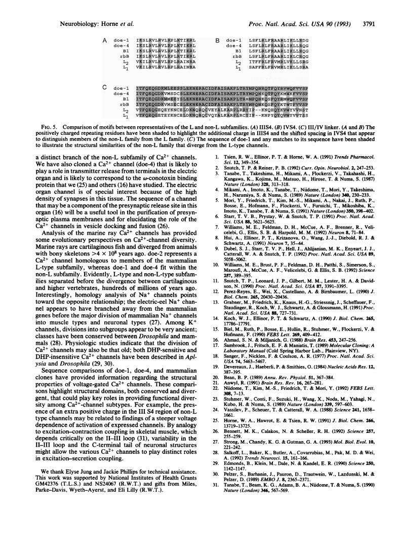
doe-1 and doe-4 belong to the non-L subfamily. The degreeof homology to BI or rbB clones ranges between 60 and 72%identity; in contrast, the homology between doe-i or doe-4and various L-type Ca2+ channels is <45%. Some distin-guishing structural features now emerge from a comparisonof the L and non-L subfamilies. In contrast to all knownL-type channels, for example, doe-i, doe-4, and BI or rbBcDNAs encode proteins containing an extra positive chargein repeat III (Fig. 5A) and a different pattern of spacing ofcharged residues in repeat IV (Fig. SB). Similar consider-ations hold for the cytoplasmic loop between repeats III andIV (Fig. SC), a segment thought to be important for inacti-vation (23, 24). doe-i, doe-4, BI, and rbB are highly homol-ogous along this stretch but display significant differenceswith respect to L-type channel cDNAs.From sequence analysis, we suggest that doe-i and BII,
which are 68% identical, represent a distinct branch of thenon-L subfamily. The deduced amino acid sequence of doe-ishows somewhat less identity to BI and rbB than they do toeach other. The overall identities are 60% for doe-i and rbB,63% for doe-i and BI, and 68% for BI and rbB. The moststriking sequence divergence between doe-i and the otherbranch of the non-L subfamily occurs in the cytosolic loopbetween repeats II and III.
doe-4 can more easily be grouped with BI and rbB thanwith doe-i and BII (68, 72, 61, and 61% identities, respec-tively). In particular, where the subfamily shows the greatestdivergence, some regions of doe-4 appear especially similarto rbB. This is most striking in the C termini of the channels:rbB and doe-4 extend, respectively, 174 and 185 aa beyondthe end of BI and are identical to each other at 110 of theseresidues. Similar comparison can be made in the cytoplasmiclinker between domains II and III.
CONCLUSIONSFrom a comparison of sequences, the doe-1 channel reportedhere and the mammalian BII clone (22) can be recognized as
Doe 1 Doe 4kb
9.5
7.5 ,.
4 4
3790 Neurobiology: Horne et al.
Proc. Natl. Acad. Sci. USA 90 (1993) 3791
...,. ..... ...,. ..,..- ...........
i*S L, LLLVPLI-TII.,.RL
IIKSLRVLRVLP LRT IEtLVKI LRVLRVLKP LRAAINaAVK£I LRVL VLaPLRAINRA
B doe-idoe-4
B1rbBL2Li
LSFLKLFRAARL IKLLRDGLSFLRLF#AARL I CLLRQLGLSFLRLFAAARL ILL,QGLSFLR LFAARLIKLCRQG
,,.... ...,. ,..
I TFF LFRVMRLVILLSAGSAFF LFRVMRLIX,LLSRA
FIG. 5. Comparison of motifs between representatives of the L and non-L subfamilies. (A) IIIS4. (B) IVS4. (C) III/IV linker. (A and B) Thepositively charged repeating residues have been shaded to highlight the additional charge in 1IIS4 and the shifted spacing in IVS4 that appear
to distinguish members of the non-L family from the L family. (C) The sequence of doe-1 and any matches to its sequence have been shadedto illustrate the structural similarities of the non-L family that diverge from the L-type channels.
a distinct branch of the non-L subfamily of Ca2+ channels.We have also cloned a Ca2+ channel (doe-4) that is likely toplay a role in transmitter release from terminals in the electricorgan and is likely to correspond to the co-conotoxin bindingprotein that we (25) and others (16) have studied. The electricorgan channel is of special interest because of the highdensity of synapses in this tissue. The sequence of a channelthat may be a component ofthe presynaptic release site in thisorgan (16) will be a useful tool in the puriflcation of presyn-aptic plasma membranes and for elucidating the role of theCa2+ channels in vesicle docking and fusion (26).
Analysis of the marine ray Ca2+ channels has providedsome evolutionary perspectives on Ca2+-channel diversity.Marine rays are cartilaginous fish and diverged from animalswith bony skeletons >4 x 10⁸ years ago. doe-2 represents a
Ca2+ channel homologous to members of the mammalianL-type subfamily, whereas doe-1 and doe-4 fit within thenon-L subfamily. Evidently, L-type and non-L-type subfam-ilies separated before the divergence between cartilaginousand higher vertebrates, hundreds of millions of years ago.Interestingly, homology analysis of Na+ channels pointstoward the opposite relationship; the electric-eel Na+ chan-nel appears to have branched away from the mammaliangenes before the major division of mammalian Na+ channelsinto muscle types and neuronal types (27). Among K+channels, divisions into subgroups appear to be very ancient;classes have been conserved between Drosophila and mam-mals (28). Physiologic studies indicate that the division ofCa2+ channels may also be that old; both DHP-sensitive andDHP-insensitive Ca2+ channels have been described in Apl-ysia and Drosophila (29, 30).
Sequence comparisons of doe-1, doe-4, and mammalianclones have provided information regarding the structuralproperties of voltage-gated Ca2+ channels. These compari-sons highlight structural domains, both conserved and diver-gent, that could play key roles in providing functional diver-sity among Ca2+-channel subtypes. For example, the pres-ence of an extra positive charge in the III S4 region of non-Ltype channels may be related to findings of a steeper voltagedependence of activation of expressed channels. By analogyto excitation-contraction coupling in skeletal muscle, whichdepends critically on the II-III loop (31), variability in the11-III loop and the C-terminal tail of neuronal structuresmight allow the various Ca2+ channels to play distinct rolesin excitation-secretion coupling.
We thank Elyse Jung and Jackie Phillips for technical assistance.This work was supported by National Institutes of Health GrantsGM42376 (T.L.S.) and NS24067 (R.W.T.) and gifts from Miles,Parke-Davis, Wyeth-Ayerst, and Eli Lilly (R.W.T.).
1. Tsien, R. W., Ellinor, P. T. & Home, W. A. (1991) Trends Pharmacol.Sci. 12, 349-354.
2. Snutch, T. P. & Reiner, P. B. (1992) Curr. Opin. Neurobiol. 2, 247-253.3. Tanabe, T., Takeshima, H., Mikami, A., Flockerzi, V., Takahashi, H.,
Kangawa, K., Kojima, M., Matsuo, H., Hirose, T. & Numa, S. (1987)Nature (London) 328, 313-318.
4. Mikami, A., Imoto, K., Tanabe, T., Niidome, T., Mori, Y., Takeshima,H., Narumiya, S. & Numa, S. (1989) Nature (London) 340, 230-233.
5. Mori, Y., Friedrich, T., Kim, M.-S., Mikami, A., Nakai, J., Ruth, P.,Bosse, E., Hofmann, F., Flockerzi, V., Furuichi, T., Mikoshiba, K.,Imoto, K., Tanabe, T. & Numa, S. (1991) Nature (London) 350,398-402.
6. Starr, T. V. B., Prystay, W. & Snutch, T. P. (1991) Proc. Natl. Acad.Sci. USA 88, 5621-5625.
7. Williams, M. E., Feldman, D. H., McCue, A. F., Brenner, R., Veli-celebi, G., Ellis, S. B. & Harpold, M. M. (1992) Neuron 8, 71-84.
8. Hui, A., Ellinor, P. T., Krizanova, O., Wang, J.-J., Diebold, R. J. &Schwartz, A. (1991) Neuron 7, 35-44.
9. Dubel, S. J., Starr, T. V. P., Hell, J., Ahlijanian, M. K., Enyeart, J. J.,Catterall, W. A. & Snutch, T. P. (1992) Proc. Natl. Acad. Sci. USA 89,5058-5062.
10. Williams, M. E., Brust, P. F., Feldman, D. H., Patthi, S., Simerson, S.,Maroufi, A., McCue, A. F., Velicelebi, G. & Ellis, S. B. (1992) Science257, 389-395.
11. Snutch, T. P., Leonard, J. P., Gilbert, M. M., Lester, H. A. & David-son, N. (1990) Proc. Nati. Acad. Sci. USA 87, 3391-3395.
12. Perez-Reyes, E., Wei, X., Castellano, A. & Birnbaumer, L. (1990) J.Biol. Chem. 265, 20430-20436.
13. Grabner, M., Friedrich, K., Knaus, H.-G., Striessnig, J., Scheffauer, F.,Staudinger, R., Koch, W. J., Schwartz, A. & Glossmann, H. (1991) Proc.Natl. Acad. Sci. USA 88, 727-731.
14. Koch, W. J., Ellinor, P. T. & Schwartz, A. (1990) J. Biol. Chem. 265,17786-17791.
15. Biel, M., Ruth, P., Bosse, E., Hullin, R., Stuhmer, W., Flockerzi, V. &Hofmann, F. (1990) FEBS Lett. 269, 409-412.
16. Ahmad, S. N. & Miljanich, G. (1988) Brain Res. 453, 247-256.17. Sambrook, J., Fritsch, E. F. & Maniatis, T. (1989) Molecular Cloning:A
Laboratory Manual (Cold Spring Harbor Lab., Plainview, NY).18. Sanger, F., Nicklen, F. & Coulson, A. R. (1977) Proc. Natl. Acad. Sci.
USA 74, 5463-5467.19. Devereaux, J., Haeberli, P. & Smithies, 0. (1984) Nucleic Acids Res. 12,
387-395.20. Bean, B. P. (1989) Annu. Rev. Physiol. 51, 367-384.21. Anwyl, R. (1991) Brain Res. Rev. 16, 265-281.22. Niidome, T., Kim, M.-S., Friedrich, T. & Mori, Y. (1992) FEBS Lett.
308, 7-13.23. Stuhmer, W., Conti, F., Suzuki, H., Wang, X., Noda, M., Yahagi, N.,
Kubo, H. & Numa, S. (1989) Nature (London) 339, 597-603.24. Vassilev, P., Scheuer, T. & Catterall, W. A. (1988) Science 241, 1658-
1661.25. Home, W. A., Hawrot, E. & Tsien, R. W. (1991) J. Biol. Chem. 266,
13719-13725.26. Bennett, M. K., Calakos, N. & Scheller, R. H. (1992) Science 257,
255-259.27. Strong, M., Chandy, K. G. & Gutman, G. A. (1993) Mol. Biol. Evol. 10,
221-242.28. Salkoff, L., Baker, K., Butler, A., Covarrubias, M., Pak, M. D. & Wei,
A. (1992) Trends Neurosci. 15, 161-166.29. Edmonds, B., Klein, M., Dale, N. & Kandel, E. R. (1990) Science 250,
1142-1147.30. Pelzer, S., Barhanin, J., Pauron, D., Trautwein, W., Lazdunski, M. &
Pelzer, D. (1989) EMBO J. 8, 2365-2371.31. Tanabe, T., Beam, K. G., Adams, B. A., Niidome, T. & Numa, S. (1990)
Nature (London) 346, 567-569.
A doe-idoe-4
BirbBL2Li
C doe-idoe-4
BirbBL2Li
Neurobiology: Home et al.