molecular switches at the synapse emerge from receptor and
TRANSCRIPT

Molecular Switches at the SynapseEmerge from Receptor and Kinase TrafficArnold Hayer
1,2¤, Upinder S. Bhalla
1*
1 National Centre for Biological Sciences, Bangalore, India, 2 Ecole Superieure de Biotechnologie de Strasbourg, Strasbourg, France
Changes in the synaptic connection strengths between neurons are believed to play a role in memory formation. Animportant mechanism for changing synaptic strength is through movement of neurotransmitter receptors andregulatory proteins to and from the synapse. Several activity-triggered biochemical events control these movements.Here we use computer models to explore how these putative memory-related changes can be stabilised long after theinitial trigger, and beyond the lifetime of synaptic molecules. We base our models on published biochemical data andexperiments on the activity-dependent movement of a glutamate receptor, AMPAR, and a calcium-dependent kinase,CaMKII. We find that both of these molecules participate in distinct bistable switches. These simulated switches areeffective for long periods despite molecular turnover and biochemical fluctuations arising from the small numbers ofmolecules in the synapse. The AMPAR switch arises from a novel self-recruitment process where the presence ofsufficient receptors biases the receptor movement cycle to insert still more receptors into the synapse. The CaMKIIswitch arises from autophosphorylation of the kinase. The switches may function in a tightly coupled manner, orrelatively independently. The latter case leads to multiple stable states of the synapse. We propose that similar self-recruitment cycles may be important for maintaining levels of many molecules that undergo regulated movement, andthat these may lead to combinatorial possible stable states of systems like the synapse.
Citation: Hayer A, Bhalla US (2005) Molecular switches at the synapse emerge from receptor and kinase traffic. PLoS Comp Biol 1(2): e20.
Introduction
Long-term storage of neuronal information is believed tooccur through alterations in synaptic efficacy. Many mecha-nisms have been identified for changes in synaptic strength,including modulation of neurotransmitter release, conduc-tivity changes in receptors, changes in numbers of receptorsor active synapses, and structural alterations of the synapse.Among these, the insertionof glutamate receptors of the alpha-amino-3-hydroxy-5-methyl-4-isoxazole propionate (AMPA)subtype into the postsynaptic membrane and the modulationof receptor conductance by phosphorylation are key eventsin modulating synaptic efficacy. A fundamental issue chal-lenges all of these mechanisms: how can they last a lifetime?
Synaptic memories can decay in at least three ways:turnover, diffusive exchange, and stochasticity. Turnover ofmajor postsynaptic molecules ranges from periods of a fewminutes to a few days and may be further enhanced bysynaptic activity [1]. One solution to loss of memory due tomolecular turnover is the concept of self-sustaining molec-ular switches [2]. These typically involve some form ofmolecular feedback giving rise to chemical systems, whichcan stably settle into one of two states. Such two-state, orbistable, systems can store information in a binary manner.Provided there is a steady supply of replacement molecules,molecular turnover can be tolerated, since newly synthesised,naıve molecules become entrained to the current state of thesystem. Some current proposals for such bistable synapticswitches include the calcium calmodulin type II kinase(CaMKII) hypothesis [3], a mitogen-activated protein kinase(MAPK) feedback loop [4,5], and, recently, the mammaliantarget of rapamycin (mTOR) protein synthesis loop [6]. Ofthese, the CaMKII model has been posed in the most detailwith the most complete structural correlates. According tothis model, CaMKII at the synapse can undergo autophos-
phorylation, which leads to activation of the kinase. Theactivated kinase molecules catalyze the phosphorylation ofyet more CaMKII molecules, resulting in a self-sustainingcycle. The MAPK feedback loop model also involves a self-sustaining cycle, but in this case several intermediatemolecules participate in the loop. The protein synthesis loopmodel is based on the observation of local protein translationmachinery associated with synapses. Messenger RNA forseveral proteins, including the ribosomes themselves, is alsopresent. Thus, high local protein synthesis creates themachinery for maintaining high levels of synthesis. Thisprotein synthetic loop is regulated by mTOR.The second mechanism for decay of synaptic memory is
diffusive exchange of synaptic proteins, leading to washout ofspecific states in the synapse. Extrapolations from freediffusion constants suggest that diffusive exchange betweenthe synaptic spine and dendrite is likely to be rapid, under 10 seven for proteins [7]. The postsynaptic density (PSD) is an
Received April 15, 2005; Accepted June 24, 2005; Published July 29, 2005DOI: 10.1371/journal.pcbi.0010020
Copyright: � 2005 Hayer and Bhalla. This is an open-access article distributedunder the terms of the Creative Commons Attribution License, which permitsunrestricted use, distribution, and reproduction in any medium, provided theoriginal author and source are credited.
Abbreviations: AMPA, alpha-amino-3-hydroxy-5-methyl-4-isoxazole propionate;AMPAR, alpha-amino-3-hydroxy-5-methyl-4-isoxazole propionate receptor; Aut-CaMKII, autonomously active CaMKII–PSD; CaM, calmodulin; CaMKII, calciumcalmodulin type II kinase; cAMP, cyclic adenosine monophosphate; GluR12,glutamate receptor heteromer of subtypes 1 and 2; GluR23, glutamate receptorheteromer of subtypes 2 and 3; LTD, long-term depression; LTP, long-termpotentiation; MAPK, mitogen-activated protein kinase; mTOR, mammalian target ofrapamycin; NMDAR, N-methyl-D-aspartate receptor; PKA, protein kinase A; PP1,protein phosphatase 1; PP2B, protein phosphatase 2B; PSD, postsynaptic density
Editor: Karl J. Friston, University College London, United Kingdom
*To whom correspondence should be addressed. E-mail: [email protected]
¤ Current address: Institute of Biochemistry, ETH Zurich, Zurich, Switzerland
PLoS Computational Biology | www.ploscompbiol.org July 2005 | Volume 1 | Issue 2 | e200137
elaborate cytoskeletal and signalling complex that providesanchors for synaptic proteins close to the region of presy-naptic neurotransmitter release. This anchoring solves theproblem of free diffusion and washout of active molecules, butintroduces the problem of regulating the insertion ofmolecules into the correct locations. There is considerableevidence for targeted trafficking of molecules to and from thePSD. One such trafficking cycle is the insertion and removal ofglutamate receptors of the AMPA subtype into the synapticmembrane [8]. A striking and physiologically importantexample of receptor insertion is the conversion of silentsynapses, lacking AMPA receptors (AMPARs), into activesynapses with a full complement of receptors (reviewed in[8]). The delivery of AMPARs to the synaptic membraneinvolves two streams: a constitutive pathway involvingglutamate receptor heteromers 2 and 3 (GluR23), and anactivity-dependent pathway involving GluR12 [9]. Based oncurrent evidence, the activity-dependent insertion of GluR12into the synaptic membrane is stimulated by phosphorylationon Ser845 [10,11]. There is also evidence for such phosphor-ylation being implicated in synaptic plasticity [12,13]. CaMKIIalso translocates to the PSD upon calmodulin (CaM) bindingand stimulation [14]. Thus, in addition to their knowninvolvement in synaptic plasticity, AMPARs and CaMKII havemechanisms for activity-dependent recruitment to the PSD ina manner that acts counter to washout processes [15]. Thiscombination of attributes makes these molecules interestingcandidates for analysing molecular memory mechanisms.Nevertheless, over the long term, even anchoring events arereversible and additional processes must be considered forstability.
A third important obstacle to stable memory formation isbiochemical stochasticity. This causes uncertainty (noise) inthe outcome of biochemical reactions involving smallnumbers of molecules. Such fluctuations are severe at thesynapse, where many important signalling molecules are
present in low numbers, that is, less than 100 molecules. In atypical synaptic volume of 0.1 fl [16] there are an estimatedfive free Ca2þ ions. Under stochastic conditions, there is afinite probability of spontaneous state flips in bistablemolecular switches [7,17]. The lifetime of the stable statesdepends both on reaction rates and on the number ofmolecules. For example, the proposed MAPK switch does notfare well in synaptic volumes, and spontaneously flips state onthe time scale of minutes [7]. Nevertheless, these timeestimates are highly dependent on assumptions aboutdiffusion, anchoring, and the levels of noise in other synapticpathways.Putting these themes together, a plausible synaptic memory
mechanism might look like a bistable molecular switch that isresistant to turnover, incorporates traffic of molecules to andfrom the PSD, and is unlikely to spontaneously flip state evenwhen small synaptic molecule numbers are taken intoaccount. In this study we report a novel glutamate (AMPA)receptor–based switch that emerges from a consideration ofits traffic and satisfies these criteria. We also examine apossible CaMKII switch in the context of these criteria.Finally, we integrate these switches to explore how multiplesynaptic states may arise [18].
Results
Our study proceeded in three stages: model constructionand exploration, then examination of regulation and bist-ability, and finally consideration of interactions between thetwo forms of bistability.
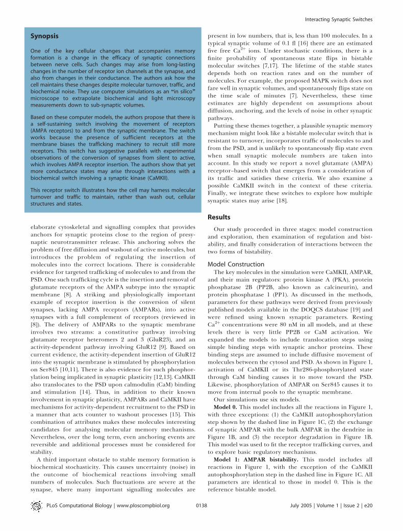
Model ConstructionThe key molecules in the simulation were CaMKII, AMPAR,
and their main regulators: protein kinase A (PKA), proteinphosphatase 2B (PP2B, also known as calcineurin), andprotein phosphatase 1 (PP1). As discussed in the methods,parameters for these pathways were derived from previouslypublished models available in the DOQCS database [19] andwere refined using known synaptic parameters. RestingCa2þ concentrations were 80 nM in all models, and at theselevels there is very little PP2B or CaM activation. Weexpanded the models to include translocation steps usingsimple binding steps with synaptic anchor proteins. Thesebinding steps are assumed to include diffusive movement ofmolecules between the cytosol and PSD. As shown in Figure 1,activation of CaMKII or its Thr286-phosphorylated statethrough CaM binding causes it to move toward the PSD.Likewise, phosphorylation of AMPAR on Ser845 causes it tomove from internal pools to the synaptic membrane.Our simulations use six models.Model 0. This model includes all the reactions in Figure 1,
with three exceptions: (1) the CaMKII autophosphorylationstep shown by the dashed line in Figure 1C, (2) the exchangeof synaptic AMPAR with the bulk AMPAR in the dendrite inFigure 1B, and (3) the receptor degradation in Figure 1B.This model was used to fit the receptor trafficking curves, andto explore basic regulatory mechanisms.Model 1: AMPAR bistability. This model includes all
reactions in Figure 1, with the exception of the CaMKIIautophosphorylation step in the dashed line in Figure 1C. Allparameters are identical to those in model 0. This is thereference bistable model.
PLoS Computational Biology | www.ploscompbiol.org July 2005 | Volume 1 | Issue 2 | e200138
Interacting Synaptic Switches
Synopsis
One of the key cellular changes that accompanies memoryformation is a change in the efficacy of synaptic connectionsbetween nerve cells. Such changes may arise from long-lastingchanges in the number of receptor ion channels at the synapse, andalso from changes in their conductance. The authors ask how thecell maintains these changes despite molecular turnover, traffic, andbiochemical noise. They use computer simulations as an ‘‘in silico’’microscope to extrapolate biochemical and light microscopymeasurements down to sub-synaptic volumes.
Based on these computer models, the authors propose that there isa self-sustaining switch involving the movement of receptors(AMPA receptors) to and from the synaptic membrane. The switchworks because the presence of sufficient receptors at themembrane biases the trafficking machinery to recruit still morereceptors. This switch has suggestive parallels with experimentalobservations of the conversion of synapses from silent to active,which involves AMPA receptor insertion. The authors show that yetmore conductance states may arise through interactions with abiochemical switch involving a synaptic kinase (CaMKII).
This receptor switch illustrates how the cell may harness molecularturnover and traffic to maintain, rather than wash out, cellularstructures and states.

Figure 1. Model Structure
(A) Overview of model, indicating key trafficking steps for AMPAR and CaMKII.(B–H) Chemical reaction schemes for pathways in model. Curved lines with arrows are enzymatic reactions catalyzed by molecules at the curves.Straight lines represent binding or unimolecular reactions.(B) Details of AMPAR model. The modelled AMPAR is a tetramer with two subunits each of GluR1 (circles) and GluR2 (triangles). There are 16phosphorylation states each in the cytosol and synaptic membrane. These are represented in expanded form in the lower portion of (B), which showsthe internalised pools of receptors. Black filling of the left half of the GluR1 circle indicates phosphorylation of Ser845, and of the right half indicatesphosphorylation of Ser831. Endocytosis occurs for the receptors with no GluR1-Ser845 phosphorylation, and exocytosis and degradation occur for thereceptors with both GluR1 subunits phosphorylated on the Ser845 site. Exchange of receptors with the bulk AMPAR pool occurs for theunphosphorylated state only, outlined in black.(C) CaMKII model. The dashed line for phosphorylation of CaMKII–PSD is applicable only for the bistable CaMKII models described in Figure 7.(D) CaM activation.(E) PP1 activation.(F) PP2B (calcineurin) activation.(G) cAMP formation. The unstimulated phosphodiesterase molecules (PDEs) also degrade cAMP, but at a lower rate than the activated forms illustrated.In the cAMP model we include diffusive exchange of cAMP with a dendritic compartment.(H) PKA activation.AMP, adenosine monophosphate; ATP, adenosine triphosphate; I1, inhibitor of PP1; Ng, neurogranin; PKA_inhib, inhibitor of PKA; PKC, protein kinase C;PP2A, protein phosphatase 2A.DOI: 10.1371/journal.pcbi.0010020.g001
PLoS Computational Biology | www.ploscompbiol.org July 2005 | Volume 1 | Issue 2 | e200139
Interacting Synaptic Switches
Model 2: Skeletal version of AMPAR bistability. This modelis used to understand bistability mechanisms.
Model 3: CaMKII bistability. This model includes reactionsfrom Figure 1C–1F. It explicitly includes the CaMKIIautophosphorylation step in Figure 1C. Rates for a few ofthe CaMKII reactions are somewhat modified compared tomodel 1.
Model 4: Combined AMPAR and CaMKII bistability. Thismodel combines models 1 and 3, using the model 3 param-eters where there are differences. The concentration of bulkAMPAR is reduced slightly. This version uses the same PP1enzyme for AMPAR and CaMKII in the PSD.
Model 5: Combined AMPAR and CaMKII bistability. Thismodel combines models 1 and 3, using model 3 parameterswhere there are differences. The concentration of bulkAMPAR is reduced slightly. This version uses distinct PP1enzymes for AMPAR and CaMKII in the PSD.
Following the initial model construction based on existingmodels, we wished to parameterize receptor trafficking rates.We represented movement of AMPAR to the PSD as a singlebinding reaction with an anchor protein. The reverse move-ment was modelled as a similar reaction releasing AMPARfrom the anchor. These simple reactions are approximationsto several cellular events, presumably including binding toanchor and diffusive or active movements of the receptor–anchor complex. As we did not have direct biochemical ratesfor these steps, we fit the reaction rates to published obser-vations of AMPAR movement from labelling and microscopystudies in live-cell preparations. We used model 0 for thesecalculations since we wished to model AMPAR movementonly between the internal and surface-membrane-anchoredstates. Model 0 does not consider exchange of AMPAR withthe bulk, or receptor degradation, and is therefore easier tomatch to the AMPAR recycling experiments.
To represent the experimental pulse-labelling of receptor,we needed to monitor the movement of a small amount ofreceptor (the pulse) without perturbing the activity levels ofthe regulatory molecules in the AMPAR trafficking cycle.Experimentally this is possible because the labelled receptorsseamlessly participate in the same reactions as the unlabelledones. In the simulations, however, a pulse of receptors wouldresult in a displacement from steady state. We thereforesimulated these experiments by first computing the steady-state levels of all molecules interacting with the receptor:CaMKII, PKA, PP1, and anchor proteins. We then numericallyfixed the levels of each of these interacting molecules to theirsteady-state levels. Finally, we introduced the test pulse ofreceptors into the PSD or internal compartment andsimulated its movement time course. Since the interactingmolecules were held fixed at their steady-state levels, thisprocedure had the same effect as a pulse of labelled receptorsthat did not perturb the steady state.
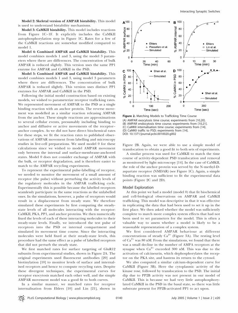
We first matched rates for surface targeting of GluR12subunits from experimental studies, shown in Figure 2A. Theoriginal experiments used fluorescent antibodies [20] andbiotinylation [10] to monitor levels of surface and internal-ised receptors and hence to compute recycling rates. Despitethese divergent techniques, the experimental curves forreceptor exocytosis matched each other well, and the simpleAMPAR movement model was a good fit to both curves.
In a similar manner, we matched rates for receptorinternalisation from Ehlers [10] and Lin [21], shown in
Figure 2B. Again, we were able to use a simple model oftranslocation to obtain a good fit to both sets of experiments.A similar process was used for CaMKII to match the time
course of activity-dependent PSD translocation and removalas monitored by light microscopy [14]. In the case of CaMKII,the role of the anchor protein was served by the N-methyl-D-aspartate receptor (NMDAR) (see Figure 1C). Again, a simplebinding reaction was sufficient to fit the experimental datapoints (Figure 2C and 2D).
Model ExplorationAt this point we had a model (model 0) that fit biochemical
and cell-biological observations on AMPAR and CaMKIItrafficking. This model was descriptive in that it was effectivein replicating the data that had been used to set it up in thefirst place. We then asked whether the model was sufficientlycomplete to match more complex system effects that had notbeen used to set parameters for the model. This is often avaluable way to assess whether a model is likely to be areasonable representation of a complex system.We first considered AMPAR behaviour at different
concentrations of steady Ca2þ (Figure 3A). The resting levelof Ca2þwas 80 nM. From the simulations, we found that therewas a small decline in the number of AMPA receptors at thesynapse when Ca2þ exceeded 300 nM. This was due to theactivation of calcineurin, which dephosphorylates the recep-tor on the PKA site, and hastens its return to the cytosol.We also computed a similar calcium-dependent curve for
CaMKII (Figure 3B). Here the cytoplasmic activity of thekinase rose, followed by translocation to the PSD. The initialdip due to PP2B activity was not present in our model ofCaMKII. This is because we had very little autophosphory-lated CaMKII in the PSD in the basal state, so there was littlesubstrate present for PP2B-activated PP1 to act upon.
Figure 2. Matching Models to Trafficking Time Course
(A) AMPAR exocytosis time course; experiments from [10,20].(B) AMPAR endocytosis time course; experiments from [10,21].(C) CaMKII internalisation time course; experiments from [14].(D) CaMKII traffic to PSD; experiments from [14].DOI: 10.1371/journal.pcbi.0010020.g002
PLoS Computational Biology | www.ploscompbiol.org July 2005 | Volume 1 | Issue 2 | e200140
Interacting Synaptic Switches

AMPAR conductance is a function both of the number ofreceptors at the surface membrane, and of their phosphor-ylation state (see Materials and Methods). In Figure 3C wecomputed conductance. At low Ca2þ, it closely tracked thenumber of membrane-inserted AMPARs. At near 1 lM Ca2þ
the conductance of the receptor rose, because CaMKII wasactivated and phosphorylated the receptor on Ser831. The neteffect of these competing events was that AMPAR conduc-tance first declined below baseline, and then rose abovebaseline. This is consistent with the theoretical Bienenstock-Cooper-Munro (BCM) curve [22], experiments using electricalstimuli [23], and Ca2þ-induced plasticity [24]. Thus, model 0was consistent with a number of experimental observationsfor which it had not been tuned. However, model 0 did nothave the capacity to retain these output changes when theinputs returned to resting levels.
We then used model 0 to analyse AMPAR responses tosustained changes in four parameters that might act as sites ofregulation of AMPAR trafficking. The parameters were the ac-tivities of CaMKII, PKA, and PP1, and recycling rates of recep-tor to and from surface membrane. Each of these parametersis implicated in synaptic change, and is a possible upstreamcontrol signal for AMPAR conductance. It is known that ap-propriate stimuli can lead to changes in synaptic conduc-tance over a range of approximately 50% to 200% of basalsynaptic transmission levels [25]. We asked which of theseparameters could control AMPAR conductance over thisrange.
In our simulated experiment, we scaled each of the fourparameters from 0.1 to ten times its basal value, one at a time.In the case of CaMKII, PKA, and PP1, this scaling was done bynumerically buffering the level of the active form of theenzyme to the desired value. In the case of the recycling rates,we scaled endocytosis and exocytosis rates as described below.In each case, the concentrations of the remaining molecularparameters (CaMKII, PKA, and PP1) were allowed to settle tonew steady-state concentrations. In biological terms, thiswould correspond to applying inhibitors or activators of theselected parameter. We recorded the resulting steady-statenumber of synaptic AMPARs (Figure 4). As in Figure 3, wealso computed the AMPAR conductance as a percent of themaximal conductance. The maximal conductance is theconductance if all the receptors were in the membrane inthe doubly Ser831-phosphorylated state. In each of thesecalculations we maintained the total number of internal plussynaptic membrane receptors at 80 molecules, and Ca2þ
concentration was at its resting level of 80 nM.The plots in Figure 4 show the results over the entire range
of scaled active inputs, from a ratio of 0.1 to ten times thebasal concentration of the input. CaMKII and PP1 (Figure 4A
Figure 3. AMPAR and CaMKII Trafficking and Dependence on Steady
Ca2þ Concentrations
(A) Number of AMPARs in internal and synaptic membrane pools;AMPARs complexed to enzymes are not counted.(B) Number of CaMKII molecules in the cytosol and PSD. The activity inthe cytosol and PSD starts to rise at about 0.5 lM Ca2þ, but translocationoccurs around 1 lM.(C) Conductance ofmembrane-insertedAMPARs. Receptor conductance iscalculated by assuming that CaMKII phosphorylation of a single GluR1-Ser831 of the tetramer gives 1.5-fold basal conductance, and of two Ser831gives 2-fold basal conductance. The conductance dips at around 300 nMCa2þ, when PP2B is active but CaMKII has yet to become fully active.DOI: 10.1371/journal.pcbi.0010020.g003
Figure 4. AMPAR Synaptic Membrane Localisation and Conductance in
Response to Sustained Inputs
Each panel is computed from a series of steady-state calculations wherethe activity of the selected input pathway was scaled with respect to itsbasal activity. The x-axis is this scaling ratio. The conductance iscalculated as in Figure 3 and is expressed as the secondary y-axis, as apercentage of maximal conductance (see Materials and Methods).(A) Changing activity of CaMKII leads to small changes in synapticmembrane localisation of AMPARs, but phosphorylation of GluR1 onSer831 gives a doubling of synaptic conductance when CaMKII activity isscaled above basal levels.(B) Low concentrations of PKA result in reduced exocytosis of AMPAR.Basal concentrations of PKA (ratio ; 1) are required to localise AMPAR tothe synaptic membrane, and higher concentrations cause a conductanceincrease. This occurs because of phosphatase saturation leadingindirectly to a rise in Ser831 phosphorylation due to CaMKII. The neteffect is that changes in PKA activity can lead to a large change inAMPAR conductance in either direction.(C) Changes in PP1 concentrations have little effect on AMPARlocalisation. However, low PP1 leads to high phosphorylation of GluR1-Ser831 by CaMKII, and hence high conductance.(D) Lower rates of receptor recycling to the internal pool lead to a smallincrease in synaptic membrane localisation. High rates bring most of thereceptor to the internal pool.DOI: 10.1371/journal.pcbi.0010020.g004
PLoS Computational Biology | www.ploscompbiol.org July 2005 | Volume 1 | Issue 2 | e200141
Interacting Synaptic Switches

and 4C) had rather little effect on synaptic membranelocalisation of receptor. Instead they acted in a complemen-tary manner in changing synaptic conductance throughphosphorylation and dephosphorylation of GluR1 on Ser831.
PKA (Figure 4B) had the largest total effect on synapticconductance, spanning a range from nearly zero to aconductance of nearly 70% of maximal. At low PKA activitythere was little insertion of AMPARs into the synapticmembrane, so the conductance was small. At high PKAactivity, most of the receptors were inserted. Additionally, thePKA active input indirectly activated CaMKII, leading toreceptor phosphorylation on Ser831, resulting in a furtherincrease in conductance. This indirect activation occursthrough two successive inhibitory steps. First PKA inhibitsPP1 because phosphorylated inhibitor 1 of PP1 binds to andblocks PP1. Second, PP1 itself inhibits CaMKII by dephos-phorylation of the kinase (see Figure 1E).
Receptor recycling has been suggested as a mechanism foraltering synaptic conductance [9,10]. In the simulations wescaled the AMPAR endocytosis rate by the specified recyclingratio, and simultaneously the exocytosis rate by its inverse.Thus, a ratio of 0.1 would have an endocytosis rate of0.1 times basal, and an exocytosis rate of ten times basal.Interestingly, it was not easy to drive more receptors into thesynaptic membrane (Figure 4D). This was partly because themodel already had most of its receptors in the membrane. Athigh values of receptor recycling rate, the endocytosis ratewas greater than the exocytosis rate, so the amount ofsynaptic-membrane-bound receptor was strongly depleted.
Thus, as an initial prediction, our simulations pointed toeither PKA or some combination of CaMKII, PP1, andrecycling as being a sufficient long-term control signal toaccount for bidirectional AMPAR changes, even in a regimewhere receptor counts did not change. These control effectswere not surprising, as these interactions with AMPARrecycling were specifically included into our model. Never-theless, the model did illustrate the amount of AMPARinsertion or removal to be expected from different manip-ulations. A similar combination of regulatory inputs has beenimplicated in learning (e.g., [26]). However, the simulations atthis stage did not address the question of how such long-termcontrol signals might be maintained.
AMPAR BistabilityThe above explorations of model responses had suggested
that the model was reasonably consistent with a range ofexperimental findings, including several that it had not beentuned for. However, these initial tests used model 0, which didnot consider molecular turnover. How might the inclusion ofreceptor synthesis and degradation alter AMPAR trafficking?
In preliminary simulations (not shown) we added orremoved AMPAR molecules from model 0 at the time ofstarting the model, and asked how the receptors redistributedwhen the model was run out to steady state. The AMPARmolecules were added to the doubly Ser845-phosphorylatedinternal pool of receptors, but the site of addition did notaffect the final steady-state distribution. Unexpectedly, wefound that the addition of receptors to model 0 actuallydecreased the number of native receptors. Native receptorsare defined as unphosphorylated receptors in the endocy-tosed pool. This suggested that receptors were beingredistributed in the model synapse in a manner that might
lead to two stable states. We proceeded to test this using aseries of simulations on the complete model includingAMPAR synthesis and degradation, that is, model 1. Detailsof model 1 parameters are in Protocol S1.In model 1 we assumed that newly synthesised AMPARs are
present in the dendrite (referred to as bulk AMPAR), and thatthey exchange with the native receptors in the spine (Figures1A, 1B, and 5A). We also assumed that there is a slow degra-dation of the doubly Ser845-phosphorylated receptor pool.Model 1 has 164 anchor proteins located in the PSD. Weperformed several tests on model 1 to examine whether itindeed exhibited two stable states with different numbers ofreceptors inserted into the synaptic membrane. We examinedinflux of receptors into the spine under different conditions.We then performed steady-state analyses, including a param-eter sensitivity analysis, to show bistability. We simulated statechanges of the model in response to stimuli and stochasticity.These results are described below and in Figure 5, and cumu-latively characterise the bistable properties of the model.We first asked whether there were two states in which there
was an influx of receptors into the spine. This influx would be anecessary condition to offset degradation, and the presence oftwo such states would be an indication of bistability. Wecomputed receptor flux between the dendrite and the nativereceptors in the spine as a function of the total number ofAMPARs in the synapse (Figure 5B). We defined the total num-ber of synaptic AMPARs as the sum of AMPARs in the internalsynaptic pool and in the synaptic membrane (Figure 5A). Inorder to compute the flux we performed the followingmanipulation: AMPAR molecules were added to the doublySer845-phosphorylated internal pool of receptors. Receptorexchange with bulk AMPAR was disabled to allow the systemto settle to steady state for 5,000 s without loss of receptors.Then receptor exchange was re-enabled and the simulationwas run for a further 1,000 s to settle. The flux of receptorsbetween bulk AMPAR and the native receptors was calculatedat this time point to obtain Figure 5B. This calculationconfirmed our preliminary observations. We found that therewere two distinct and widely separated regions of receptorinflux, one where there were fewer than 20 total synapticAMPARs, and one where there were 180 or more.This suggested that there were indeed two stable regimes
where receptor influx from the dendrite might balance outreceptor degradation. In the regime of low numbers of sy-naptic AMPARs, there was a simple equilibration between thebulk AMPA receptor pool and the native receptor pool. In thehigh-number regime, the receptors in the spine were redis-tributed to the membrane so that the native receptor pool wasdepleted, again leading to receptor influx. The presence of tworegimes of receptor influx, depending on the number of sy-naptic AMPARs,may be an experimentally testable prediction.To confirm that this formed a bistable system, we
computed stable states of the system under differentregulatory conditions (Figure 5C and 5D). We used Ca2þ
and cyclic adenosine monophosphate (cAMP) as regulatoryinputs. We obtained the stable states by running model 1 tosteady state (120,000 s), starting from either a low state (lownumbers of synaptic AMPARs) or a high state (high numbersof synaptic AMPARs). In cases where there was only onestable state, the two runs converged to the same steady value.In cases where there were distinct stable states, the two runssettled to their respective high or low stable states. We also
PLoS Computational Biology | www.ploscompbiol.org July 2005 | Volume 1 | Issue 2 | e200142
Interacting Synaptic Switches

found the unstable fixed point (the threshold for stateswitching) by using an iterative bisection method describedin Materials and Methods.These simulations give us dose-dependence curves that
illustrate the bistable nature of the system. The Ca2þ dosedependence of the switch is interesting and unusual (Figure5C). In the low Ca2þ regime, the system is bistable. The systemsettles into either a high or low state depending on initialconditions. In the 0.12 to 0.6 lM range, the system goes into asingle low state of activity because of the action of calcineurin(PP2B). Calcineurin is activated at these concentrations ofCa2þ, and is able to rapidly dephosphorylate AMPAR. Theunphosphorylated receptor moves into the native receptorpool, and then out to the dendrite. At Ca2þ concentrationsover 0.8 lM, the system goes into a single state of high activitybecause of CaM activation. The activity of CaM leads toincreased AMPAR phosphorylation both through PKA andCaMKII. CaM-activated adenylyl cyclase (see Figure 1)produces cAMP, increasing PKA activity. CaM also directlyactivates CaMKII. These events are similar to those describedfor Figure 3.In this manner, applied Ca2þ can flip the state of the switch
in either direction, depending on Ca2þ amplitudes. Theswitch is unusual because both states are reached by anincrease in the regulatory concentrations of Ca2þ. Asconsidered below for cAMP, it is much more common forone state to be triggered by low regulator concentrations, andthe other state by high regulator concentrations. Thisbidirectional regulation by increases in Ca2þ is also observedin the findings shown in Figure 3A, but is not as striking. It ispossible that there may be a very narrow bistable region at
Figure 5. AMPAR Translocation and Bistability for Model 1
(A) Simplified schematic of receptor recycling.(B) Bistability analysis. The flux of AMPARs from the bulk AMPAR pool tothe native AMPAR pool is plotted against the total number of synapticreceptors. Receptor influx into the spine occurs both at very low and athigh numbers of synaptic AMPARs.(C) States of the system as a function of Ca2þ concentration. Upper curveis obtained by starting system in state with high numbers of AMPARs in
the synapse; lower curve with low numbers of AMPARs. The intermediatethreshold curve is calculated using successive bisection as described inthe Materials and Methods. Bistability is present when Ca2þ concen-tration is less than 0.12 lM. Between 0.12 and 0.6 lM the system settlesto a state of low AMPAR numbers. Above 0.8 lM the system is in thehigh state.(D) States of the system as a function of cAMP concentration. Steady-state number of AMPARs is calculated as in (C). There is hysteresis as thehigh and low states coexist for the bistable region of the curve.Threshold points (open circles) complete the characteristic S-shapedcurve for a bistable system.(E) Parameter sensitivity analysis. The bars represent the range ofparameter values over which the system remains bistable. Other thankey regulators, the system tolerates a 2-fold or greater range of mostparameters without losing bistability.(F) Time course of AMPAR showing two stable states. A pulse of 0.2 lMcAMP is applied for 1,000 s to trigger translocation of AMPARs to thesynaptic membrane. Following this, cAMP is restored to resting levels,and the system settles to the state of high membrane AMPAR. The ‘‘off’’stimulus is provided by reducing cAMP to 0 lM for 6,000 s. Followingthis, the system settles back to the basal state of AMPAR.(G) Stochastic runs in low and high states. The high state is triggered byan initial cAMP pulse from t¼ 0 to 4,000 s. The state spontaneously turnsoff at around 20 h in this run, but the low state does not flip.(H) Average stability time of low and high states for different numbers ofbulk receptors (mean 6 standard error of the mean). Twenty-foursimulations for each state were run, as in (G). Stability time is calculatedas total simulation time in selected state, divided by number oftransitions out of that state. Large symbols represent cases where notransitions occurred over the entire set of simulations. As expected, ahigher level of bulk receptor increases the likelihood that the spine willspontaneously turn ‘‘on’’, and vice versa.(I) Stability time in low and high states for different numbers of anchorproteins. As the number of anchor proteins increases the stability timefor both states also rises. At very large numbers of anchor proteins thesynapse occasionally turns ‘‘on’’ spontaneously. Symbols and calcu-lations as in (H).DOI: 10.1371/journal.pcbi.0010020.g005
PLoS Computational Biology | www.ploscompbiol.org July 2005 | Volume 1 | Issue 2 | e200143
Interacting Synaptic Switches

around 0.7 lM Ca2þ in the model, as suggested by the smallseparation between the low and high curves, but this was notwithin the numerical resolution of our calculations. We donot expect that such a fine separation would be biologicallyrelevant. We also estimated the thresholds for the bistableswitch (open circles in Figure 5C). These were not verydependent on Ca2þ concentrations. In biological terms, thesynapse could be switched to the low state by raising Ca2þ tothe low regime (0.12 to 0.6 lM), allowing the flux of receptorsto be initiated, and rapidly lowering Ca2þ back to the bistableregime. To attain the high state, the Ca2þ input should beover 0.8 lM for long enough for the switch to settle, and thenCa2þ should rapidly fall below 0.1 lM into the bistable region.As we discuss later, the details of biological Ca2þ dynamics arebeyond the scope of our current steady-state models.
The cAMP dose dependence of the switch was moreconventional. It took the shape of a simple hysteresis curve,where the low state resulted from a decrease in cAMP and thehigh state from an increase (Figure 5D). The bistable regionof the switch is in an intermediate range of cAMP. In this casethe synapse switches to the low state if cAMP is reduced below20 nM, and to the high state if cAMP is raised above 35 nM.To complete the analysis, we found the unstable fixed pointsof the bistable switch (open circles in Figure 5D). Thesepoints can be interpreted as thresholds for switching fromone state to the other. As expected for a bistable system, theseunstable fixed points curve back toward the limits of thehysteresis cycle in an S-shaped curve (Figure 5D).
How ‘‘robust’’ is the bistability of model 1? One measure ofthis is to ask whether the bistable effects persist whenimportant model parameters are varied. We systematicallyvaried important model parameters and looked for bist-ability. To test for bistability, we started the model off ineither the upper or lower state, then ran it out to steady statewith the altered parameters. If the model switched state it wasno longer bistable. We found that model 1 retained itsbistable behaviour over a wide range of most parameters,illustrated in Figure 5E. The key ‘‘sensitive’’ parameters wereexactly those identified in Figure 4 as key regulators ofsteady-state synaptic conductance: CaMKII, PKA, recycling,and Ca2þ. Most parameters were able to scale a factor of atleast two up or down without losing bistability.
As a simpler signature of bistability we ran a time-coursesimulation in which the model explicitly switched betweentwo steady states (Figure 5F). Here the model first settled tothe low state where there were few AMPARs in the synapticmembrane. Following a cAMP pulse (0.2 lM, 1,000 s), themodel switched to the high state, with many AMPARs insertedinto the synaptic membrane. The system was switched back tothe low state by numerically reducing cAMP to zero (6,000 s).While this switching of the number of membrane-insertedAMPARs is possibly a testable prediction, it only indicates thepresence of bistability and does not shed much light on themechanism, which is analysed below.
The time course of switching was slow, of the order of anhour. This was consistent with the receptor trafficking ratesin the model, which were derived from steady-state measure-ments. More rapid transient rates may be applicable duringsynaptic stimulation, as we consider in the Discussion.
Another manifestation of robustness is the ability of themodel to retain state information despite stochasticity.Stochasticity in the synapse originates from the probabilistic
occurrence of reactions among small numbers of moleculesand gives rise to apparent biochemical noise. This noisinessimposes severe constraints on the reliability of any proposedsynaptic signalling mechanism [7]. In particular, bistablebiochemical systems are subject to spontaneous state flipsbecause of biochemical noise [7,17]. We tested stochasticresponses by simulating model 1 using the Gillespie exactstochastic method [27]. The entire model was simulatedstochastically, including all molecules in the dendrite, spinehead, and PSD (details in Materials and methods). We startedthe model in the low state, where few AMPARs were insertedinto the synaptic membrane (Figure 5G). In half the runs weapplied a cAMP stimulus to switch the model to the high stateat around 1 h (black line in Figure 5G). We then simulatedthe model for a period of 120,000 s (.33 h) to test itsresistance to spontaneous switching from either state. In theexample in Figure 5G, the low state (gray line) did notchange, but the high state (black line) spontaneously turnedoff at around 16 h.Based on our analysis shown in Figure 5B, the stability of
each state should be a function of the concentration of bulkAMPAR. If the concentration of bulk AMPAR is high, thenreceptor influx increases. Under these conditions, a relativelysmall fluctuation should push the system past the effluxregime into the upper influx regime (Figure 5B). Conversely,at low bulk AMPAR it should be easy to flip from the high tothe low state. We repeated our stochastic simulations for arange of bulk AMPAR concentrations while otherwiseretaining the parameters of model 1. At each concentrationof bulk AMPAR we repeated the simulations at least 24 timesto build up a profile of the switching times (Figure 5H). Atour reference range of 11.11 nM of bulk AMPAR, the off statewas stable for more than 360 h on average, and the on statewas stable for about 42 h. As we discuss below, other synapticprocesses may take over the job of maintaining stateinformation, within this time frame.At low bulk AMPAR the high state was very short-lived, but
the low state did not flip at all during the entire duration ofour simulations (indicated by the large symbols in Figure 5H).Conversely, at high bulk AMPAR, the low state was unstablebut the high state lasted for very long times.We then considered how the lifetime of model states might
scale with the number of anchor proteins in the PSD. As thenumber of anchor proteins sets the maximum number ofreceptors that can be inserted into the membrane, thisparameter is important for the robustness and stability of themodel. We simulated the stability time of the switch for arange of anchor protein numbers (Figure 5I). The defaultnumber of anchor proteins in the model was 164. At loweranchor protein numbers the switch lifetime was rather short,of the order of a few hours. At higher anchor numbers theswitch lifetime increased rapidly for both states. The lifetimeof the high state continued to rise and exceeded 2 mo whenthere were more than 320 anchor proteins (large symbols inFigure 5I indicate that the model did not change state duringthe entire duration of our simulations). The lifetime of thelow state was greatest (approximately 2 mo) at 240 anchorproteins, and then declined to around 200 h when moreanchor proteins were present. This may have occurredbecause the presence of additional anchor proteins biasedthe movement of the receptor toward the spine. Biologically,an increase in anchor protein number may correlate with the
PLoS Computational Biology | www.ploscompbiol.org July 2005 | Volume 1 | Issue 2 | e200144
Interacting Synaptic Switches
size of the spine head. Thus, our model predicts that largerspines should be more stable, an observation that has someexperimental support [28,29].
At this stage of the study we had extensively analysed theproperties of the bistability of model 1 with respect to thenumber of AMPARs at the synapse. We had considered statedependence on flux and regulators. We had shown that thebistability was robust, and in particular had shown how longthe model could retain state information when stochasticitywas taken into account. Based on these calculations, wesuggest that the model might be a candidate for retainingsynaptic state information for hours to months.
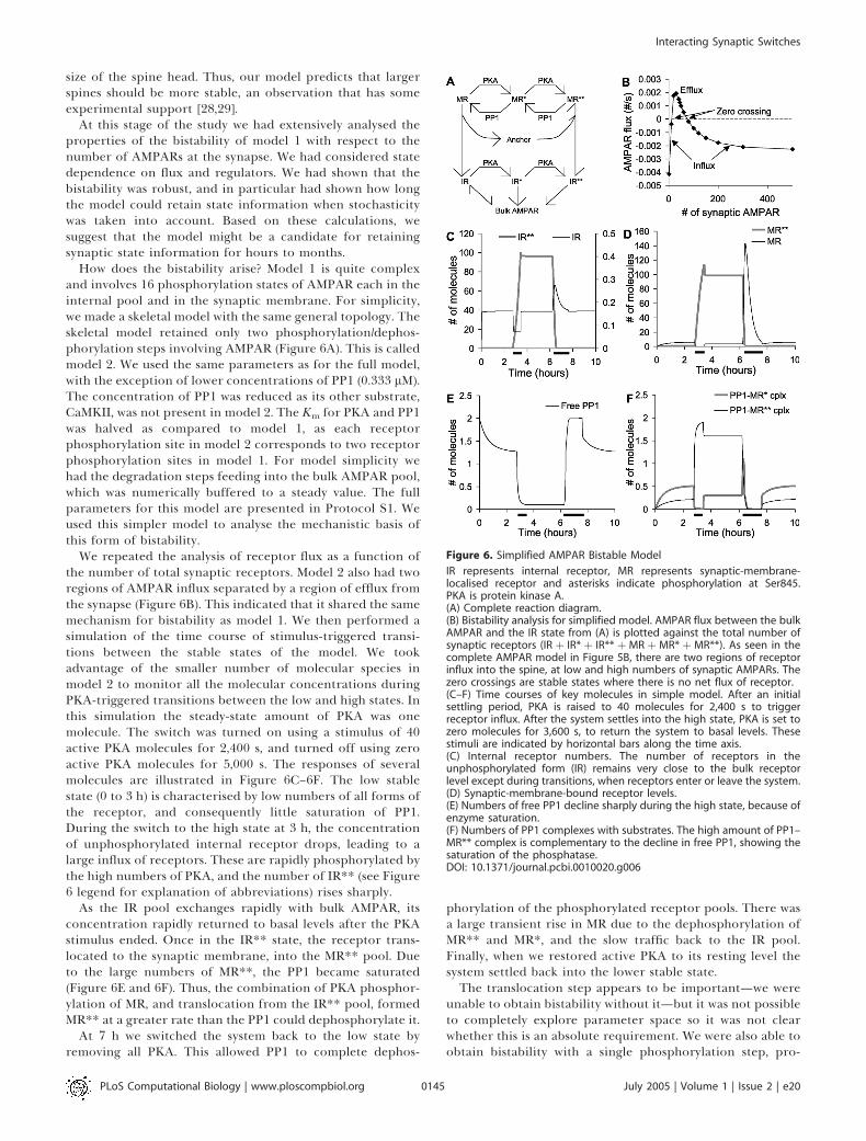
How does the bistability arise? Model 1 is quite complexand involves 16 phosphorylation states of AMPAR each in theinternal pool and in the synaptic membrane. For simplicity,we made a skeletal model with the same general topology. Theskeletal model retained only two phosphorylation/dephos-phorylation steps involving AMPAR (Figure 6A). This is calledmodel 2. We used the same parameters as for the full model,with the exception of lower concentrations of PP1 (0.333 lM).The concentration of PP1 was reduced as its other substrate,CaMKII, was not present in model 2. The Km for PKA and PP1was halved as compared to model 1, as each receptorphosphorylation site in model 2 corresponds to two receptorphosphorylation sites in model 1. For model simplicity wehad the degradation steps feeding into the bulk AMPAR pool,which was numerically buffered to a steady value. The fullparameters for this model are presented in Protocol S1. Weused this simpler model to analyse the mechanistic basis ofthis form of bistability.
We repeated the analysis of receptor flux as a function ofthe number of total synaptic receptors. Model 2 also had tworegions of AMPAR influx separated by a region of efflux fromthe synapse (Figure 6B). This indicated that it shared the samemechanism for bistability as model 1. We then performed asimulation of the time course of stimulus-triggered transi-tions between the stable states of the model. We tookadvantage of the smaller number of molecular species inmodel 2 to monitor all the molecular concentrations duringPKA-triggered transitions between the low and high states. Inthis simulation the steady-state amount of PKA was onemolecule. The switch was turned on using a stimulus of 40active PKA molecules for 2,400 s, and turned off using zeroactive PKA molecules for 5,000 s. The responses of severalmolecules are illustrated in Figure 6C–6F. The low stablestate (0 to 3 h) is characterised by low numbers of all forms ofthe receptor, and consequently little saturation of PP1.During the switch to the high state at 3 h, the concentrationof unphosphorylated internal receptor drops, leading to alarge influx of receptors. These are rapidly phosphorylated bythe high numbers of PKA, and the number of IR** (see Figure6 legend for explanation of abbreviations) rises sharply.
As the IR pool exchanges rapidly with bulk AMPAR, itsconcentration rapidly returned to basal levels after the PKAstimulus ended. Once in the IR** state, the receptor trans-located to the synaptic membrane, into the MR** pool. Dueto the large numbers of MR**, the PP1 became saturated(Figure 6E and 6F). Thus, the combination of PKA phosphor-ylation of MR, and translocation from the IR** pool, formedMR** at a greater rate than the PP1 could dephosphorylate it.
At 7 h we switched the system back to the low state byremoving all PKA. This allowed PP1 to complete dephos-
phorylation of the phosphorylated receptor pools. There wasa large transient rise in MR due to the dephosphorylation ofMR** and MR*, and the slow traffic back to the IR pool.Finally, when we restored active PKA to its resting level thesystem settled back into the lower stable state.The translocation step appears to be important—we were
unable to obtain bistability without it—but it was not possibleto completely explore parameter space so it was not clearwhether this is an absolute requirement. We were also able toobtain bistability with a single phosphorylation step, pro-
Figure 6. Simplified AMPAR Bistable Model
IR represents internal receptor, MR represents synaptic-membrane-localised receptor and asterisks indicate phosphorylation at Ser845.PKA is protein kinase A.(A) Complete reaction diagram.(B) Bistability analysis for simplified model. AMPAR flux between the bulkAMPAR and the IR state from (A) is plotted against the total number ofsynaptic receptors (IR þ IR* þ IR** þMR þMR* þ MR**). As seen in thecomplete AMPAR model in Figure 5B, there are two regions of receptorinflux into the spine, at low and high numbers of synaptic AMPARs. Thezero crossings are stable states where there is no net flux of receptor.(C–F) Time courses of key molecules in simple model. After an initialsettling period, PKA is raised to 40 molecules for 2,400 s to triggerreceptor influx. After the system settles into the high state, PKA is set tozero molecules for 3,600 s, to return the system to basal levels. Thesestimuli are indicated by horizontal bars along the time axis.(C) Internal receptor numbers. The number of receptors in theunphosphorylated form (IR) remains very close to the bulk receptorlevel except during transitions, when receptors enter or leave the system.(D) Synaptic-membrane-bound receptor levels.(E) Numbers of free PP1 decline sharply during the high state, because ofenzyme saturation.(F) Numbers of PP1 complexes with substrates. The high amount of PP1–MR** complex is complementary to the decline in free PP1, showing thesaturation of the phosphatase.DOI: 10.1371/journal.pcbi.0010020.g006
PLoS Computational Biology | www.ploscompbiol.org July 2005 | Volume 1 | Issue 2 | e200145
Interacting Synaptic Switches
vided that the translocation step was second order in thereceptor (data not shown). In all these processes, it wasassumed that the bulk AMPAR was constant, that is, that thebalance of synthesis and degradation was sufficient to rapidlyadd and remove receptors from the spine. In the biologicalsystem the situation is more complex and synthesis itself maybe activity dependent [30–32].In summary, the simple model retains the fundamental
features of the AMPAR translocation-based bistability andfacilitates an analysis of its mechanism. The low state of thisform of bistability occurs when few synaptic AMPARmolecules are present, so that PKA can act on only a fewsubstrate molecules, and PP1 is not saturated. Therefore, fewinternal AMPARs are in the phosphorylated state and only afew AMPARs are translocated to the surface. The upper stateof activity is characterised by high numbers of AMPARs in thephosphorylated states both internally and in the synapticmembrane. This state persists because of the higher basalactivity of PKA as compared to PP1. This leads to PP1saturation. The final step in maintaining the upper state is thetranslocation of phosphorylated receptor to the membrane.This removes receptors from the internal pools and keeps thenative receptor (internal receptor) at sufficiently low levelsthat receptor influx is favoured. A similar PP1 saturationeffect is seen in the complete model (model 1) when it is inthe high state (data not shown). This is a possible testableprediction of the model.
CaMKII BistabilityHaving characterised AMPAR translocation bistability, we
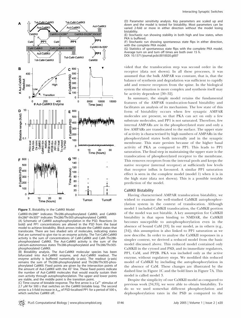
wished to examine the well-studied CaMKII autophosphor-ylation system in the context of translocation. Althoughmodel 1 included CaMKII translocation, the CaMKII portionof the model was not bistable. A key assumption for CaMKIIbistability is that upon binding to NMDAR, the CaMKIIbecomes susceptible to autophosphorylation even in theabsence of bound CaM [33]. In our model, as in others (e.g.,[34]), this assumption is also linked to PP1 saturation as wenow describe. In order to analyse the CaMKII responses in asimpler context, we derived a reduced model from the basicmodel discussed above. This reduced model contained onlyCaMKII in the cytosol and PSD, and its immediate regulators,PP1, CaM, and PP2B. PKA was included only as the activeenzyme, without regulatory steps. We modified this reducedmodel of CaMKII by including the autophosphorylation inthe absence of CaM. These changes are illustrated by thedashed line in Figure 1C and the bold lines in Figure 7A. Thismodel is called model 3.Despite the simplicity of our CaMKII model as compared to
previous work [34,35], we were able to obtain bistability. Todo so we used somewhat different phosphorylation anddephosphorylation rates in the PSD as compared to the
Figure 7. Bistability in the CaMKII Model
CaMKII-thr286* indicates Thr286-phosphorylated CaMKII, and CaMKII-thr286*-thr305* indicates Thr286/Thr305-phosphorylated CaMKII.(A) Schematic of CaMKII autophosphorylation in the PSD. Reactions (inbold) and PP1 concentrations are altered in the PSD from the basalmodel to achieve bistability. Block arrows indicate the CaMKII states thattranslocate. There are two shaded sets of molecules, indicating statesthat are summed to give rise to an enzyme activity. The Tot-CaM-CaMKIIactivity is the sum of concentrations of CaM-CaMKII and CaM–Thr286-phosphorylated CaMKII. The Aut-CaMKII activity is the sum of thecalcium-autonomous states Thr286-phosphorylated and Thr286/Thr305-phosphorylated CaMKII.(B) Bistability analysis. The Aut-CaMKII molecular species has beenbifurcated into Aut-CaMKII enzyme, and Aut-CaMKII readout. Theenzyme activity is buffered numerically (x-axis). The readout (y-axis)remains the sum of Thr286-phosphorylated and Thr286/Thr305-phos-phorylated CaMKII. Fixed points are given by the intersection points ofthe amount of Aut-CaMKII with the 458 line. These fixed points indicatethe number of Aut-CaMKII molecules that would exactly sustain theirown activity through autophosphorylation. The upper and lower pointsare stable, and the middle point is the transition point.(C) Time course of bistable response. The first arrow is a Ca2þ stimulus of2.7 lM for 500 s that switches on the CaMKII bistable loop. The secondarrow is a 5-fold increase in kcat of PSD-localised PP1 for a period of 500 s,which switches CaMKII off.
(D) Parameter sensitivity analysis. Key parameters are scaled up anddown and the model is tested for bistability. Most parameters can bevaried 2-fold or more in either direction without the model losingbistability.(E) Stochastic run showing stability in both high and low states, whenPKA is buffered.(F) Stochastic run showing spontaneous state flips in either direction,with the complete PKA model.(G) Statistics of spontaneous state flips with the complete PKA model.Average turn on and turn off times are both over 15 h.DOI: 10.1371/journal.pcbi.0010020.g007
PLoS Computational Biology | www.ploscompbiol.org July 2005 | Volume 1 | Issue 2 | e200146
Interacting Synaptic Switches

model 1 (see Protocol S1). Since CaMKII phosphorylatesitself, we had to adapt our previous analysis, which relies onseparate input and output molecules [4]. We did this bynumerically bifurcating the autonomously active CaMKII–PSD (Aut-CaMKII; Figure 7A) into two simulated molecularpools: Aut-CaMKII enzyme and Aut-CaMKII readout. We setthe number of Aut-CaMKII enzyme pools to specified values,and monitored the number of molecules in the Aut-CaMKIIreadout pool (Figure 7B). This manipulation was facilitatedbecause the level of Aut-CaMKII is computed as the sum ofthe autonomously active states of Thr286-phosphorylatedand Thr286/Thr305-phosphorylated CaMKII, indicated ingray in Figure 7A. So our enzyme assignment bypassed thissummation, and directly set the number of Aut-CaMKIImolecules. Our readout number was the sum of Thr286-phosphorylated and Thr286/Thr305-phosphorylated CaMKII.
The results of these calculations for a range of Aut-CaMKIIenzyme values are shown in Figure 7B. The intersectionpoints of this curve with the 458 line are stable points of thesystem, because at these points the enzyme and readoutactivities of Aut-CaMKII are identical. In other words, atthese points the autonomous CaMKII would exactly sustainits own activity. The upper and lower intersection pointsdefine the stable numbers of Aut-CaMKII, and the inter-mediate point is a transition point. This behaviour can beseen by considering a small increase in the number of Aut-CaMKII enzyme above one of the stable points. The resultingAut-CaMKII readout (read off from the y-axis) would besmaller than the new input number. This would tend torestore the CaMKII activity toward the stable point. A similarargument applies to small negative deflections from thestable points. Around the transition point the situation isreversed: any small deflection will be amplified until thesystem converges to either the upper or lower stable point.
We confirmed the presence of bistability by simulating atime series in which the system was turned on with a calciumpulse of 2.7 lM for 500 s and later turned off by raising thekcat of the PSD-localised PP1 by 5-fold for 500 s (Figure 7C).Two distinct stable states were observed, which correspondedto the upper and lower intersection points in Figure 7B.There is a small offset between the two calculations becauseFigure 7C reports all forms of CaMKII in the PSD, whereasFigure 7B refers only to Aut-CaMKII.
We evaluated the robustness of the CaMKII model (model3) using the same approach as for the AMPAR model (model1). We found that the CaMKII bistability was highly robustwith respect to variation of parameters (Figure 7D). Manyparameters could be varied from 0.1 to ten times thereference value without losing bistability. Most of theremaining parameters could be varied from 0.5 to two timesthe reference value, and only PP1 and PKA were moresensitive. This sensitivity reflects the key role of PP1 indephosphorylating CaMKII, and the role of PKA in control-ling the activity of PP1.
We checked the robustness of model 3 in synapticvolumes by simulating it using stochastic numerical meth-ods. Model 3 was resistant to spontaneous switching and wedid not observe any switches to either state in over 300cumulative hours of simulation time. A 33-h sample of thehigh and low states is shown in Figure 7E. This state stabilityturned out to be an artefact of our reduced model forCaMKII, which only used the final active concentration of
PKA as one of the key regulatory inputs. The PKA pathwaymodel output was quite noisy in small volumes [36]. Whenwe incorporated the full PKA pathway into model 3, wefound it introduced a considerable amount of additionalstochasticity into the system and did lead to bidirectionalstate flips (Figure 7F). We repeated these simulations 50times and found that the off state endured for 17.3 6 2.5 h,and the on state for 37.2 6 6.9 h (Figure 7G). Thus, theremay be a marked reduction in bistable state lifetimes whennoisy inputs are taken into account. This issue is consideredin the Discussion.Overall, our CaMKII translocation model (model 3) also
exhibited bistability coupled with translocation, such that theactive state led to accumulation of CaMKII in the PSD. Understochastic conditions, the lifetime of stable states in themodel was sensitive to noise from the PKA input pathway.
Bistability Interactions: Tight CouplingAt this point we had a reasonably constrained model of
AMPAR and CaMKII trafficking, and had shown that undercertain conditions both molecules could be bistable. In thefinal part of the study we considered interactions betweenthese two forms of bistability. We first made model 4 bymerging model 1 and model 3, while sharing the same PP1molecule in the PSD (Figure 8A). This scenario assumes thatthe PP1 is free to move between its CaMKII and AMPARsubstrates while remaining in the PSD. Thus, there is a tightcoupling between the two forms of bistability, mediated bothby PP1 and by CaMKII phosphorylation of Ser831 of AMPAR,as in model 1. We asked how the system would respond tostimuli designed to activate the AMPAR and CaMKII switchesindependently.In our first test we stimulated the model 4 with Ca2þ (2.7 lM,
500 s) then allowed the system to settle for 3 h, then stimulatedit with cAMP (108 nM, 2,000 s) (Figure 8B). Following the Ca2þ
stimulus, the CaMKII switch turned on transiently, but soonreturned to baseline. The AMPAR switch did not turn on untilthe cAMP stimulus was applied, and at this time CaMKII alsoturned on. When the cAMP stimulus was applied first, itrapidly turned on both the AMPAR and CaMKII switches(Figure 8C). Together, these simulations show that in model 4the two forms of bistability function in lockstep. That is,sustained activation of CaMKII is contingent upon the activityof AMPAR. If AMPAR is activated, it saturates PP1, and thisleads to activation of the CaMKII bistable switch. As shown inFigure 8C, CaMKII is also activitated by the cAMP stimulusindependently of the PP1-mediated cross-activation, leadingto rapid turn on. This is because cAMP activates PKA, whichrelieves the PP1 inhibition of CaMKII. We ran separatesimulations (not shown) that showed that even in the absenceof this cAMP activation of CaMKII, the activation of AMPARcaused CaMKII to turn on as well.We ran model 4 using stochastic methods to test its
propensity to spontaneously change state. The off state wasvery stable though it did exhibit occasional transient spikes ofactivity (Figure 8D). The model spontaneously turned on onlyonce in a cumulative total of over 1,000 h of simulation time.As before, we tested high-state durations by applying aninitial cAMP stimulus at about 1 h to turn the system on, andthen ran the simulation for about 33 h. We repeated theseruns 24 times to obtain the distribution. The high state wasnot as stable, and was subject to occasional spontaneous flips
PLoS Computational Biology | www.ploscompbiol.org July 2005 | Volume 1 | Issue 2 | e200147
Interacting Synaptic Switches

to the off state with an average time of 101 6 79 h (mean 6
standard error of the mean). As expected from the lockstepmechanism, both CaMKII and AMPAR underwent a state flipat nearly the same time (Figure 8E).
Bistability Interactions: Weak CouplingIn our final model (model 5), we considered the situation
where CaMKII and AMPAR interacted only weakly. This is incontrast to model 4, where CaMKII and AMPAR were tightlycoupled through a shared pool of PP1. In model 5, wecombined the CaMKII and AMPAR bistable models (models 1and 3) while keeping an independent pool of PP1 for each(Figure 9A). Such a scenario might arise if PP1 were bound todistinct scaffold proteins for each of its targets, and wererestricted in its mobility across targets. The CaMKII-coupledpool of PP1 was treated as independent of PKA activity.There was one indirect form of coupling still present fromCaMKII to the AMPAR bistability, since CaMKII phosphor-ylates AMPAR on Ser831. While this does not alter trafficrates directly, the Ser831 is a substrate for the AMPAR-
associated pool of PP1 in model 5. Thus, CaMKII activity didcontribute to the saturation of the AMPAR-associated PP1.As before, we examined the interactions between the
CaMKII and AMPAR switches using stimuli designed to turneach switch on independently of the other. When we firstturned on the CaMKII switch alone using a Ca2þ stimulus, weobserved a slow activation of the AMPAR switch (Figure 9B).The Ca2þ stimulus indirectly increased AMPAR insertionthrough the following steps: Ca2þ ! CaM ! CaMKII !phosphorylation of Ser831 ! saturation of AMPAR-specificPP1 ! reduced endocytosis of AMPAR. We did not modelany changes in recycling or internalisation rates due toreceptor phosphorylation on Ser831.A particularly interesting effect was seen when the AMPAR
switch was activated first using cAMP (Figure 9C). Thisstimulus turned on the AMPAR switch without affecting thestate of CaMKII. At about 4 h we applied a Ca2þ stimulus thatturned on the CaMKII switch as well. Thus, in model 5, the
Figure 9. Nested Bistability for Weakly Coupled Switches
(A) Schematic of independent PSD-localised PP1 enzyme activities forCaMKII and AMPAR. The two PP1 activities are labelled PP1-PSD-CaMKIIand PP1-PSD-AMPAR, respectively. The asterisks represent phosphor-ylation.(B) Time course of system response to Ca2þ (2.7 lM, 500-s duration), thencAMP (0.108 lM, 2,000-s duration) stimulus. The initial activation ofCaMKII leads to a slow turn on of the AMPAR system.(C) Time course of system response to cAMP (0.108 lM, 2,000-s duration),then Ca2þ (2.7 lM, 500-s duration) stimulus. First the AMPAR systemturns on, then, following the Ca2þ stimulus, the CaMKII turns on. Theconductance of the synapse has different levels in each of these states.(D) Stochastic run for 60 h, showing resting, AMPAR only, and AMPARþCaMKII activity states.DOI: 10.1371/journal.pcbi.0010020.g009
Figure 8. Bistability for Tightly Coupled Switches
(A) Schematic of PSD-localised PP1 acting on both CaMKII and AMPARsubstrates in the PSD. The asterisks on CaMKII and AMPAR representphosphate groups.(B) Time course of response to Ca2þ (2.7 lM, 500-s duration), then cAMP(0.108 lM, 2,000-s duration) stimuli. The initial Ca2þ stimulus turns onCaMKII transiently, but it eventually returns to baseline. The subsequentcAMP stimulus turns on both switches.(C) Time course of response to cAMP (0.108 lM, 2,000-s duration), thenCa2þ (2.7 lM, 500-s duration) stimuli. The initial AMPAR stimulus (cAMPelevation) is sufficient to turn both the AMPAR and the CaMKII switches on.(D) Stochastic run in the low state. The figure illustrates a transient eventthat did not result in complete turn on.(E) Stochastic run in the high state. There is a spontaneous turn off, butthe average on time is over 100 h.DOI: 10.1371/journal.pcbi.0010020.g008
PLoS Computational Biology | www.ploscompbiol.org July 2005 | Volume 1 | Issue 2 | e200148
Interacting Synaptic Switches

two switches were able to coexist in three combinations ofstates: both off, only AMPAR on, or both on. The fourthpossible combination, of CaMKII on and AMPAR off, was notstable because of weak coupling between CaMKII andAMPAR, which slowly turned the latter on as shown inFigure 9B. The weak coupling is due to the phosphorylationof GluR1 on Ser831 by CaMKII. While this does not directlyaffect translocation, it does engage the AMPAR-specific poolof PP1, leading to eventual phosphatase saturation and turnon of the AMPAR switch.
Overall, this model exhibited nested bistability. Thefundamental switch took place when AMPAR turned on oroff. Nested within this was the capacity for CaMKII to turn onor off. A possible physiological outcome of the nesting ofCaMKII activation is that the phosphorylation state and hencethe conductance of the synapse can settle to three levels: (1) noAMPAR, (2) AMPAR with low levels of Ser831 phosphoryla-tion, and (3) AMPAR with high Ser831 phosphorylation andconsequently a higher conductance (Figure 9C). As shown inFigures 3 and 4, the equivalent conductance is expressed interms of the number of unphosphorylated AMPAR channelsthat would have the same conductance.
The three states of the model were quite stable understochastic conditions (Figure 9D). The only state that showedany transitions over the entire cumulative duration ofsimulations tested was that in which AMPAR was on andCaMKII was off (time to switch was 24.8 6 3.5 h). We wereparticularly interested in the long-term stability of the stateswith both switches off, and both switches on. To examinethese we performed several hundred independent stochasticsimulation runs on a cluster, each representing 120,000 s(33 h) of simulated time. No state transitions were observed ineither direction, over a cumulative duration of over a year ofsimulated time for each state.
Transient Responses of ModelsHow does the model respond to stimuli that induce
changes in synaptic efficacy? We tested two synaptic plasticityprotocols on each of the models 1, 3, 4, and 5 (Figures S1 andS2). These tests were only qualitative, as the models in thecurrent study were parameterized using steady-state ratherthan transient experiments. Nevertheless, they are useful inshowing model behaviour under transient conditions. Thefirst protocol had been used to elicit long-term potentiation(LTP) of synaptic efficacy and consisted of three bursts of 100impulses at 100 Hz, each separated by 600 s. The second
protocol was used to induce long-term depression (LTD) atthe synapse, and consisted of 900 impulses at 1 Hz. Werepresented each stimulus as a computed calcium transientwith an exponential build-up and decay of Ca2þ using theformulation of Zhabotinsky [34] (Figure S1). The LTPstimulus gave calcium peaks of 12 lM, and the LTD stimulushad peaks of 0.5 lM. We found that the LTP stimulus was ableto cause a switch to the on state only in the CaMKII model(model 3) that incorporated the PKA activation pathway(Figure S2). This is interesting, as it suggests that even in itscurrent form the CaMKII model is reasonably sensitive tophysiological stimuli that may play a role in synapticplasticity. The LTD stimulus did not turn off any of themodels, indicating that the current models are missing somekey interactions. These tests highlight some of the unknownsin our models, in particular, the specific transient interac-tions that are needed to trigger the steady-state effects wehave analysed. As discussed below, more experimental detailwill be needed to extend the models to include transientresponse characteristics.
Summary of Bistable BehaviourIn summary, we analysed four bistable synaptic trafficking
models (models 1, 3, 4, and 5). The remaining two models inthis study (models 0 and 2) were used to characterise model 1.Model 3 included CaMKII alone, but models 1, 4, and 5included both AMPAR and CaMKII. As described above, weperformed a number of steady-state and transient tests tocharacterise the stable states of each model. Properties ofthese models are summarised in Table 1.
Discussion
In this study we have developed a model of the movementof a glutamate receptor (AMPAR) and a calcium-activatedkinase (CaMKII) to and from the synaptic membrane, usingsteady-state trafficking rates as a major experimental con-straint. We find that the AMPAR trafficking cycle may lead toa form of switching or bistability where the presence ofsufficient receptors at the synapse leads to recruitment ofmore receptors. This process may be a candidate for thetransition from silent to active synapses, and early phases oftheir subsequent maintenance. When combined with apreviously proposed mechanism for a CaMKII molecularswitch, we observe interesting interactions between these twoforms of synaptic bistability. Depending on the degree of
Table 1. Summary of Model Properties
Model Number of
Stable States
Spontaneous Stochastic
Turn On Time
Spontaneous Stochastic
Turn Off Time
Turn On by LTP
Stimulus
Turn Off by LTD
Stimulus
Model 1—basic AMPAR 2 360 h 42 h No No
Model 3—CaMKII alone 2 .17 h .37 h Yes, if PKA is modelled
explicitly
No
Model 4—tight coupling 2 ;1,000 h .100 h No No
Model 5—weak coupling 3 .1 y .24 h for state with AMPAR on
and CaMKII off
.1 y for both on
No No
DOI: 10.1371/journal.pcbi.0010020.t001
PLoS Computational Biology | www.ploscompbiol.org July 2005 | Volume 1 | Issue 2 | e200149
Interacting Synaptic Switches

coupling between these switches, we predict that AMPARrecruitment and CaMKII activation may either occur in atightly coordinated manner, or nearly independent of eachother. The latter may give rise to multiple stable synapticstates. Stochastic calculations suggest that these stable statespersist for many hours, and in some cases over a year, despitebiochemical fluctuations due to small numbers of moleculesin the synapse.
Structural BistabilityLong-term storage of information at the synapse is
intimately connected to structural changes [8,15]. Suchchanges arise from the insertion, removal, and reorganisationof synaptic molecules. For example, many types of glutama-tergic synapses initially lack postsynaptic AMPARs and areunresponsive to moderate synaptic input. These ‘‘silentsynapses’’ become active when AMPARs are inserted. Thechange from silent to active synapses is a major mechanismfor increases in synaptic efficacy [8]. Similarly, formation ofdendritic spines is facilitated by the presence of the GluR2subunit of the AMPAR [29]. Several signalling events areknown that may affect these structural processes, but it is notalways clear how persistent changes may be maintained. Asingle brief pulse of insertion of molecules, or formation of asynaptic spine, will have a transient effect unless some self-sustaining mechanism is also available to keep the changes inplace. Formally, bistable systems are a possible mechanism forsuch structural memory, as such systems can withstandmolecular turnover [2]. The AMPAR model analysed in thecurrent paper shows that synaptic membrane insertion (aform of structural change) can be self-sustaining even whenmolecular turnover and noise are considered.
There are several proposed forms of synaptic bistability,and each has some structural correlates. The classical form ofsynaptic bistability is the CaMKII autophosphorylationsystem (reviewed in [3]). This is a biochemical bistability thatrelies on autophosphorylation leading to self-activation ofCaMKII. This self-activation leads to bistability if PP1 ispresent at sufficiently low levels that it can be saturated. Theinvolvement of PP1 saturation is also an important aspect ofour model for AMPAR bistability. CaMKII activation hasseveral structural correlates, including translocation to thePSD and formation of complexes with NMDA and other PSDproteins [33]. There is also recent evidence that CaMKIIactivation leads to an increase in AMPAR numbers [37].Another synaptic biochemical bistability involving a MAPKfeedback loop has been proposed [4] and tested in a fibroblastmodel system [38]. MAPK is known to be important insynaptic plasticity and may also play a role in structuralchanges at the synapse [39,40]. However, the MAPK bistablefeedback mechanism is vulnerable to biochemical noise andmay not be plausible in synaptic volumes [7]. A more recentproposal for a self-sustaining synaptic plasticity mechanisminvolves the mTOR system and local protein synthesis. mTORphosphorylation increases protein synthesis at the dendrites,and the synthesis machinery itself is one of the products.Several other proteins have also been identified that mayparticipate in such a feedback loop [6]. Such a protein-synthesis-dependent bistability would be very interesting forstructural change at the synapse, as it could account forincreased availability of many synaptic and PSD proteins.
In the current study we propose a novel form of synaptic
bistability involving self-recruitment of AMPARs to thesynapse. The mechanism is particularly interesting for thesynapse in three ways: (1) it has parallels with the conversionof silent to active synapses, (2) it works at basal levels ofactivity of synaptic enzymes, and (3) it intimately involves atranslocation and synaptic membrane insertion process. Asanalysed in Figures 5 and 6, this form of bistability is afunction of the number of molecules of receptor in thesynapse, rather than biochemical activation. The bistabilityinvolves phosphatase saturation of PP1 due to its action onAMPAR in the PSD. This is a specific prediction of this study.However, the prediction of AMPAR-specific PP1 activity is yetto be tested in detail. We discuss the issue of PP1 access toother substrates below.It should be stressed that this mechanism for synaptic state
maintenance is by no means exclusive. We discuss severalother possible mechanisms above. There are also importantdetails about AMPAR cycling that invite further analysis. Forexample, the AMPARs in Ser831/Ser845 double phosphomu-tant mice would not sustain this form of bistability. Never-theless, these animals form synapses and retain some degreeof synaptic plasticity [12]. Furthermore, our model considersactivity-dependent changes in GluR12, and does not accountfor a presumed hand-over of synaptic state to some long-termprocess involving GluR23 insertion. Mechanisms for main-tenance of GluR23 levels are still poorly understood, but wespeculate that this too involves a self-recruiting bistableprocess.Phosphatase saturation is a key part of our self-recruitment
model. This has parallels with a distinct form of bistabilityanalysed for the MAPK system by Markevich et al. [41]. In thisMAPK model, there are two stable states of MAPK activity.The high state is sustained in part because of the saturation ofphosphatases that reverse its activity. The distinction, again,is that the AMPAR bistability involves translocation withoutsustained biochemical activation whereas the Markevichmodel involves biochemical activation without intrinsicstructural effects.In a broader context, the AMPAR structural bistability
could be generalised as a state-dependent translocation ofmolecules coupled to a saturable interconversion betweenthese states. Many cellular trafficking events have a similargeneral form, including the Rab-mediated system of smallGTPases and nuclear transport control. By our analysis inFigures 5 and 6, translocation bistability should exhibit twoclearly separated regimes in which traffic occurs into one ofthe compartments, and a regime in which addition of thetranslocated molecule actually decreases its number in one ofthe cellular compartments. This might be an experimentallyaccessible signature for such behaviour in the cell.
Stochasticity and RobustnessThe typical synapse has a volume of 0.1 fl [16], and
contains rather small numbers of key signalling molecules.This introduces fluctuations in reactions taking place in thesynapse. We have previously analysed small-volume signal-ling for several pathways using simple assumptions aboutscaling and diffusion, and find that stochastic effects are sosevere that some conventional signalling mechanisms simplydo not work in these volumes [7,36]. Stable retention ofsynaptic state is a potential victim of stochasticity, asbiochemical noise can lead to spontaneous state transitions.
PLoS Computational Biology | www.ploscompbiol.org July 2005 | Volume 1 | Issue 2 | e200150
Interacting Synaptic Switches

Bialek [17] and Miller et al. [42] have previously analysedsynaptic bistability and suggest that in principle anautophosphorylation mechanism can give molecular stabilityof the order of hundreds of years even with a small numberof CaMKII holoenzymes. Our current study is both coarserand more detailed than these analyses. Our representationof CaMKII does not consider individual holoenzymes, but onthe other hand we explicitly represent translocation andseveral important signalling interactions at the synapse. Insome models, and for some states, we did not observe anytransitions over a year of simulation time. For other statesthe predicted lifetime is of the order of tens of hours (Table1). Thus, in terms of state stability, our models are quiterobust.
While it is heartening that our models are stable for manyhours, it is clear that this may change in either direction ifadditional interactions are considered. For example, weignore the diffusive exchange of most spine molecules withthe PSD and dendrite. Over the time scales of oursimulations, all the spine molecules would in fact exchangeor degrade. This introduces further challenges to synapticstability mechanisms that are beyond the scope of this study.A further illustration of the importance of context onstability comes from the CaMKII analysis in Figure 7. Herethe inclusion of a more detailed PKA signalling modelreduces the predicted lifetime of CaMKII states fromhundreds of hours to tens. Additional input pathways maycontribute to increased noise, but scaffold anchoring ofenzymes such as PKA may create local signalling environ-ments that could potentially reduce stochastic effects. Directexperimental measurements of synaptic stochasticity aredifficult to perform [43], but such measurements would beinvaluable for comparison with our simulated estimates ofstochasticity.
Another commonly used measure of robustness is to askwhether the model retains some critical attribute over a widerange of parameters. In our analysis, we use bistability as theattribute of interest. We perform parameter sensitivityanalyses by varying key reaction rates and enzyme parametersover a 100-fold range. Our parameter sensitivity analyses forthe AMPAR and CaMKII systems suggest that our models arefairly robust by this measure. Most parameters can be variedat least 0.5- to 2-fold their original values without the modellosing bistability (see Figures 5 and 7).
A particularly stringent measure of the robustness of amechanism is to see how well essential features are preservedas reaction details are changed. We have performed a rathersevere pruning of the AMPAR bistable model to a bareskeleton (see Figure 6) and with very little parameter tuningthe simplified model is also bistable. Thus, by severalmeasures, the bistable processes we have analysed areresistant to stochasticity, parameter uncertainty, and evenchanges in reaction mechanisms.
Coupled Bistable SwitchesIndividual synapses are likely to exist in many states [18].
Given the short life of synaptic molecules discussed above, itseems possible that one mechanism for stabilising such statesmight be to associate bistable switches with them. Multiplestates may be achieved if the individual switches are coupledloosely, so that combinations of states become possible. Herewe have shown (model 5; Figure 9) that distinct forms of
bistability may coexist to give rise to three possible synapticstates. The AMPAR switch is the major one, as it brings thereceptors to the synaptic membrane in the first place. TheCaMKII switch is nested within this as it can fine-tune theconductance of the receptors. This situation of nestedbistable states is possible if the mobility of PP1 at the synapseis limited so that each PP1 molecule has exclusive accesseither to CaMKII or to AMPAR. The alternate assumption(model 4) is that PP1 is mobile within the PSD, and can accessboth CaMKII and AMPAR. This assumption causes the twoforms of bistability to function in lockstep, where theactivation of the AMPAR switch causes the CaMKII switchto turn on. This occurs because the two switches share thesame pool of PP1 enzymes, and phosphatase saturationresulting from the AMPAR switch activates CaMKII. It islikely that the biological situation involves different degreesof PP1 mobility between multiple possible synaptic targets,and may even differ for the same synapse in differentcontexts. In our study, we obtain distinct outcomes for twocases of PP1 mobility and targeting. This sensitivity ofsynaptic state to PP1 mobility is a testable prediction andhighlights the possible importance of subtle details of PSDanchoring on synaptic function.An alternative proposal for multiple levels of synaptic
activation is that the CaMKII–NMDAR complex may act in ahighly modular manner (reviewed by Lisman and McIntyre[3]). In this scenario, each CaMKII–NMDAR complex canindependently persist in a high or low state of activity. Thissituation would make it possible for an individual synapse topresent graded levels of conductance depending on thenumber of active CaMKII complexes. Our model of CaMKIIis at the bulk rather than holoenzyme level, and is toocoarse-grained to address this possibility. The CaMKIIphosphorylation of AMPAR plays two roles in our study.First, it directly increases the conductance of individualreceptor tetramers. Second, it indirectly leads to an increasein the number of receptors at the synapse, by producingadditional phosphorylation states of AMPAR for PP1 to acton. In the model, this leads to further saturation of thephosphatase and ultimately to an increase in surfaceAMPAR. There is recent evidence that CaMKII activity mayalso affect AMPAR numbers [37]. This would provideanother mechanism for coupling between our proposedbistable mechanisms.In addition to forming multiple synaptic states, our
simulations show that coexisting bistable mechanisms mayfunction to ‘‘hand-over’’ information about synaptic statefrom one switch to another. For example, in model 5 (weaklycoupled synaptic switches), a rather brief Ca2þ input issufficient to activate CaMKII, which can then turn on theAMPAR switch over a timescale of hours (Figure 9B). This isloosely analogous to different forms of computer informationstorage, where information is initially stored in fast butvolatile form (e.g., RAM) and is later transferred to slow butstable forms of memory (e.g., hard disk).Our study illustrates how two mechanisms for synaptic
bistability may coexist to give rise to multiple possiblesynaptic states. We propose that the synapse may exhibit acombinatorial set of states through the interactions of severalmolecular switches. These may include local protein synthesisfeedback loops involving mTOR, self-assembly processes atthe synapse, or presynaptic switches. From the cell-biological
PLoS Computational Biology | www.ploscompbiol.org July 2005 | Volume 1 | Issue 2 | e200151
Interacting Synaptic Switches

perspective, we have considered synaptic recruitment mech-anisms for only two of the hundreds of postsynapticmolecules. All these molecules undergo turnover, and manyexperience regulated movement similar to that in the switcheswe have analysed. We suggest that there are many forms ofself-recruitment, coordinated self-assembly, and other poten-tially switch-like processes that contribute to the maintenanceof different constituents and states of the synapse.
Materials and Methods
Model development. Our model was developed to closely tie withexperimental observations and to build on existing, well-docu-mented, and experimentally constrained models. Two moleculartrafficking cycles form the core of the model: (1) the trafficking ofAMPARs, and in particular GluR12, between internal vesicular poolsand the synaptic membrane associated with the PSD and (2) themovement of CaMKII to and from the spine cytosol and the PSD (seeFigure 1). As elaborated below, we developed the models usingpublished experimental observations on these trafficking processes,and considerable specific data on the biochemistry of the phosphor-ylation of these molecules. A few regulatory pathways were alsomodelled to provide signalling input and context. Reactions in themodel take place in two compartments: the PSD and the bulkcytosolic volume of the spine. The receptors are membrane-associated, so the PSD-associated synaptic membrane is included inthe PSD compartment. Likewise, the internalised, vesicular pool ofreceptors is included in the cytosolic compartment. The PSD volumeis taken as 0.01 fl, and the spine head volume as 0.09 fl. There is athird, dendritic compartment of 5 fl that is occupied only bydiffusible cAMP and by a bulk AMPAR pool. The bulk AMPAR pool isassumed to be at a steady level and is meant to represent synthesisand degradation of the receptor. No reactions occur in the dendriticcompartment as it is meant only to couple diffusively with the spine.
AMPAR model. Due to a combinatorial proliferation of states, thereaction diagram of the AMPAR steps appears complex. As describedbelow, we modelled 16 phosphorylation states of the receptor each inthe internal and synaptic-membrane-associated pools. However, mostof the reactions involving these 16 states were symmetric as theyinvolved independent phosphorylation sites. This simplifies themodel definition. We assumed that symmetric reactions had thesame rates, so our model relies on only a few trafficking andphosphorylation parameters. AMPARs occur as tetrameric structures[44] with most AMPARs composed of two subunits each of GluR1 andGluR2 (GluR12) or GluR2 and GluR3 (GluR23). GluR23 receptorsshow constitutive trafficking and are responsible for basal synaptictransmission whereas GluR12 receptor insertion can be altered bystimuli [10,20]. We considered only GluR12 receptors in the model tofocus on activity-stimulated events. The dynamics of GluR12 AMPARtrafficking were determined by kinase/phosphatase activities at theSer845 sites of the two GluR1 subunits in the tetrameric GluR12complex. Dephosphorylation of these sites by phosphatases triggersendocytosis whereas phosphorylation by PKA is required for synapticmembrane targeting [10,11]. We modelled the phosphorylation/dephosphorylation as a two-step reaction, where phosphorylationor dephosphorylation of both GluR1 subunits is necessary forsynaptic membrane targeting or internalisation, respectively.Through simulations we found that basal activities of PP1 and PKAcan account for the constitutive cycling of receptors in our model,consistent with experimental studies [8,45]. We assumed that PKAand PP1 were the relevant enzymes, but the model does not excludethe possibility that the same cycling effects might be mediated byother phosphorylation enzymes.
There is evidence that the membrane-associated PP1 dephosphor-ylates AMPARs only in the PSD, as loading neurons with active PP1does not alter basal synaptic strength transmission [46]. Hence, weassumed that PP1 acts on GluR1 only in the PSD whereas PKAphosphorylates GluR1 in both compartments. In both compartments,Ser845 of GluR1 was also dephosphorylated by PP2B [10,47,48], whichitself is inactive at basal Ca2þ concentrations.
Phosphorylation of Ser831 of the GluR1 subunit by CaMKII alterschannel properties of the receptor in that the phosphorylationincreases channel conductance approximately 2-fold [49]. As for theSer845 sites, we modelled Ser831 phosphorylation of GluR1 so thatboth sites of a tetrameric complex could be phosphorylatedindividually by CaMKII. Dephosphorylation of Ser831 was modelled
to occur only in the PSD, as internalisation was reported not to alterthe phosphorylation state of AMPARs at Ser831 [10].
In the bistable models we explicitly modelled protein turnoverthrough activity-dependent degradation [1]. The activity dependencewas introduced by restricting the turnover to the doubly Ser845-phosphorylated states in the internal pool of AMPARs (see Figure5A). There are around 150 receptors in an active dendritic spine [50].We represented this constraint in the model as an anchor protein(possibly GRIP [8]) required for AMPAR insertion into the synapticmembrane.
The synaptic AMPAR conductance is a function both of thenumber of synaptic membrane receptors, and of their phosphor-ylation state. We assumed that if a single GluR1 subunit wasphosphorylated on the CaMKII site (Ser831), the channel conduc-tance was 1.5 times the basal level, and if two GluR1 subunits werephosphorylated the channel conductance doubled. In the figures,conductances are expressed as percent maximal conductance. Weobtained the maximal conductance by considering that all the anchorprotein was occupied, and that all the AMPARs were doublyphosphorylated and hence had double the basal conductance.
CaMKII model. The CaMKII model was derived closely from apreviously developed single-compartment model of CaMKII activity[4,51]. This model is duplicated for the cytosol and the PSD andtrafficking steps included. There is evidence that PP2A dephosphor-ylates CaMKII in the cytosol and PP1 in the PSD [52–54]. Because oflimited data about the PP2A activity, we represented the cytosolicdephosphorylation step as involving a distinct phosphatase from thePP1 in the PSD, but using the same kinetics as PP1.
Binding of Ca2þ/CaM is necessary and sufficient for the kinase totranslocate to the PSD [55], where it binds to the NMDAR [56,57]. Aswe lacked direct association constants between CaMKII and NMDAR,we used time course information to constrain translocation ofCaMKII to the PSD [14]. NMDAR was modelled as a putative bindingsite within the PSD [58]. Robust translocation away from the PSDoccurs upon removal of the Ca2þ stimulus, and phosphorylation ofThr305 is required in this process [14]. However, only simultaneousdephosphorylation at Thr286 is sufficient for effective dissociation ofCaMKII from the PSD [14,59].
Other pathways. There are numerous regulatory inputs, which aretaken fromapre-existing library of signallingpathwaymodels ([19]; theDOQCS database [http://doqcs.ncbs.res.in]). The parameters of thesemodels are substantially the same, with the exception of PP2B (calci-neurin), the cAMPpathway, and some scaling of phosphatase activities.
In the case of PP2B we did not vary any rates, but we eliminated thecatalytic activity of two substates (Ca2.CaM.Ca4.CaN and Ca3.CaM.Ca4.CaN) as their contribution to the total was small (data notshown), and since the inclusion of these additional phosphorylationsteps for all states of AMPAR would have substantially increased thenumber of reactions in the model.
In the case of cAMP we increased cyclase concentrations by afactor of approximately 4-fold, to get integral numbers of moleculesin the model and to compensate for the reduction in assumed ATPconcentrations from earlier model values of 5 mM to 2 mM.Phosphodiesterase concentrations have also been scaled up tomaintain effective cAMP concentrations. A diffusion step is modelledfor cAMP exchange with the dendrite, using a diffusion constantfrom frog olfactory neurons [60].
The phosphatase rates were scaled to obtain correct steady-statephosphorylation levels of inhibitor 1 of PP1 and CaMKII. PP1 ratesand concentrations were also scaled, as described in the Resultssection, for the model of CaMKII bistability.
Most molecules in the simulation were modelled as independentpools for the PSD and cytosol. Only PKA was assumed to have accessto both the cytosolic and PSD volumes. The adenylyl cyclase pathwaywas modelled only in the cytosolic volume. Because of its rapiddiffusion, cAMP was modelled as exchanging between the spine headcytosol and the dendrite. We make an implicit assumption that theconcentration of spine head constituents is maintained over the longperiods of our simulations, through unspecified trafficking or otherprocesses.
Computations. Simulations were performed on Linux workstationsand on a Linux cluster (Atipa Technologies, Lawrence, Kansas,United States) for stochastic calculations. Models were implementedusing Kinetikit/GENESIS [61], and solved using the Exponential Eulermethod [61]. Enzyme reactions were modelled with an explicitenzyme–substrate complex, with the exception of the adenylyl cyclaseactivity (see Figure 1G), which used the Michaelis-Menten form toimprove numerical stability.
Stochastic calculations were done using an adaptive stochasticmethod [62] and using the Gillespie exact stochastic method as
PLoS Computational Biology | www.ploscompbiol.org July 2005 | Volume 1 | Issue 2 | e200152
Interacting Synaptic Switches

implemented in GENESIS 3/MOOSE [27]. The exact stochasticcalculations used the Mersenne Twister random number generator[63]. When using the exact stochastic method, the entire model wassimulated with the Gillespie method. Thus, all reactions led tointegral changes in the numbers of the variable molecules. A fewmolecules in the model are buffered. The numbers of these bufferedmolecules were folded into the corresponding rate terms forefficiency. For example, if we have the reaction A ,¼¼. B and A isbuffered, then the propensity of formation of B is dnB/dt ¼ kf.nA �kb.nB, where nA and nB are the numbers of molecules of A and Brespectively. Since A is buffered, the value of nA is fixed and wereplaced this equation with dnB/dt ¼ kf9 � kb.nB, where kf9 ¼ kf.nA.This substitution also meant that it was possible to use nonintegralnumbers for buffered molecules. This was meant to representsituations where the chemical buffering system on average gave riseto a nonintegral number of molecules.
Stochastic transition time calculations. Several lengthy stochasticruns were performed to estimate transition times between states ofthe models. In order to obtain longer samples, we set off manyindependent simulations in parallel on a cluster using distinctrandom number seeds, typically for a simulation time of 120,000 s(approximately 33 h) each. Transition times were estimated for a setof independent simulations as follows. Let T be the time to firsttransition in a given run, or total time of the run if there were notransitions. Let N be the number of runs where there weretransitions. Then transition time for the entire sample is RT/N. Insome cases there were no transitions at all, even for a large sample ofruns. In these cases N was zero, so we could only set a lower bound tothe transition time to be of the order of RT.
In some cases (e.g., Figure 7G) we estimated individual transitiontimes by summing T for successive runs until the first run that had atransition. This sum gave the estimated transition time. Then the sumwas reset to zero and the process repeated for the next transition.
Estimation of thresholds (unstable fixed points). Thresholds fortransition between lower and upper states of AMPAR in the spinewere estimated using an iterative bisection method. The range ofpossible values was known from the upper and lower steady states ofthe bistable model (model 1). These were set as the upper and lowerlimits U and L, respectively. The first estimate E of the threshold washalfway between U and L: E¼ (UþL)/2. The model was equilibrated atE receptors by blocking the AMPAR exchange with the bulk and
AMPAR degradation. Then the exchange and degradation reactionswere unblocked, and the model was run out for 10,000 s. Dependingon whether E was above or below the actual threshold point, themodel settled toward U or L, respectively. If E was high, then U wasreassigned to E. If E was low, then L was reassigned to E. This processwas repeated seven times to obtain an approximately 1% accurateestimate of the threshold.
Model and simulator availability. Complete model parameters andreaction schemes are presented in Protocol S1. All models,demonstration simulations, and the GENESIS/Kinetikit simulatorare freely available at http://www.ncbs.res.in/;bhalla/AMPAR_switch/index.html. Models 0 to 5 (including the buffered PKA version ofmodel 3) are deposited in the DOQCS database (http://doqcs.ncbs.res.in) as accession numbers 59 to 65.
Supporting Information
Figure S1. Transient Stimuli and Responses of Model 0 with the TotalNumber of AMPARs Set to 80
Found at DOI: 10.1371/journal.pcbi.0010020.sg001 (51 KB PDF).
Figure S2. Responses of Different Bistable Models to Transient Inputs
Found at DOI: 10.1371/journal.pcbi.0010020.sg002 (60 KB PDF).
Protocol S1. Model Equations and Parameters
Found at DOI: 10.1371/journal.pcbi.0010020.sd001 (240 KB PDF).
Acknowledgments
We acknowledge many helpful discussions with Mike Ehlers andDennis Bray. AH received support from the Conseil Regional Alsace,USB from the Wellcome Trust and NCBS/TIFR. The development ofGENESIS 3/MOOSE was supported in part by Biophase Systems.
Competing interests. The authors have declared that no competinginterests exist.
Author contributions. AH and USB conceived and designed theexperiments, performed the experiments, and analysed the data. USBcontributed reagents/materials/analysis tools. AH and USB wrote thepaper. &
References1. Ehlers MD (2003) Activity level controls postsynaptic composition and
signaling via the ubiquitin-proteasome system. Nat Neurosci 6: 231–242.2. Roberson ED, Sweatt JD (1999) A biochemical blueprint for long-term
memory. Learn Mem 6: 381–388.3. Lisman JE, McIntyre CC (2001) Synaptic plasticity: A molecular memory
switch. Curr Biol 11: R788–R791.4. Bhalla US, Iyengar R (1999) Emergent properties of networks of biological
signaling pathways. Science 283: 381–387.5. Kuroda S, Schweighofer N, Kawato M (2001) Exploration of signal
transduction pathways in cerebellar long-term depression by kineticsimulation. J Neurosci 21: 5693–5702.
6. Blitzer RD, Iyengar R, Landau EM (2005) Postsynaptic signaling networks:Cellular cogwheels underlying long-term plasticity. Biol Psychiatry 57: 113–119.
7. Bhalla US (2004) Signaling in small subcellular volumes. II. Stochastic anddiffusion effects on synaptic network properties. Biophys J 87: 745–753.
8. Malinow R, Malenka RC (2002) AMPA receptor trafficking and synapticplasticity. Annu Rev Neurosci 25: 103–126.
9. Shi S, Hayashi Y, Esteban JA, Malinow R (2001) Subunit-specific rulesgoverning AMPA receptor trafficking to synapses in hippocampalpyramidal neurons. Cell 105: 331–343.
10. Ehlers MD (2000) Reinsertion or degradation of AMPA receptorsdetermined by activity-dependent endocytic sorting. Neuron 28: 511–525.
11. Esteban JA, Shi SH, Wilson C, Nuriya M, Huganir RL, et al. (2003) PKAphosphorylation of AMPA receptor subunits controls synaptic traffickingunderlying plasticity. Nat Neurosci 6: 136–143.
12. Lee HK, Takamiya K, Han JS, Man H, Kim CH, et al. (2003) Phosphorylationof the AMPA receptor GluR1 subunit is required for synaptic plasticity andretention of spatial memory. Cell 112: 631–643.
13. Wikstrom MA, Matthews P, Roberts D, Collingridge GL, Bortolotto ZA(2003) Parallel kinase cascades are involved in the induction of LTP athippocampal CA1 synapses. Neuropharmacology 45: 828–836.
14. Shen K, Teruel MN, Connor JH, Shenolikar S, Meyer T (2000) Molecularmemory by reversible translocation of calcium/calmodulin-dependentprotein kinase II. Nat Neurosci 3: 881–886.
15. Meyer T, Shen K (2000) In and out of the postsynaptic region: Signallingproteins on the move. Trends Cell Biol 10: 238–244.
16. Harris KM, Kater SB (1994) Dendritic spines: Cellular specializationsimparting both stability and flexibility to synaptic function. Annu RevNeurosci 17: 341–371.
17. Bialek W (2001) Stability and noise in biochemical switches. Adv Neural InfProcess Sys 13: 103–109.
18. Montgomery JM, Madison DV (2004) Discrete synaptic states define a majormechanism of synapse plasticity. Trends Neurosci 27: 744–750.
19. Sivakumaran S, Hariharaputran S, Mishra J, Bhalla US (2003) The databaseof quantitative cellular signaling: Repository and analysis tools for chemicalkinetic models of signaling networks. Bioinformatics 19: 408–415.
20. Passafaro M, Piech V, Sheng M (2001) Subunit-specific temporal and spatialpatterns of AMPA receptor exocytosis in hippocampal neurons. NatNeurosci 4: 917–926.
21. Lin JW, Ju W, Foster K, Lee SH, Ahmadian G, et al. (2000) Distinctmolecular mechanisms and divergent endocytotic pathways of AMPAreceptor internalization. Nat Neurosci 3: 1282–1290.
22. Bienenstock EL, Cooper LN, Munro PW (1982) Theory for the developmentof neuron selectivity: Orientation specificity and binocular interaction invisual cortex. J Neurosci 2: 32–48.
23. Dudek SM, Bear MF (1993) Bidirectional long-term modification ofsynaptic effectiveness in the adult and immature hippocampus. J Neurosci13: 2910–2918.
24. Yang SN, Tang YG, Zucker RS (1999) Selective induction of LTP and LTDby postsynaptic [Ca2þ]i elevation. J Neurophysiol 81: 781–787.
25. Bliss TVP, Collingridge GL (1993) A synaptic model of memory: Long-termpotentiation in the hippocampus. Nature 361: 31–39.
26. Genoux D, Haditsch U, Knobloch M, Michalon A, Storm D, et al. (2002)Protein phosphatase 1 is a molecular constraint on learning and memory.Nature 418: 970–975.
27. Gillespie DT (1977) Exact stochastic simulation of coupled chemicalreactions. J Phys Chem 81: 2340–2361.
28. Grutzendler J, Kasthuri N, Gan WB (2002) Long-term dendritic spinestability in the adult cortex. Nature 420: 812–816.
29. Passafaro M, Nakagawa T, Sala C, Sheng M (2003) Induction of dendriticspines by an extracellular domain of AMPA receptor subunit GluR2.Nature 424: 677–681.
30. Nayak A, Zastrow DJ, Lickteig R, Zahniser NR, Browning MD (1998)Maintenance of late-phase LTP is accompanied by PKA-dependentincrease in AMPA receptor synthesis. Nature 394: 680–683.
PLoS Computational Biology | www.ploscompbiol.org July 2005 | Volume 1 | Issue 2 | e200153
Interacting Synaptic Switches

31. Ju W, Morishita W, Tsui J, Gaietta G, Deerinck TJ, et al. (2004) Activity-dependent regulation of dendritic synthesis and trafficking of AMPAreceptors. Nat Neurosci 7: 244–253.
32. Smith WB, Starck SR, Roberts RW, Schuman EM (2005) Dopaminergicstimulation of local protein synthesis enhances surface expression ofGluR1 and synaptic transmission in hippocampal neurons. Neuron 45:765–779.
33. Bayer KU, De Koninck P, Leonard AS, Hell JW, Schulman H (2001)Interaction with the NMDA receptor locks CaMKII in an activeconformation. Nature 411: 801–805.
34. Zhabotinsky AM (2000) Bistability in the Ca(2þ)/calmodulin-dependentprotein kinase-phosphatase system. Biophys J 79: 2211–2221.
35. Holmes WR (2000) Models of calmodulin trapping and CaM kinase IIactivation in a dendritic spine. J Comput Neurosci 8: 65–85.
36. Bhalla US (2004) Signaling in small subcellular volumes. I. Stochastic anddiffusion effects on individual pathways. Biophys J 87: 733–744.
37. Tomita S, Stein V, Stocker TJ, Nicoll RA, Bredt DS (2005) Bidirectionalsynaptic plasticity regulated by phosphorylation of stargazin-like TARPs.Neuron 45: 269–277.
38. Bhalla US, Ram PT, Iyengar R (2002) MAP Kinase phosphatase as a locus offlexibility in a mitogen-activated protein kinase signaling network. Science297: 1018–1023.
39. Wu GY, Deisseroth K, Tsien RW (2001) Spaced stimuli stabilize MAPKpathway activation and its effects on dendritic morphology. Nat Neurosci4: 151–158.
40. Ajay SM, Bhalla US (2004) A role for ERKII in synaptic pattern selectivityon the time-scale of minutes. Eur J Neurosci 20: 2671–2680.
41. Markevich NI, Hoek JB, Kholodenko BN (2004) Signaling switches andbistability arising from multisite phosphorylation in protein kinasecascades. J Cell Biol 164: 353–359.
42. Miller P, Zhabotinsky AM, Lisman JE, Wang XJ (2005) The stability of astochastic CaMKII switch: Dependence on the number of enzymemolecules and protein turnover. PLoS Biol 3: e107. DOI: 10.1371/journal.pbio.0030107
43. Groc L, Heine M, Cognet L, Brickley K, Stephenson FA, et al. (2004)Differential activity-dependent regulation of the lateral mobilities ofAMPA and NMDA receptors. Nat Neurosci 7: 695–696.
44. Rosenmund C, Stern-Bach Y, Stevens CF (1998) The tetrameric structure ofa glutamate receptor channel. Science 280: 1596–1599.
45. Rosenmund C, Carr DW, Bergeson SE, Nilaver G, Scott JD, et al. (1994)Anchoring of protein kinase A is required for modulation of AMPA/kainatereceptors on hippocampal neurons. Nature 368: 853–856.
46. Morishita W, Connor JH, Xia H, Quinlan EM, Shenolikar S, et al. (2001)Regulation of synaptic strength by protein phosphatase 1. Neuron 32:1133–1148.
47. Zeng H, Chattarji S, Barbarosie M, Rondi-Reig L, Philpot BD, et al. (2001)Forebrain-specific calcineurin knockout selectively impairs bidirectionalsynaptic plasticity and working/episodic-like memory. Cell 107: 617–629.
48. Beattie EC, Carroll RC, Yu X, Morishita W, Yasuda H, et al. (2000)Regulation of AMPA receptor endocytosis by a signaling mechanism sharedwith LTD. Nat Neurosci 3: 1291–1300.
49. Derkach V, Barria A, Soderling TR (1999) Ca2þ/calmodulin-kinase IIenhances channel conductance of alpha-amino-3-hydroxy-5-methyl-4-isoxazolepropionate type glutamate receptors. Proc Natl Acad Sci U S A96: 3269–3274.
50. Matsuzaki M, Ellis-Davies GC, Nemoto T, Miyashita Y, Iino M, et al. (2001)Dendritic spine geometry is critical for AMPA receptor expression inhippocampal CA1 pyramidal neurons. Nat Neurosci 4: 1086–1092.
51. Bhalla US (2002) Mechanisms for temporal tuning and filtering bypostsynaptic signaling pathways. Biophys J 83: 740–752.
52. Colbran RJ, Bass MA, McNeill RB, Bollen M, Zhao S, et al. (1997)Association of brain protein phosphatase 1 with cytoskeletal targeting/regulatory subunits. J Neurochem 69: 920–929.
53. Sim AT, Ratcliffe E, Mumby MC, Villa-Moruzzi E, Rostas JA (1994)Differential activities of protein phosphatase types 1 and 2A in cytosolicand particulate fractions from rat forebrain. J Neurochem 62: 1552–1559.
54. Strack S, Barban MA, Wadzinski BE, Colbran RJ (1997) Differentialinactivation of postsynaptic density-associated and soluble Ca2þ/Cal-modulin-dependent protein kinase II by protein phosphatases 1 and 2A.J Neurochem 68: 2119–2128.
55. Shen K, Meyer T (1999) Dynamic control of CaMKII translocation andlocalization in hippocampal neurons by NMDA receptor stimulation.Science 284: 162–166.
56. Gardoni F, Caputi A, Cimino M, Pastorino L, Cattabeni F, et al. (1998)Calcium/calmodulin-dependent protein kinase II is associated with NR2A/Bsubunits of NMDA receptor in postsynaptic densities. J Neurochem 71:1733–1741.
57. Gardoni F, Schrama LH, van Dalen JJ, Gispen WH, Cattabeni F, et al. (1999)AlphaCaMKII binding to the C-terminal tail of NMDA receptor subunitNR2A and its modulation by autophosphorylation. FEBS Lett 456: 394–398.
58. Suzuki T, Okumura-Noji K, Tanaka R, Tada T (1994) Rapid translocation ofcytosolic Ca2þ/calmodulin-dependent protein kinase II into postsynapticdensity after decapitation. J Neurochem 63: 1529–1537.
59. Elgersma Y, Fedorov NB, Ikonen S, Choi ES, Elgersma M, et al. (2002)Inhibitory autophosphorylation of CaMKII controls PSD association,plasticity, and learning. Neuron 36: 493–505.
60. Chen C, Nakamura T, Koutalos Y (1999) Cyclic AMP diffusion coefficient infrog olfactory cilia. Biophys J 76: 2861–2867.
61. Bhalla US (2002) Use of Kinetikit and GENESIS for modeling signalingpathways. In: Hildebrandt JD, Iyengar R, editors. Methods in enzymology,Volume 345. San Diego: Academic Press. pp. 3–23
62. Vasudeva K, Bhalla US (2004) Adaptive stochastic-deterministic chemicalkinetic simulations. Bioinformatics 20: 78–84.
63. Matsumoto M, Nishimura T (1998) Mersenne Twister: A 623-dimensionallyequidistributed uniform pseudorandom number generator. ACM TransModel Comput Simul 8: 3–30.
PLoS Computational Biology | www.ploscompbiol.org July 2005 | Volume 1 | Issue 2 | e200154
Interacting Synaptic Switches