molecular size of a sodium-dependent amino acid transporter in ehrlich ascites cell plasma membranes...
TRANSCRIPT
3704 Biochemistry 1991, 30, 3704-3709
Molecular Size of a Na+-Dependent Amino Acid Transporter in Ehrlich Ascites Cell Plasma Membranes Estimated by Radiation Inactivation+
John I. McCormick,*,t Marc Jetti,$ Michel Potier,” Richard Btliveau,§ and Rose M. Johnstone$ Department of Biochemistry, McGill University, Montreal, Quebec, Canada H3G I Y6, Laboratoire de Membranologie
Mol?culaire. DPpartement de Chimie, UniversitP du QuPbec d MontrPal, CP 8888, Succ. A, MontrPal, Quebec, Canada H3C 3P8, and Groupe de Recherche en Transport Membranare and Section de GPnPtique MPdicale, Hapita1
Sainte- Justine, UniversitP de MontrPal, MontrPal, QuPbec, Canada HI W 2C4 Received July 27, 1990; Revised Manuscript Received January 18, I991
ABSTRACT: Radiation inactivation was used to estimate the molecular size of a Na+-dependent amino acid transport system in Ehrlich ascites cell plasma membrane vesicles. Na+-dependent a-aminoisobutyric acid uptake was measured after membranes were irradiated at -78.5 OC in a cryoprotective medium. Twenty-five percent of the transport activity was lost a t low radiation doses (<OS Mrad), suggesting the presence of a high molecular weight transport complex. The remaining activity (-75% of total) decreased exponentially with increasing radiation dose, and a molecular size of 347 kDa was calculated for the latter carrier system. Vesicle permeability and intravesicular volume were measured to verify that losses in transport activity were due to a direct effect of radiation on the transporter and not through indirect effects on the structural integrity of membrane vesicles. Radiation doses 2-3-fold higher than those required to inactivate amino acid transport were needed to cause significant volume changes (> 15%). Vesicle permeability was unchanged by the irradiation. The structural integrity of plasma membrane vesicles was therefore maintained a t radiation doses where there was a dramatic decrease in amino acid transport. The relationship between the frag- mentation of a 120-130-kDa peptide, a putative component of the Na+-dependent amino acid carrier [McCormick, J . I., & Johnstone, R. M. (1988) Proc. Nutl. Acad. Sci. U.S.A. 85, 7877-78811, and loss of transport activity in irradiated membranes was also examined. Peptide loss was quantitated by Western blot analysis. The rate of disappearance of the 120-1 30-kDa peptide and the rate of loss of transport activity from irradiated membranes gave similar values of 13.4%/Mrad and 10.9%/Mrad, respectively, when expressed as percent decrease per unit amount of radiation. A value of -350 kDa was obtained from the rate of loss of transport activity and the disappearance of the 120-1 30-kDa peptide. In contrast, loss of nucleoside binding activity (which measures the nucleoside transporter) decreased a t a rate of 4%/Mrad, and a size of 56 kDa was derived. The data support the conclusion that fragmentation of the 120-130-kDa peptide is related to loss of amino acid transport in irradiated Ehrlich cell plasma membranes.
E h r l i c h ascites cells take up amino acids by both Na+-de- pendent and Na’hdependent transport systems (Johnstone, 1979; Shotwell et al., 1983). Although the carrier systems have been extensively characterized (Oxender & Christensen, 1963; Christensen et al., 1967; Johnstone, 1979), the protein components involved in the transport process have not been positively identified. Using purification procedures, we (McCormick & Johnstone, 1988) have recently shown that a 120-1 30-kDa peptide plays a role in Na+-dependent amino acid transport in the Ehrlich cell. Further attempts at puri- fication of the putative component of the transporter have been hampered by rapid losses in transport activity. In addition to traditional methods for the structural characterization of proteins, radiation inactivation offers further insight into the functional size of proteins. The principle of this technique relies on the inactivation of protein function by ionizing ra- diation and the relationship between loss of function and the molecular size of the target molecule (Lea et al., 1944; Jung, 1984, 1988; Bdiveau & Potier, 1989; Stevens et al., 1990). For many years, radiation inactivation has been used to de- termine the molecular mass of soluble enzymes (Kempner &
+This work was supported by grants to R.M.J. from the Medical Research Council of Canada (MT-1984) and to R.B. from the Kidney Foundation of Canada.
*Department of Biochemistry, McGill University. 8 Dtpartement de Chimie, Universitt du Qutbec 5 Montrtal, and
Groupe de Recherche en Transport Membranare, Universite de Montrtal.
Section de Gtnttique Mtdicale, Universitb de Montreal.
0006-296019 110430-3704%02.50/0
Schegel, 1979; Jung, 1988). However, more recently the method has been applied to the characterization of mem- brane-associated proteins (Jung, 1988; BEliveau & Potier, 1989). Radiation inactivation provides a method for deter- mination of the size in situ of the functional unit of a transport protein. This information may support data derived from isolation of the transport protein by solubilization and puri- fication in detergent, allowing assessment of the difference between “subunit” size and functional size of the protein. Furthermore, the determination of functional size takes place in the native membrane environment, in the presence of lipids and other protein molecules that may play a role in the es- tablishment of a functional quaternary structure.
In the present study, we have used radiation inactivation to determine the molecular size of the A system transporter in Ehrlich cell plasma membranes. Concomitantly, we have found that the quantity of the 120-1 30-kDa peptide in plasma membranes decreases with irradiation. Virtually identical values were determined for the rate of loss of transport activity and the rate of fragmentation of the 120-1 30-kDa peptide in irradiated plasma membranes.
EXPERIMENTAL PROCEDURES
Plasma Membrane Preparation. Plasma membranes were prepared from Ehrlich ascites cells and stored at -70 ‘C in dimethyl sulfoxide (Johnson & Johnstone, 1982; Colombini & Johnstone, 1973; Bardin & Johnstone, 1978). For radiation inactivation experiments, membranes were washed twice in
0 1991 American Chemical Society

Radiation Inactivation of Amino Acid Transport
buffer A [ 100 mM KCI, 5 mM Tris, pH 7.4, 0.1 mM CaC12, 0. I mM MgCI2, 14% glycerol (v/v), and 1.4% sorbitol (w/v)], resuspended in the same buffer at 2 mg of protein/100 pL, incubated at 37 "C for 30 min, and stored at -70 "C prior to irradiation.
Radiation Inactivation. The membrane samples ( 1 00-pL aliquots) were irradiated at -78.5 "C in a Gammacell (Model 220) at a dose rate of about 2 Mrad/h (Bbliveau et al., 1988). I n control samples, membranes were prepared in the same manner but were not irradiated. The molecular size [radiation inactivation size ( R E ) ] I was determined from the equation:
where D3,., is the dose in megarads required to inactivate transport or binding activity to 37% of the initial value at an irradiation temperature t , in degrees centigrade. The D37,t values were determined from semilogarithmic plots of transport or binding activity versus dose in megarads using a least- squares fit (BEliveau et al., 1988). The RIS is defined as the mass of 1 mol of protein whose activity is destroyed by a radiation hit (Beauregard et al., 1987).
Measurement of Na+-Dependent Amino Acid Uptake. Following irradiation, membranes were thawed and diluted with an equal volume of buffer A. Na+ gradient stimulated transport activity was measured by dilution of 12.5-pL aliquots of the membrane suspension ( N 120 pg of protein) into 62.5 pL of buffer B [ 100 mM NaCI, 5 mM Tris, pH 7.4,O.l mM CaCI2, 0.1 mM MgC12, 14% glycerol (v/v), and 1.4% sorbitol (w/v)] containing 0.12 mM [I4C]AIB (7.3 X lo4 cpm/nmol) (ICN, St. Laurent, Quebec, Canada) and incubation at 22 "C for I O or 15 s. Uptake was stopped by the addition of 1 mL of buffer B (4 " C ) containing 0.1 mM unlabeled AIB and application of the suspension to a GF/B filter under vacuum. The filter was washed with IO mL of buffer B and dried, and the radioactivity was determined by scintillation counting. Uptake in the absence of a Na+ gradient was determined in the same manner except that membranes were diluted into buffer A containing 0.12 mM [I4C]AIB.
Measurement of lntravesicular Volume. Aliquots of the treated membranes (12.5 pL) were diluted into 62.5 pL of buffer A containing 0.12 mM [I4C]AIB and incubated at 22 "C for 60 min. Following addition of 1 mL of buffer B (4 " C ) containing 0. I mM unlabeled AIB, the suspension was applied to a GF/B filter under vacuum and processed as de- scribed above. For determination of nonspecific binding, membranes were diluted into stopping solution [buffer B (4 " C ) and 0. I mM AIB] containing [I4C]AIB and filtered im- mediately. All the intravesicular volumes represent values corrected for nonspecific binding.
Measurement of [3H]NBMPR Binding. In a typical ex- periment, 1 0 - p L aliquots of irradiated plasma membrane suspensions (70 pg of protein) were diluted into 1.5 mL of buffer A containing 1 mM EDTA, 6 pM phenylmethane- sulfonyl fluoride, 0.005% soybean trypsin inhibitor, and 1 nM [3H]NBMPR (Moravek Biochemicals Inc., Brea, CA) (spe- cific activity = 2.1 X I O 4 cpm/pmol). Following incubation for 30 min at 22 "C, 4 mL of buffer A (4 "C) was added and the suspension applied to a GF/B filter under vacuum. The filter was washed with 4 mL of buffer A (4 "C) and dried and the radioactivity measured. For determination of nonspecific binding, incubations were carried out in the presence of 10 pM NBMPR.
log RIS = 5.89 - log D37,r - 0.0028t
' Abbreviations: RIS, radiation inactivation size: AIB a-aminoiso- butyric acid; EDTA, ethylenediaminetetraacetic acid; SDS, sodium do- decyl sulfatc; NBMPR. (nitrobenzy1)thioinosine.
Biochemistry, Vol. 30, No. 15, 1991 3705
0 2 3 1 5 e
DODL (Mtad)
FIGURE 1: Effect of irradiation on Na+-dependent AIB transport in plasma membrane vesicles. Ehrlich cell plasma membranes were irradiated and Na+-dependent AIB transport was determined as described under Experimental Procedures. Results are expressed as the log of the percentage of activity remaining after irradiation relative to an unirradiated control. The inset shows the percent decrease in transport activity with increasing radiation dose. The data were obtained from five separate experiments. For each experiment, membrane samples were irradiated in duplicate, and transport activity was determined in triplicate or quadruplicate.
SDS Gel Electrophoresis and Western Blot Analysis. Gradient polyacrylamide gels (5-1 5%) were run following the procedure of Laemmli (1 970). Samples were boiled for 5 min in 2% SDS (w/v), 0.062 M Tris-HC1, pH 6.8, 10% @-mer- captoethanol (v/v), and 10% glycerol (v/v), and conventional staining and destaining methods were used.
For Western blotting, proteins were electrophoretically transferred to nitrocellulose paper after gel electrophoresis (Towbin et al., 1979). The nitrocellulose membranes were probed with immune rabbit serum containing antibody against the 120-1 30-kDa peptide of Ehrlich cell plasma membranes (McCormick & Johnstone, 1988). Goat anti-rabbit IgG conjugated to alkaline phosphatase and AP color developing reagents (Bio-Rad) were used to detect antibody binding. For quantitation of antibody binding, photographs of the Western blots were scanned with a Laser Densitometer (LKB).
Protein Determination. Protein concentration was deter- mined by a modified Lowry procedure (Lowry et al., 1951; Markwell et al., 1981) using bovine serum albumin as standard protein.
RESULTS Radiation Inactivation of Amino Acid Transport Activity
in Plasma Membrane Vesicles. Ehrlich cell plasma mem- branes form stable vesicular structures which support Na+- dependent amino acid transport (Colombini & Johnstone, 1974). Since membranes were irradiated in 14% glycerol/l.4% sorbitol, initial experiments determined if this cryoprotective medium had any adverse effects on amino acid transport. Membranes were frozen and thawed in the presence of 14% glycerol/ 1.4% sorbitol, and AIB transport was measured in the presence and absence of a Na+ gradient where all solutions, including filter washes, contained glycerol and sorbitol. The levels of AIB uptake were very similar to those found in membranes prepared in the absence of glycerol and sorbitol, and Na+-dependent AIB uptake was linear for 15 s (results not shown). The cryoprotective medium then had no major effect on Na+-dependent AIB transport in plasma membrane vesicles.
When membranes were irradiated, Na+-dependent amino acid transport activity decreased by approximately 25-30% at low radiation doses (<OS Mrad) (inset, Figure 1). At doses
3706 Biochemistry, Vol. 30, No. 15, 1991 McCormick et al.
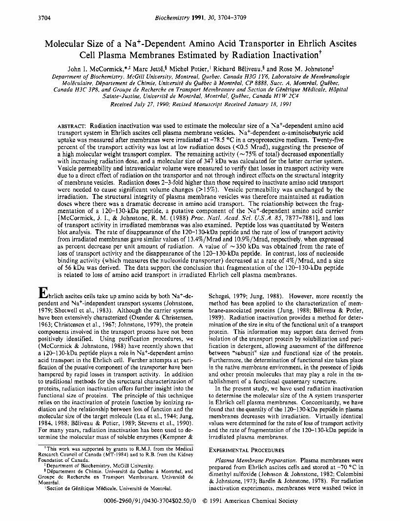
Table I: Effect of Irradiation on Membrane Permeability"
0 54.5 f 3.6 272.7 f 28 218.2 (loo)* 2.3 51.8 f 2.7 139.1 & 4.7 87.3 (40) 5 .36 55.7 f 10.9 103.2 f 8.5 47.5 (22)
Membrancs wcrc prepared in glycerol/sorbitol and irradiated as described under Experimental Procedures. [I4C]AIB uptake was then measured in the presence of K+ or Nat as described in Figure 1. Nat-dependent uptake represents the difference between total uptake in Nat and uptake in Kt. *Percent.
>0.5 Mrad, there was a further, slower decline in transport activity (Figure 1). The latter component showed an expo- nential loss of activity with radiation dose (Figure l), a result characteristic of a single transport component. From the slope of this line derived from radiation values > O S Mrad, a D37,-78,5 value for the transport system of 3.71 f 0.84 Mrad was calculated from which an RlS value of 347 f 69 kDa was determined. I n making this derivation from radiation values >0.5 Mrad, it is important to note that -25% of the total transport activity, rapidly lost at low radiation doses, suggested the presence of a second high molecular size component active in amino acid transport. Attempts to define the molecular size of this component by using lower doses of radiation were not successful.
The loss of transport activity with irradiation could be the result of a radiation-induced increase in vesicle permeability. This would lead to a decrease in the driving force of the Na+ gradient and a subsequent decrease in Na+-dependent AIB transport. As a measure of vesicle permeability, [14C]AIB uptake was measured in the presence of K+. AIB uptake in K+ is not a carrier-mediated process (Johnson & Johnstone, 1982), and an alteration in vesicle permeability would be expected to lead to changes in the rate of [I4C]AIB uptake. The data in Table 1 show that the rate of AIB uptake in K+ remained constant as membranes were irradiated while AIB uptake in Na+ decreased. Thus, vesicle permeability was unaltered by irradiation under these conditions. In a more detailed study of vesicle permeability using brush border membrane vesicles, Btliveau et al. (1988) also concluded that membrane permeability was unaffected by low-dose irradiation in the presence of glycerol and sorbitol.
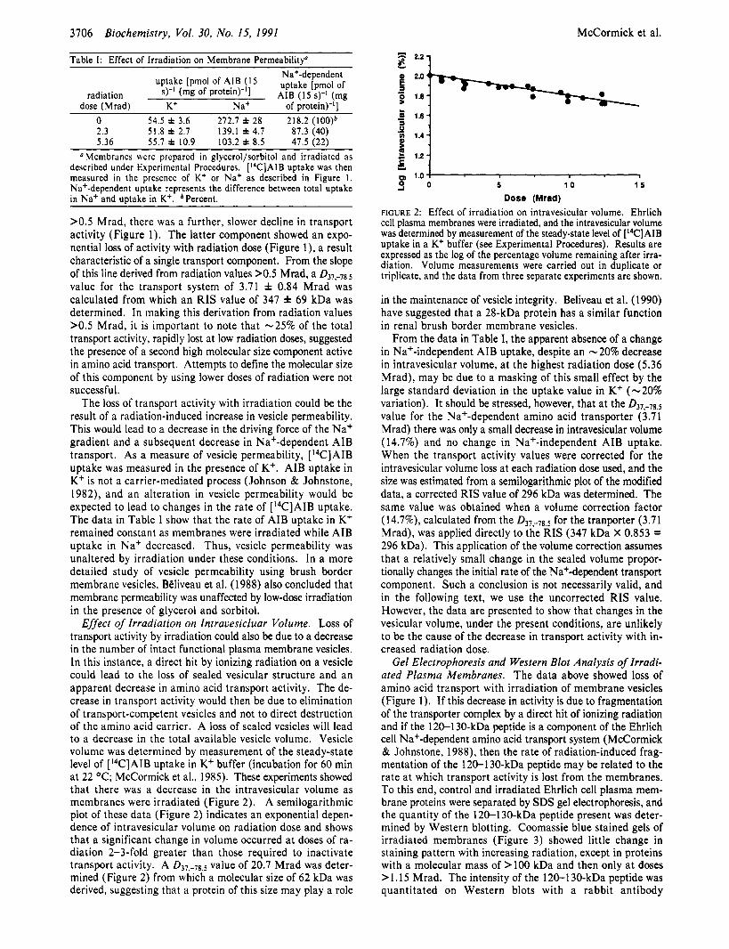
Effect of Irradiation on Intravesicluar Volume. Loss of transport activity by irradiation could also be due to a decrease in the number of intact functional plasma membrane vesicles. In this instance, a direct hit by ionizing radiation on a vesicle could lead to the loss of sealed vesicular structure and an apparent decrease in amino acid transport activity. The de- crease in transport activity would then be due to elimination of transport-competent vesicles and not to direct destruction of the amino acid carrier. A loss of sealed vesicles will lead to a decrease in the total available vesicle volume. Vesicle volume was determined by measurement of the steady-state level of [I4C]AIB uptake in K+ buffer (incubation for 60 min at 22 "C; McCormick et al., 1985). These experiments showed that there was a decrease in the intravesicular volume as membranes were irradiated (Figure 2). A semilogarithmic plot of these data (Figure 2) indicates an exponential depen- dence of intravesicular volume on radiation dose and shows that a significant change in volume occurred at doses of ra- diation 2-3-fold greater than those required to inactivate transport activity. A D37,-78,5 value of 20.7 Mrad was deter- mined (Figure 2) from which a molecular size of 62 kDa was derived, suggesting that a protein of this size may play a role
Dose (Mrad) FIGURE 2: Effect of irradiation on intravesicular volume. Ehrlich cell plasma membranes were irradiated, and the intravesicular volume was determined by measurement of the steady-state level of [I4C]AIB uptake in a Kt buffer (see Experimental Procedures). Results are expressed as the log of the percentage volume remaining after irra- diation. Volume measurements were carried out in duplicate or triplicate, and the data from three separate experiments are shown.
in the maintenance of vesicle integrity. Beliveau et al. (1990) have suggested that a 28-kDa protein has a similar function in renal brush border membrane vesicles.
From the data in Table I, the apparent absence of a change in Na+-independent AIB uptake, despite an -20% decrease in intravesicular volume, at the highest radiation dose (5.36 Mrad), may be due to a masking of this small effect by the large standard deviation in the uptake value in K+ (-20% variation). It should be stressed, however, that at the D37,-78,s value for the Na+-dependent amino acid transporter (3.71 Mrad) there was only a small decrease in intravesicular volume (14.7%) and no change in Na+-independent AIB uptake. When the transport activity values were corrected for the intravesicular volume loss at each radiation dose used, and the size was estimated from a semilogarithmic plot of the modified data, a corrected RIS value of 296 kDa was determined. The same value was obtained when a volume correction factor (14.7%), calculated from the D37,-78,5 for the tranporter (3.71 Mrad), was applied directly to the RIS (347 kDa X 0.853 = 296 kDa). This application of the volume correction assumes that a relatively small change in the sealed volume propor- tionally changes the initial rate of the Na+-dependent transport component. Such a conclusion is not necessarily valid, and in the following text, we use the uncorrected RIS value. However, the data are presented to show that changes in the vesicular volume, under the present conditions, are unlikely to be the cause of the decrease in transport activity with in- creased radiation dose.
Gel Electrophoresis and Western Blot Analysis of Irradi- ated Plasma Membranes. The data above showed loss of amino acid transport with irradiation of membrane vesicles (Figure 1). If this decrease in activity is due to fragmentation of the transporter complex by a direct hit of ionizing radiation and if the 120-1 30-kDa peptide is a component of the Ehrlich cell Na+-dependent amino acid transport system (McCormick & Johnstone, 1988), then the rate of radiation-induced frag- mentation of the 120-130-kDa peptide may be related to the rate at which transport activity is lost from the membranes. To this end, control and irradiated Ehrlich cell plasma mem- brane proteins were separated by SDS gel electrophoresis, and the quantity of the 120-1 30-kDa peptide present was deter- mined by Western blotting. Coomassie blue stained gels of irradiated membranes (Figure 3) showed little change in staining pattern with increasing radiation, except in proteins with a molecular mass of > lo0 kDa and then only at doses > 1.15 Mrad. The intensity of the 120-1 30-kDa peptide was quantitated on Western blots with a rabbit antibody

Radiation Inactivation of Amino Acid Transport Biochemistry, Vol. 30, No. 15, 1991 3707
g4kDa- - - _
3OkDa- - 1 2 3 4 5 6 7 8 9
FIGURE 3: SDS-polyacrylamide gel electrophoresis of irradiated plasma membranes. Plasma membranes were irradiated at 0 (lane 2), 0.38 (lane 3), 0.77 (lane 4), 1.15 (lane 5), 2.3 (lane 6), 3.07 (lane 7), 3.83 (lane 8), or 5.36 (lane 9) Mrad and prepared for SDS gel electrophoresis as described under Experimental Procedures. One hundred micrograms of membrane protein was applied to each lane. Gels were stained with Coomassie blue. Standard proteins were run in lane 1. The arrow indicates the position of the 120-1 30-kDa peptide.
a - b
2.2 7
l a + . , . , . , . , . . 0 1 2 3 4 5 6
Doaa (Mrad) 1 2 3 4 5 6
FIGURE 4: (a) Western blot analysis of irradiated plasma membranes. lrradiatcd plasma membranes were resolved on SDS-polyacrylamide gels ( 1 50 pg of protein/lane) and electrophoretically transferred to nitrocellulose paper. The paper was incubated overnight with immune serum containing antibody against the 120-1 30-kDa peptide, and bound antibody was located by incubation with goat anti-rabbit IgG conjugated to alkaline phosphatase. Membranes were irradiated at 5.05 (lane I ) , 4.27 (lanc 2), 3.1 1 (lane 3), 1.94 (lane 4), 1.17 (lane 5), and 0 (lane 6) Mrad. (b) Fragmentation of the 120-130-kDa peptide and loss of amino acid transport activity in irradiated mem- branes. The Western blot shown in (a) was quantitated by laser densitomctry, and the results were expressed as log of the percent area remaining undcr cach peak with respect to an unirradiated control (0). Na+-dependent AIB transport activity was measured in aliquots of the same membranes, and the semilogarithmic plot of these data is also included in the figure (0).
(McCormick & Johnstone, 1988). Figure 4a clearly shows that when plasma membranes were irradiated there was a decrease in the quantity of the 120-1 30-kDa peptide, apparent from the decreased binding of antibody. The data in Figure 4a were quantitated by laser scanning of the 120-130-kDa molecular mass region of the blot. A semilogarithmic plot of the data from this one experiment (Figure 4a) shows a simple exponential decrease in antibody binding with increased ra- diation dose, reflecting the loss of a single immunoreactive species (Figure 4b). When the decrease in antibody binding with radiation dose was compared with the decrease in Na+-dependent amino acid transport activity, for the same membranes (Figure 4b), the two events clearly decreased at a similar rate. I n addition, the best-fit line through all the data points (Figure 4b) had a correlation coefficient of -0.97, and a 037,-78.5 value of 3.22 Mrad was determined from this slope. A molecular size of 399 kDa was calculated from the combined Western blot and transport activity data, a value close to the size determined from transport activity alone (Figure 1; 347 kDa). The pooled data from four experiments for loss of immunoreactivity (by Western blot) against radi- ation dose are shown in Figure 5. The 037,-78.5 value for these data, using computer-generated estimates of the best slope for the regression line was 3.69 Mrad, giving a target size of 348 kDa. When the percent decrease in antibody binding per unit
6 2! Y
0 '"t- 1.8 - 1.8
1.6 1.6 1 :::I . , , , . 8 . , 1 .o
0 1 2 3 4
Dose (Mrad) FIGURE 5 : Western blot analysis of irradiated plasma membranes. The decrease in the 120-130-kDa peptide in irradiated plasma membranes from four different experiments was quantitated by Western blot analysis and laser densitometry. The 037,-78,5 value was 3.69 Mrad.
U
!k m 3 cn A
0 5 1 0 1 5
Dose (Mrad) FIGURE 6: Effect of irradiation on NBMPR binding activity in Ehrilch cell plasma membranes. Plasma membranes were irradiated and NBMPR binding activity was determined as described under Ex- perimental Procedures. Results are expressed as the percentage of binding activity remaining relative to unirradiated membranes. The D37,-78,5 value was 23.19 Mrad.
of radiation (Figure 5) and the percent decrease in transport activity (Figure 1) per unit of radiation were compared, the values were 13.4%/Mrad and 10.9%/Mrad, respectively. In contrast, the value for NBMPR binding activity, presumably an unrelated process, was 4%/Mrad (see Figure 6). The similar values for the rate of loss of the 120-1 30-kDa peptide and Na+-dependent amino acid transport activity in irradiated plasma membranes suggest that the two processes are related. Evidence for a structural relationship between peptide loss and transport activity is also suggested by the identical target sizes determined either by Western blotting (348 kDa) or by transport activity measurements (347 kDa).
Effect of Irradiation on NBMPR Binding Activity in Ehrlich Cell Plasma Membranes. To assess the reliability of the present technique, the radiation inactivation of a trans- porter of known molecular mass was examined. Nucleoside transport in mammalian cells occurs by way of a facilitated transport mechanism (Wohlhueter & Plagemann, 1980). NBMPR is a potent, specific inhibitor of the transport system (Jarvis, 1986), and the binding of radiolabeled NBMPR to plasma membranes can be used to quantitate the nucleoside transporter content (Jarvis et al., 1980). Hammond and Johnstone (1 989) have characterized the nucleoside transport system in the Ehrlich cell, and using photoaffinity labeling have identified the transporter as a protein with a molecular mass of 42 kDa on SDS-polyacrylamide gels. A molecular size of 56 kDa (Figure 6) was determined from radiation inactivation of [3H]NBMPR binding activity in Ehrlich cell plasma membranes. The data show (Figure 6) that the decrease in [3H]NBMPR binding is characteristic of the loss of a single

3708
membrane binding component.
DISCUSSION In the present study, Ehrlich cell plasma membranes were
irradiated in a cryoprotective medium containing glycerol and sorbitol at -78.5 OC. Binding and transport activities were subsequently assayed at 22 OC, in the presence of the same cryoprotective agents. The validity of this procedure for membrane irradiation has been demonstrated by BCliveau et al. (1988), who used rat brush border membrane vesicles. These authors showed that the molecular size of the brush border membrane enzyme, alkaline phosphatase, determined by radiation inactivation, was 104 kDa, virtually identical with the size (1 20 kDa) of the purified enzyme determined by SDS gel electrophoresis (Colbeau & Maroux, 1978). Biliveau and his colleagues (BCliveau et al., 1988) have also shown that the structural integrity of brush border membrane vesicles was unaffected by low-dose irradiation and that the experimental system was suitable for the determination of the moleclar size of a number of transport systems. It appears that the presence of cryoprotective agents prevents large changes in vesicle structure, so that the successful measurement of a transport system is not compromised (Kinne et al., 1984; Jung, 1988; BCliveau et al., 1988, 1990).
These conclusions are consistent with the present observa- tions. There is little evidence in the present study to suggest that Ehrlich cell membrane vesicles themselves were severely affected by either the cryoprotective medium or the irradiation. Amino acid transport in membrane vesicles was identical in the presence or absence of glycerol and sorbitol. In addition, there was no indication that structural integrity, as measured by permeability, was altered at doses of 5-6 Mrad. However, measurement of intravesicular volume showed that at higher radiation doses (6-1 2 Mrad) there was a significant loss of vesicle volume, suggesting that a direct hit by ionizing radiation could destroy the sealed vesicle structure. However, over the range of radiation doses where the Na+-dependent amino acid transporter was affected (1-5 Mrad), there were only relatively small decreases (10-1 5%) in intravesicular volume.
In the present study, NBMPR binding activity was used as an intrinsic marker for the irradiation of Ehrlich cell plasma membranes. A molecular size of 56 kDa was determined by radiation inactivation (Figure 6), suggesting that the NBMPR binding component of the nucleoside transporter functions as a monomer in Ehrlich cell plasma membranes. The agreement between the radiation inactivation size (56 kDa) and the molecular size of the nucleoside transporter determined by SDS gel analyses (43 kDa; Hammond & Johnstone, 1989) supports the validity of the procedures used in the present study, From radiation inactivation of the human erythrocyte transporter, Jarvis et a!. (1 980, 1984) have reported an RIS value of 122 kDa and a dimeric in situ structure for this nucleoside transporter.
The decrease in Na+-dependent amino acid transport in irradiated plasma membranes occurred in two stages. In the first stage, 25-30% of the transport activity was lost at low radiation doses (<0.5 Mrad). During the second stage, the remaining transport activity decreased at a slower rate in a manner characteristic of a single radiation target (Jung, 1984, 1988). The RIS value for the latter component was 347 kDa.
The sensitivity of amino acid transport to low radiation doses of < O S Mrad suggests that some of this transporter (or an- other transporter) is present as a high molecular weight com- plex or aggregate. Although AIB is transported mainly by the A system in the Ehrlich cell, other transport systems such as the ASC system may also transport AIB and exist as high
Biochemistry, Vol. 30, No. 15, 1991 McCormick et al.
molecular weight transport components, sensitive to low doses of radiation. The majority of AIB transport activity had a radiation inactivation target size of 347 kDa. This target value may be contrasted to those obtained in rat kidney brush border membrane vesicles by BCliveau et al. (1990) for Na+-dependent transport of alanine, glutamate, proline, and L-leucine, namely, 274, 250, 224, and 293 kDa, respectively. However, using different conditions for irradiation, Takahashi et al. (1985) have reported that the molecular size for Na+-dependent alanine transport in rabbit kidney brush border vesicles is 1200 kDa. Direct comparison of radiation inactivation sizes in Ehrlich cell plasma membrane vesicles and brush border membrane vesicles is probably unwarranted because the classical amino acid carriers present in Ehrlich cells are not present in brush border membranes (Stevens et al., 1984). However, the closeness of our values and those of BEliveau et al. (1990), who used identical radiation conditions, is note- worthy.
We have proposed (McCormick & Johnstone, 1988) that the 120-1 30-kDa peptide is a component of the amino acid transporter. If this conclusion is correct, the structural decay of this peptide and loss of transport activity during irradiation may be related. The 120-130-kDa peptide fragmented as a protein with a molecular size of 348 kDa. Since a single radiation hit on the complex destroys the total oligomeric structure, efficient energy transfer between closely associated subunits is indicated. Thus, either the 348-kDa oligomer consists of three subunits of the 120-130-kDa monomer or the 120-1 30-kDa peptide is part of a heteropolymer associated with other membrane proteins to give the 348-kDa oligomer. The identity of molecular size values determined either from fragmentation of the 120-130-kDa peptide or from loss of transport activity in irradiated membranes (348 and 347 kDa, respectively) suggests that fragmentation of the 120-1 30-kDa peptide is related to loss of amino acid transport. These data support the conclusion that the 120-130-kDa peptide is an integral component of the A-system transporter.
ACKNOWLEDGMENTS
in the preparation of Ehrlich cell plasma membranes.
REFERENCES Bardin, C., & Johnstone, R. M. (1978) J . Biol. Chem. 253,
Beauregard, G. , Maret, A,, Salvayre, R., & Potier, M. (1987)
Biliveau, R., & Potier, M. (1989) News Physiol. Sci. 4 ,
Biliveau, R., Demeule, M., Ibnoul-Khatib, H., Bergeron, M., Beauregard, G. , & Potier, M. (1988) Biochem. J . 252,
Biliveau, R., Demeule, M., JettE, M., & Potier, M. (1990)
Colbeau, A., & Maroux, S . (1978) Biochim. Biophys. Acta
Colombini, M., & Johnstone, R. M. (1974) J . Membr. Biol.
Hammond, J. R., & Johnstone, R. M. (1989) Biochem. J. 262,
Jarvis, S. M. (1986) Mol. Pharmacol. 30, 659-665. Jarvis, S. M., Young, J. D., & Ellory, J. C. (1980) Biochem.
Jarvis, S . M., Fincham, D. A., Ellory, J. C., Paterson, A. R. P., & Young, J. 0. (1984) Biochim. Biophys. Acta 772,
We thank Anoush Cotchikian for expert technical assistance
1725-1 732.
Methods Biochem. Anal. 32, 313-343.
134-138.
807-813.
Biochem. J . 268, 195-200.
511, 39-51.
15, 261-276.
109-1 18.
J . 190, 313-376.
227-230.

Biochemistry 1991, 30, 3709-371 5 3709
Johnson, P. A,, & Johnstone, R. M. (1982) Membr. Biochem.
Johnstone, R. M. (1979) Can. J . Physiol. Pharmacol. 57,
Jung, C. Y. (1984) in Receptor Biochemistry and Metho- dology (Venter, J . C., & Harrison, L. C., Eds.) Vol. 3, pp 113-208, Alan R. Liss, New York.
Jung, C. Y. ( 1 988) in Methods for Studying Membrane Fluidity (Aloia, R. C., Curtain, C. C., & Gordon, L. M., Eds.) pp 107-1 26, Alan R. Liss, New York.
Kempner, E. S . , & Schlegel, W. (1979) Anal. Biochem. 92, 2- 10.
Kinne, R., Da Cruz, M. T. M., & Lin, J. T. (1984) Curr. Top. Membr. Transp. 20, 248-250.
Laemmli, U. K . (1970) Nature (London) 227, 680-685. Lea, D., Smith, K. M., Holmes, B., & Markham, R. (1944)
Lowry, 0. H., Rosebrough, N. J., Farr, A. L., & Randall, R.
4, 189-218.
1-15.
Parasitology 36, 1 10-1 18.
J. (1951) J . Biol. Chem. 193, 265-275.
Markwell, M. A. K., Hass, S . M., Tolbert, N. E., & Bieber, L. L. (1 98 1) Methods Enzymol. 72, 296-303.
McCormick, J. I., & Johnstone, R. M. (1988) Proc. Narl. Acad. Sci. U.S.A. 85, 7877-78 1 1.
McCormick, J. I., Silvius, J. R., & Johnstone, R. M. (1 985) J . Biol. Chem. 260, 5706-5714.
Shotwell, M. A., Kilberg, M. S. , & Oxender, D. L. (1983) Biochim. Biophys. Acta 737, 267-284.
Stevens, B. R., Kaunitz, J. D., & Wright, E. M. (1984) Annu. Rev. Physiol. 46, 417-433.
Stevens, B. R., Fernandez, A,, Hirayama, B., Wright, E. M., & Kempner, E. S . (1990) Proc. Natl. Acad. Sci. U.S.A. 87,
Takahashi, M., Malathi, P., Preiser, H., & Jung, C. Y . ( 1 985)
Towbin, H., Staehelin, T., & Gordon, J. (1979) Proc. Natl .
Wohlhueter, R. M., & Plagemann, P. G. W. (1980) Cum.
1456-1 460.
J . Biol. Chem. 260, 10551-10556.
Acad. Sci. U.S.A. 76, 4350-4354.
Top. Membr. Transp. 14, 225-330.
Interaction of Fluorescently Labeled Dideoxynucleotides with HIV- 1 Reverse Transcriptase?
Barbara Muller, Tobias Restle, Joachim Reinstein, and Roger S . Goody* Abteilung Biophysik. Max- Planck-lnstitut fur Medizinische Forschung, Jahnstrasse 29, 6900 Heidelberg, FRG
Received June 19, 1990; Revised Manuscript Received December 3, 1990
ABSTRACT: Succinylfluorescein-labeled dideoxyTTP has been used as a substrate for reverse transcriptase from HIV- 1. On addition to the 3’-end of a primer molecule, there is a reduction of fluorescence yield of a factor of ca. 4. Release of a fluorescent DNA/DNA primer/template duplex from its complex with reverse transcriptase results in a reduction of fluorescence by a further factor of 2. The fluorescent nucleotide is incorporated somewhat less efficiently than 3’-azidoTMP and TMP, which show similar incorporation kinetics. Fluorescent chain-terminated primers have been used to investigate the interaction of normal and chain- terminated primer/template complexes with reverse transcriptase. The dissociation constant of a 36/ 18-mer was 0.65 nM, whereas that of the same complex after the addition of the fluorescent chain-terminating nucleotide to the primer was 3 nM at 25 “C. The rate of dissociation of the latter complex from the enzyme was 0.04 s-’. This was decreased by a factor of ca. 10 a t high concentrations (>200 p M ) of the nucleotide triphosphate complementary to the next position of the template. The results obtained suggest that potent inhibition of reverse transcriptase activity in in vitro assays results from formation of a slowly dissociating complex between the enzyme and chain-terminated primer/template complexes. However, arguments are presented that lead to the conclusion that this is not the mode of inhibition in cells invaded by HIV. At the prevailing relative concentrations in this situation, chain termination resulting in incomplete transcription is likely to be the major factor.
A detailed knowledge of the structure of an enzyme and its catalytic mechanism is the basis for any approach to a rational design of potent and specific inhibitors. Crucial information can be provided by investigations of the dynamics and ther- modynamics of the interactions of substrates with the protein. The availability of pure HIV-1 reverse transcriptase from bacterial expression systems (Tanese et al,. 1986; Larder et al., 1987; Hizi et al., 1988; Mous et al., 1988; Muller et al., 1989) has made this enzyme amenable to detailed biochemical and biophysical characterization. In the last few years, several
‘This work was supported by the Bundesministerium fur Forschung
* To whom correspondence should be addressed. und Technologie.
0006-296019 110430-3709$02.50/0
studies examining the steady-state kinetics of the enzyme have been published (Cheng et al., 1987; Majumdar et al., 1988, 1989; Huber et a]., 1989). While this method has the ad- vantage that only catalytic amounts of enzyme are needed, it has the disadvantage that conclusions drawn concerning the mechanism are often indirect and that the rate and equilibrium constants of discrete steps in the mechanism cannot be de- termined. This limitation does not apply to studies using “substrate concentrations” of enzyme, which are now available.
Fluorescent substrate analogues are useful tools for inves- tigation of enzymesubstrate interactions. Their use for kinetic studies often allows a more direct monitoring of certain steps in the catalytic process (e.g., binding of the substrate to the enzyme) than the more conventional biochemical approaches.
0 1991 American Chemical Society