molecular mechanisms of colorectal carcinogenesis
TRANSCRIPT

25K.M. Haigis (ed.), Molecular Pathogenesis of Colorectal Cancer, DOI 10.1007/978-1-4614-8412-7_2, © Springer Science+Business Media New York 2013
Abstract Colorectal cancer (CRC) presents in three major forms: inherited, spo-radic, and familial. Although the mechanisms underlying familial CRC are poorly understood, a large body of evidence suggests that inherited and sporadic CRC are caused by sequential genetic and molecular events. There are three distinct path-ways of CRC pathogenesis: the chromosomal instability pathway (CIN), the micro-satellite instability pathway (MSI), and the serrated pathway. The majority of CRCs arise from the CIN pathway, which is characterized by defects in chromosomal segregation, telomere stability, and the DNA damage response. Microsatellite insta-bility derives from the loss of DNA mismatch repair and is found in about 15 % of all CRCs, 3 % of which are associated with Lynch syndrome. The serrated pathway, recognized only in the last 15 years, describes the progression of serrated polyps to CRC. The goal of this chapter is to discuss the key genetic and molecular elements of each pathway from a historical perspective and to describe the relevance of this knowledge to the care of patients with CRC.
Keywords Colorectal cancer • Chromosomal instability • Microsatellite instability • Mouse models • Serrated pathway
2.1 Introduction
Colorectal cancer (CRC) continues to be an enormous public health burden. It is the third most common cancer in men and second most common cancer in women worldwide, with nearly 1.2 million new cases yearly, and the third leading cause of
Chapter 2 Molecular Mechanisms of Colorectal Carcinogenesis
Jatin Roper and Kenneth E. Hung
J. Roper , M.D. • K. E. Hung , M.D., Ph.D. (*) Division of Gastroenterology, Department of Medicine , Tufts Medical Center , 800 Washington Street, Box #233 , Boston , MA 02111 , USA e-mail: [email protected]

26
cancer-related mortality, with approximately 600,000 deaths each year. The 5-year prognosis for patients with newly diagnosed metastatic colon cancer continues to be less than 20 % (Jemal et al. 2011 ). The underlying causes of CRC are complex and heterogeneous. Both environmental factors and genetic events contribute to CRC risk. Among the environmental risk factors for CRC are diets rich in unsaturated fats and red meat, total energy intake, excessive alcohol consumption, and reduced physical activity. Many studies have examined other exposures for their effects on CRC risk but have yielded ambiguous results (Chan and Giovannucci 2010 ). In contrast, there has been signifi cant progress in identifi cation of the specifi c genetic defects underlying the majority of CRCs. To develop effective CRC prevention, diagnosis, and treatment strategies, an understanding of the pathways and molecular events that drive CRC carcinogenesis is essential.
CRC presents in one of three patterns: inherited, familial, and sporadic. Inherited and familial CRC derive, at least in part, from germline mutations. Inherited CRC accounts for 10 % of cases and presents as well-characterized cancer predisposition syndromes including Lynch syndrome and familial adenomatous polyposis (FAP). Familial CRC accounts for 25 % of CRCs and presents without precisely defi ned Mendelian inheritance patterns or genetic etiology (Pino and Chung 2010 ). Sporadic CRC derives from somatic mutation, accounts for approximately 70 % of CRCs, and is not associated with family history. This chapter focuses on the genetic and molecular events underlying the three major pathways for sporadic and inherited colorectal carcinogenesis: chromosomal instability (CIN), microsatellite instability (MSI), and the CpG island methylator phenotype (CIMP) pathways.
2.2 CIN: Chromosomal Instability Pathway
2.2.1 The Adenoma–Carcinoma Sequence
By the mid-1970s, several pieces of indirect evidence suggested that colorectal adenocarcinomas may progress from adenomas (1) residual benign adenomatous tissue was found in carcinomas, (2) malignant foci were observed in larger polyps, and (3) there were rare observations of a benign-appearing polyp developing into an invasive carcinoma (Morson 1974 ). In 1987, Stryker and colleagues reported the natural history of unresected colonic polyps >1 cm in size in 226 patients who declined surgical resection. After 20-year follow-up, they found a 24 % risk of invasive adenocarcinoma at the site of the index polyp, and a 35 % risk of carci-noma at any colonic site (Stryker et al. 1987 ). Individuals affected by cancer pre-disposition syndromes, such as FAP, invariably develop CRC by the third or fourth decade of life if their colons are not removed (Lynch and de la Chapelle 2003 ). The National Polyp Study confi rmed the hypothesis that colorectal carcinomas arise from adenomas through showing that polypectomy by colonoscopy reduces the
J. Roper and K.E. Hung

27
risk of subsequent CRC (Winawer et al. 1993 ). In 1990, Fearon and Vogelstein proposed a multistep genetic model of colorectal carcinogenesis in which inactiva-tion of the adenomatous polyposis coli ( APC ) tumor suppressor gene occurs fi rst in normal colonic mucosa, followed by activating mutations in the KRAS gene and subsequent additional mutations (e.g., PIK3CA , TP53 , and TGF - β pathway genes) (Fig. 2.1 and Table 2.1 ) (Fearon and Vogelstein 1990 ; Vogelstein et al. 1988 ; Fearon 2011 ). Several key principles of the so-called adenoma–carcinoma sequence have been established (1) multiple genetic hits are required, (2) there are discrete intermediaries in the progression to cancer (Pino and Chung 2010 ; Haigis et al. 2008 ), and (3) adenomas arise from aberrant crypt foci in the colonic epithelium (Takayama et al. 1998 ).
2.2.2 Genomic Instability and Cancer
The mutation rate per nucleotide base pair is far too low (estimated to be approxi-mately 10 −9 per cell generation) to account for the multiple genetic mutations required for tumorigenesis (Albertini et al. 1990 ). Therefore, it has been proposed that cancer cells must acquire a “mutator phenotype” that increases the rate of spon-taneous mutations (Loeb et al. 2003 ). 65–70 % of sporadic colorectal cancers exhibit
Fig. 2.1 The adenoma–carcinoma sequence. The initial step in colorectal carcinogenesis is thought to be the formation of aberrant crypt foci (ACF). Activation of the Wnt pathway occurs during this step as a result of inactivating mutations in the APC gene. Progression to adenoma and carcinoma is usually mediated by activating mutations in KRAS and loss of TP53 expression, respectively. A subset of advanced adenomas may progress due to mutations in PIK3CA and loss of 18q. Reproduced with permission from: Pino MS, Chung DC (2010) The chromosomal instabil-ity pathway (CIN) in colon cancer. Gastroenterology 138(6):2059–2072
2 Molecular Mechanisms of Colorectal Carcinogenesis

Table 2.1 Somatic mutations in oncogenes and tumor suppressor genes implicated in colorectal carcinogenesis
Gene Chromosomal location Type of mutation
Prevalence (%) Function of gene product
Oncogenes KRAS 12p12 Point mutation (codons
12, 13 of exon 2) 40 Cell proliferation and survival
PIK3CA 3q26 Point mutations (E545K on exon 9, H1047R on exon 20)
15–30 Cell proliferation and survival
CDK8 13q12 Gene amplifi cation 10–15 β-catenin activation EGFR 7p12 Gene amplifi cation 5–15 Cell proliferation and
survival BRAF 7q34 Point mutations activating
kinase activity (most commonly V600E)
5–10 Cell proliferation and survival
CMYC 8q24 Gene amplifi cation 5–10 Cell proliferation and survival CCNE1 19q12 Gene amplifi cation 5 NRAS 1p13 Point mutation <5 Cell proliferation and survival CTNNB1 3p22 Stabilizing point mutations
and in-frame deletions near N terminus
<5 Regulation of Wnt pathway target genes that promote tumor growth and invasion
ERBB2 ( HER2 )
17q21 Gene amplifi cation <5 Cell proliferation and survival
MYB 6q22-q23 Gene amplifi cation <5 Stimulates growth of intestinal stem cells
Tumor-suppressor genes APC 5q21 Frameshift, point mutation,
deletion, allele loss leading to truncated protein
70–80 Inhibition of Wnt signaling
TP53 17q13 Point mutation (missense), allele loss
60–70 Cell cycle arrest, apoptosis and autophagy induction
DCC 18q21 Point mutation 50 Cell surface receptor for netrin-1, triggers tumor cell apoptosis
TGFβRII ( TGFBR2 )
3p22 Frameshift, nonsense 25 Inhibition of cell growth
SMAD4 18q21 Nonsense, missense, allele loss
10–15 Intracellular mediator of the TGF-β pathway
PTEN 10q23 Nonsense, deletion 10 Inhibition of PI3K activity ACVR2A 2q22 Frameshift 10 Cellular growth SMAD2 18q21 Nonsense, deletion,
allele loss 5–10 Intracellular mediator of the
TGF-β pathway FBXW7 4q31 Nonsense, missense,
deletion 9 Targets oncoproteins for
ubiquitin- mediated degradation
SMAD3 15q22 Nonsense, deletion 5 Intracellular mediator of the TGF-β pathway
TCF7L2 10q25 Frameshift, nonsense 5 Regulation of the Wnt signaling
BAX 19q13 Frameshift 5 Apoptotic activator LKB1 ( STK11 )
19p13 Deletion Rare (limited to PJS)
Regulation of cell polarity
Modifi ed from Fearon ER. Molecular genetics of colorectal cancer. Annu Rev Pathol 2011;6:479–507

29
an accelerated rate of gains or losses of whole or large portions of chromosomes that result in karyotypic variability between cells. This chromosomal instability (CIN) appears to be a dominant trait (Lengauer et al. 1997 , 1998 ). Consequences of CIN include an imbalance in chromosomal number (aneuploidy), subchromosomal genomic amplifi cations, and a high frequency of loss of heterozygosity (LOH). There are currently no standardized measures of chromosomal instability; hence, CIN-positive vs. CIN-negative tumors cannot be clearly defi ned (Pino and Chung 2010 ). One theory views cancers as clonal in origin, in that they arise from a single, genomically unstable cell, but develop genetic heterogeneity due to CIN. This explains observed heterogeneity within single tumors with regard to DNA content, chromosomal number, gene expression, metabolism, resistance to cytotoxic drugs, and metastatic potential (Duesberg et al. 2004 ).
2.2.3 Mechanisms Leading to Chromosomal Instability
2.2.3.1 Defects in Chromosomal Segregation
The CIN phenotype can result from defects in pathways that regulate chromosomal segregation. The mitotic or spindle checkpoint ensures proper chromosome segre-gation by delaying the metaphase-to-anaphase transition until all pairs of dupli-cated chromatids are properly aligned on the spindle. Genes that encode proteins operating as spindle checkpoint regulators include mitotic arrest - defi cient ( MAD1L1 and MAD2L1 ), budding uninhibited by benzimidazoles 1 ( BUB1 ), and kinesin fam-ily member 11 ( KIF11 ). Mutations in BUB1 result in abnormal spindle checkpoint and CIN in chromosomally stable cell lines (Bardelli et al. 2001 ). Cells from dominant- negative mBub1 mutant mice demonstrate escape from apoptosis, con-tinued cell cycle progression, and disrupted spindles (Taylor and McKeon 1997 ). Kinesin spindle protein, also known as Eg5, is a motor protein responsible for mitotic spindle formation and chromosomal separation during mitosis. Overexpression of Eg5 in mice leads to spindle defects, CIN, and solid tumor for-mation (Castillo et al. 2007 ). Mutations in the hZw10, hZwilch/FLJ10036, and hROD/KNTC genes, which encode kinetochore proteins, have been reported in CRC (Wang et al. 2004 ).
Chromosomal missegregation due to defects in the mitotic checkpoint may lead to aneuploidy, a concept fi rst proposed by Theodor Boveri in 1902—well before the advent of chromosomal karyotyping (Boveri 2008 ). The aneuploidy hypothesis pro-poses a two-step mechanism for tumor initiation. The fi rst step is an event (i.e., a defect in spindle formation) that promotes chromosomal missegregation and aneu-ploidy. In the second step, aneuploidy destabilizes the genome, gives rise to poly-clonal mutations, and results in heterogeneous karyotypes. Aneuploidy therefore stimulates tumorigenesis either by increasing the chances of LOH of a tumor- suppressor gene or by amplifying an oncogene through chromosomal duplication (Duesberg et al. 2004 ; Castillo et al. 2007 ).
2 Molecular Mechanisms of Colorectal Carcinogenesis

30
2.2.3.2 Centromere Dysfunction
Another proposed cause of CIN is abnormal centromere number and function. Centrosomes serve to anchor cytoplasmic microtubules as they are arranged into a mitotic spindle apparatus. Extra centrosomes in cancer cell lines may lead to the for-mation of multiple spindle poles during mitosis, resulting in unequal distribution of chromosomes and CIN (Ganem et al. 2009 ). Polo-like kinases (Plk) are serine/threo-nine kinases, which regulate centrosome duplication. Elevated expression of Plk1 has been observed in 73 % of CRCs and correlate with tumor invasion, lymph node involvement, and stage (Takahashi et al. 2003 ). The centrosome-associated Aurora A protein is amplifi ed and positively associated with CIN in CRC, but metastatic CRC patients with increased Aurora A gene copy number have longer overall and progres-sion-free survival, particularly in KRAS wild-type tumors (Dotan et al. 2002 ; Herz et al. 2011 ). The related Aurora B protein regulates chromatid segregation, and its expression is correlated with advanced stages of CRC (Katayama et al. 1999a ).
2.2.3.3 Telomere Dysfunction
CIN may also be driven by telomere dysfunction. Telomeres are hexameric DNA repeats (TTAGGG in humans) that protect the ends of eukaryotic chromosomes from fusing and breaking during segregation. A portion of telomeric DNA is lost after each round of DNA replication due the inability of DNA polymerase to com-pletely synthesize the 3′ end of chromosomes. Cells with suffi ciently shortened telomeres are targeted for senescence and apoptosis by DNA damage checkpoints. Cells that survive the checkpoint activate telomerase, which elongates telomeres. In mice defi cient in the RNA component of telomerase ( Terc −/−), telomere shortening results in aberrant crypt foci, adenomas, and gastrointestinal tumors (Rudolph et al. 2001 ; Plentz et al. 2003 ). 77–90 % of CRCs harbor shorter telomeres, compared to adjacent normal tissue, but increased telomerase activity has also been reported (Engelhardt et al. 1997 ; Takagi et al. 1999 ; Katayama et al. 1999b ; Nakamura et al. 2000 ; Gertler et al. 2004 ; Chadeneau et al. 1995 ; Tatsumoto et al. 2000 ). These fi nd-ings suggest that telomere shortening promotes CIN that initiates carcinogenesis, whereas telomerase activation in established carcinomas leads to immortality of cancer cells.
2.2.3.4 Loss of Heterozygosity
LOH is a key feature of CIN-positive tumors and distinguishes tumors arising from the CIN pathway from tumors arising from the MSI pathway. Approximately 25–30 % of alleles are lost in tumors (Lengauer et al. 1998 ). Mitotic nondisjunction, recombination between homologous chromosomes, and chromosomal deletion are among the implicated mechanisms. One study found that the majority of losses on chromosome 18 involved the whole chromosome and were caused by mitotic
J. Roper and K.E. Hung

31
nondisjunction. Losses limited to a part of a chromosome were thought to be due to interchromosomal recombinations and deletions associated with DNA double- strand breaks (Thiagalingam et al. 2001 ).
2.2.3.5 Defi ciencies in DNA Damage Response
Defi ciencies in DNA damage response have been linked to human cancer. Inactivating mutations in ataxia telangiectasia mutated ( ATM ) and ataxia telangiec-tasia and Rad3-related ( ATR ) protein kinases lead to the ataxia telangiectasia and Seckel syndromes, respectively (Khanna and Jackson 2001 ). Other syndromes linked to impaired DNA damage response include Li–Fraumeni ( TP53 mutations) and hereditary breast–ovarian cancer ( BRCA1 and BRCA2 mutations). Of these genes, only TP53 has been directly implicated in human colorectal cancer. Haploinsuffi ciency of histone H2AX, an ATM and ATR substrate, leads to genomic instability and tumor susceptibility in a p53-defi cient background, and mouse embryonic fi broblasts derived from ATM- and H2Ax-defi cient mice show severe genomic instability (Bassing et al. 2003 ; Celeste et al. 2003 ; Zha et al. 2008 ). Defi ciency in Chk1 , a DNA damage checkpoint protein, causes mitotic defects and disrupts Aurora B during mitosis, resulting in failure of cytokinesis and multinucle-ation (Peddibhotla et al. 2009 ).
2.2.4 Genetic Abnormalities Implicated in the Chromosomal Instability Pathway
Recent comprehensive sequencing studies have identifi ed over 80 somatic muta-tions in exons of colorectal tumors. However, a limited number of these mutations are found in a signifi cant percentage of tumors. Wood et al. predicted that perhaps 15 or fewer of these mutations in any given CRC are critical drivers of tumor initia-tion, progression, and/or maintenance (Wood et al. 2007 ). Many of the genes identi-fi ed by sequencing analysis were already well known to be somatically mutated in CRC (e.g., APC , KRAS , and TP53 ). Table 2.1 describes data on oncogenes and tumor suppressor genes that are somatically mutated in CRC.
2.2.4.1 APC and the Wnt Pathway
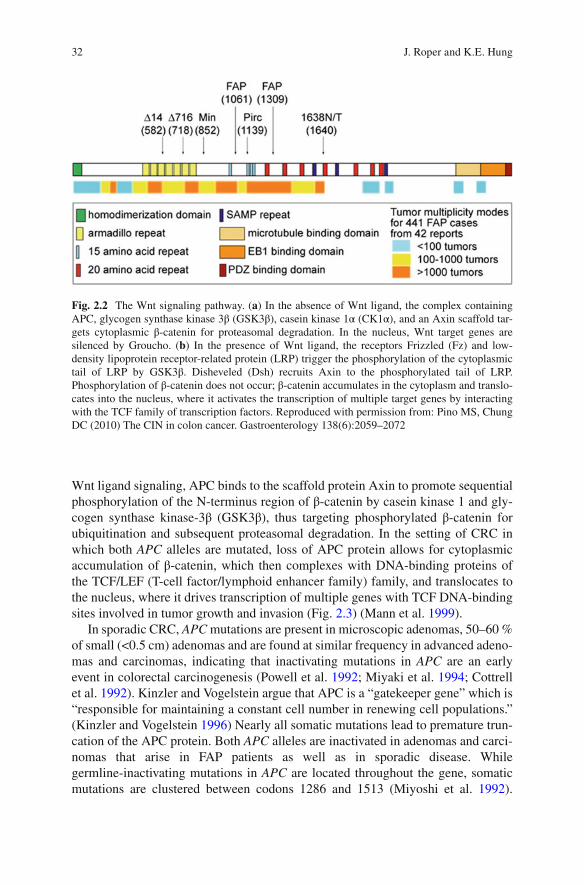
The earliest genetic event in colorectal carcinogenesis is activation of the Wnt path-way, typically via disruption of APC on 5q21 (Powell et al. 1992 ). The APC gene product is an approximately 300-kDa protein with multiple functional domains that regulates differentiation, adhesion, polarity, migration, development, apoptosis, and chromosomal segregation (Fig. 2.2 ). Restoration of APC protein expression in CRC cells that lack endogenous APC expression promotes apoptosis. In the absence of
2 Molecular Mechanisms of Colorectal Carcinogenesis
32
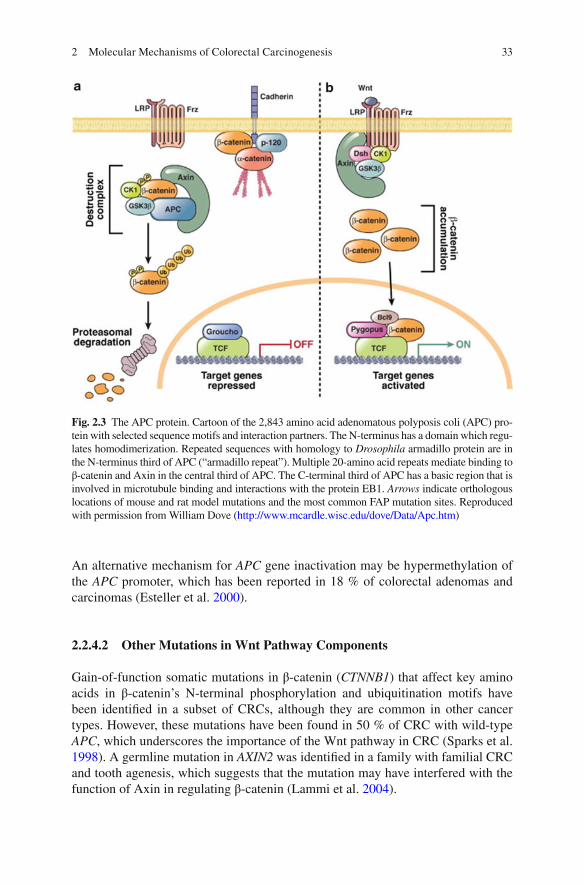
Wnt ligand signaling, APC binds to the scaffold protein Axin to promote sequential phosphorylation of the N-terminus region of β-catenin by casein kinase 1 and gly-cogen synthase kinase-3β (GSK3β), thus targeting phosphorylated β-catenin for ubiquitination and subsequent proteasomal degradation. In the setting of CRC in which both APC alleles are mutated, loss of APC protein allows for cytoplasmic accumulation of β-catenin, which then complexes with DNA-binding proteins of the TCF/LEF (T-cell factor/lymphoid enhancer family) family, and translocates to the nucleus, where it drives transcription of multiple genes with TCF DNA-binding sites involved in tumor growth and invasion (Fig. 2.3 ) (Mann et al. 1999 ).
In sporadic CRC, APC mutations are present in microscopic adenomas, 50–60 % of small (<0.5 cm) adenomas and are found at similar frequency in advanced adeno-mas and carcinomas, indicating that inactivating mutations in APC are an early event in colorectal carcinogenesis (Powell et al. 1992 ; Miyaki et al. 1994 ; Cottrell et al. 1992 ). Kinzler and Vogelstein argue that APC is a “gatekeeper gene” which is “responsible for maintaining a constant cell number in renewing cell populations.” (Kinzler and Vogelstein 1996 ) Nearly all somatic mutations lead to premature trun-cation of the APC protein. Both APC alleles are inactivated in adenomas and carci-nomas that arise in FAP patients as well as in sporadic disease. While germline-inactivating mutations in APC are located throughout the gene, somatic mutations are clustered between codons 1286 and 1513 (Miyoshi et al. 1992 ).
Fig. 2.2 The Wnt signaling pathway. ( a ) In the absence of Wnt ligand, the complex containing APC, glycogen synthase kinase 3β (GSK3β), casein kinase 1α (CK1α), and an Axin scaffold tar-gets cytoplasmic β-catenin for proteasomal degradation. In the nucleus, Wnt target genes are silenced by Groucho. ( b ) In the presence of Wnt ligand, the receptors Frizzled (Fz) and low- density lipoprotein receptor-related protein (LRP) trigger the phosphorylation of the cytoplasmic tail of LRP by GSK3β. Disheveled (Dsh) recruits Axin to the phosphorylated tail of LRP. Phosphorylation of β-catenin does not occur; β-catenin accumulates in the cytoplasm and translo-cates into the nucleus, where it activates the transcription of multiple target genes by interacting with the TCF family of transcription factors. Reproduced with permission from: Pino MS, Chung DC (2010) The CIN in colon cancer. Gastroenterology 138(6):2059–2072
J. Roper and K.E. Hung
33
An alternative mechanism for APC gene inactivation may be hypermethylation of the APC promoter, which has been reported in 18 % of colorectal adenomas and carcinomas (Esteller et al. 2000 ).
2.2.4.2 Other Mutations in Wnt Pathway Components
Gain-of-function somatic mutations in β-catenin ( CTNNB1 ) that affect key amino acids in β-catenin’s N-terminal phosphorylation and ubiquitination motifs have been identifi ed in a subset of CRCs, although they are common in other cancer types. However, these mutations have been found in 50 % of CRC with wild-type APC , which underscores the importance of the Wnt pathway in CRC (Sparks et al. 1998 ). A germline mutation in AXIN2 was identifi ed in a family with familial CRC and tooth agenesis, which suggests that the mutation may have interfered with the function of Axin in regulating β-catenin (Lammi et al. 2004 ).
Fig. 2.3 The APC protein. Cartoon of the 2,843 amino acid adenomatous polyposis coli (APC) pro-tein with selected sequence motifs and interaction partners. The N-terminus has a domain which regu-lates homodimerization. Repeated sequences with homology to Drosophila armadillo protein are in the N-terminus third of APC (“armadillo repeat”). Multiple 20-amino acid repeats mediate binding to β-catenin and Axin in the central third of APC. The C-terminal third of APC has a basic region that is involved in microtubule binding and interactions with the protein EB1. Arrows indicate orthologous locations of mouse and rat model mutations and the most common FAP mutation sites. Reproduced with permission from William Dove ( http://www.mcardle.wisc.edu/dove/Data/Apc.htm )
2 Molecular Mechanisms of Colorectal Carcinogenesis

34
2.2.4.3 KRAS
The RAS family of small G-proteins consists of K-RAS4A, K-RAS4B, H-RAS, and N-RAS, which are molecular switches downstream of growth factor receptors such as the epidermal growth factor receptor (EGFR) (Malumbres and Barbacid 2003 ). EGFR is affected by somatic mutations (e.g., point mutations or gene amplifi cation) in fewer than 5 % of CRCs. In contrast, the KRAS oncogene is mutated in 40 % of CRC. Single nucleotide point mutations in codons 12 and 13 of exon 2, codon 146 in exon 4, and rarely in codon 61 of exon 3, lock the enzyme in the guanosine tri-phosphate bound (GTP), activated form, which leads to constitutive activation of RAS. A small number of CRCs have NRAS mutations at codon 12, 13, or 61. KRAS mutations are frequently found in aberrant crypt foci but are not required for ade-noma initiation (Pretlow and Pretlow 2005 ). KRAS mutations are demonstrated in 10 % of adenomas smaller than 1 cm and 40–50 % of adenomas >1 cm, suggesting that KRAS plays a role in colorectal adenoma progression (Vogelstein et al. 1988 ). Targeted disruption of mutant KRAS alleles in CRC cell lines reduced cell growth, and activating Kras G12D mutation accelerated tumor growth in a mouse model for sporadic CRC (Shirasawa et al. 1993 ; Hung et al. 2010 ).
The best characterized effector of KRAS is the Raf-mitogen-activated protein kinase (MEK)-extracellular signal-regulated kinase (ERK) pathway. The Raf fam-ily includes three serine–threonine kinases (A-RAF, B-RAF, and C-RAF) that phos-phorylate MEK1 and MEK2, which then activate ERK1 and ERK2. ERK in turn activates substrates such as JUN and ELK1, transcription factors that regulate genes such as cyclin D1, which is involved in cell cycle control (Pruitt and Der 2001 ). RAS is linked to nuclear factor-kB (NF-kB), a transcription factor that regulates immune response and cell survival. TBK1 can activate NF-kB by phosphorylating its inhibitor IkB. TBK1 and NF-kB signaling are essential in KRAS-mediated tumors; suppression of TBK1 induced apoptosis specifi cally in KRAS-transformed cancer cell lines, whereas inhibition of NF-kB blocked RAS-induced formation of lung tumors in mice (Barbie et al. 2009 ; Meylan et al. 2009 ).
2.2.4.4 PIK3CA and PTEN
PIK3CA , the gene encoding the catalytic p110α subunit of type I PI3Ks, is somati-cally mutated in 15–30 % of CRCs, most commonly in exons 9 (E532K, E545K) and 20 (H1047R) (Samuels et al. 2004 ). These PIK3CA mutations are oncogenic in CRC cell lines (Samuels et al. 2005 ). PIK3CA mutations predict reduced progres-sion free survival in response to EGFR-inhibitor therapy (Souglakos et al. 2009 ). The PTEN protein is a phospholipid phosphatase that mediates dephosphorylation from PIP 3 to PIP 2 . Germline mutations in the PTEN tumor suppressor gene are found in patients with Cowden syndrome, who demonstrate benign GI tumors but not an increased risk for CRC. However, approximately 10 % of sporadic CRCs exhibit somatic PTEN mutations, and loss of PTEN likely enhances PIP3-mediated
J. Roper and K.E. Hung

35
activation of AKT, which in turn acts on downstream antiapoptotic factors and the mTOR pathway. However, the signifi cance of PTEN mutations in sporadic CRC is still unclear (Chalhoub and Baker 2009 ).
2.2.4.5 TP53
TP53 is located on chromosome 17p and encodes a transcription factor that is a tumor suppressor and master regulator of hundreds of genes involved in DNA metab-olism, apoptosis, autophagy, cell cycle regulation, senescence, angiogenesis, immune response, cell differentiation, motility, and migration. P53 dysfunction is almost uni-versal in human tumors, and loss of p53 function is reported in 4–26 % of adenomas, 50 % of adenomas with foci of carcinoma, and 50–75 % of CRC, which suggests that mutation and LOH of TP53 plays a major role in the transition from adenoma to carcinoma (Leslie et al. 2002 ). Selection for TP53 defects at the adenoma–carcinoma transition may refl ect the fact that stresses such as DNA-strand breakage, telomere erosion, and hypoxia may activate apoptotic and cell-cycle arrest pathways in tumor cells with wild-type TP53 function. As such, mutations in TP53 may facilitate con-tinued growth and invasion in the setting of stresses that might otherwise hinder tumor cell survival at the adenoma–carcinoma transition. Approximately 80 % of TP53 mutations are missense mutations, which lead to the synthesis of a partially inactive protein. TP53 is induced by oncogenic proteins such as c-Myc, RAS, and adenovirus E1A. TP53 is normally negatively regulated by MDM2, E3-ubiquitin ligase, and MDM4, which target TP53 for ubiquitination, while in stress situations TP53 is allowed function (Levine 1997 ; Vogelstein et al. 2000 ).
2.2.4.6 Aneuploidy: 18q Loss
Allelic loss at chromosome 18q has been identifi ed in as many as 70 % of CRCs, particularly at advanced stages. Candidate tumor suppressors located on 18q include deleted in colorectal carcinoma ( DCC ), SMAD2 , SMAD4 , and Cables . DCC gene expression is absent or markedly reduced in a majority of advanced colorectal can-cers (Fearon et al. 1990 ; Takagi et al. 1996 ; Mehlen and Fearon 2004 ). DCC encodes a receptor for netrin-1 and induces apoptosis unless bound to its ligand (Mehlen et al. 1998 ). However, a DCC mutant mouse model did not develop cancer, so doubts were raised about the role of DCC in carcinogenesis (Fazeli et al. 1997 ). A group led by Patric Mehlen recently reported that mice in which the proapoptotic activity of DCC is genetically silenced develop spontaneous intestinal neoplasia and, in an Apc mutant background, more invasive adenocarcinoma. Thus, DCC sup-presses colorectal tumor formation via induction of tumor cell apoptosis (Castets et al. 2011 ). SMAD2 and SMAD4 mutations have been found in 10 % and 15 % of CRCs, respectively (Takagi et al. 1998 ). Mutations in SMAD4 are found in a subset of patients with juvenile polyposis syndrome (JPS), which is characterized by
2 Molecular Mechanisms of Colorectal Carcinogenesis

36
childhood onset of multiple hamartomatous polyps throughout the GI tract and an increased incidence of stomach, small intestinal, colon, and pancreatic cancers (Merg and Howe 2004 ). Cables protein increases tyrosine phosphorylation of cyclin-dependent kinases (cdk2, cdk3, and cdk5) by nonreceptor tyrosine kinases (Src, Abl, and Wee1). Loss of Cables expressions is found in 60–70 % of sporadic CRC, and loss of Cables in mice potentiates carcinogen-induced colonic tumorigen-esis (Park et al. 2007 ; Kirley et al. 2005 ).
2.2.4.7 TGF-β Type II Receptor
Inactivating mutations in the TGF-β type II receptor ( TGFβIIR or TGFBR2 ) are found in approximately 25 % of CRCs, principally in those with MSI (see Sect. 3.5 ). In addition to MSI-associated tumors, somatic TGFβIIR mutations are found in 15 % of MSS tumors. TGF-β-mediated receptor phosphorylation regulates the func-tion of the SMAD2 and SMAD3 proteins (Grady et al. 1999 ).
2.2.4.8 Aneuploidy: Inactivation of CDC4 and Chromosome 1p Deletion
Chromosome 1p deletions occur at an early stage of colorectal carcinogenesis (Lothe et al. 1995 ; Bomme et al. 1994 ; Di Vinci et al. 1996 ) and are linked to karyo-typic evolution during CRC development (Höglund et al. 2002 ). Introduction of chromosomal band 1p36 into CRC cell lines suppressed tumorigenicity (Tanaka et al. 1993 ). Interestingly, 76 % of patients with deletions in chromosome 1p in colorectal cancers were reported to harbor similar 1p deletions in distant normal- appearing mucosa (Cianciulli et al. 2004 ). Chromosome 1p deletions may infl uence carcinogenesis via loss of genes associated with DNA repair, spindle checkpoint function, apoptosis, miRNAs, the Wnt signaling pathway, tumor suppression, anti-oxidant functions, and defense against environmental toxins (Roschke et al. 2008 ; Negrini et al. 2010 ).
2.2.4.9 CMYC, CCNE1, and FBW7
The role of the CMYC gene in human cancer was fi rst identifi ed in the early 1980s, in the setting of chromosomal translocation in lymphoma and gene amplifi cations in small-cell lung cancer (Eilers and Eisenman 2008 ). The c-Myc protein is a tran-scription factor that regulates genes involved in cell-cycle progression and cellular survival. High and moderate copy amplifi cation of the CMYC gene is seen in 10 % and 30 % of CRCs, respectively (Camps et al. 2009 ; Leary et al. 2008 ). Expression of CMYC is repressed by wild-type APC and activated by β-catenin, and this effect is mediated by TCF-4 binding sites in the CMYC promoter. APC inactivation may thus in part explain amplifi cations in CMYC expression (He et al. 1998 ).
J. Roper and K.E. Hung

37
High copy amplifi cation of the cyclin E gene ( CCNE1 ) is found in 5 % of CRCs, although modest increases are found in 15–20 % of CRCs (Leary et al. 2008 ; Bondi et al. 2005 ). More commonly, elevated cyclin E protein expression is due to inacti-vating mutations in the FBXW7 gene, the human homologue of yeast gene Cdc4 . Fbxw7/hCdc4 is a member of the F-box family of proteins, which acts as a substrate recognition component for the SCG ubiquitin ligase complex. Inactivation of Fbxw7/hCdc4 in CRC cells results in a CIN phenotype due to a defect in execution of metaphase (Rajagopalan et al. 2004 ). Fbxw7/hCdc4 mediates the ubiquitin- dependent proteolysis of several oncoproteins including cyclin E, c-Myc, c-Jun, and Notch (Tan et al. 2008 ). Somatic mutations that inactivate FBXW7 are found in 9 % of CRCs (Akhoondi et al. 2007 ). Low tumor FBXW7 mRNA expression corre-sponds to signifi cantly poorer prognosis in CRC patients (Iwatsuki et al. 2010 ). Together, these data implicate FBXW7 as a tumor suppressor in CRC.
2.2.4.10 CDK8
The CKD8 oncogene, located at 13q12, is amplifi ed in approximately 10–15 % of CRCs. CDK8 is a cyclin-dependent kinase that complexes with cyclin C to phos-phorylate substrates such as RNA polymerase II and DNA-binding transcription factors. CDK8 kinase activity is necessary for β-catenin activity and for expression of several β-catenin transcriptional targets (Firestein et al. 2008 ). Overexpression of the CDK8 gene is associated with increased CRC-related mortality (Firestein and Hahn 2009 ; Firestein et al. 2010 ).
2.2.4.11 COX2
Overexpression of cyclooxygenase-2 (COX2) is believed to play a role in CRC tumorigenesis. The COX2 gene is overexpressed in 43 % of adenomas and 86 % of carcinomas (Eberhart et al. 1994 ), which is consistent with epidemiologic data for a protective role of aspirin and other nonsteroidal anti-infl ammatory drugs (NSAIDs) in CRC (Hahn et al. 2010 ; Garcia-Albeniz and Chan 2011 ; Ruder et al. 2011 ). Direct evidence for a role for COX2 in CRC carcinogenesis came from a study in which the number of small intestinal polyps in APC Δ716 knockout mice was reduced by 34 % when one copy of COX2 was knocked out and by 86 % when both alleles were deleted (Oshima et al. 2001 ). A recent meta-analysis found that aspirin users in four randomized, placebo-controlled trials had a pooled risk ratio of 0.83 (95 % CI, 0.72–0.96) for any adenoma and 0.72 (95 % CI, 0.57–0.90) for advanced adeno-mas (Cole et al. 2009 ). Three randomized trials showed that the COX-2 selective inhibitors celecoxib and rofecoxib prevent adenoma recurrence among patients with a history of adenoma (Arber et al. 2006 ; Bertagnolli et al. 2006 ; Baron et al. 2006 ), but enthusiasm for chemoprevention of CRC with COX2 inhibitors was dampened after reports of increased cardiovascular mortality in the COX2 arms of these trials (Bresalier et al. 2005 ; Curfman et al. 2005 ).
2 Molecular Mechanisms of Colorectal Carcinogenesis
38
2.2.4.12 LKB1
LKB1 is a tumor suppressor gene that encodes a ubiquitously expressed and evolu-tionarily conserved serine–threonine kinase, which in turn regulates and number of downstream kinases. LKB1 inactivation leads to stimulation of the mammalian tar-get of rapamycin (mTOR) pathway, which promotes cell division and growth. Deletion mutations in LKB1 are found in a majority of cases of Peutz–Jeghers syn-drome (PJS), a rare autosomal dominant syndrome. Loss of function LKB1 muta-tions have also been identifi ed in 5–15 % of sporadic nonsmall lung cancers and 5 % of pancreatic cancers and melanomas (Hezel and Bardeesy 2008 ).
2.2.5 Timing of CIN
Is CIN the cause or a consequence of colorectal carcinogenesis? A number of stud-ies have found allelic imbalances in early stages of tumorigenesis; Shih et al. found allelic imbalances of at least one chromosomal arm in over 90 % of adenomas 2 mm in size (Bardi et al. 1997 ; Stoler et al. 1999 ; Shih et al. 2001 ; Cardoso et al. 2006 ). However, these studies did not ask whether CIN occurred before or after APC inac-tivation. Nowak et al. used a stochastic mathematical model to conclude that under a variety of conditions, CIN mutation is likely the initiating event or the second event following mutation of one allele of APC (Nowak et al. 2002 ). APC inactiva-tion has been proposed as a potential initiator of CIN. Mouse embryonic stem cells with APC mutations, but not wild-type cells, became aneuploid and accumulated chromosomal abnormalities (Fodde et al. 2001 ; Kaplan et al. 2001 ), while other studies have found that Wnt signaling might contribute to CIN (Aoki et al. 2007 ; Hadjihannas et al. 2006 ). Chromosomal instability has not been conclusively linked to acquisition of key mutations required for colorectal carcinogenesis but is com-mon in the early stages of malignancy and likely increases mutation rate and facili-tates CRC progression.
2.2.6 Clinical Implications of CIN
Our insights into the genetic basis for CRC have allowed the identifi cation of prog-nostic molecular markers. Patients with activating KRAS and BRAF mutations may experience worse overall survival outcomes compared to wild-type patients (Van Cutsem et al. 2011 ; Ogino et al. 2009a , 2011 ). Patients with tumor harboring KRAS and PIK3CA mutations are more likely to develop liver metastases compared to wild-type patients (Li et al. 2011 ). TP53 mutation may be associated with greater mortality, but this risk may be limited to patients with metastatic disease (Munro et al. 2005 ; Russo et al. 2005 ). There are contradictory reports on whether deletion of chromosome 18q is associated with poor outcomes; individual chromosomal
J. Roper and K.E. Hung

39
deletions are currently used as molecular markers for CRC prognosis (Zhou et al. 2002 ; Diep et al. 2003 ; Ogino et al. 2009b ).
Years of research on the molecular mechanisms of CRC are slowly translating into the clinic. Patients with KRAS mutant tumors do not appreciably respond to inhibition of the EGFR; use of agents such as Cetuximab is thus limited to patients with KRAS wild-type cancer (Karapetis et al. 2008 ). A recent phase I clinical trial examined treatment of BRAF V600E CRC with Vemurafenib, a specifi c inhibitor of the BRAF V600E protein and demonstrated mixed results, which suggest the presence of primary resistance mechanisms (Tol et al. 2009 ). Inhibition of the PI3K and down-stream mTOR pathways has shown effi cacy in a mouse model for PIK3CA wild-type CRC, and phase I clinical trials are planned (Roper et al. 2011 ). Small molecule inhibitors of Aurora kinase, Plks, and the spindle motor protein Eg5 have shown promise in preclinical studies and have demonstrated safety and antitumor effi cacy in phase I human trials (Jani et al. 2010 ; Schöffski et al. 2012 ; Infante et al. 2012 ).
2.3 Microsatellite Instability in Colorectal Cancer
In 1993, Manuel Perucho and colleagues performed PCR amplifi cation of thousands of sequences in colon cancer and matched normal tissue samples using randomly chosen primers. His group found that 12 % of the tumors had bands that were shorter in length. The sequences from these bands contained simple repetitive elements (i.e., microsatellites), primarily in polyadenine (A n ) tracts associated with Alu sequences. Further work revealed that tumors with these somatic mutations were associated with distinct clinical characteristics. The tumors were signifi cantly more likely to arise in the proximal colon, less likely to be invasive, less likely to harbor mutations in KRAS or TP53 , more likely to be poorly differentiated, and were found in younger patients (Ionov et al. 1993 ). Concurrently, the laboratory of Stephen Thibodeau identifi ed deletion mutations in [CA] n sequences in chromosomes 5q, 15q, 17p, and 18q in colorectal tumors and coined the term microsatellite instability . Similar to Perucho’s fi ndings, Thibodeau’s group reported MSI in 28 % of colorectal tumors and found that 89 % of tumors with MSI were located in the proximal colon and were associated with a better prognosis than MSS tumors (Blake et al. 2001 ; Thibodeau et al. 1993 ). Allotyping studies of CRC found that 15 % of CRCs had no apparent LOH; these tumors were later found to harbor MSI (Thibodeau et al. 1993 ; Vogelstein et al. 1989 ). Both the Perucho and Thibodeau groups recognized that microsatellite instability represents a unique pathway to CRC development.
2.3.1 DNA MMR System
Further investigations revealed that MSI arises from defects in the DNA mismatch repair (MMR) system, which is one of a number of DNA repair systems. In
2 Molecular Mechanisms of Colorectal Carcinogenesis

40
prokaryotes, the MMR system consists of a family of enzymes encoded by the mutS and mutL genes that detect DNA replication errors in which the newly synthesized strand has incorporated the wrong nucleotide. These single base-pair mismatches usually result in point mutations. DNA polymerase is more likely to make such errors during replication of long repetitive DNA sequences such as microsatellites. Slippage during replication of a repetitive sequence results in formation of an inser-tion–deletion loop that can be identifi ed and corrected by the MMR system. If this loop is not repaired a frameshift mutation results, which can produce a truncated, nonfunctional protein. This results in MSI (Boland and Goel 2010 ). In yeast, MMR is encoded by the genes Mut S homologue ( MSH ), Mut L homologue ( MLH ), and postmeiotic segregation - 1 ( PMS1 ). Homologous copies of these genes are desig-nated MSH1 to MSH6 , and MLH1 through MLH3 .
2.3.2 Lynch Syndrome
Lynch syndrome (also known as hereditary nonpolyposis CRC or HNPCC), one of the fi rst inherited disease syndromes to be identifi ed, was fi rst described in 1913 by Warthin ( 1913 ). Many years later, Henry Lynch and colleagues further character-ized kindreds with autosomal dominant patterns of CRC that lacked extensive pol-yposis. Patients with Lynch syndrome develop CRC at early ages, at a mean age of 40, and also present with extracolonic tumors of the endometrium, stomach, ovary, urinary tract, small intestine, and other sites (Vasen 2005 ). Without a putative genetic etiology to defi ne the syndrome, the Amsterdam Criteria were developed to facilitate clinical diagnosis and research on families with clustering of CRC. According to these criteria, Lynch syndrome is defi ned as three CRC cases in a fam-ily in which one individual is a fi rst-degree relative of the other two, CRC in at least two generations (in which FAP is excluded), and one affected family member younger than age 50 (Vasen et al. 1991 ). The Amsterdam II Criteria were developed in 1999 to include the presence of noncolonic tumors (i.e., cancer of the endome-trium or small bowel, and transitional cell carcinoma of the ureter or renal pelvis) in the diagnosis (Vasen et al. 1999 ).
2.3.3 Sporadic MSI
Two of the three initial descriptions of MSI were made in samples from sporadic colon cancers, rather than tumors from patients with familial CRC (Ionov et al. 1993 ; Blake et al. 2001 ). Approximately 12–17 % of all colorectal tumors have MSI, whereas only 3 % of CRCs are identifi ed in Lynch syndrome families; thus, most CRCs with MSI are sporadic (Ward et al. 2001 ; Hampel et al. 2005 ). Characteristically, sporadic CRC with MSI is associated with (1) absence of
J. Roper and K.E. Hung

41
signifi cant clustering in families, (2) biallelic methylation of the MLH1 promoter (Veigl et al. 1998 ), (3) absence of MLH1 and PMS2 proteins (not MSH2), (4) dip-loidy (74 %), (5) frequent mutation in BRAF (usually V600E) (Carragher et al. 2010 ), and (6) better prognosis than MSS tumors (Sinicrope et al. 2006 ). Nevertheless, MSI is associated with poorer survival in metastatic CRC in the con-text of BRAF mutation (Tran et al. 2011 ). Patients with sporadic CRC with MSI tend to be older than those with microsatellite stable sporadic CRC, and loss of MLH1 expression increases with age (Kakar et al. 2003 ).
2.3.4 Epigenetic Changes in CRC and CpG Island Methylator Phenotype
Unlike colorectal tumors from Lynch syndrome, sporadic CRC with MSI arises via a mechanism involving the CIMP (Toyota et al. 1999 ). The combination of a cyto-sine nucleotide followed by a guanine nucleotide (CpG dinucleotide) is relatively uncommon in the human genome. However, pockets of CpG dinucleotides, termed CpG islands, are found in the promoter regions of approximately 50 % of all genes (Bird 1986 ). The addition of a methyl group to cytosine bases in these CpG regions (i.e., DNA methylation) has been associated with silencing of genes that encode tumor suppressors (e.g., p16 , insulin - like growth factor 2 , and HIC1 ), DNA repair genes such as methylguanine methyltransferase ( MGMT ) and MLH1 , and Wnt sig-naling antagonists known as SFRPs (secreted Frizzled-related proteins), leading to cancer (Jones and Laird 1999 ; Kim et al. 2010a ). Hypermethylation of MLH1 is the major cause of MSI in sporadic CRC (Kane et al. 1997 ). Other tumor suppressor genes are also more commonly silenced by methylation in MSI associated, com-pared to MSS-associated CRC; this lead to the observation that 20–30 % of colorec-tal cancers are associated with hypermethylation of CpG islands—a phenomenon that was termed CIMP (Benatti et al. 2005 ; Des Guetz et al. 2010 ). A subsequent study used a more sensitive method for detecting methylation to develop a more specifi c classifi cation of CIMP and found the phenotype in 18 % of colorectal tumors (Weisenberger et al. 2006 ). Although most sporadic MSI-associated tumors have CIMP, half of all tumors with CIMP do not have methylation of MLH1 or MSI (Samowitz et al. 2005a ; Hawkins et al. 2002 ). Many of these tumors carry BRAF mutations and arise from the serrated pathway (discussed later in this chapter) (Leggett and Whitehall 2010 ).
In contrast to the specifi c hypermethylation found in CpG islands, in benign and malignant colorectal tumors there is an overall decrease in total DNA methylation (i.e., hypomethylation) compared to adjacent normal tissue, perhaps leading to acti-vation of oncogenes, though the functional signifi cance of this fi nding is still unclear (Goelz et al. 1985 ; Feinberg et al. 1988 ). DNA hypomethylation of pericentrosomic sequences may impair chromosomal segregation, a theory that would link hypo-methylation to the CIN pathway (Ji et al. 1997 ).
2 Molecular Mechanisms of Colorectal Carcinogenesis
42
2.3.5 Pathophysiology of Colorectal Carcinogenesis with MSI
In 1995, Markowitz et al. examined the role of transforming growth factor-β (TGF- β) in MSI; TGF-β signaling inhibits proliferation of colonic epithelial cells. They found that transforming growth factor B ( TGF - β ) type II receptor ( TGFβR2 ) was not expressed in CRC cell lines with MSI but was expressed in MSS cell lines. Those cell lines without TGFβR2 expression did not slow proliferation in response to TGF-β. The group further demonstrated that a single base-pair deletion in a repet-itive A 10 sequence in TGFβR2 was found in 90 % of 111 MSI-positive colorectal tumor samples, which suggested a model in which repetitive DNA sequences are sensitive to loss of DNA MMR activity, leading to frameshift mutations, premature stop codons, and gene inactivation (Markowitz et al. 1995 ). An additional tumor suppressor gene in MSI-H CRC is ACVR2A , which encodes the activin type II receptor. Both alleles of the ACVR2A gene are somatically mutated in a polyadenine repeat tract at exon 10 in approximately 85 % of MSI-H CRCs. The resulting frame-shift mutation is associated with loss of the activin type II receptor and poorer prog-nosis. MSI-H CRC cells in which ACVR2 or TGFβR2 function has been restored exhibit slower growth (Jung et al. 2006 , 2007 ). Approximately one-third of MSI-H CRCs harbor mutations in a repeat tract of the TCF7L2 gene, which encodes the TCF4 protein. TCF4 suppresses DNA transcription of Wnt pathway target genes in the setting of stabilized β-catenin, which may provide an additional pathway for Wnt activation in MSH-H cancer (Cuilliere-Dartigues et al. 2006 ).
Several other genes affected by MSI have since been identifi ed that encode regu-lators of cellular proliferation ( GTB1 , TCG - 4 , WISP3 , insulin - like growth factor - 2 receptor , axin - 2 , and CDX2 ), cell cycle ( BAX , caspase - 5 , RIZ , BCL - 10 , PTEN , hG4 - 1 , and FAS ), and DNA repair ( MBD - 4 , BLM , CHK1 , MLH3 , RAD50 , MSH3 , and MSH6 ) (O’Brien et al. 2006 ). However, it is unclear which of these mutations are of functional signifi cance (as has been determined for TGFβR2 ) and which are simply markers of MSI, because biallelic inactivation of these genes has not been documented in all of the tumors. For instance, a recent retrospective study found no association between BAX mutations in MSI-H tumors and patient survival (Shima et al. 2011 ). Genes associated with MSI in CRC are summarized in Table 2.2 . The key steps in the MSI pathway to CRC are outlined in Fig. 2.4 .
The discovery of multiple genetic targets of MMR defi ciency that differ from the classic Fearon and Vogelstein model indicates that MSI-associated CRC occurs via a different biological pathway than conventional MSS tumors. Tumors in the CIN path-way arise from a combination of genetic mutations and LOH, resulting in biallelic inactivation of APC. Colorectal tumors with MSI, on the other hand, harbor an increased number of point mutations compared to MSS cancers, are more likely to be diploid, and do not exhibit widespread LOH. A vast majority of MSI-associated tumors have normal expression of APC but have mutations in β-catenin that prevent binding to the APC protein and degradation, which is functionally equivalent to loss of the APC protein (Miyaki et al. 1999 ; Mirabelli-Primdahl et al. 1999 ). Other MSI- associated tumors have neither inactivated APC nor mutated β - catenin but instead have frameshift mutations in other Wnt pathway factors such as TCF - 4 (Boland and Goel 2010 ).
J. Roper and K.E. Hung
43
2.3.6 MSI and Infl ammatory Bowel Disease
CRC risk is increased in infl ammatory bowel disease, but the mechanisms are not well established. Infl ammation may increase mutagenesis via generation of oxida-tive stress and free radicals that may promote proliferation of colorectal cells. Although seemingly paradoxical, oxidative stress can inactivate the DNA MMR system and is associated with an increased mutation rate (Lee et al. 2003 ; Chang et al. 2002 ; Gasche et al. 2001 ). MSI has been identifi ed in colorectal cancers of patients with ulcerative colitis; 21 % of 63 colitis-associated tumors and areas of dysplasia had at least 1 of 5 dinucleotide repeat markers mutated (Suzuki et al. 1994 ). Interestingly, MSI has been found in at least 1 of 7 dinucleotide repeat
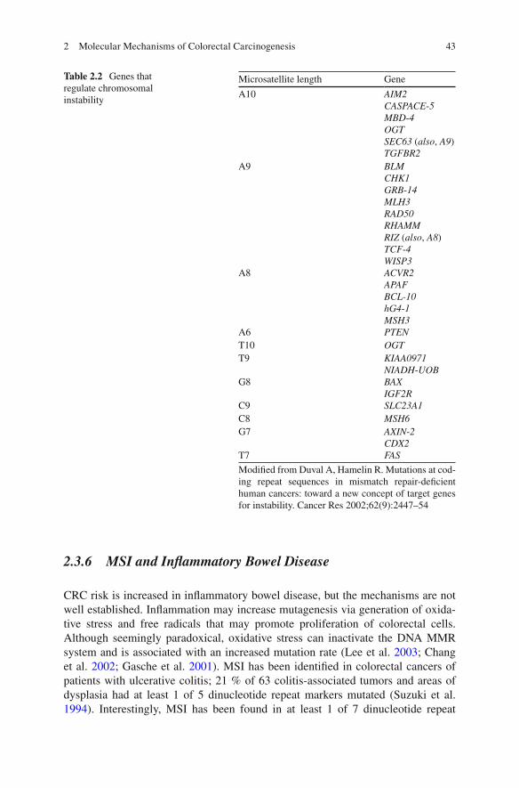
Table 2.2 Genes that regulate chromosomal instability
Microsatellite length Gene
A10 AIM2 CASPACE - 5 MBD - 4 OGT SEC63 ( also , A9 ) TGFBR2
A9 BLM CHK1 GRB - 14 MLH3 RAD50 RHAMM RIZ ( also , A8 ) TCF - 4 WISP3
A8 ACVR2 APAF BCL - 10 hG4 - 1 MSH3
A6 PTEN T10 OGT T9 KIAA0971
NIADH - UOB G8 BAX
IGF2R C9 SLC23A1 C8 MSH6 G7 AXIN - 2
CDX2 T7 FAS
Modifi ed from Duval A, Hamelin R. Mutations at cod-ing repeat sequences in mismatch repair-defi cient human cancers: toward a new concept of target genes for instability. Cancer Res 2002;62(9):2447–54
2 Molecular Mechanisms of Colorectal Carcinogenesis

44
Fig. 2.4 Molecular pathways to MSI-associated colorectal cancer (CRC). Approximately 25 % of MSI-associated CRC arises from the Lynch syndrome, in which inactivating germline mutations in MMR genes are inherited in an autosomal dominant pattern. Additional “second hits” to the wild- type copy of the gene (inherited from the unaffected parent) in the form of somatic mutations, dele-tions, and methylation lead to MSI. 75 % of MSI-associated CRC is sporadic. These cancers are associated with CIMP and undergo MMR gene inactivation via hypermethylation of the MLH1 promoter. Both Lynch syndrome and sporadic CIMP positive-associated defects in MMR lead to MSI and rapid accumulation of somatic mutations in genes with coding “microsatellite repeats.” Many of these microsatellite repeats may not contribute to carcinogenesis but provide a signature that can be used for identifi cation of MSI. Lynch-associated CRCs often harbor KRAS mutations, whereas sporadic CIMP-associated tumors are often BRAF mutant. Modifi ed from Boland CR, Goel A (2010) Microsatellite instability in colorectal cancer. Gastroenterology 138(6):2073–2087.e3
J. Roper and K.E. Hung
45
markers in 50 % of nonneoplastic tissue in patients with chronic ulcerative colitis, but not in controls with acute infectious colitis (Brentnall et al. 1996 ).
2.3.7 Clinical Diagnosis of MSI
The defi nition of MSI was standardized at an international consensus meeting in 1997. The term “MSI” refers to MSI-high, in which >30 % of a defi ned microsatel-lite marker panel is mutated. Those CRCs in which at least 1, but <30 %, of the markers are mutated are called MSI-low and have clinical features of MSS tumors (Boland et al. 1998 ). Another type of MSI has been recognized, called “elevated microsatellite alterations at selected tetranucleotide repeats” (EMAST). EMAST is largely found in noncolonic tumors such as lung, is associated with TP53 mutations, and is not caused by inactivation of the MMR system (Ahrendt et al. 2000 ).
MSI testing is used clinically to identify patients with Lynch syndrome, which comprises 2–3 % of all CRCs. MSI identifi es MMR-defi cient colorectal tumors with 93 % sensitivity, whereas the sensitivity and specifi city of immunohistochemi-cal analysis of MLH1 and MSH2 is 92.3 % and 100 %, respectively (Shia 2008 ). The sensitivity of IHC improves with expression of MSH6 and PMS2 are included in the analysis. Staining of tumors for MMR proteins can be heterogeneous, which may limit sensitivity (Shia 2008 ; Zhang 2008 ). MSI-H tumors can also be distin-guished from MSS tumors by the presence of tumor-infi ltrating cytotoxic lympho-cytes on histologic examination, the degree of which independently confers improved survival (Ogino et al. 2009c ; Phillips et al. 2004 ).
2.3.8 MSI and Response to Chemotherapy
The MMR phenotype is associated with resistance to cytotoxic agents in human CRC cell lines such as HCT-116 (Bhattacharyya et al. 1994 ). Stable restoration of MMR activity in cell lines increases sensitivity to alkylating agents, 6-thioguanine, 5-fl uorouracil, and platinum compounds (Mäkinen et al. 2001 ; Samowitz et al. 2005b ; Chan et al. 2002 ; Wynter et al. 2004 ; O’Brien et al. 2004 ; Minoo et al. 2006 ). With the exception of one study with potential methodological fl aws (Elsaleh et al. 2000 ), multiple studies, including two meta-analyses, have shown no benefi t for chemotherapy among patients with MSI-associated colorectal tumors (de Vos tot Nederveen Cappel et al. 2004 ; Ribic et al. 2003 ; Storojeva et al. 2005 ; Benatti et al. 2005 ; Popat et al. 2005 ; Lanza et al. 2006 ; Jover et al. 2006 ; Kim et al. 2007 ; Des Guetz et al. 2010 ). The largest such study, a prospective, multicenter, randomized controlled trial, a threefold increased mortality was found in Stage II CRC patients with MSI-associated tumors compared to without (Ribic et al. 2003 ). However, MSI is associated with improved response to regimens containing a topoisomerase I inhibitor, irinotecan (Bertagnolli et al. 2009 ; Fallik et al. 2003 ).
2 Molecular Mechanisms of Colorectal Carcinogenesis
46
2.4 The Serrated Pathway in Colorectal Cancer Pathogenesis
Colorectal polyps have traditionally been classifi ed as either hyperplastic or adeno-matous, with only the latter progressing into carcinoma. However, beginning in the late 1980s, an increasing number of reports suggested that CRC can arise from hyperplastic polyps in the setting of what is now known as the hyperplastic polypo-sis syndrome (HPS), in which a large number of hyperplastic polyps are found throughout the colon (these polyps are distinguished from typical hyperplastic pol-yps, which are small, left sided, and benign) (Samowitz et al. 2006 ; Ji et al. 2006 ; Pérez et al. 2010 ; Shrubsole et al. 2008 ; Chirieac et al. 2005 ; Ogino et al. 2006b ; Ward et al. 2003 ; Glazer et al. 2008 ). These studies identifi ed a 35 % risk of CRC in patients with HPS, as well as increased risk of synchronous cancers (Boparai et al. 2010 ). Polyps in patients with HPS are characterized by gland serrations, which led pathologists to reexamine the malignant potential of other polyps with histologic serrations. Data from screening colonoscopy cohorts have demonstrated that ser-rated polyps are strongly associated with the development of synchronous and metachronous advanced adenoma and CRC (Li et al. 2009 ; Schreiner et al. 2010 ).
2.4.1 Classifi cation of Serrated Polyps
Serrated polyps are characterized by a “sawtooth” pattern, or serrations, in the colonic crypts. In 1990, Longacre and Fenoglio-Presiser proposed the term “serrated adenoma” for polyps exhibiting features of both adenomatous and hyperplastic pol-yps (Longacre and Fenoglio-Preiser 1990 ). In 1996, Torlakovic and Snover fi rst showed that polyps in HPS have serrated features similar to serrated adenomas, though with less atypia, and were more likely to be sessile than standard hyperplas-tic polyps (Torlakovic and Snover 1996 ). Further detailed work identifi ed a subset of serrated polyps with abnormal proliferation, crypt distortion, and dilation that were typically sessile and found on the right side of the colon. These polyps were distin-guished from traditional serrated adenomas (TSAs), which more closely resembled conventional adenomas (Torlakovic et al. 2003 ). These fi ndings eventually led to a proposal for a new nomenclature for serrated polyps in 2005 (Snover et al. 2005 ).
The use of the term “adenoma” to describe sessile lesions has been controversial because conventional adenomas are dysplastic, whereas SSAs lack cytological dys-plasia, though they manifest disordered proliferation and crypt architecture. Robert Odze and colleagues have thus opted for the term “sessile serrated polyp” in a recent pathology textbook (Hornick and Odze 2009 ), whereas a recent European publica-tion chose the term “sessile serrated lesion.” (Lambert et al. 2009 ) As the term SSA has grown in research and clinical practice, we will use it in this chapter.
J. Roper and K.E. Hung

47
2.4.1.1 Hyperplastic Polyp
Hyperplastic polyps (HPs) have a narrow crypt base lined with proliferative cells and serrations in the upper third of the gland. HPs have been subdivided into goblet cell-rich type, microvesicular type (which are precursers to SSAs), and the rare mucin-poor variant. Overall, HPs are highly prevalent sessile lesions that are com-monly located in the distal colon and rectum (Tedesco et al. 1982 ; Imperiale et al. 2002 ). Endoscopically, HPs are identifi ed by their smooth, symmetrical, and pale appearance. Microvesicular type HPs are precursor lesions to SSAs and, like SSAs, harbor BRAFV 600E mutations. Goblet cell HPs, on the other hand, often contain KRAS mutations (43 % in one study), which are mutually exclusive of BRAF muta-tions (O’Brien et al. 2006 ). Large goblet cell HPs may progress into KRAS mutant dysplastic serrated polyps (Boparai et al. 2008 ).
2.4.1.2 Sessile Serrated Polyp
Sessile serrated adenomas (SSAs) are characterized by crypt architectural altera-tions that refl ect disordered growth. These include serration of the epithelium, often at the base of the crypts; dilation of the base of the crypts; and T- or L-shaped crypts. SSAs may contain areas of cytologic dysplasia and adenocarcinoma; tumors with neoplastic progression tend to lose serrated features (Fujita et al. 2011 ). SSAs likely evolve from preexisting microvesicular type HPs (Spring et al. 2006 ). Endoscopically, SSAs are usually larger than 5 mm, fl at or sessile (height one half or less than width), and often mucous covered (Jaramillo et al. 2005 ). They are generally larger than HPs and located in the proximal colon. The surface is often smooth, and the edges are poorly defi ned and irregular. These features make SSAs diffi cult to detect endoscopically (Higuchi et al. 2005 ).
2.4.1.3 Dysplastic Serrated Polyps
Dysplastic serrated polyps contain gland serrations and cytologic dysplasia. There are two categories of dysplastic serrated polyp (1) SSA with dysplasia, which exhibits SSA morphologic characteristics contiguous to an area of conventional dysplasia and (2) TSA, which has not only serrations but also dysplastic epithelial cells and ectopic crypts with bases not adjacent to the muscularis mucosa, in con-trast to SSAs in which new crypts are generally anchored to the muscularis mucosa. TSAs differ from SSAs in that they are typically distally located, polypoid, contain tubulovillous architecture, and marked cytoplasmic eosinophilia (O’Brien 2007 ; Torlakovic et al. 2008 ). TSAs not only are frequently KRAS mutant (which heralds an aggressive phenotype) but may also be KRAS / BRAF wild-type or BRAF mutant (Kim et al. 2010b ).
2 Molecular Mechanisms of Colorectal Carcinogenesis

48
2.4.2 Epidemiology
It is estimated that up to 20 % of CRCs arise from the serrated pathway, or nearly 30,000 cases annually (Jass 2007 ). One study reported a prevalence of 29 % HPs, 9 % SSAs, 1.7 % mixed polyps, and 0.7 % TSAs from a cohort of colonoscopy- resected specimens (Spring et al. 2006 ). Dysplastic serrated polyps are much less common than conventional polyps or HPs, representing 1–2 % of all polyps (Higuchi et al. 2005 ; Jass et al. 2006 ).
The molecular basis of the serration of the crypt epithelium has not been deter-mined, though it has been proposed that serrations occur due to cell crowding or because of failure of apoptosis or anoikis (Tateyama et al. 2002 ). Crypt serration is strongly associated with the presence of BRAF mutation; hyperplastic polyps with KRAS rather than BRAF mutation have less, or absence of, gland serration.
2.4.3 Serrated Polyps, MSI, CIMP, and BRAF
Tumors associated with HPS have a higher than expected incidence of MSI (Leggett and Whitehall 2010 ; Jeevaratnam et al. 1996 ; Rashid et al. 2000 ; Jass et al. 2000 ). Serrated polyps from colectomy specimens were more likely to have MSI than MSS, and another study found that MSI was more common in serrated adenomas than in control tumors (37.5 % vs. 11 %, respectively) (Hawkins and Ward 2001 ; Mäkinen et al. 2001 ). O’Brien et al. found MSI only in the areas of advanced SSAs with carcinoma, which suggests that MSI develops late in the serrated pathway. Epigenetic silencing of MLH1 is the underlying cause of MSI in serrated lesions and is an important driver of the progression to invasive cancer (O’Brien et al. 2006 ). A large proportion serrated cancers are MSS and frequently have TP53 mutation, which may explain their more aggressive phenotype and poorer prognosis than MSI-associated tumors [hazard rate ratio (HRR), 2.97; 95 % CI, 2.05–4.32] (Samowitz et al. 2005b ).
CIMP is commonly observed in both HPs and in proximal SSA (Chan et al. 2002 ; Wynter et al. 2004 ; O’Brien et al. 2004 ). Yang et al. detected CIMP in microvesicular HP (47 %), SSA (75 %), and TSA (80 %). Using a narrower defi ni-tion of methylation (≥4/5 markers), CIMP was detected in 11 % of MVHP, com-pared to 40 % of SSA 284 . CIMP has even been detected in histologically normal colonic mucosa of HPS patients (Minoo et al. 2006 ). Higher CIMP levels (four or more markers positive) were more frequently found in SSAs (with or without carci-noma) than in conventional adenomas or carcinomas (O’Brien et al. 2006 ). Together, these data indicate that methylation of specifi c CIMP loci may facilitate the transi-tion from microvesicular HP to SSA.
In a systematic genome-wide screen for genes affecting cell proliferation and death, activating mutations in BRAF were identifi ed in a high proportion of melano-mas and in a small fraction of other cancers including colon. BRAF is a serine/
J. Roper and K.E. Hung

49
threonine kinase that is part of the mitogen-activated protein kinase (MAPK) cell signaling pathway; mutations in BRAF result in constitutive activation of the MAPK pathway and transcription of genes promoting cell growth and proliferation (Davies et al. 2002 ). Rajagopalan et al. sequenced BRAF and KRAS mutations in colorectal tumors and found that (1) 10 % of tumors harbored somatic mutations in BRAF and (2) no tumors exhibited mutations in both BRAF and KRAS (Rajagopalan et al. 2002 ). Another group confi rmed these fi ndings (Yuen et al. 2002 ). Chan et al. exam-ined BRAF and KRAS mutations in a series of serrated polyps and found BRAF muta-tions in 36 % of HPs and 100 % of SSAs. The BRAF V600E substitution is the most common BRAF mutation in human cancers including serrated CRCs (Davies et al. 2002 ). Using current histologic defi nitions, 70–76 % of MVHPs and 75–83 % of SSAs have BRAF V600E mutations. BRAF and KRAS mutations are mutually exclusive (O’Brien et al. 2006 ). The BRAF V600E mutation was found in 5 % of a cohort of MSS tumors and 52 % of MSI-associated tumors (Samowitz et al. 2005b ). However, his-tological reviews have confi rmed that BRAF is almost never mutated in conventional adenomas or in Lynch syndrome, highlighting the association of BRAF mutation with the serrated pathway rather than MSI (O’Brien et al. 2006 ; Kambara et al. 2004 ; Wang et al. 2003 ). Mutation of BRAF strongly correlates with CIMP (Weisenberger et al. 2006 ). These fi ndings support the role of CIMP and the MAPK pathway via activating mutation in BRAF or KRAS in the serrated adenoma pathway.
2.4.4 Initiation and Progression of the Serrated Pathway
Activation of BRAF in normal melanocyte epithelium and in mouse gastrointestinal epithelium results in an initial burst of proliferation followed by cell senescence (Carragher et al. 2010 ; Campisi 2005 ). Methylation-induced silencing of p16INK4a is an early event in the serrated pathway and may be suffi cient to allow colorectal cells (and possible microvesicular HPs) to escape BRAF-induced senescence (Chen et al. 2005 ). In melanocytes, activated BRAF is suffi cient for synthesis and secre-tion of insulin-like growth factor binding protein 7 (IGFBP7) which in turn inhibits MAPK signaling and induces senescence and apoptosis (Wajapeyee et al. 2008 ). The large columnar vacuolated cells of the upper crypts of the microvesicular HP and SSA are a manifestation of cell senescence (Minoo and Jass 2006 ). Silencing via methylation of IGFBP7 in BRAF -mutant, CIMP-positive CRC cells permits unrestrained cell proliferation and progression to SSA by enabling escape from p53-induced senescence (Suzuki et al. 2010 ). Therefore, the additive tumorigenic effects of mutated BRAF and CIMP may result from silencing of tumor suppressor genes such as p16INK4a and IGFBP7 via hypermethylation.
The Wnt signaling pathway is another major regulator of cellular proliferation in CRC. In the absence of APC protein, β-catenin accumulates in the cell nucleus instead of undergoing degradation. Three studies found positive nuclear β-catenin immunostaining in 0–50 % of HPs and 38–67 % of SSAs, 36 % of TSAs, and 100 % of tubular adenomas (TA). 29 % of SSAs without dysplasia and all SSAs with
2 Molecular Mechanisms of Colorectal Carcinogenesis

50
dysplasia displayed aberrant β-catenin staining. Nuclear β-catenin was identifi ed only in the setting of BRAF V600E mutation (Wu et al. 2008 ; Yachida et al. 2009 ; Sandmeier et al. 2009 ). A recent histological study found that nuclear β-catenin staining in SSA was limited to dysplastic areas of the polyps, and histologically these dysplastic areas lost serrated features and become more tubulovillous (Fujita et al. 2011 ). Unlike conventional adenomas, however, APC mutation is found in only a minority (19 %) of serrated polyps, and β-catenin gain-of-function mutation in CTNNB1 has not been identifi ed in serrated polyps (Yachida et al. 2009 ; de Vogel et al. 2009 ). A mouse model for BRAF V600E CRC demonstrated that expression of BRAF V600E in intestinal crypts was suffi cient for β-catenin nuclear localization via MAPK-dependent, Akt-independent phosphorylation of Gsk3β (Carragher et al. 2010 ). However, this mechanism of Wnt activation has not been confi rmed in the human serrated pathway. These fi ndings suggest that activation of the Wnt signaling pathway follows BRAF mutation and plays an important role in the progression (but not initiation) of the serrated pathway. The molecular steps in the initiation and progression of sessile serrated adenomas are summarized in Fig. 2.5 .
2.4.5 An Alternate Serrated Pathway
Recognition of the heterogeneity of serrated polyps has led to the hypothesis that there are two parallel serrated pathways to colorectal carcinogenesis: one driven by BRAF mutation and the other driven by KRAS mutation (O’Brien et al. 2006 ; O’Brien 2007 ; Yang et al. 2004 ). The BRAF pathway has been discussed in detail above. KRAS mutant serrated carcinomas have relatively low levels of CIMP, but it is possi-ble that rather than being a true CIMP-low group, these cancers are methylated at different loci (Weisenberger et al. 2006 ). Silencing of the DNA repair gene MGMT by promoter hypermethylation has been associated with KRAS mutation and CIMP-low status (Ogino et al. 2006a , 2007 ; Whitehall et al. 2001 ). However, no specifi c panel of markers has been validated to study this pathway. No precursor lesion to KRAS mutant serrated carcinomas has been identifi ed, though it has been proposed that large goblet cell HPs, tubulovillous adenomas, and/or serrated polyps with dysplasia may be relevant to the “alternate pathway” (Boparai et al. 2008 ; Jass et al. 2006 ).
2.4.6 Risk Factors for Serrated CRC
Susceptibility to serrated neoplasia may be associated with a genetic predisposition to hypermethylation of gene promoters. Rare families with multiple members affected by HPS have been described (Jeevaratnam et al. 1996 ; Rashid et al. 2000 ; Chow et al. 2006 ). Most cases of CIMP-high, BRAF mutant serrated polyps appear to be sporadic, although a few families with high incidences of CRC and serrated polyps have been identifi ed (Des Guetz et al. 2010 ) 306,307 . However, residents of
J. Roper and K.E. Hung

51
Fig. 2.5 The sessile serrated pathway. Activation of BRAF induces the formation of ACF with serrated features and microvesicular hyperplastic polyp (MVHP). Further cell proliferation is con-trolled by cell senescence, which is mediated by p16INK4a expression and IGFBP7 secretion. Methylation-induced silencing of p16INK4a or loss p53 function allows early polyps to escape the senescent state and develop into sessile serrated adenomas. Endoscopic images courtesy of Moises Guelrud, Tufts Medical Center; pathology images courtesy of Barbara Weinstein, Tufts Medical Center. Modifi ed from Leggett B, Whitehall V (2010) Role of the serrated pathway in CRC patho-genesis. Gastroenterology 138(6):2088–2100
2 Molecular Mechanisms of Colorectal Carcinogenesis

52
Melbourne, Australia of Anglo-Celtic origin were found to have a higher risk of CIMP and BRAF mutant CRC compared to those of southern European origin, and serrated polyps were more frequent in Caucasians compared to Hispanics and African Americans (English et al. 2008 ; Wallace et al. 2009 ). Cigarette smoking has been strongly associated with CIMP and BRAF mutation and is a stronger risk fac-tor for HPs than adenomatous polyps in multiple studies, although one report found no association between smoking and HPs (Samowitz et al. 2006 ; Ji et al. 2006 ; Pérez et al. 2010 ; Shrubsole et al. 2008 ). Aspirin is protective against serrated pol-yps, as with conventional polyps (Wallace et al. 2009 ). A study on risk factors for CRC found that obesity, smoking, dietary fat, caloric intake, and red meat intake were associated with increased risk for distal, but not proximal, serrated polyps (Wallace et al. 2009 ).
2.4.7 Clinical Characteristics of Serrated CRC
The presence or absence of BRAF mutation does not affect the excellent prognosis of MSI-associated CRC (Samowitz et al. 2005b ). Cancers that arise via the serrated pathway, whether with or without MSI, tend to be proximal, mucinous, occur in older individuals, and present at more advanced stage (Samowitz et al. 2005a ; Hawkins et al. 2002 ; Chirieac et al. 2005 ; Ogino et al. 2006b ). In the context of MSS, increased DNA methylation and BRAF mutation is associated with worse prognosis (Weisenberger et al. 2006 ; Ward et al. 2003 ). Serrated polyps are strongly associated with synchronous advanced neoplasia (defi ned as invasive carcinoma, tubular adenoma 1 cm, or adenoma with any villous histology or high-grade dyspla-sia), particularly proximal CRCs, in large colonoscopy cohort studies (Glazer et al. 2008 ; Hiraoka et al. 2010 ; Li et al. 2009 ; Schreiner et al. 2010 ).
HPS is an uncommon condition characterized by multiple and/or large HPs. Several reports of CRC in patients with HPS led to the hypothesis that serrated pol-yps may develop into CRC (Jeevaratnam et al. 1996 ). The incidence of CRC in HPS is estimated to be 40–50 % (Buchanan et al. 2010 ; Leggett et al. 2001 ; Rubio et al. 2006 ). Type I HPS is defi ned multiple (fi ve or more), large, proximally located SSAs. There is a high frequency of CIMP and mutated BRAF. Type II HPS, a more heterogeneous condition, describes the fi nding of numerous (≥30) small HPS dis-tributed throughout the colon, and is believed to have a lower risk of CRC than type I HPS (Ferrández et al. 2004 ). Although the syndrome has no proven genetic basis, there are reports of familial HPS and ethnic associations in population studies (Young and Jass 2006 ; Young et al. 2007 ).
2.4.8 Detection and Surveillance of Serrated Polyps
Detection of serrated polyps via currently available screening modalities may be dif-fi cult. Serrated polyps are less likely to bleed, and hence may not be detected by
J. Roper and K.E. Hung

53
fecal occult blood testing (East et al. 2008 ). CT Colonography may be less likely to detect fl at or sessile lesions, though this has not been studied. Colonoscopy performs relatively poorly in the detection of serrated polyps, which may partly explain fi nd-ings that morality rates from left-sided CRC, but not right-sided CRC, have decreased in recent years (Baxter et al. 2009 ; Brenner et al. 2010 ). This may be due to poor colonic prep on the right side of the colon and/or poor visualization of fl at, mucous-covered lesions. Randomized trials have demonstrated that chromoendoscopy improves detection of serrated polyps by twofold. The importance of detection and removal of serrated polyps is highlighted by the fi ndings that interval cancers (found despite appropriate screening or surveillance colonoscopy) were four times as likely to be associated with MSI (Sawhney et al. 2006 ) and CIMP, and more likely to be proximal and mucinous, which are all features suggestive of BRAF mutation (Leggett et al. 1997 ; Farrar et al. 2006 ; Bressler et al. 2004 ; Arain et al. 2010 ).
2.4.9 Models of the Serrated Pathway
Isogenic BRAF V600E human CRC cell lines (VACO432 and RKO) have been devel-oped in which either the endogenous wild-type or mutant allele has been inactivated through targeted homologous recombination (Yun et al. 2009 ). Carragher et al. pub-lished a Cre–lox-regulated knockin mouse in which Braf V600E is expressed from the endogenous Braf gene in the proliferative cells of the intestinal crypts. They showed that intestinal Braf V600E is not only suffi cient for formation of hyperplastic crypts via activation of the MAPK and Wnt pathways but also induces cell senescence, and that inactivation of p16INK4a through DNA methylation is necessary for tumor progression. However, polyps in this model are adenomas, not carcinomas, and are confi ned to the small bowel (Carragher et al. 2010 ). Kenneth Hung and colleagues developed a novel genetically engineered mouse model in which mice with a condi-tional Apc allele were crossed with those with a latent Braf V600E allele. They showed that combination treatment with BRAF and PI3K/mTOR inhibitors was required to induce apoptosis and tumor regression. This model offers several advantages for preclinical drug testing (1) solitary tumors develop rapidly along a reproducible time line in the colon; (2) tumors can be continuously monitored throughout drug treatment via colonoscopy; and (3) tumors recapitulate the serrated pathway seen in humans, including HPs, SSAs, SSAs with dysplasia, and SSAs with congruent inva-sive adenocarcinoma (Coffee et al., manuscript under review).
2.5 Conclusions
CRC continues to be a signifi cant public health burden. Whereas there have been signifi cant advances in the development of targeted therapies, the 5-year prognosis for metastatic CRC still continues to be less than 10 %. However, our increased understanding of the molecular events underlying CRC carcinogenesis will enable
2 Molecular Mechanisms of Colorectal Carcinogenesis

54
the development of new targeted therapies and the identifi cation of clinical biomark-ers that will inform their effective usage. This is an exciting time for cancer medicine and we believe that the fi eld is poised to make signifi cant therapeutic breakthroughs.
References
Ahrendt SA, Decker PA, Doffek K et al (2000) Microsatellite instability at selected tetranucleotide repeats is associated with p53 mutations in non-small cell lung cancer. Cancer Res 60(9):2488–2491
Akhoondi S, Sun D, von der Lehr N et al (2007) FBXW7/hCDC4 is a general tumor suppressor in human cancer. Cancer Res 67(19):9006–9012
Albertini RJ, Nicklas JA, O’Neill JP, Robison SH (1990) In vivo somatic mutations in humans: measurement and analysis. Annu Rev Genet 24:305–326
Aoki K, Aoki M, Sugai M et al (2007) Chromosomal instability by β-catenin/TCF transcription in APC or β-catenin mutant cells. Oncogene 26(24):3511–3520
Arain MA, Sawhney M, Sheikh S et al (2010) CIMP status of interval colon cancers: another piece to the puzzle. Am J Gastroenterol 105(5):1189–1195
Arber N, Eagle CJ, Spicak J et al (2006) Celecoxib for the prevention of colorectal adenomatous polyps. N Engl J Med 355(9):885–895
Barbie DA, Tamayo P, Boehm JS et al (2009) Systematic RNA interference reveals that oncogenic KRAS-driven cancers require TBK1. Nature 462(7269):108–112
Bardelli A, Cahill DP, Lederer G et al (2001) Carcinogen-specifi c induction of genetic instability. Proc Natl Acad Sci U S A 98(10):5770–5775
Bardi G, Parada LA, Bomme L et al (1997) Cytogenetic comparisons of synchronous carcinomas and polyps in patients with colorectal cancer. Br J Cancer 76(6):765–769
Baron JA, Sandler RS, Bresalier RS et al (2006) A randomized trial of rofecoxib for the chemopre-vention of colorectal adenomas. Gastroenterology 131(6):1674–1682
Bassing CH, Suh H, Ferguson DO et al (2003) Histone H2AX: a dosage-dependent suppressor of oncogenic translocations and tumors. Cell 114(3):359–370
Baxter NN, Goldwasser MA, Paszat LF, Saskin R, Urbach DR, Rabeneck L (2009) Association of colonoscopy and death from colorectal cancer. Ann Intern Med 150(1):1–8
Benatti P, Gafà R, Barana D et al (2005) Microsatellite instability and colorectal cancer prognosis. Clin Cancer Res 11(23):8332–8340
Bertagnolli MM, Eagle CJ, Zauber AG et al (2006) Celecoxib for the prevention of sporadic colorectal adenomas. N Engl J Med 355(9):873–884
Bertagnolli MM, Niedzwiecki D, Compton CC et al (2009) Microsatellite instability predicts improved response to adjuvant therapy with irinotecan, fl uorouracil, and leucovorin in stage III colon cancer: Cancer and Leukemia Group B Protocol 89803. J Clin Oncol 27(11):1814–1821
Bhattacharyya NP, Skandalis A, Ganesh A, Groden J, Meuth M (1994) Mutator phenotypes in human colorectal carcinoma cell lines. Proc Natl Acad Sci U S A 91(14):6319–6323
Bird AP (1986) CpG-rich islands and the function of DNA methylation. Nature 321(6067):209–213 Blake C, Tsao J-L, Wu A, Shibata D (2001) Stepwise deletions of PolyA sequences in mismatch
repair-defi cient colorectal cancers. Am J Pathol 158(5):1867–1870 Boland CR, Goel A (2010) Microsatellite instability in colorectal cancer. Gastroenterology
138(6):2073–2087.e3 Boland CR, Thibodeau SN, Hamilton SR et al (1998) A National Cancer Institute workshop on
microsatellite instability for cancer detection and familial predisposition: development of inter-national criteria for the determination of microsatellite instability in colorectal cancer. Cancer Res 58(22):5248–5257
Bomme L, Bardi G, Pandis N, Fenger C, Kronborg O, Heim S (1994) Clonal karyotypic abnor-malities in colorectal adenomas: clues to the early genetic events in the adenoma-carcinoma sequence. Genes Chromosomes Cancer 10(3):190–196
J. Roper and K.E. Hung
55
Bondi J, Husdal A, Bukholm G, Nesland JM, Bakka A, Bukholm IRK (2005) Expression and gene amplifi cation of primary (A, B1, D1, D3, and E) and secondary (C and H) cyclins in colon adenocarcinomas and correlation with patient outcome. J Clin Pathol 58(5):509–514
Boparai KS, Dekker E, Van Eeden S et al (2008) Hyperplastic polyps and sessile serrated adeno-mas as a phenotypic expression of MYH-associated polyposis. Gastroenterology 135(6):2014–2018
Boparai KS, Mathus-Vliegen EMH, Koornstra JJ et al (2010) Increased colorectal cancer risk dur-ing follow-up in patients with hyperplastic polyposis syndrome: a multicentre cohort study. Gut 59(8):1094–1100
Boveri T (2008) Concerning the origin of malignant tumours by Theodor Boveri. Translated and annotated by Henry Harris. J Cell Sci 121(Suppl 1):1–84
Brenner H, Hoffmeister M, Arndt V, Stegmaier C, Altenhofen L, Haug U (2010) Protection from right- and left-sided colorectal neoplasms after colonoscopy: population-based study. J Natl Cancer Inst 102(2):89–95
Brentnall TA, Crispin DA, Bronner MP et al (1996) Microsatellite instability in nonneoplastic mucosa from patients with chronic ulcerative colitis. Cancer Res 56(6):1237–1240
Bresalier RS, Sandler RS, Quan H et al (2005) Cardiovascular events associated with rofecoxib in a colorectal adenoma chemoprevention trial. N Engl J Med 352(11):1092–1102
Bressler B, Paszat LF, Vinden C, Li C, He J, Rabeneck L (2004) Colonoscopic miss rates for right- sided colon cancer: a population-based analysis. Gastroenterology 127(2):452–456
Buchanan DD, Sweet K, Drini M et al (2010) Phenotypic diversity in patients with multiple ser-rated polyps: a genetics clinic study. Int J Colorectal Dis 25(6):703–712
Campisi J (2005) Suppressing cancer: the importance of being senescent. Science 309(5736):886–887
Camps J, Nguyen QT, Padilla-Nash HM et al (2009) Integrative genomics reveals mechanisms of copy number alterations responsible for transcriptional deregulation in colorectal cancer. Genes Chromosomes Cancer 48(11):1002–1017
Cardoso J, Molenaar L, De Menezes RX et al (2006) Chromosomal instability in MYH- and APC- mutant adenomatous polyps. Cancer Res 66(5):2514–2519
Carragher LAS, Snell KR, Giblett SM et al (2010) V600EBraf induces gastrointestinal crypt senescence and promotes tumour progression through enhanced CpG methylation of p16INK4a. EMBO Mol Med 2(11):458–471
Castets M, Broutier L, Molin Y et al (2011) DCC constrains tumour progression via its depen-dence receptor activity. Nature 482(7386):534–537, Available from: http://www.ncbi.nlm.nih.gov/pubmed/22158121
Castillo A, Morse HC III, Godfrey VL, Naeem R, Justice MJ (2007) Overexpression of Eg5 causes genomic instability and tumor formation in mice. Cancer Res 67(21):10138–10147
Celeste A, Difi lippantonio S, Difi lippantonio MJ et al (2003) H2AX haploinsuffi ciency modifi es genomic stability and tumor susceptibility. Cell 114(3):371–383
Chadeneau C, Hay K, Hirte HW, Gallinger S, Bacchetti S (1995) Telomerase activity associated with acquisition of malignancy in human colorectal cancer. Cancer Res 55(12):2533–2536
Chalhoub N, Baker SJ (2009) PTEN and the PI3-kinase pathway in cancer. Annu Rev Pathol 4:127–150
Chan AT, Giovannucci EL (2010) Primary prevention of colorectal cancer. Gastroenterology 138(6):2029–2043.e10
Chan AO-O, Issa J-PJ, Morris JS, Hamilton SR, Rashid A (2002) Concordant CpG island methyla-tion in hyperplastic polyposis. Am J Pathol 160(2):529–536
Chang CL, Marra G, Chauhan DP et al (2002) Oxidative stress inactivates the human DNA mis-match repair system. Am J Physiol Cell Physiol 283(1):C148–C154
Chen Z, Trotman LC, Shaffer D et al (2005) Crucial role of p53-dependent cellular senescence in suppression of Pten-defi cient tumorigenesis. Nature 436(7051):725–730
Chirieac LR, Shen L, Catalano PJ, Issa J-P, Hamilton SR (2005) Phenotype of microsatellite-stable colorectal carcinomas with CpG island methylation. Am J Surg Pathol 29(4):429–436
Chow E, Lipton L, Lynch E et al (2006) Hyperplastic polyposis syndrome: phenotypic presenta-tions and the role of MBD4 and MYH. Gastroenterology 131(1):30–39
2 Molecular Mechanisms of Colorectal Carcinogenesis

56
Cianciulli A, Cosimelli M, Marzano R et al (2004) Genetic and pathologic signifi cance of 1p, 17p, and 18q aneusomy and the ERBB2 gene in colorectal cancer and related normal colonic mucosa. Cancer Genet Cytogenet 151(1):52–59
Cole BF, Logan RF, Halabi S et al (2009) Aspirin for the chemoprevention of colorectal adenomas: meta-analysis of the randomized trials. J Natl Cancer Inst 101(4):256–266
Cottrell S, Bicknell D, Kaklamanis L, Bodmer WF (1992) Molecular analysis of APC mutations in familial adenomatous polyposis and sporadic colon carcinomas. Lancet 340(8820):626–630
Cuilliere-Dartigues P, El-Bchiri J, Krimi A et al (2006) TCF-4 isoforms absent in TCF-4 mutated MSI-H colorectal cancer cells colocalize with nuclear CtBP and repress TCF-4-mediated tran-scription. Oncogene 25(32):4441–4448
Curfman GD, Morrissey S, Drazen JM (2005) Expression of concern: Bombardier et al., “Comparison of upper gastrointestinal toxicity of rofecoxib and naproxen in patients with rheu-matoid arthritis”, N Engl J Med 2000;343:1520–8. N Engl J Med 353(26):2813–2814
Davies H, Bignell GR, Cox C et al (2002) Mutations of the BRAF gene in human cancer. Nature 417(6892):949–954
de Vogel S, Weijenberg MP, Herman JG et al (2009) MGMT and MLH1 promoter methylation versus APC, KRAS and BRAF gene mutations in colorectal cancer: indications for distinct pathways and sequence of events. Ann Oncol 20(7):1216–1222
de Vos tot Nederveen Cappel WH, Meulenbeld HJ, Kleibeuker JH et al (2004) Survival after adju-vant 5-FU treatment for stage III colon cancer in hereditary nonpolyposis colorectal cancer. Int J Cancer 109(3):468–471
Des Guetz G, Lecaille C, Mariani P et al (2010) Prognostic impact of microsatellite instability in colorectal cancer patients treated with adjuvant FOLFOX. Anticancer Res 30(10):4297–4301
Di Vinci A, Infusini E, Peveri C, Risio M, Rossini FP, Giaretti W (1996) Deletions at chromosome 1p by fl uorescence in situ hybridization are an early event in human colorectal tumorigenesis. Gastroenterology 111(1):102–107
Diep CB, Thorstensen L, Meling GI, Skovlund E, Rognum TO, Lothe RA (2003) Genetic tumor markers with prognostic impact in Dukes’ stages B and C colorectal cancer patients. J Clin Oncol 21(5):820–829
Dotan E, Meropol NJ, Zhu F et al (2012) Relationship of increased aurora kinase A gene copy number, prognosis and response to chemotherapy in patients with metastatic colorectal cancer. Br J Cancer 106(4):748–755, Available from: http://www.ncbi.nlm.nih.gov/pubmed/22240781
Duesberg P, Fabarius A, Hehlmann R (2004) Aneuploidy, the primary cause of the multilateral genomic instability of neoplastic and preneoplastic cells. IUBMB Life 56(2):65–81
East JE, Saunders BP, Jass JR (2008) Sporadic and syndromic hyperplastic polyps and serrated adenomas of the colon: classifi cation, molecular genetics, natural history, and clinical manage-ment. Gastroenterol Clin North Am 37(1):25–46, v
Eberhart CE, Coffey RJ, Radhika A, Giardiello FM, Ferrenbach S, Dubois RN (1994) Up-regulation of cyclooxygenase 2 gene expression in human colorectal adenomas and adenocarcinomas. Gastroenterology 107(4):1183–1188
Eilers M, Eisenman RN (2008) Myc’s broad reach. Genes Dev 22(20):2755–2766 Elsaleh H, Joseph D, Grieu F, Zeps N, Spry N, Iacopetta B (2000) Association of tumour site and
sex with survival benefi t from adjuvant chemotherapy in colorectal cancer. Lancet 355(9217):1745–1750
Engelhardt M, Drullinsky P, Guillem J, Moore MAS (1997) Telomerase and telomere length in the development and progression of premalignant lesions to colorectal cancer. Clin Cancer Res 3(11):1931–1941
English DR, Young JP, Simpson JA et al (2008) Ethnicity and risk for colorectal cancers showing somatic BRAF V600E mutation or CpG island methylator phenotype. Cancer Epidemiol Biomarkers Prev 17(7):1774–1780
Esteller M, Sparks A, Toyota M et al (2000) Analysis of adenomatous polyposis coli promoter hypermethylation in human cancer. Cancer Res 60(16):4366–4371
Fallik D, Borrini F, Boige V et al (2003) Microsatellite instability is a predictive factor of the tumor response to irinotecan in patients with advanced colorectal cancer. Cancer Res 63(18):5738–5744
J. Roper and K.E. Hung

57
Farrar WD, Sawhney MS, Nelson DB, Lederle FA, Bond JH (2006) Colorectal cancers found after a complete colonoscopy. Clin Gastroenterol Hepatol 4(10):1259–1264
Fazeli A, Dickinson SL, Hermiston ML et al (1997) Phenotype of mice lacking functional deleted in colorectal cancer (Dec) gene. Nature 386(6627):796–804
Fearon ER (2011) Molecular genetics of colorectal cancer. Annu Rev Pathol 6:479–507 Fearon ER, Vogelstein B (1990) A genetic model for colorectal tumorigenesis. Cell
61(5):759–767 Fearon ER, Cho KR, Nigro JM et al (1990) Identifi cation of a chromosome 18q gene that is altered
in colorectal cancers. Science 247(4938):49–56 Feinberg AP, Gehrke CW, Kuo KC, Ehrlich M (1988) Reduced genomic 5-methylcytosine content
in human colonic neoplasia. Cancer Res 48(5):1159–1161 Ferrández A, Samowitz W, DiSario JA, Burt RW (2004) Phenotypic characteristics and risk of
cancer development in hyperplastic polyposis: case series and literature review. Am J Gastroenterol 99(10):2012–2018
Firestein R, Hahn WC (2009) Revving the throttle on an oncogene: CDK8 takes the driver seat. Cancer Res 69(20):7899–7901
Firestein R, Bass AJ, Kim SY et al (2008) CDK8 is a colorectal cancer oncogene that regulates [bgr]-catenin activity. Nature 455(7212):547–551
Firestein R, Shima K, Nosho K et al (2010) CDK8 expression in 470 colorectal cancers in relation to β-catenin activation, other molecular alterations and patient survival. Int J Cancer 126(12):2863–2873
Fodde R, Kuipers J, Rosenberg C et al (2001) Mutations in the APC tumour suppressor gene cause chromosomal instability. Nat Cell Biol 3(4):433–438
Fujita K, Yamamoto H, Matsumoto T et al (2011) Sessile serrated adenoma with early neoplastic progression: a clinicopathologic and molecular study. Am J Surg Pathol 35(2):295–304
Ganem NJ, Godinho SA, Pellman D (2009) A mechanism linking extra centrosomes to chromo-somal instability. Nature 460(7252):278–282
Garcia-Albeniz X, Chan AT (2011) Aspirin for the prevention of colorectal cancer. Best Pract Res Clin Gastroenterol 25(4–5):461–472
Gasche C, Chang CL, Rhees J, Goel A, Boland CR (2001) Oxidative stress increases frameshift mutations in human colorectal cancer cells. Cancer Res 61(20):7444–7448
Gertler R, Rosenberg R, Stricker D et al (2004) Telomere length and human telomerase reverse transcriptase expression as markers for progression and prognosis of colorectal carcinoma. J Clin Oncol 22(10):1807–1814
Glazer E, Golla V, Forman R, Zhu H, Levi G, Bodenheimer HC Jr (2008) Serrated adenoma is a risk factor for subsequent adenomatous polyps. Dig Dis Sci 53(8):2204–2207
Goelz SE, Vogelstein B, Hamilton SR, Feinberg AP (1985) Hypomethylation of DNA from benign and malignant human colon neoplasms. Science 228(4696):187–190
Grady WM, Myeroff LL, Swinler SE et al (1999) Mutational inactivation of transforming growth factor beta receptor type II in microsatellite stable colon cancers. Cancer Res 59(2):320–324
Hadjihannas MV, Brückner M, Jerchow B, Birchmeier W, Dietmaier W, Behrens J (2006) Aberrant Wnt/β-catenin signaling can induce chromosomal instability in colon cancer. Proc Natl Acad Sci U S A 103(28):10747–10752
Hahn E, Kraus S, Arber N (2010) Role of cyclooxygenase-2 in pathogenesis and prevention of colorectal cancer. Dig Dis 28(4–5):585–589
Haigis KM, Kendall KR, Wang Y et al (2008) Differential effects of oncogenic K-Ras and N-Ras on proliferation, differentiation and tumor progression in the colon. Nat Genet 40(5):600–608
Hampel H, Frankel WL, Martin E et al (2005) Screening for the Lynch syndrome (hereditary non-polyposis colorectal cancer). N Engl J Med 352(18):1851–1860
Hawkins NJ, Ward RL (2001) Sporadic colorectal cancers with microsatellite instability and their possible origin in hyperplastic polyps and serrated adenomas. J Natl Cancer Inst 93(17):1307–1313
Hawkins N, Norrie M, Cheong K et al (2002) CpG island methylation in sporadic colorectal can-cers and its relationship to microsatellite instability. Gastroenterology 122(5):1376–1387
2 Molecular Mechanisms of Colorectal Carcinogenesis

58
He T-C, Sparks AB, Rago C et al (1998) Identifi cation of c-MYC as a target of the APC pathway. Science 281(5382):1509–1512
Herz C, Schlürmann F, Batarello D et al (2011) Occurrence of Aurora A positive multipolar mito-ses in distinct molecular classes of colorectal carcinomas and effect of Aurora A inhibition. Mol Carcinog 51(9):696–710, Available from: http://www.ncbi.nlm.nih.gov/pubmed/21739483
Hezel AF, Bardeesy N (2008) LKB1; linking cell structure and tumor suppression. Oncogene 27(55):6908–6919
Higuchi T, Sugihara K, Jass JR (2005) Demographic and pathological characteristics of serrated polyps of colorectum. Histopathology 47(1):32–40
Hiraoka S, Kato J, Fujiki S, et al (2010) The presence of large serrated polyps increases risk for colorectal cancer. Gastroenterology 139(5):1503–1510, 1510.e1–3
Höglund M, Gisselsson D, Hansen GB, Säll T, Mitelman F, Nilbert M (2002) Dissecting karyo-typic patterns in colorectal tumors: two distinct but overlapping pathways in the adenoma- carcinoma transition. Cancer Res 62(20):5939–5946
Hornick JL, Odze RD (2009) Polyps of the large intestine, 2nd edn. Elsevier, Philadelphia Hung KE, Maricevich MA, Richard LG et al (2010) Development of a mouse model for sporadic
and metastatic colon tumors and its use in assessing drug treatment. Proc Natl Acad Sci U S A 107(4):1565–1570
Imperiale TF, Wagner DR, Lin CY, Larkin GN, Rogge JD, Ransohoff DF (2002) Results of screen-ing colonoscopy among persons 40 to 49 years of age. N Engl J Med 346(23):1781–1785
Infante JR, Kurzrock R, Spratlin J et al (2012) A phase I study to assess the safety, tolerability, and pharmacokinetics of AZD4877, an intravenous Eg5 inhibitor in patients with advanced solid tumors. Cancer Chemother Pharmacol 69(1):165–172
Ionov Y, Peinado MA, Malkhosyan S, Shibata D, Perucho M (1993) Ubiquitous somatic mutations in simple repeated sequences reveal a new mechanism for colonic carcinogenesis. Nature 363(6429):558–561
Iwatsuki M, Mimori K, Ishii H et al (2010) Loss of FBXW7, a cell cycle regulating gene, in colorectal cancer: clinical signifi cance. Int J Cancer 126(8):1828–1837
Jani JP, Arcari J, Bernardo V et al (2010) PF-03814735, an orally bioavailable small molecule aurora kinase inhibitor for cancer therapy. Mol Cancer Ther 9(4):883–894
Jaramillo E, Tamura S, Mitomi H (2005) Endoscopic appearance of serrated adenomas in the colon. Endoscopy 37(3):254–260
Jass JR (2007) Classifi cation of colorectal cancer based on correlation of clinical, morphological and molecular features. Histopathology 50(1):113–130
Jass JR, Iino H, Ruszkiewicz A et al (2000) Neoplastic progression occurs through mutator path-ways in hyperplastic polyposis of the colorectum. Gut 47(1):43–49
Jass JR, Baker K, Zlobec I et al (2006) Advanced colorectal polyps with the molecular and mor-phological features of serrated polyps and adenomas: concept of a “fusion” pathway to colorec-tal cancer. Histopathology 49(2):121–131
Jeevaratnam P, Cottier DS, Browett PJ, Van De Water NS, Pokos V, Jass JR (1996) Familial giant hyperplastic polyposis predisposing to colorectal cancer: a new hereditary bowel cancer syndrome. J Pathol 179(1):20–25
Jemal A, Bray F, Center MM, Ferlay J, Ward E, Forman D (2011) Global cancer statistics. CA Cancer J Clin 61(2):69–90
Ji W, Hernandez R, Zhang XY et al (1997) DNA demethylation and pericentromeric rearrange-ments of chromosome 1. Mutat Res 379(1):33–41
Ji B-T, Weissfeld JL, Chow W-H, Huang W-Y, Schoen RE, Hayes RB (2006) Tobacco smoking and colorectal hyperplastic and adenomatous polyps. Cancer Epidemiol Biomarkers Prev 15(5):897–901
Jones PA, Laird PW (1999) Cancer epigenetics comes of age. Nat Genet 21(2):163–167 Jover R, Zapater P, Castells A et al (2006) Mismatch repair status in the prediction of benefi t from
adjuvant fl uorouracil chemotherapy in colorectal cancer. Gut 55(6):848–855 Jung B, Smith EJ, Doctolero RT et al (2006) Infl uence of target gene mutations on survival, stage
and histology in sporadic microsatellite unstable colon cancers. Int J Cancer 118(10): 2509–2513
J. Roper and K.E. Hung

59
Jung BH, Beck SE, Cabral J et al (2007) Activin type 2 receptor restoration in MSI-H colon cancer suppresses growth and enhances migration with activin. Gastroenterology 132(2):633–644
Kakar S, Burgart LJ, Thibodeau SN et al (2003) Frequency of loss of hMLH1 expression in colorectal carcinoma increases with advancing age. Cancer 97(6):1421–1427
Kambara T, Simms LA, Whitehall VLJ et al (2004) BRAF mutation is associated with DNA meth-ylation in serrated polyps and cancers of the colorectum. Gut 53(8):1137–1144
Kane MF, Loda M, Gaida GM et al (1997) Methylation of the hMLH1 promoter correlates with lack of expression of hMLH1 in sporadic colon tumors and mismatch repair-defective human tumor cell lines. Cancer Res 57(5):808–811
Kaplan KB, Burds AA, Swedlow JR, Bekir SS, Sorger PK, Näthke IS (2001) A role for the adeno-matous polyposis coli protein in chromosome segregation. Nat Cell Biol 3(4):429–432
Karapetis CS, Khambata-Ford S, Jonker DJ et al (2008) K-ras mutations and benefi t from cetux-imab in advanced colorectal cancer. N Engl J Med 359(17):1757–1765
Katayama H, Ota T, Jisaki F et al (1999a) Mitotic kinase expression and colorectal cancer progres-sion. J Natl Cancer Inst 91(13):1160–1162
Katayama S, Shiota G, Oshimura M, Kawasaki H (1999b) Clinical usefulness of telomerase activ-ity and telomere length in the preoperative diagnosis of gastric and colorectal cancer. J Cancer Res Clin Oncol 125(7):405–410
Khanna KK, Jackson SP (2001) DNA double-strand breaks: signaling, repair and the cancer con-nection. Nat Genet 27(3):247–254
Kim GP, Colangelo LH, Wieand HS et al (2007) Prognostic and predictive roles of high-degree microsatellite instability in colon cancer: a National Cancer Institute-National Surgical Adjuvant Breast and Bowel Project Collaborative Study. J Clin Oncol 25(7):767–772
Kim MS, Lee J, Sidransky D (2010a) DNA methylation markers in colorectal cancer. Cancer Metastasis Rev 29(1):181–206
Kim K-M, Lee EJ, Kim Y-H, Chang DK, Odze RD (2010b) KRAS mutations in traditional serrated adenomas from Korea herald an aggressive phenotype. Am J Surg Pathol 34(5):667–675
Kinzler KW, Vogelstein B (1996) Lessons from hereditary colorectal cancer. Cell 87(2):159–170 Kirley SD, D’Apuzzo M, Lauwers GY, Graeme-Cook F, Chung DC, Zukerberg LR (2005) The
cables gene on chromosome 18Q regulates colon cancer progression in vivo. Cancer Biol Ther 4(8):861–863
Lambert R, Kudo SE, Vieth M et al (2009) Pragmatic classifi cation of superfi cial neoplastic colorectal lesions. Gastrointest Endosc 70(6):1182–1199
Lammi L, Arte S, Somer M et al (2004) Mutations in AXIN2 cause familial tooth agenesis and predispose to colorectal cancer. Am J Hum Genet 74(5):1043–1050
Lanza G, Gafà R, Santini A, Maestri I, Guerzoni L, Cavazzini L (2006) Immunohistochemical test for MLH1 and MSH2 expression predicts clinical outcome in stage II and III colorectal cancer patients. J Clin Oncol 24(15):2359–2367
Leary RJ, Lin JC, Cummins J et al (2008) Integrated analysis of homozygous deletions, focal amplifi cations, and sequence alterations in breast and colorectal cancers. Proc Natl Acad Sci 105(42):16224–16229
Lee S-H, Chang DK, Goel A et al (2003) Microsatellite instability and suppressed DNA repair enzyme expression in rheumatoid arthritis. J Immunol 170(4):2214–2220
Leggett B, Whitehall V (2010) Role of the serrated pathway in colorectal cancer pathogenesis. Gastroenterology 138(6):2088–2100
Leggett BA, Cornwell M, Thomas LR et al (1997) Characteristics of metachronous colorectal carcinoma occurring despite colonoscopic surveillance. Dis Colon Rectum 40(5):603–608
Leggett BA, Devereaux B, Biden K, Searle J, Young J, Jass J (2001) Hyperplastic polyposis: asso-ciation with colorectal cancer. Am J Surg Pathol 25(2):177–184
Lengauer C, Kinzler KW, Vogelstein B (1997) Genetic instability in colorectal cancers. Nature 386(6625):623–627
Lengauer C, Kinzler KW, Vogelstein B (1998) Genetic instabilities in human cancers. Nature 396(6712):643–649
Leslie A, Carey FA, Pratt NR, Steele RJC (2002) The colorectal adenoma-carcinoma sequence. Br J Surg 89(7):845–860
2 Molecular Mechanisms of Colorectal Carcinogenesis

60
Levine AJ (1997) p53, the cellular gatekeeper for growth and division. Cell 88(3):323–331 Li D, Jin C, McCulloch C et al (2009) Association of large serrated polyps with synchronous
advanced colorectal neoplasia. Am J Gastroenterol 104(3):695–702 Li H-T, Lu Y-Y, An Y-X, Wang X, Zhao Q-C (2011) KRAS, BRAF and PIK3CA mutations in
human colorectal cancer: relationship with metastatic colorectal cancer. Oncol Rep 25(6):1691–1697, Available from: http://www.ncbi.nlm.nih.gov/pubmed/21424126
Loeb LA, Loeb KR, Anderson JP (2003) Multiple mutations and cancer. Proc Natl Acad Sci U S A 100(3):776–781
Longacre TA, Fenoglio-Preiser CM (1990) Mixed hyperplastic adenomatous polyps/serrated ade-nomas. A distinct form of colorectal neoplasia. Am J Surg Pathol 14(6):524–537
Lothe RA, Andersen SN, Hofstad B et al (1995) Deletion of 1p loci and microsatellite instability in colorectal polyps. Genes Chromosomes Cancer 14(3):182–188
Lynch HT, de la Chapelle A (2003) Hereditary colorectal cancer. N Engl J Med 348(10):919–932 Mäkinen MJ, George SM, Jernvall P, Mäkelä J, Vihko P, Karttunen TJ (2001) Colorectal carci-
noma associated with serrated adenoma—prevalence, histological features, and prognosis. J Pathol 193(3):286–294
Malumbres M, Barbacid M (2003) RAS oncogenes: the fi rst 30 years. Nat Rev Cancer 3(6):459–465
Mann B, Gelos M, Siedow A et al (1999) Target genes of β-catenin-T cell-factor/lymphoid-enhancer- factor signaling in human colorectal carcinomas. Proc Natl Acad Sci U S A 96(4):1603–1608
Markowitz S, Wang J, Myeroff L et al (1995) Inactivation of the type II TGF-beta receptor in colon cancer cells with microsatellite instability. Science 268(5215):1336–1338
Mehlen P, Fearon ER (2004) Role of the dependence receptor DCC in colorectal cancer pathogen-esis. J Clin Oncol 22(16):3420–3428
Mehlen P, Rabizadeh S, Snipas SJ, Assa-Munt N, Salvesen GS, Bredesen DE (1998) The DCC gene product induces apoptosis by a mechanism requiring receptor proteolysis. Nature 395(6704):801–804
Merg A, Howe JR (2004) Genetic conditions associated with intestinal juvenile polyps. Am J Med Genet C Semin Med Genet 129C(1):44–55
Meylan E, Dooley AL, Feldser DM et al (2009) Requirement for NF-B signalling in a mouse model of lung adenocarcinoma. Nature 462(7269):104–107
Minoo P, Jass JR (2006) Senescence and serration: a new twist to an old tale. J Pathol 210(2):137–140
Minoo P, Baker K, Goswami R et al (2006) Extensive DNA methylation in normal colorectal mucosa in hyperplastic polyposis. Gut 55(10):1467–1474
Mirabelli-Primdahl L, Gryfe R, Kim H et al (1999) Beta-catenin mutations are specifi c for colorec-tal carcinomas with microsatellite instability but occur in endometrial carcinomas irrespective of mutator pathway. Cancer Res 59(14):3346–3351
Miyaki M, Konishi M, Kikuchi-Yanoshita R et al (1994) Characteristics of somatic mutation of the adenomatous polyposis coli gene in colorectal tumors. Cancer Res 54(11):3011–3020
Miyaki M, Iijima T, Kimura J et al (1999) Frequent mutation of beta-catenin and APC genes in primary colorectal tumors from patients with hereditary nonpolyposis colorectal cancer. Cancer Res 59(18):4506–4509
Miyoshi Y, Nagase H, Ando H et al (1992) Somatic mutations of the APC gene in colorectal tumors: mutation cluster region in the APC gene. Hum Mol Genet 1(4):229–233
Morson B (1974) President’s address. The polyp-cancer sequence in the large bowel. Proc R Soc Med 67(6 Pt 1):451–457
Munro AJ, Lain S, Lane DP (2005) P53 abnormalities and outcomes in colorectal cancer: a sys-tematic review. Br J Cancer 92(3):434–444
Nakamura K-I, Furugori E, Esaki Y et al (2000) Correlation of telomere lengths in normal and cancers tissue in the large bowel. Cancer Lett 158(2):179–184
Negrini S, Gorgoulis VG, Halazonetis TD (2010) Genomic instability—an evolving hallmark of cancer. Nat Rev Mol Cell Biol 11(3):220–228
J. Roper and K.E. Hung

61
Nowak MA, Komarova NL, Sengupta A et al (2002) The role of chromosomal instability in tumor initiation. Proc Natl Acad Sci U S A 99(25):16226–16231
O’Brien MJ (2007) Hyperplastic and serrated polyps of the colorectum. Gastroenterol Clin North Am 36(4):947–968, viii
O’Brien MJ, Yang S, Clebanoff JL et al (2004) Hyperplastic (serrated) polyps of the colorectum: relationship of CpG island methylator phenotype and K-ras mutation to location and histologic subtype. Am J Surg Pathol 28(4):423–434
O’Brien MJ, Yang S, Mack C et al (2006) Comparison of microsatellite instability, CpG island methylation phenotype, BRAF and KRAS status in serrated polyps and traditional adenomas indicates separate pathways to distinct colorectal carcinoma end points. Am J Surg Pathol 30(12):1491–1501
Ogino S, Kawasaki T, Kirkner GJ, Loda M, Fuchs CS (2006a) CpG island methylator phenotype- low (CIMP-low) in colorectal cancer: possible associations with male sex and KRAS muta-tions. J Mol Diagn 8(5):582–588
Ogino S, Odze RD, Kawasaki T et al (2006b) Correlation of pathologic features with CpG island methylator phenotype (CIMP) by quantitative DNA methylation analysis in colorectal carci-noma. Am J Surg Pathol 30(9):1175–1183
Ogino S, Kawasaki T, Kirkner GJ, Suemoto Y, Meyerhardt JA, Fuchs CS (2007) Molecular cor-relates with MGMT promoter methylation and silencing support CpG island methylator phenotype- low (CIMP-low) in colorectal cancer. Gut 56(11):1564–1571
Ogino S, Nosho K, Kirkner GJ et al (2009a) CpG island methylator phenotype, microsatellite instability, BRAF mutation and clinical outcome in colon cancer. Gut 58(1):90–96
Ogino S, Nosho K, Irahara N et al (2009b) Prognostic signifi cance and molecular associations of 18q loss of heterozygosity: a cohort study of microsatellite stable colorectal cancers. J Clin Oncol 27(27):4591–4598
Ogino S, Nosho K, Irahara N et al (2009c) Lymphocytic reaction to colorectal cancer is associated with longer survival, independent of lymph node count, microsatellite instability, and CpG island methylator phenotype. Clin Cancer Res 15(20):6412–6420
Ogino S, Shima K, Meyerhardt J et al (2011) Predictive and prognostic roles of BRAF mutation in stage III colon cancer: results from intergroup trial CALGB 89803. Clin Cancer Res 18(3):890–900, Available from: http://www.ncbi.nlm.nih.gov/pubmed/22147942
Oshima M, Murai N, Kargman S et al (2001) Chemoprevention of intestinal polyposis in the ApcΔ716 mouse by rofecoxib, a specifi c cyclooxygenase-2 inhibitor. Cancer Res 61(4):1733–1740
Park DY, Sakamoto H, Kirley SD et al (2007) The cables gene on chromosome 18q is silenced by promoter hypermethylation and allelic loss in human colorectal cancer. Am J Pathol 171(5):1509–1519
Peddibhotla S, Lam MH, Gonzalez-Rimbau M, Rosen JM (2009) The DNA-damage effector checkpoint kinase 1 is essential for chromosome segregation and cytokinesis. Proc Natl Acad Sci U S A 106(13):5159–5164
Pérez R, Lasa A, Toro DH, Cruz-Correa M, Martinez-Souss J (2010) Relationship between spo-radic hyperplastic polyps and colorectal neoplasia in Hispanic veterans. P R Health Sci J 29(4):372–376
Phillips SM, Banerjea A, Feakins R, Li SR, Bustin SA, Dorudi S (2004) Tumour-infi ltrating lym-phocytes in colorectal cancer with microsatellite instability are activated and cytotoxic. Br J Surg 91(4):469–475
Pino MS, Chung DC (2010) The chromosomal instability pathway in colon cancer. Gastroenterology 138(6):2059–2072
Plentz RR, Wiemann SU, Flemming P et al (2003) Telomere shortening of epithelial cells charac-terises the adenoma-carcinoma transition of human colorectal cancer. Gut 52(9):1304–1307
Popat S, Hubner R, Houlston RS (2005) Systematic review of microsatellite instability and colorectal cancer prognosis. J Clin Oncol 23(3):609–618
Powell SM, Zilz N, Beazer-Barclay Y et al (1992) APC mutations occur early during colorectal tumorigenesis. Nature 359(6392):235–237
2 Molecular Mechanisms of Colorectal Carcinogenesis
62
Pretlow TP, Pretlow TG (2005) Mutant KRAS in aberrant crypt foci (ACF): initiation of colorectal cancer? Biochim Biophys Acta 1756(2):83–96
Pruitt K, Der CJ (2001) Ras and Rho regulation of the cell cycle and oncogenesis. Cancer Lett 171(1):1–10
Rajagopalan H, Bardelli A, Lengauer C, Kinzler KW, Vogelstein B, Velculescu VE (2002) Tumorigenesis: RAF/RAS oncogenes and mismatch-repair status. Nature 418(6901):934
Rajagopalan H, Jallepalli PV, Rago C et al (2004) Inactivation of hCDC4 can cause chromosomal instability. Nature 428(6978):77–81
Rashid A, Houlihan PS, Booker S, Petersen GM, Giardiello FM, Hamilton SR (2000) Phenotypic and molecular characteristics of hyperplastic polyposis. Gastroenterology 119(2):323–332
Ribic CM, Sargent DJ, Moore MJ et al (2003) Tumor microsatellite-instability status as a predictor of benefi t from fl uorouracil-based adjuvant chemotherapy for colon cancer. N Engl J Med 349(3):247–257
Roper J, Richardson MP, Wang WV et al (2011) The dual PI3K/mTOR inhibitor NVP-BEZ235 induces tumor regression in a genetically engineered mouse model of PIK3CA wild-type colorectal cancer. PLoS One 6(9):e25132
Roschke AV, Glebov OK, Lababidi S, Gehlhaus KS, Weinstein JN, Kirsch IR (2008) Chromosomal instability is associated with higher expression of genes implicated in epithelial-mesenchymal transition, cancer invasiveness, and metastasis and with lower expression of genes involved in cell cycle checkpoints, DNA repair, and chromatin maintenance. Neoplasia 10(11):1222–1230
Rubio CA, Stemme S, Jaramillo E, Lindblom A (2006) Hyperplastic polyposis coli syndrome and colorectal carcinoma. Endoscopy 38(3):266–270
Ruder EH, Laiyemo AO, Graubard BI, Hollenbeck AR, Schatzkin A, Cross AJ (2011) Non- steroidal anti-infl ammatory drugs and colorectal cancer risk in a large, prospective cohort. Am J Gastroenterol 106(7):1340–1350
Rudolph KL, Millard M, Bosenberg MW, DePinho RA (2001) Telomere dysfunction and evolu-tion of intestinal carcinoma in mice and humans. Nat Genet 28(2):155–159
Russo A, Bazan V, Iacopetta B, Kerr D, Soussi T, Gebbia N (2005) The TP53 colorectal cancer international collaborative study on the prognostic and predictive signifi cance of p53 mutation: infl uence of tumor site, type of mutation, and adjuvant treatment. J Clin Oncol 23(30):7518–7528
Samowitz WS, Albertsen H, Herrick J et al (2005a) Evaluation of a large, population-based sample supports a CpG island methylator phenotype in colon cancer. Gastroenterology 129(3):837–845
Samowitz WS, Sweeney C, Herrick J et al (2005b) Poor survival associated with the BRAF V600E mutation in microsatellite-stable colon cancers. Cancer Res 65(14):6063–6069
Samowitz WS, Albertsen H, Sweeney C et al (2006) Association of smoking, CpG island methyl-ator phenotype, and V600E BRAF mutations in colon cancer. J Natl Cancer Inst 98(23):1731–1738
Samuels Y, Wang Z, Bardelli A et al (2004) High frequency of mutations of the PIK3CA gene in human cancers. Science 304(5670):554
Samuels Y, Diaz LA, Schmidt-Kittler O et al (2005) Mutant PIK3CA promotes cell growth and invasion of human cancer cells. Cancer Cell 7(6):561–573
Sandmeier D, Benhattar J, Martin P, Bouzourene H (2009) Serrated polyps of the large intestine: a molecular study comparing sessile serrated adenomas and hyperplastic polyps. Histopathology 55(2):206–213
Sawhney MS, Farrar WD, Gudiseva S et al (2006) Microsatellite instability in interval colon can-cers. Gastroenterology 131(6):1700–1705
Schöffski P, Awada A, Dumez H et al (2012) A phase I, dose-escalation study of the novel Polo- like kinase inhibitor volasertib (BI 6727) in patients with advanced solid tumours. Eur J Cancer 48(2):179–186
Schreiner MA, Weiss DG, Lieberman DA (2010) Proximal and large hyperplastic and nondysplas-tic serrated polyps detected by colonoscopy are associated with neoplasia. Gastroenterology 139(5):1497–1502
J. Roper and K.E. Hung
63
Shia J (2008) Immunohistochemistry versus microsatellite instability testing for screening colorec-tal cancer patients at risk for hereditary nonpolyposis colorectal cancer syndrome. J Mol Diagn 10(4):293–300
Shih I-M, Zhou W, Goodman SN, Lengauer C, Kinzler KW, Vogelstein B (2001) Evidence that genetic instability occurs at an early stage of colorectal tumorigenesis. Cancer Res 61(3):818–822
Shima K, Morikawa T, Yamauchi M et al (2011) TGFBR2 and BAX mononucleotide tract muta-tions, microsatellite instability, and prognosis in 1072 colorectal cancers. PLoS One 6(9):e25062
Shirasawa S, Furuse M, Yokoyama N, Sasazuki T (1993) Altered growth of human colon cancer cell lines disrupted at activated Ki-ras. Science 260(5104):85–88
Shrubsole MJ, Wu H, Ness RM, Shyr Y, Smalley WE, Zheng W (2008) Alcohol drinking, cigarette smoking, and risk of colorectal adenomatous and hyperplastic polyps. Am J Epidemiol 167(9):1050–1058
Sinicrope FA, Rego RL, Halling KC et al (2006) Prognostic impact of microsatellite instability and DNA ploidy in human colon carcinoma patients. Gastroenterology 131(3):729–737
Snover DC, Jass JR, Fenoglio-Preiser C, Batts KP (2005) Serrated polyps of the large intestine: a morphologic and molecular review of an evolving concept. Am J Clin Pathol 124(3):380–391
Souglakos J, Philips J, Wang R et al (2009) Prognostic and predictive value of common mutations for treatment response and survival in patients with metastatic colorectal cancer. Br J Cancer 101(3):465–472
Sparks AB, Morin PJ, Vogelstein B, Kinzler KW (1998) Mutational analysis of the APC/β-catenin/Tcf pathway in colorectal cancer. Cancer Res 58(6):1130–1134
Spring KJ, Zhao ZZ, Karamatic R et al (2006) High prevalence of sessile serrated adenomas with BRAF mutations: a prospective study of patients undergoing colonoscopy. Gastroenterology 131(5):1400–1407
Stoler DL, Chen N, Basik M et al (1999) The onset and extent of genomic instability in sporadic colorectal tumor progression. Proc Natl Acad Sci U S A 96(26):15121–15126
Storojeva I, Boulay J-L, Heinimann K et al (2005) Prognostic and predictive relevance of micro-satellite instability in colorectal cancer. Oncol Rep 14(1):241–249
Stryker SJ, Wolff BG, Culp CE, Libbe SD, Ilstrup DM, MacCarty RL (1987) Natural history of untreated colonic polyps. Gastroenterology 93(5):1009–1013
Suzuki H, Harpaz N, Tarmin L et al (1994) Microsatellite instability in ulcerative colitis-associated colorectal dysplasias and cancers. Cancer Res 54(18):4841–4844
Suzuki H, Igarashi S, Nojima M et al (2010) IGFBP7 is a p53-responsive gene specifi cally silenced in colorectal cancer with CpG island methylator phenotype. Carcinogenesis 31(3):342–349
Takagi Y, Kohmura H, Futamura M et al (1996) Somatic alterations of the DPC4 gene in human colorectal cancers in vivo. Gastroenterology 111(5):1369–1372
Takagi Y, Koumura H, Futamura M et al (1998) Somatic alterations of the SMAD-2 gene in human colorectal cancers. Br J Cancer 78(9):1152–1155
Takagi S, Kinouchi Y, Hiwatashi N et al (1999) Telomere shortening and the clinicopathologic characteristics of human colorectal carcinomas. Cancer 86(8):1431–1436
Takahashi T, Sano B, Nagata T et al (2003) Polo-like kinase 1 (PLK1) is overexpressed in primary colorectal cancers. Cancer Sci 94(2):148–152
Takayama T, Katsuki S, Takahashi Y et al (1998) Aberrant crypt foci of the colon as precursors of adenoma and cancer. N Engl J Med 339(18):1277–1284
Tan Y, Sangfelt O, Spruck C (2008) The Fbxw7/hCdc4 tumor suppressor in human cancer. Cancer Lett 271(1):1–12
Tanaka K, Yanoshita R, Konishi M et al (1993) Suppression of tumourigenicity in human colon carcinoma cells by introduction of normal chromosome 1p36 region. Oncogene 8(8):2253–2258
Tateyama H, Li W, Takahashi E, Miura Y, Sugiura H, Eimoto T (2002) Apoptosis index and apoptosis- related antigen expression in serrated adenoma of the colorectum: the saw-toothed structure may be related to inhibition of apoptosis. Am J Surg Pathol 26(2):249–256
Tatsumoto N, Hiyama E, Murakami Y et al (2000) High telomerase activity is an independent prognostic indicator of poor outcome in colorectal cancer. Clin Cancer Res 6(7):2696–2701
2 Molecular Mechanisms of Colorectal Carcinogenesis
64
Taylor SS, McKeon F (1997) Kinetochore localization of murine Bub1 is required for normal mitotic timing and checkpoint response to spindle damage. Cell 89(5):727–735
Tedesco FJ, Hendrix JC, Pickens CA, Brady PG, Mills LR (1982) Diminutive polyps: histopathol-ogy, spatial distribution, and clinical signifi cance. Gastrointest Endosc 28(1):1–5
Thiagalingam S, Laken S, Willson JKV et al (2001) Mechanisms underlying losses of heterozy-gosity in human colorectal cancers. Proc Natl Acad Sci U S A 98(5):2698–2702
Thibodeau SN, Bren G, Schaid D (1993) Microsatellite instability in cancer of the proximal colon. Science 260(5109):816–819
Tol J, Nagtegaal ID, Punt CJA (2009) BRAF mutation in metastatic colorectal cancer. N Engl J Med 361(1):98–99
Torlakovic E, Snover DC (1996) Serrated adenomatous polyposis in humans. Gastroenterology 110(3):748–755
Torlakovic E, Skovlund E, Snover DC, Torlakovic G, Nesland JM (2003) Morphologic reappraisal of serrated colorectal polyps. Am J Surg Pathol 27(1):65–81
Torlakovic EE, Gomez JD, Driman DK et al (2008) Sessile serrated adenoma (SSA) vs. traditional serrated adenoma (TSA). Am J Surg Pathol 32(1):21–29
Toyota M, Ahuja N, Ohe-Toyota M, Herman JG, Baylin SB, Issa JP (1999) CpG island methylator phenotype in colorectal cancer. Proc Natl Acad Sci U S A 96(15):8681–8686
Tran B, Kopetz S, Tie J et al (2011) Impact of BRAF mutation and microsatellite instability on the pattern of metastatic spread and prognosis in metastatic colorectal cancer. Cancer 117(20):4623–4632, Available from: http://www.ncbi.nlm.nih.gov/pubmed/21456008
Van Cutsem E, Köhne C-H, Láng I et al (2011) Cetuximab plus irinotecan, fl uorouracil, and leu-covorin as fi rst-line treatment for metastatic colorectal cancer: updated analysis of overall sur-vival according to tumor KRAS and BRAF mutation status. J Clin Oncol 29(15):2011–2019
Vasen HFA (2005) Clinical description of the Lynch syndrome [hereditary nonpolyposis colorectal cancer (HNPCC)]. Fam Cancer 4(3):219–225
Vasen HFA, Mecklin J-P, Meera Khan P, Lynch HT (1991) The International Collaborative Group on Hereditary Non-Polyposis Colorectal Cancer (ICG-HNPCC). Dis Colon Rectum 34(5):424–425
Vasen HFA, Watson P, Mecklin J-P, Lynch HT (1999) New clinical criteria for hereditary nonpol-yposis colorectal cancer (HNPCC, Lynch syndrome) proposed by the International Collaborative Group on HNPCC. Gastroenterology 116(6):1453–1456
Veigl ML, Kasturi L, Olechnowicz J et al (1998) Biallelic inactivation of hMLH1 by epigenetic gene silencing, a novel mechanism causing human MSI cancers. Proc Natl Acad Sci U S A 95(15):8698–8702
Vogelstein B, Fearon ER, Hamilton SR et al (1988) Genetic alterations during colorectal-tumor development. N Engl J Med 319(9):525–532
Vogelstein B, Fearon E, Kern S et al (1989) Allelotype of colorectal carcinomas. Science 244(4901):207–211
Vogelstein B, Lane D, Levine AJ (2000) Surfi ng the p53 network. Nature 408(6810):307–310 Wajapeyee N, Serra RW, Zhu X, Mahalingam M, Green MR (2008) Oncogenic BRAF induces
senescence and apoptosis through pathways mediated by the secreted protein IGFBP7. Cell 132(3):363–374
Wallace K, Grau MV, Ahnen D et al (2009) The association of lifestyle and dietary factors with the risk for serrated polyps of the colorectum. Cancer Epidemiol Biomarkers Prev 18(8):2310–2317
Wang L, Cunningham JM, Winters JL et al (2003) BRAF mutations in colon cancer are not likely attributable to defective DNA mismatch repair. Cancer Res 63(17):5209–5212
Wang Z, Cummins JM, Shen D et al (2004) Three classes of genes mutated in colorectal cancers with chromosomal instability. Cancer Res 64(9):2998–3001
Ward R, Meagher A, Tomlinson I et al (2001) Microsatellite instability and the clinicopathological features of sporadic colorectal cancer. Gut 48(6):821–829
Ward RL, Cheong K, Ku S-L, Meagher A, O’Connor T, Hawkins NJ (2003) Adverse prognostic effect of methylation in colorectal cancer is reversed by microsatellite instability. J Clin Oncol 21(20):3729–3736
J. Roper and K.E. Hung

65
Warthin A (1913) Heredity with reference to carcinoma—as shown by the study of the cases exam-ined in the pathological laboratory of the University of Michigan, 1895–1913. Arch Intern Med 12(5):546–555
Weisenberger DJ, Siegmund KD, Campan M et al (2006) CpG island methylator phenotype underlies sporadic microsatellite instability and is tightly associated with BRAF mutation in colorectal cancer. Nat Genet 38(7):787–793
Whitehall VL, Walsh MD, Young J, Leggett BA, Jass JR (2001) Methylation of O-6-methylguanine DNA methyltransferase characterizes a subset of colorectal cancer with low-level DNA micro-satellite instability. Cancer Res 61(3):827–830
Winawer SJ, Zauber AG, Ho MN et al (1993) Prevention of colorectal cancer by colonoscopic polypectomy. N Engl J Med 329(27):1977–1981
Wood LD, Parsons DW, Jones S et al (2007) The genomic landscapes of human breast and colorec-tal cancers. Science 318(5853):1108–1113
Wu JM, Montgomery EA, Iacobuzio-Donahue CA (2008) Frequent beta-catenin nuclear labeling in sessile serrated polyps of the colorectum with neoplastic potential. Am J Clin Pathol 129(3):416–423
Wynter CVA, Walsh MD, Higuchi T, Leggett BA, Young J, Jass JR (2004) Methylation patterns defi ne two types of hyperplastic polyp associated with colorectal cancer. Gut 53(4):573–580
Yachida S, Mudali S, Martin SA, Montgomery EA, Iacobuzio-Donahue CA (2009) Beta-catenin nuclear labeling is a common feature of sessile serrated adenomas and correlates with early neoplastic progression after BRAF activation. Am J Surg Pathol 33(12):1823–1832
Yang S, Farraye FA, Mack C, Posnik O, O’Brien MJ (2004) BRAF and KRAS Mutations in hyper-plastic polyps and serrated adenomas of the colorectum: relationship to histology and CpG island methylation status. Am J Surg Pathol 28(11):1452–1459
Young J, Jass JR (2006) The case for a genetic predisposition to serrated neoplasia in the colorec-tum: hypothesis and review of the literature. Cancer Epidemiol Biomarkers Prev 15(10): 1778–1784
Young J, Jenkins M, Parry S et al (2007) Serrated pathway colorectal cancer in the population: genetic consideration. Gut 56(10):1453–1459
Yuen ST, Davies H, Chan TL et al (2002) Similarity of the phenotypic patterns associated with BRAF and KRAS mutations in colorectal neoplasia. Cancer Res 62(22):6451–6455
Yun J, Rago C, Cheong I et al (2009) Glucose deprivation contributes to the development of KRAS pathway mutations in tumor cells. Science 325(5947):1555–1559
Zha S, Sekiguchi J, Brush JW, Bassing CH, Alt FW (2008) Complementary functions of ATM and H2AX in development and suppression of genomic instability. Proc Natl Acad Sci U S A 105(27):9302–9306
Zhang L (2008) Immunohistochemistry versus microsatellite instability testing for screening colorectal cancer patients at risk for hereditary nonpolyposis colorectal cancer syndrome. Part II. The utility of microsatellite instability testing. J Mol Diagn 10(4):301–307
Zhou W, Goodman SN, Galizia G et al (2002) Counting alleles to predict recurrence of early-stage colorectal cancers. Lancet 359(9302):219–225
2 Molecular Mechanisms of Colorectal Carcinogenesis

http://www.springer.com/978-1-4614-8411-0