molecular footprints of aquatic adaptation including bone ... (papd) , and nih grants gm065204 a nd...
TRANSCRIPT

Molecular Footprints of Aquatic Adaptation Including Bone
Mass Changes in Cetaceans
Xuming Zhou1,2,†, Di Sun1,†, Xuanmin Guang3,4,†, Siming Ma5, Xiaodong Fang3, Marco Mariotti2, RasmusNielsen6, Vadim N. Gladyshev2,*, and Guang Yang1,*1Jiangsu Key Laboratory for Biodiversity and Biotechnology, College of Life Sciences, Nanjing Normal University, China2Division of Genetics, Department of Medicine, Brigham and Women’s Hospital, Harvard Medical School, Boston, Massachusetts3BGI-Shenzhen, Shenzhen, China4The Key Laboratory of Conservation Biology for Endangered Wildlife of the Ministry of Education, College of Life Sciences, Zhejiang University,
Hangzhou, China5Genome Institute of Singapore, Singapore6Department of Integrative Biology, University of California, Berkeley
†These authors contributed equally to this work.
*Corresponding authors: E-mails: [email protected]; [email protected].
Accepted: March 13, 2018
Abstract
Cetaceans (whales, dolphins, and porpoises) are a group of specialized mammals that evolved from terrestrial ancestors and are fully
adapted to aquatic habitats. Taking advantage of the recently sequenced finless porpoise genome, we conducted comparative
analysesof thegenomesof sevencetaceansandrelated terrestrial species toprovide insight into themolecularbasesofadaptationof
these aquatic mammals. Changes in gene sequences were identified in main lineages of cetaceans, offering an evolutionary picture
of cetacean genomes that reveal new pathways that could be associated with adaptation to aquatic lifestyle. We profiled bone
microanatomical structures across 28 mammals, including representatives of cetaceans, pinnipeds, and sirenians. Subsequent
phylogenetic comparative analyses revealed genes (including leptin, insulin-like growth factor 1, and collagen type I alpha 2 chain)
with the root-to-tip substitution rate significantly correlated with bone compactness, implicating these genes could be involved in
bonemasscontrol.Overall, this studydescribedadjustmentsof thegenomesofcetaceansaccordingto lifestyle,phylogeny,andbone
mass.
Key words: cetacean, comparative genomics, aquatic adaptation, bone microanatomical structure.
Introduction
Whales, dolphins, and porpoises are collectively termed ceta-
ceans and have evolved from terrestrial ancestors to occupy
aquatic niches and fully aquatic life histories (Thewissen et al.
2007). These mammals are a fascinating example of evolution
because the return to a fully aquatic mode of life required
wholesale anatomical rearrangements. For example, they lost
hindlimbs, their forelimbs transformed into fins, and some
species can dive and tolerate low levels of oxygen (Gatesy
et al. 2013). In addition, the specialization to aquatic life oc-
curred concurrently with the further divergence of modern
whales to two suborders, Odontoceti (toothed whales) and
Mysticeti (baleen whales), around 34 Ma (Gingerich et al.
1983; Zhou et al. 2011). Whereas the toothed whales have
evolved echolocation (Oelschl€ager 1992), baleen whales have
evolved a novel filter-feeding apparatus consisting of kerati-
nized baleen plates (Dem�er�e et al. 2008). Other features
reported in cetaceans include extreme longevity (>200 years
in the case of the bowhead whale, Balaena mysticetus;
George et al. 1999; George and Bockstoce 2008), reduced
cancer incidence (e.g., in the blue whale, Balaenoptera mus-
culus; Caulin and Maley 2011), and resistance to insulin (bot-
tlenose dolphin, Tursiops truncatus; Venn-Watson et al.
2013). Recent sequencing efforts of several whales and dol-
phins have provided many insights into the potential evolu-
tionary adaptations to aquatic lifestyle and physiology
� The Author(s) 2018. Published by Oxford University Press on behalf of the Society for Molecular Biology and Evolution.
This isanOpenAccessarticledistributedunderthetermsoftheCreativeCommonsAttributionNon-CommercialLicense(http://creativecommons.org/licenses/by-nc/4.0/),whichpermitsnon-
commercial re-use, distribution, and reproduction in any medium, provided the original work is properly cited. For commercial re-use, please contact [email protected]
Genome Biol. Evol. 10(3):967–975. doi:10.1093/gbe/evy062 Advance Access publication March 14, 2018 967
GBE
Downloaded from https://academic.oup.com/gbe/article-abstract/10/3/967/4935247by gueston 28 June 2018

(Lindblad-Toh et al. 2011; Zhou et al. 2013, 2015; Yim
et al. 2014; Foote et al. 2015; Keane et al. 2015;
Tsagkogeorga et al. 2015). However, most of the
genome-scale studies of molecular adaptation in cetaceans
to date have been restricted to single or few cetacean spe-
cies and have not included the porpoises (family
Phocoenidae), which include six recognized species, repre-
senting a distinct group of smallest cetaceans characterized
by spade-shaped teeth, a triangular dorsal fin and a short,
blunt snout (Barnes 1984).
Although the most obvious morphological changes ob-
served in cetaceans relate to their skeletons, dramatic
modifications also occurred in the structural properties
of bone tissue (Ricqles and Buffrenil 2001). The structural
modifications of bone can be due to ecological, biome-
chanical, and behavioral characteristics of particular spe-
cies. Bone microanatomy was documented to resolve
locomotor mode of extant and extinct amniotes and tet-
rapods, including early cetaceans (e.g., Ricqles and
Buffrenil 2001; Amson et al. 2014; Houssaye et al.
2015; Canoville et al. 2016). It has been suggested that
osteological adaptations to aquatic life in cetaceans are a
strategy for managing buoyancy (Taylor 2000).
Nevertheless, recent discoveries have documented molec-
ular variations that are potential responses to phenotypes
(such as sensory adaptations, hairless, and echolocation)
in cetaceans (Chen et al. 2013; Xu et al. 2013; Zhou et al.
2013; Zhu et al. 2014). The genetic footprints of the struc-
tural properties of the cetacean bone have not been
examined.
Here, to identify genes putatively associated with the major
morphological and physiological changes required for an obli-
gate aquatic niche, we contrasted whales, dolphins, and por-
poises with their terrestrial relatives. We first focused on gene
family changes in the ancestral branch leading to cetaceans
(toothed and baleen whales), the ancestral branch to toothed
whales, and the ancestral branches to baleen whales and por-
poises (represented by the Yangtze River finless porpoise,
Neophocaena asiaeorientalis). In addition, the availability of a
large number of cetacean genomes allowed us to test if the
bone microanatomical structure underwent adaptive evolu-
tion in the cetaceans, and we further identified candidate
genes associated with the major skeletal changes facilitating
an aquatic lifestyle.
Materials and Methods
Gene Family and Positively Selected Genes
All mammalian genomes with significant sequence coverage
from the current Entrez Genome Project at NCBI were used in
this study (a total of 24 organisms). The genome sequence of
the bowhead whale was retrieved from The Bowhead Whale
Genome Resource (http://www.bowhead-whale.org/). Gene
families were constructed using TreeFam (Li et al. 2006). The
phylogenetic tree was estimated using 3,911 single-copy
genes shared by the finless porpoise (107� coverage),
bowhead whale (150� coverage), minke whale
Balaenoptera acutorostrata (128� coverage), sperm
whale Physeter macrocephalus (72� coverage), Yangtze
river dolphin Lipotes vexillifer (115� coverage), killer
whale Orcinus orca (200� coverage) and bottlenose dol-
phin (2.59� coverage), and 10 other mammals (cow Bos
taurus, sheep Ovis aries, pig Sus scrofa, Alpaca Vicugna
pacos, horse Equus caballus, dog Canis lupus familiaris,
cat Felis catus, large flying fox Pteropus vampyrus, hedge-
hog Erinaceus europaeus, shrew Sorex araneus) using the
maximum-likelihood algorithm as implemented in RAxML
software (Stamatakis 2006). The divergence time for the
analyzed taxa was estimated with Reltime (Tamura et al.
2012) on the basis of 4-fold-degenerate codon sites. The
branch-site model (Zhang et al. 2005) was used to detect
positive selection along a target branch. P value of each
gene was computed using the likelihood ratio tests (LRTs)
and corrected for multiple testing by the false discovery
rate (FDR) method.
Technical Processing of the Ribs and Compactness ProfileParameters
Ribs from eight mammals (minke whale, Yangtze river dol-
phin, finless porpoise, bottlenose dolphin, cow, sheep, pig,
and dog) were sampled and were further photographed. The
sample preparation was following the standard procedure
suggested by Canoville et al. (2016) and de Buffr�enil et al.
(2010). Using binary images of thin-section, quantification
analysis of the distribution of bone density were performed
by software Bone Profiler (Girondot and Laurin 2003) to ob-
tain parameters S (relative width of the transition zone be-
tween the medullary and the cortical regions) and P
(proportional to the size of the medullary cavity) for each sec-
tion. Compactness of center/periphery/whole of the bone
sections was calculated by the image analysis software
Image-Pro Plus (Version 6.2, Media Cybernetics). Bone sec-
tion pictures of other mammalian species analyzed in this
study, including platypus (Ornithorhynchus anatinus), tam-
mar wallaby (Macropus eugenii), African elephant
(Loxodonta africana), manatee (Trichechus manatus), rat
(Rattus norvegicus), marmoset (Callithrix jacchus), western
gorilla (Gorillagorilla), hedgehog,David’smyotis (Myotisdavi-
dii), big brown bat (Eptesicus fuscus), large flying fox, black
flying fox (Pteropus alecto), Weddell seal (Leptonychotes
weddellii), walrus (Odobenus rosmarus), polar bear (Ursus
maritimus), cat, horse, white rhinoceros (Ceratotherium
simum), camel (Camelus dromedarius), and killer whale
(O. orca) were retrieved from Canoville et al. (2016) and mea-
sured using the method described above.
Zhou et al. GBE
968 Genome Biol. Evol. 10(3):967–975 doi:10.1093/gbe/evy062 Advance Access publication March 14, 2018Downloaded from https://academic.oup.com/gbe/article-abstract/10/3/967/4935247by gueston 28 June 2018

Analysis of Associations between dN/dS and Bone GlobalCompactness
Genes with significant associations between dN/dS and bone
microstructure were identified across the mammalian phylog-
eny. The “root-to-tip” dN/dS, which was defined as accumu-
lated dN/dS extending from the last common ancestor of
mammals to terminal branch, was estimated for each of the
28 species scored for ribs data. This measurement was recog-
nized as an index of selection that takes the entire evolution-
ary history of a lineage from a common ancestor into account
and negates the issue of temporal effects on dN/dS
(Montgomery et al. 2011). Using Orthomcl Software-v2.0.9
(Li et al. 2003), we generated a base set of 3,293 single copy
orthologues shared by 28 mammals. We also searched for
genes that underlie processes such as skeletal system devel-
opment, ossification, bone remodeling, osteoblast prolifera-
tion, osteoclast differentiation, and osteoclast proliferation in
Gene Ontology and pathway such as osteoclast differentia-
tion in KEGG database (Kanehisa et al. 2016). We identified
350 genes associated with possible roles in bone development
and formation. The coding sequences were aligned and se-
lected by PRANK (Loytynoja and Goldman 2008) and Gblocks
(Castresana 2000). Root-to-tip dN/dS values were estimated
with PAML 4.4 (Yang 2007) using the free ratio model. Bone
compactness was measured and analyzed using Bone Profiler
(Girondot and Laurin 2003). Phylogenetic and statistical anal-
yses were performed using R (R Core Team 2013) and R pack-
ages “phytools” (Revell 2012) and “phylolm” (Ho and An�e
2014). Regression by generalized least-squares method was
used to identify a correlation between bone traits and root-to-
tip dN/dS of genes. Robustness of regression was evaluated
using a two-step verification procedure. First, regression was
repeated by excluding a point with the largest residue error
(“P value.robust”), so the potential effect from outliers on the
overall relationship could be reduced. Next, we repeated re-
gression by removing one species at a time to calculate the
maximal (i.e., least significant) P value (“P value.max”) for
each gene, ensuring that the correlation was not dependent
on a particular species (Ma et al. 2015).
Results and Discussion
Evolution of Genes in the Ancestor Branch of Cetaceans
We compiled 3,911 single-copy orthologs that shared by
seven cetaceans and 10 other mammals (fig. 1a). In the an-
cestral branch leading to all cetaceans, we identified 140 ex-
panded and 851 contracted gene families. The expanded
gene families were involved in “cellular response to oxidative
stress” (3 genes, P¼ 4.4� 10�3), “oxidation reduction” (10
genes, P¼ 2.5� 10�2), and “response to hydrogen
peroxide” (3 genes, P¼ 9.1� 10�3; supplementary fig. S1
and table S1, Supplementary Material online). Under hypoxic
conditions, reactive oxygen species (ROS) are generated, that
may damage cellular molecules, such as DNA, proteins, and
lipids. We also detected an expansion of the peroxiredoxin
(PRDX) family, which has an important role in eliminating
peroxides and in redox signaling. This expansion has been
previously reported for the minke whale and dolphin
genomes (Yim et al. 2014). Our analysis showed that all ceta-
ceans harbored several copies of PRDX1, though most of
these appear to be pseudogenes (ranged one copy in bottle-
nose dolphin to four copies in killer whale). A similar situation
was observed in the case of the glutathione peroxidase (GPx)
family (fig. 1b and c), which also degrades hydroperoxides
and protects against oxidative damage. For example, a partial
extra copy of GPx1 was identified in most of the cetacean
genomes. The overall conservation of these genes with GPx1
was quite low, and their ratio of nonsynonymous to synony-
mous substitutions, dN/dS, was 0.87, approaching the value
expected under neutral evolution (dN/dS¼ 1.0). Together with
a lack of supporting evidence from transcriptome data, these
observations suggest that the extra copies are unlikely to be
pseudogenes that are not functional.
Contracted gene families in the ancestral cetacean lineage
were mainly associated with sensory perception, including
“sense of smell” (106 genes, P¼ 5.2� 10�99) and “taste”
(4 genes, P¼ 6.5� 10�3). These findings are consistent with
the recent studies proposing that cetaceans have a greatly
reduced sense of smell, and cannot detect basic taste modal-
ities (sweet, sour, and umami; the umami, also known as
savory taste, described as meaty) except for salt, and have a
reduced ability to sense bitter taste (Zhou et al. 2013; Zhu
et al. 2014). Genes associated with “immune response” (25
genes, P¼ 4.8� 10�5) were also contracted in cetaceans.
These were further categorized into different groups accord-
ing to their biological function: “defense response to
bacterium” (10 genes), “cellular defense response” (4 genes),
“acute inflammatory response” (5 genes), and “innate im-
mune response” (6 genes; supplementary table S2,
Supplementary Material online). Similarly, a recent study dem-
onstrated that toothed whales (i.e., dolphins and porpoises)
have lost two genes associated with antiviral activity (MX1
and MX2; Braun et al. 2015). We speculate that the immune
system of cetaceans is modified to fit the aquatic environ-
ment. Recent studies support this hypothesis. For example,
cetacean Toll-like receptor 4 has undergone adaptive evolu-
tion, against a background of purifying selection in a lipopoly-
saccharide (LPS) interaction domain (Shen et al. 2012), and
LPS treatment rapidly increases the expression of immune
genes in leukocytes from the bottlenose dolphin (Ohishi
et al. 2011).
Adaptive evolution can also be manifested by rapid evolu-
tion of genes. Thirty-nine PSGs (positively selected genes; sup-
plementary table S3, Supplementary Material online) were
identified in the common branch leading to cetaceans using
the branch-site model (Zhang et al. 2005). Three
reproduction-related categories, “development of primary
Molecular Footprints of Aquatic Adaptation GBE
Genome Biol. Evol. 10(3):967–975 doi:10.1093/gbe/evy062 Advance Access publication March 14, 2018 969Downloaded from https://academic.oup.com/gbe/article-abstract/10/3/967/4935247by gueston 28 June 2018

female sexual characteristics” (P¼ 5.2� 10�4), “ovarian fol-
licle development” (P¼ 3.9� 10�3), and “multicellular or-
ganism reproduction” (P¼ 2.4� 10�2) were significantly
enriched by these PSGs (supplementary table S4,
Supplementary Material online). In particular, PSGs included
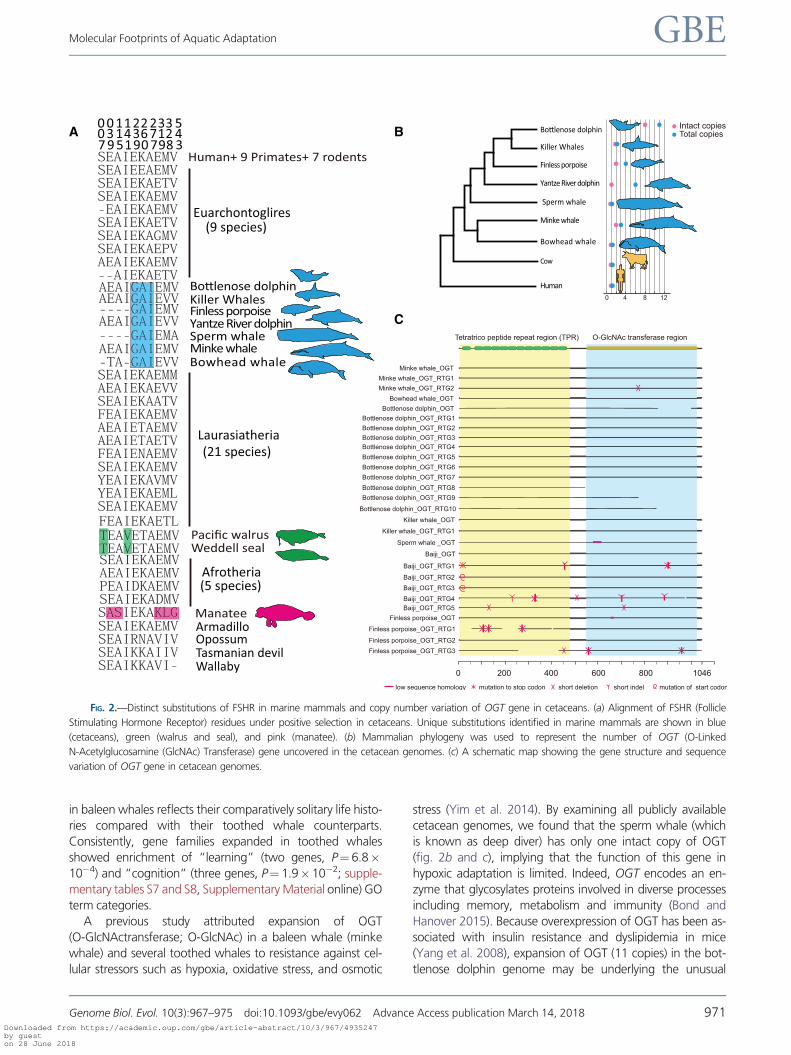
follicle-stimulating hormone receptor (FSHR) and INHBA
genes that encode subunits of the FSH inhibitor, and FSHR
is essential for reproduction and mutations in this gene were
found to be associated with human pathologies manifested
by altered ovarian function (Beau et al. 1998; De Leener et al.
2008; Kuechler et al. 2010). Intriguingly, compared with 60
terrestrial mammals, we found several nonsynonymous sub-
stitutions in FSHR that were unique to cetaceans and other
marine mammals (fig. 2a). For example, an isoleucine present
at position 277 in cetacean FSHR is located at a repeat domain
critical for activation of the receptor by FSH (Jiang et al. 2012).
Whereas the reproductive organs of marine mammals are
clearly mammalian in appearance, some specialization to ac-
commodate body streamlining and marine environment is
evident (Berta and Sumich 1999). For example, the duration
of a recognizable corpus albicans, which can increase FSH
secretion, varies between species and individuals across
marine mammals; and remnants may persist for several years
in some species (Laws and Sinha 1993).
In the common branch leading to toothed whales, genes
associated with “intermediate filament organization” (two
genes, KRT9 and KRT20, P¼ 1.4� 10�3) were significantly
contracted (supplementary table S5, Supplementary
Material online). Both genes play significant role in maintain-
ing keratin filament organization (Szeverenyi et al. 2008).
Toothed whales are nearly hairless, whereas baleen is formed
from keratin. Our findings are consistent with the recent stud-
ies attributing the loss of keratin genes in cetacean genomes
to their hairless phenotype (Nery et al. 2014; Strasser et al.
2015). Contraction of genes associated with “neuron differ-
entiation” (eight genes, APC, CNTNAP2, CTNNA2, DMD,
HES5, HOXA2, RELN, and STMN3; P¼ 1.3� 10�2) was ob-
served in baleen whales (supplementary table S6,
Supplementary Material online). Cetaceans are believed to
be highly intelligent, possessing large brains and complex be-
havioral patterns, with toothed whale brains being larger and
having significantly more neuronal cells than baleen whales
(Marino et al. 2007; Mortensen et al. 2014). We speculate
that the loss of genes associated with neuronal differentiation
A
B
C
FIG. 1.—Gene families and aquatic adaptations in cetacean genomes. (a) Phylogenetic tree and divergence times estimated for the cetaceans and their
relatives. Numbers associated with each branch designate the number of gene families that have expanded (red) and contracted (blue) since the split from
the common ancestor. (b) Phylogenetic tree of PRDX1 family in mammals showing expanded PRDX1 in cetaceans (highlighted by blue background).
Positions of introns along the protein sequence are shown with black lines. (c) Phylogenetic tree of the GPx family in mammals showing expanded GPX1
sequences in cetaceans.
Zhou et al. GBE
970 Genome Biol. Evol. 10(3):967–975 doi:10.1093/gbe/evy062 Advance Access publication March 14, 2018Downloaded from https://academic.oup.com/gbe/article-abstract/10/3/967/4935247by gueston 28 June 2018
in baleen whales reflects their comparatively solitary life histo-
ries compared with their toothed whale counterparts.
Consistently, gene families expanded in toothed whales
showed enrichment of “learning” (two genes, P¼ 6.8�10�4) and “cognition” (three genes, P¼ 1.9�10�2; supple-
mentary tables S7 and S8, Supplementary Material online) GO
term categories.
A previous study attributed expansion of OGT
(O-GlcNActransferase; O-GlcNAc) in a baleen whale (minke
whale) and several toothed whales to resistance against cel-
lular stressors such as hypoxia, oxidative stress, and osmotic
stress (Yim et al. 2014). By examining all publicly available
cetacean genomes, we found that the sperm whale (which
is known as deep diver) has only one intact copy of OGT
(fig. 2b and c), implying that the function of this gene in
hypoxic adaptation is limited. Indeed, OGT encodes an en-
zyme that glycosylates proteins involved in diverse processes
including memory, metabolism and immunity (Bond and
Hanover 2015). Because overexpression of OGT has been as-
sociated with insulin resistance and dyslipidemia in mice
(Yang et al. 2008), expansion of OGT (11 copies) in the bot-
tlenose dolphin genome may be underlying the unusual
A B
C
FIG. 2.—Distinct substitutions of FSHR in marine mammals and copy number variation of OGT gene in cetaceans. (a) Alignment of FSHR (Follicle
Stimulating Hormone Receptor) residues under positive selection in cetaceans. Unique substitutions identified in marine mammals are shown in blue
(cetaceans), green (walrus and seal), and pink (manatee). (b) Mammalian phylogeny was used to represent the number of OGT (O-Linked
N-Acetylglucosamine (GlcNAc) Transferase) gene uncovered in the cetacean genomes. (c) A schematic map showing the gene structure and sequence
variation of OGT gene in cetacean genomes.
Molecular Footprints of Aquatic Adaptation GBE
Genome Biol. Evol. 10(3):967–975 doi:10.1093/gbe/evy062 Advance Access publication March 14, 2018 971Downloaded from https://academic.oup.com/gbe/article-abstract/10/3/967/4935247by gueston 28 June 2018

response of this species to insulin (Venn-Watson et al. 2013).
Taken together, these findings provide insights into how evo-
lution shaped genes associated with aquatic adaptation of
cetaceans.
Association of Root-to-Tip dN/dS and Bone Compactness
It has been suggested that bone microanatomy is critical for
habitat preferences, facilitating transition of terrestrial mam-
mals to the aquatic environment (see Introduction, Gray et al.
2007; Canoville et al. 2016). While most studies use appendic-
ular skeleton, such as the humerus, femur, or vertebral column
to measure their effects of lifestyle, the ribs are an important
part of the axial skeleton are rarely analyzed (Canoville et al.
2016). In addition to the environmental pressures and respira-
tory function, biomechanical constraints linked to locomotion
and body support would strongly affect the ribs and rib cage,
particular in limbless tetrapods (e.g.,BrambleandCarrier1983;
Carrier1996;Houssayeetal. 2013,2014;Fujiwaraetal. 2009).
The buoyant force of water relaxes constraints on skeletal
mass; however, cetaceans have adapted to diverse habitats.
Therefore, investigating microstructural features of bone over
the main lineages of cetaceans as well as other marine mam-
mals can help revealing the adaptive changes of bone micro-
anatomy in aquatic environment.
We compiled data on rib compactness from eight marine
mammals (finless porpoise, minke whale, Yangtze river dol-
phin, killer whale, bottlenose dolphin, Weddell seal, walrus,
and manatee) and another 23 mammals (fig. 3a and b), for
which we obtained full genome data. We employed the root-
to-tip dN/dS (the ratio of nonsynonymous to synonymous sub-
stitutions) for each species to test the association between
selection pressure and phenotype. Such “accumulated” dN/
dS values from root to each tip branch allow taking a common
ancestor of each node into account. Of 3,293 single gene
copy orthologues shared by these 28 mammals, 134 were
found with a significant correlation (with P value, P value.ro-
bust [from regression by excluding the point with largest res-
idue error] and P value.maximum [defined as the maximal P
value from regression with species excluded separate-
ly]< 0.05) between rib globe compactness (Cg) and root-to-
tip dN/dS rations.
A
B
C
FIG. 3.—Root-to-tip dN/dS correlates with global compactness of ribs across 28 mammals. (a) Diagrammatic sketch showing bone microanatomy of the
rib and parameters measured in this study. (b) Variation in global compactness of ribs across 28 mammals in a phylogenetic context. Representatives from
marine, terrestrial, and flying mammals are in green, black, and red, respectively. (c) Root-to-tip dN/dS of genes LEP (top panel) and IGF1 (bottom panel)
negatively correlates with global compactness. Points are colored according to taxonomical orders (same color scheme as in b).
Zhou et al. GBE
972 Genome Biol. Evol. 10(3):967–975 doi:10.1093/gbe/evy062 Advance Access publication March 14, 2018Downloaded from https://academic.oup.com/gbe/article-abstract/10/3/967/4935247by gueston 28 June 2018
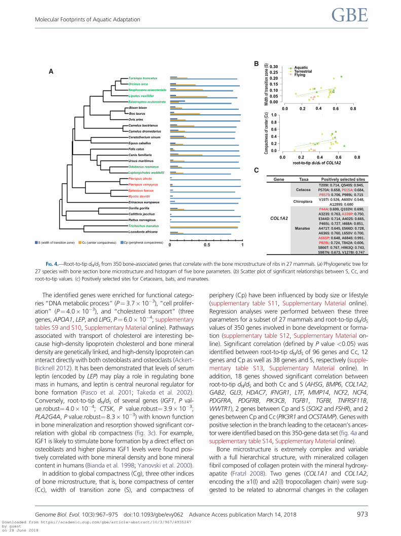
The identified genes were enriched for functional catego-
ries “DNA metabolic process” (P¼ 3.7� 10�3), “cell prolifer-
ation” (P¼ 4.0� 10�3), and “cholesterol transport” (three
genes, APOA1, LEP, and LIPG, P¼ 6.0� 10�4; supplementary
tables S9 and S10, Supplementary Material online). Pathways
associated with transport of cholesterol are interesting be-
cause high-density lipoprotein cholesterol and bone mineral
density are genetically linked, and high-density lipoprotein can
interact directly with both osteoblasts and osteoclasts (Ackert-
Bicknell 2012). It has been demonstrated that levels of serum
leptin (encoded by LEP) may play a role in regulating bone
mass in humans, and leptin is central neuronal regulator for
bone formation (Pasco et al. 2001; Takeda et al. 2002).
Conversely, root-to-tip dN/dS of several genes (IGF1, P val-
ue.robust¼ 4.0� 10�4; CTSK, P value.robust¼ 3.9� 10�3;
PLA2G4A, P value.robust¼ 8.3� 10�3) with known function
in bone mineralization and resorption showed significant cor-
relation with global rib compactness (fig. 3c). For example,
IGF1 is likely to stimulate bone formation by a direct effect on
osteoblasts and higher plasma IGF1 levels were found posi-
tively correlated with bone mineral density and bone mineral
content in humans (Bianda et al. 1998; Yanovski et al. 2000).
In addition to global compactness (Cg), three other indices
of bone microstructure, that is, bone compactness of center
(Cc), width of transition zone (S), and compactness of
periphery (Cp) have been influenced by body size or lifestyle
(supplementary table S11, Supplementary Material online).
Regression analyses were performed between these three
parameters for a subset of 27 mammals and root-to-tip dN/dS
values of 350 genes involved in bone development or forma-
tion (supplementary table S12, Supplementary Material on-
line). Significant correlation (defined by P value <0.05) was
identified between root-to-tip dN/dS of 96 genes and Cc, 12
genes and Cp as well as 38 genes and S, respectively (supple-
mentary table S13, Supplementary Material online). In
addition, 18 genes showed significant correlation between
root-to-tip dN/dS and both Cc and S (AHSG, BMP6, COL1A2,
GAB2, GLI3, HDAC7, IFNGR1, LTF, MMP14, NCF2, NCF4,
PDGFRA, PDGFRB, PIK3CB, TGFB1, TGFBI, TNFRSF11B,
WWTR1), 2 genes between Cp and S (SOX2 and FSHR), and 2
genes between Cp and Cc (PIK3R1 and OCSTAMP). Genes with
positive selection in the branch leading to the cetacean’s ances-
tor were identified based on this 350-gene data set (fig. 4a and
supplementary table S14, Supplementary Material online).
Bone microstructure is extremely complex and variable
with a full hierarchical structure, with mineralized collagen
fibril composed of collagen protein with the mineral hydroxy-
apatite (Fratzl 2008). Two genes (COL1A1 and COL1A2,
encoding the a1(I) and a2(I) tropocollagen chain) were sug-
gested to be related to abnormal changes in the collagen
AB
C
FIG. 4.—Root-to-tip dN/dS from 350 bone-associated genes that correlate with the bone microstructure of ribs in 27 mammals. (a) Phylogenetic tree for
27 species with bone section bone microstructure and histogram of five bone parameters. (b) Scatter plot of significant relationships between S, Cc, and
root-to-tip values. (c) Positively selected sites for Cetaceans, bats, and manatees.
Molecular Footprints of Aquatic Adaptation GBE
Genome Biol. Evol. 10(3):967–975 doi:10.1093/gbe/evy062 Advance Access publication March 14, 2018 973Downloaded from https://academic.oup.com/gbe/article-abstract/10/3/967/4935247by gueston 28 June 2018

triple-helical structure (Fratzl et al. 2004). In the present study,
root-to-tip dN/dS values of COL1A2 were significantly corre-
lated with Cc (P value.robust< 0.001, P value.max< 0.01)
and S (P value.robust< 0.01, P value.max< 0.05; fig. 4b).
In addition, COL1A2 appeared to be a positively selected
gene in cetaceans (P< 0.01, x¼ 197.77), manatee (T. man-
atus; P< 0.05, x¼ 4.785), and the ancestor of Chiroptera
(P< 0.05, x¼ 131.667; fig. 4c). Manatees are fully aquatic
herbivores that feed on plants in shallow waters and are char-
acterized by compact bones (de Buffr�enil et al. 2010). The
bats, as the only group of flying mammals, exhibit simple
bone microanatomy, with thin cortices, and few (or none)
trabeculae bones in the medullary region. It is reasonable to
hypothesize that these substitutions in fully aquatic cetaceans
may have modified the collagen triple helix related to bone
mass. The overall evolutionary pattern suggests that gene
level changes potentially drive phenotypic alterations in the
mammalian bone structure. Future studies aiming to disen-
tangling the observed changes by studying the expression and
function of these genes in diverse taxa, could be rewarding.
Conclusions
Comparative genomic analyses of cetaceans and their terres-
trial relatives provided several novel insights into the distinct
evolutionary scenarios of adaptation to a fully aquatic lifestyle.
Genes associated with oxidation–reduction and immune pro-
cess were found to be accompanied by pseudogene copies.
Genes under positive selection in the cetaceans were related
to reproduction, keratin protein, learning, and energy turn-
over. This was interesting given their special lifestyle com-
pared with other mammals. Our study also documented the
bone microstructure across mammals and marine mammals,
and for the first time, revealed the benefit of using a phylo-
genetic comparative approach to study the evolution of bone
compactness. Our findings offer valuable information on
genes critical for adaptation to aquatic life of mammals in
diverse environments.
Supplementary Material
Supplementary data are available at Genome Biology and
Evolution online.
Acknowledgments
This work was supported by the Key Project of the National
Natural Science Foundation of China (NSFC) (grant no.
31630071 to G.Y.), the National Science Fund for
Distinguished Young Scholars (grant no. 31325025 to G.Y.),
Ministry of Science and Technology of China (grant no.
2016YFC0503200), the Priority Academic Program
Development of Jiangsu Higher Education Institutions
(PAPD), and NIH grants GM065204 and AG047200.
Literature CitedAckert-Bicknell CL. 2012. HDL cholesterol and bone mineral density: is
there a genetic link? Bone 50(2):525–533.
Amson E, de Muizon C, Laurin M, Argot C, de Buffr�enil V. 2014. Gradual
adaptation of bone structure to aquatic lifestyle in extinct sloths from
Peru. Proc Biol Sci. 281(1782):20140192.
Barnes LG. 1984. Fossil odontocetes (Mammalia: cetacea) from the
Almejas Formation, Isla Cedros, Mexico. PaleoBios 42:1–46.
Beau I, et al. 1998. A novel phenotype related to partial loss of function
mutations of the follicle stimulating hormone receptor. J Clin Invest.
102(7):1352–1359.
Berta A, Sumich JL. 1999. Marine mammals. Evolutionary biology. San
Diego (CA): Academic Press.
BiandaT,GlatzY,BouillonR,FroeschER,SchmidC.1998.Effectsofshort-term
insulin-like growth factor-I (IGF-I) or growth hormone (GH) treatment
on bone metabolism and on production of 1, 25-dihydroxycholecalci-
ferol in GH-deficient adults 1. J Clin Endocrinol Metab. 83(1):81–87.
Bond MR, Hanover JA. 2015. A little sugar goes a long way: the cell
biology ofO-GlcNAc. J Cell Biol. 208(7):869–880.
Bramble DM, Carrier DR. 1983. Running and breathing in mammals.
Science 219(4582):251–256.
Braun BA, Marcovitz A, Camp JG, Jia R, Bejerano G. 2015. Mx1 and Mx2
key antiviral proteins are surprisingly lost in toothed whales. Proc Natl
Acad Sci USA. 112(26):8036–8040.
Canoville A, de Buffr�enil V, Laurin M. 2016. Microanatomical diversity of
amniote ribs: an exploratory quantitative study. Biol J Linn Soc Lond.
118(4):706–733.
Castresana J. 2000. Selection of conserved blocks from multiple align-
ments for their use in phylogenetic analysis. Mol Bio Evol.
17(4):540–552.
Caulin AF, Maley CC. 2011. Peto’s Paradox: evolution’s prescription for
cancer prevention. Trends Ecol Evol. 26(4):175–182.
Carrier DR. 1996. Ontogenetic limits on locomotor performance. Physiol
Zool. 69(3):467–488.
Chen Z, Wang Z, Xu S, Zhou K, Yang G. 2013. Characterization of hairless
(Hr) and FGF5 genes provides insights into the molecular basis of hair
loss in cetaceans. BMC Evol Biol. 13:34.
De Leener A, et al. 2008. Identification of the first germline mutation in the
extracellular domain of the follitropin receptor responsible for sponta-
neous ovarian hyper stimulation syndrome. Hum Mutat. 29(1):91–98.
de Buffr�enil V, Canoville A, D’Anastasio R, Domning DP. 2010. Evolution
of sirenian pachyosteosclerosis, a model-case for the study of bone
structure in aquatic tetrapods. J Mamm Evol. 17(2):101–120.
Dem�er�e TA, McGowen MR, Berta A, Gatesy J. 2008. Morphological and
molecular evidence for a stepwise evolutionary transition from teeth to
baleen in mysticete whales. Syst Biol. 57(1):15–37.
Foote AD, et al. 2015. Convergent evolution of the genomes of marine
mammals. Nat Genet. 47(3):272–275.
Fratzl P, Gupta HS, Paschalis EP, Roschger P. 2004. Structure and mechan-
ical quality of the collagen–mineral nano-composite in bone. J Mater
Chem. 14(14):2115–2123.
Fratzl P. 2008. Collagen: structure and mechanics, an introduction. New
York: Springer.
Fujiwara SI, Kuwazuru O, Inuzuka N, Yoshikawa N. 2009. Relationship
between scapular position and structural strength of rib cage in quad-
ruped animals. J Morphol. 270(9):1084–1094.
Gatesy J, et al. 2013. A phylogenetic blueprint for a modern whale. Mol
Phylogenet Evol. 66(2):479–506.
George JC, et al. 1999. Age and growth estimates of bowhead whales
(Balaena mysticetus) via aspartic acid racemization. Can J Zool.
77(4):571–580.
George JC, Bockstoce JR. 2008. Two historical weapon fragments as an
aid to estimating the longevity and movements of bowhead whales.
Polar Biol. 31(6):751–754.
Zhou et al. GBE
974 Genome Biol. Evol. 10(3):967–975 doi:10.1093/gbe/evy062 Advance Access publication March 14, 2018Downloaded from https://academic.oup.com/gbe/article-abstract/10/3/967/4935247by gueston 28 June 2018
Gingerich PD, Wells NA, Russell DE, Shah SI. 1983. Origin of whales in
epicontinental remnant seas: new evidence from the early Eocene of
Pakistan. Science 220(4595):403–406.
Girondot M, Laurin M. 2003. Bone Profiler: a tool to quantify, model, and
statistically compare bone-section compactness profiles. J Vert
Paleontol. 23:458–461.
Gray NM, Kainec K, Madar S, Tomko L, Wolfe S. 2007. Sink or swim?
Bone density as a mechanism for buoyancy control in early cetaceans.
Anat Rec (Hoboken). 290(6):638–653.
Tung Ho LS, An�e C. 2014. A linear-time algorithm for Gaussian and
nonGaussian trait evolution models. Syst Biol. 63(3):397–408.
Houssaye A, Boistel R, Bohme W, Herrel A. 2013. Jack-of-all-trades master
of all? Snake vertebrae have a generalist inner organization.
Naturwissenschaften 100(11):997–1006.
Houssaye A, Tafforeau P, Herrel A. 2014. Amniote vertebral microanatomy
– what are the major trends? Biol J Linnean Soc. 112(4):735–746.
Houssaye A, Tafforeau P, De Muizon C, Gingerich PD. 2015. Transition of
Eocene whales from land to sea: evidence from bone microstructure.
PLOS ONE. 10(2):e0118409.
Jiang X, et al. 2012. Structure of follicle-stimulating hormone in complex
with the entire ectodomain of its receptor. Proc Natl Acad Sci USA.
109(31):12491–12496.
Kanehisa M, Sato Y, Kawashima M, Furumichi M, Tanabe M. 2016. KEGG
as a reference resource for gene and protein annotation. Nucleic Acids
Res. 44(D1):D457–D462.
Keane M, et al. 2015. Insights into the evolution of longevity from the
bowhead whale genome. Cell Rep. 10(1):112–122.
Kuechler A, et al. 2010. An unbalanced translocation unmasks a recessive
mutation in the follicle-stimulating hormone receptor (FSHR) gene and
causes FSH resistance. Eur J Hum Genet. 18(6):656–661.
Laws RM, Sinha AA. 1993. Reproduction. In: Laws, RM, editor. Antarctic
seals. Cambridge: Cambridge University Press. p. 228–267.
Li H, et al. 2006. TreeFam: a curated database of phylogenetic trees of
animal gene families. Nucleic Acids Res. 34:D572–D580.
Li L, Stoeckert CJ, Roos DS. 2003. OrthoMCL: identification of ortholog
groups for eukaryotic genomes. Genome Res. 13(9):2178–2189.
Lindblad-Toh K, et al. 2011. A high-resolution map of human evolutionary
constraint using 29 mammals. Nature 478(7370):476–482.
Loytynoja A, Goldman N. 2008. Phylogeny-aware gap placement prevents
errors in sequence alignment and evolutionary analysis. Science
320(5883):1632–1635.
Ma S, et al. 2015. Organization of the mammalian metabolome according
to organ function, lineage specialization, and longevity. Cell Metab.
22(2):332–343.
Marino L, et al. 2007. Cetaceans have complex brains for complex cogni-
tion. PLoS Biol. 5(5):e139.
Montgomery SH, Capellini I, Venditti C, Barton RA, Mundy NI. 2011.
Adaptive evolution of four microcephaly genes and the evolution of
brain size in anthropoid primates. Mol Biol Evol. 28(1):625–638.
Mortensen HS, et al. 2014. Quantitative relationships in delphinid neocor-
tex. Front Neuroanat. 8:132.
Nery MF, Arroyo JI, Opazo JC. 2014. Increased rate of hair keratin gene
loss in the cetacean lineage. BMC Genomics. 15:869.
Oelschl€ager HA. 1992. Development of the olfactory and terminalis sys-
tems in whales and dolphins. In: Doty RL, Muller-Schwarze D. (Eds.),
Chemical Signals in Verterbrates VI. New York: Plenum Press,
pp. 141–147.
Ohishi K, et al. 2011. Lipopolysaccharide-induced innate immune factors
in the bottlenose dolphin (Tursiops truncates) detected in expression
sequence tag analysis. Microbiol Immunol. 55(11):790–797.
Pasco JA, et al. 2001. Serum leptin levels are associated with bone mass in
nonobese women 1. J Clin Endocrinol Metab. 86(5):1884–1887.
R Core Team. 2013. R: a language and environment for statistical com-
puting. R Foundation for Statistical Computing, Vienna, Austria.
Revell LJ. 2012. Phytools: an R package for phylogenetic comparative bi-
ology (and other things). Methods Ecol Evol. 3(2):217–223.
Ricqles AD, Buffr�enil VD. 2001. Bone histology, heterochronies and the
return of tetrapods to life in water: where are we. In: Mazin JM,
Buffr�enil V, editors. Secondary adaptation of tetrapods to life in water.
Munchen (Germany): Verlag. p. 289–310.
Shen T, et al. 2012. Adaptive evolution and functional constraint at TLR4
during the secondary aquatic adaptation and diversification of ceta-
ceans. BMC Evol Biol. 12(1):39.
Stamatakis A. 2006. RaxML-VI-HPC: maximum likelihood-based phyloge-
netic analyses with thousands of taxa and mixed models.
Bioinformatics 22(21):2688–2690.
Strasser B, Mlitz V, Fischer H, Tschachler E, Eckhart L. 2015. Comparative
genomics reveals conservation of filaggrin and loss of caspase-14 in
dolphins. Exp Dermatol. 24(5):365–369.
Szeverenyi I, et al. 2008. The Human Intermediate Filament Database:
comprehensive information on a gene family involved in many human
diseases. Hum Mutat. 29(3):351–360.
Takeda S, et al. 2002. Leptin regulates bone formation via the sympathetic
nervous system. Cell 111(3):305–317.
Tamura K, et al. 2012. Estimating divergence times in large molecular
phylogenies. Proc Natl Acad Sci USA. 109(47):19333–19338.
Taylor MA. 2000. Functional significance of bone ballastin in the evolution
of buoyancy control strategies by aquatic tetrapods. Histor Biol. 14(1–
2):15–31.
Thewissen JGM, Cooper LN, Clementz MT, Bajpai S, Tiwari BN. 2007.
Whales originated from aquatic artiodactyls in the Eocene epoch of
India. Nature 450(7173):1190–1194.
Tsagkogeorga G, et al. 2015. A phylogenomic analysis of the role and
timing of molecular adaptation in the aquatic transition of cetartio-
dactyl mammals. R Soc Open Sci. 2(9):150156.
Venn-Watson S, et al. 2013. Blood-based indicators of insulin resistance
and metabolic syndrome in bottlenose dolphins (Tursiops truncatus).
Front Endocrinol. 4:136.
Xu S, et al. 2013. Adaptive evolution of the osmoregulation-related genes
in cetaceans during secondary aquatic adaptation. BMC Evol Biol.
13:189.
Yang X, et al. 2008. Phosphoinositide signalling links O-GlcNAc transferase
to insulin resistance. Nature 451(7181):964–969.
Yang Z. 2007. PAML 4: phylogenetic analysis by maximum likelihood. Mol
Biol Evol. 24(8):1586–1591.
Yanovski JA, Sovik KN, Nguyen TT, Sebring NG. 2000. Insulin-like growth
factors and bone mineral density in African American and White girls. J
Pediatr. 137(6):826–832.
Yim HS, et al. 2014. Minke whale genome and aquatic adaptation in
cetaceans. Nat Genet. 46(1):88–92.
Zhang J, Nielsen R, Yang Z. 2005. Evaluation of an improved branch-site
likelihood method for detecting positive selection at the molecular
level. Mol Biol Evol. 22(12):2472–2479.
Zhu K, et al. 2014. The loss of taste genes in cetaceans. BMC Evol Biol.
14:218.
Zhou X, Seim I, Gladyshev VN. 2015. Convergent evolution of marine
mammals is associated with distinct substitutions in common genes.
Sci Rep. 5:16550.
Zhou X, et al. 2013. Baiji genomes reveal low genetic variability and
new insights into secondary aquatic adaptations. Nat Commun.
4:2708.
Zhou X, Xu S, Yang Y, Zhou K, Yang G. 2011. Phylogenomic analyses and
improved resolution of Cetartiodactyla. Mol Phylogenet Evol.
61(2):255–264.
Associate editor: Marta Barluenga
Molecular Footprints of Aquatic Adaptation GBE
Genome Biol. Evol. 10(3):967–975 doi:10.1093/gbe/evy062 Advance Access publication March 14, 2018 975Downloaded from https://academic.oup.com/gbe/article-abstract/10/3/967/4935247by gueston 28 June 2018