molecular epidemiology and phylogeny reveals complex spatial
TRANSCRIPT
Molecular epidemiology and phylogeny reveals complex spatial dynamics of endemic canine 1
parvovirus 2
3
S.R. Clegg1, K.P. Coyne
1, J. Parker
3, S. Dawson
1, S.A. Godsall
2, G. Pinchbeck
1, P.J. Cripps
1, R.M Gaskell
1, 4
A.D. Radford 1 5
1. Institute of Infection and Global health, School of Veterinary Science, University of Liverpool, 6
Leahurst Campus, Chester High Road, Neston, South Wirral, CH64 7TE, UK. 7
2. PDSA, PDSA Regional Office, 556 Bath Road, Brislington, Bristol, BS4 3JZ, UK. 8
3. School of Biological and Chemical Sciences, Queen Mary, University of London, E1 4NS, UK. 9
10
Running title: Spatial dynamics of canine parvovirus 11
12
Correspondence address. Simon Clegg, Faculty of Health and Life Science, School of Veterinary 13
Science, University of Liverpool, Leahurst Campus, Chester High Road, Neston, South Wirral, UK, 14
CH64 7TE. 0151 794 6012. [email protected] 15
16
17
18
19
20
Copyright © 2011, American Society for Microbiology and/or the Listed Authors/Institutions. All Rights Reserved.J. Virol. doi:10.1128/JVI.01576-10 JVI Accepts, published online ahead of print on 18 May 2011
on March 30, 2018 by guest
http://jvi.asm.org/
Dow
nloaded from
21
Abstract 22
Canine parvovirus 2 (CPV-2) is a severe enteric pathogen of dogs, causing high mortality in 23
unvaccinated dogs. After emerging, CPV-2 spread rapidly worldwide. However, there is now some 24
evidence to suggest that international transmission appears to be more restricted. 25
In order to investigate the transmission and evolution of CPV-2 both nationally and in relation to the 26
global situation, we have used a long range PCR to amplify and sequence the full VP2 gene of 150 27
canine parvoviruses obtained from a large cross-sectional sample of dogs presenting with severe 28
diarrhoea to veterinarians in the UK, over a two year period. 29
Amongst these 150 strains, 50 different DNA sequence types were identified, and apart from one 30
case, all appeared unique to the UK. Phylogenetic analysis provided clear evidence for spatial 31
clustering at the international level, and for the first time also at the national level, with the 32
geographical range of some sequence types appearing to be highly restricted within the UK. 33
Evolution of the VP2 gene in this dataset was associated with a lack of positive selection. In addition, 34
the majority of predicted amino acid sequences were identical to those found elsewhere in the 35
world, suggesting CPV VP2 has evolved a highly fit conformation. Based on typing systems using key 36
amino acid mutations, 43% of viruses were CPV 2a, 57% CPV 2b, with no type 2 or 2c found. 37
However phylogenetic analysis suggested complex antigenic evolution of this virus, with both type 38
2a and 2b viruses appearing polyphyletic. As such, typing based on specific amino acid mutations 39
may not reflect the true epidemiology of this virus. 40
The geographical restriction we observed both within the UK, and between the UK and other 41
countries, together with the lack of CPV-2c in this population, strongly suggest the spread of CPV 42
within its population may be heterogeneously subject to limiting factors. This cross-sectional study 43
on March 30, 2018 by guest
http://jvi.asm.org/
Dow
nloaded from

of national and global CPV phylogeographic segregation reveals a substantially more complex 44
epidemic structure than previously described. 45
on March 30, 2018 by guest
http://jvi.asm.org/
Dow
nloaded from
Introduction 46
Sequence analysis has revolutionised our knowledge of the spatial and temporal dynamics of 47
infection, allowing a greater understanding of the evolution and molecular epidemiology of 48
pathogens. This is particularly important for rapidly evolving pathogens such as RNA viruses (e.g. 49
feline calicivirus (9), foot and mouth disease virus (8), and influenza virus (34), and also for certain 50
single stranded DNA viruses with high mutation rates, such as canine parvovirus (38). 51
Canine parvovirus 2 (CPV) consists of a 5.5kb single stranded linear DNA genome (5) encoding the 52
non-structural (NS1 and 2) and capsid (VP1,2 and 3) proteins at the 5’ and 3’ ends respectively (1). 53
The three structural proteins are derived from the same open reading frame (ORF) by proteolytic 54
cleavage and alternate RNA splicing, with full, infectious capsid consisting predominately of VP2 (35). 55
The virus first emerged as a new causative agent of severe enteritis in dogs in 1978 (21, 3), and 56
rapidly spread worldwide. This virus, which was named CPV-2 to distinguish it from the unrelated 57
parvovirus, minute virus of canines (MVC), is thought to have emerged from a related virus, feline 58
panleukopenia (FPV), possibly via some wildlife intermediate such as a fox (45). This initial species 59
jump was mapped to amino acid mutations in VP2 when compared to FPV, which resulted in gain of 60
receptor affinity for the canine host. Subsequently, antigenic variants (CPV types 2a, 2b and 2c) have 61
been described largely based on mouse monoclonal antibody reactivity, and the mutations 62
responsible have been mapped to specific residues in VP2 (CPV2a - Met-87-Leu, Ile-101-Thr, Ala-63
300-Gly, Asp-305- Tyr and Val-555-Ile ; CPV2b – Asp-426-Asn and Ile-555-Val reversion; CPV2c – Asp-64
426-Glu) (6). This has led to several experimental studies showing that CPV vaccines based on CPV-2 65
or CPV- 2b, are able to cross-protect against all four antigenic types, including the newly emerged 66
CPV - 2c (39, 41). Mutations in VP2 have also been shown to influence host range (28), 67
haemaglutination spectrum (31) and affinity of receptor binding (27). 68
Currently, the original CPV type 2 is thought not to circulate in the general dog population although 69
it is present within certain live vaccines. The distribution of CPV types 2a, 2b and 2c seem to differ 70
on March 30, 2018 by guest
http://jvi.asm.org/
Dow
nloaded from

across different regions of the world (14, 16, 19, 32, 36, 46). It has recently been suggested that the 71
initial rapid global spread of CPV-type 2 was a feature of the naïve dog population it gained access 72
to, and is in contrast to the current more endemic phase of disease, where the international range of 73
new strains is more restricted (21). 74
Whilst these studies are collectively improving our knowledge of the spatial and temporal dynamics 75
of CPV transmission, they are generally based on relatively unstructured sampling strategies of 76
national collections and / or are limited to using only partial VP2 gene sequence analysis or typing by 77
key amino acid mutations (7, 10, 12, 16, 22, 47). In this paper, we have used a cross sectional study 78
of clinically ill dogs, and full VP2 sequence analysis, to investigate in depth the evolution and spread 79
of virus at national and local levels, and in relation to the global situation, to specifically test the 80
hypothesis that currently there is limited international spread of CPV. 81
82
Methods 83
Samples 84
Faecal samples were obtained from 25 People’s Dispensary for Sick Animals (PDSA) Petaid hospitals 85
across mainland UK (Figure 1) from 373 clinically ill dogs presenting with diarrhoea of unknown 86
aetiology that required more than conservative treatment. Clinical information and signalment were 87
supplied with the samples and presented elsewhere (18). In addition, 16 CPV positive samples from 88
11 different locations within the UK (one post mortem sample, 12 from a commercial diagnostic 89
laboratory and three potential vaccine breakdowns), and viruses from the six commercial vaccines 90
currently used within the UK (coded A-F), were also obtained. All samples and vaccines were 91
collected over a two year period (2006-2008). Samples were stored at -80 C until used. 92
Amplification and sequence of VP2 93
on March 30, 2018 by guest
http://jvi.asm.org/
Dow
nloaded from
DNA was extracted as described previously (13) with a slight modification. Briefly samples were 94
homogenised (10% W/V) in phosphate buffered saline (PBS) and centrifuged for 15 minutes at 9300 95
rpm. The supernatant was boiled for 15 minutes, chilled on ice, and then centrifuged again as before 96
for 5 minutes. The supernatant was stored at 4°C. Primers were chosen to amplify the full VP2 97
region, using both full genomes of CPV and FPV, and VP2 sequences available on Genbank, aligned 98
using CLUSTAL as implemented in Mega 4 (43). Two primers were used: EF (2748-2765) 99
GCCGGTGCAGGACAAGTA and JS2R (25) (4818-4799) CAACCCACACCATAACAACA (all primer 100
sequences numbered based on ref 34) to obtain full VP2 sequence. In order to minimise PCR 101
inhibition, DNA extraction supernatant was diluted 1 in 10 before being amplified by PCR (37). 102
Amplification was carried out in 25 µl reactions, consisting of 12.5 µl extensor PCR master mix 103
(Abgene), 8.5 µl of molecular water (Sigma), (12.5 pmoles of primer EF, 12.5 pmoles primer JS2R) 104
(Meers et al, 2007) and 2 µl DNA. Negative controls were processed alongside faecal samples 105
throughout all stages (1 negative control (water or PBS) per 2 faecal samples). All samples were 106
processed in order of arrival at the laboratory and not batched according to location. The PCR 107
cycling conditions were 1 min at 94°C, followed by 40 cycles of denaturation at 94°C, annealing at 108
52°C and extension at 72°C, followed by a final extension phase of 68°C for 10 minutes. 109
Amplicons were purified using Qiaquick PCR purification kit (Qiagen) according to manufacturer’s 110
instructions. Sequencing of full VP2 was generated in three overlapping fragments using the two PCR 111
primers (EF and JS2R) and four internal sequencing primers MF (TACCATCTCATACTGGAACTAGTGG; 112
(3441 – 3466), ER (TGTTCCTGTAGCAAATTCATCACC; 3581-3558), 555F (6); 113
CAGGAAGATATCCAGAAGGA; 4003-4022) and MR (GTATAGTTAATTCCTGTTTTACCTCC; 4140 – 4118), 114
All sequences were aligned into a double-stranded consensus sequence using Chromas Pro 1.41 115
(Technelysium Pty Ltd). All external primer sites were removed, giving a final consensus sequence of 116
1755 bp. All sequences were homozygous, with no evidence to suggest dual or mixed infections. 117
Sequence analysis 118
on March 30, 2018 by guest
http://jvi.asm.org/
Dow
nloaded from
Consensus sequences were aligned by Clustal W. These alignments included sequences available for 119
feline panleucopaenia virus (Genbank accession number M24004), and also included published 120
sequences for two CPV type2 (M23255 and U22186). The most appropriate evolution model was 121
predicted using MODELTEST as implemented in Topali (26). The final model for nucleotide 122
substitutions chosen was the TrN model (44), which was used to infer bootstrapped maximum 123
likelihood trees using PHYML implemented on the ATGC bioinformatics platform (20). Amino acid 124
trees were drawn using Mega 4, rooted using FPV and drawn using the Dayhoff PAM matrix. The 125
final alignment was screened for evidence of recombination and selection using GARD and SLAC 126
available through the Datamonkey web server (22). 127
To seek correlations between the geographical origin of a particular sequence and its position within 128
the phylogeny, a posterior set of trees was obtained through Bayesian MCMC analysis using BEAST 129
v1.4 (17). This analysis implemented the most-favoured model identified in the earlier step (TrN + γ 130
+ I); a comparison of alternative MCMC models (HKY + γ + I ; GTR + γ + I) by Bayes’ factor (42) 131
confirmed the TrN model was also the most appropriate in a Bayesian MCMC context. We also 132
compared the fit of the strict and ‘relaxed’ (uncorrelated exponential distribution; ‘UCED’) molecular 133
clock models; the UCED model provided a better fit. Similarly, the constant population-size model 134
was preferred over an exponential-growth model (available on request). The MCMC trace was 135
inspected in TRACER v1.5 (33) for convergence. This posterior set of trees was subjected to Bayesian 136
Tip-association Significance testing implemented by BaTS (29). In addition, Simpson’s equitability 137
index (E) was also used as a measure of population diversity (4, 40), where Pi is the proportion of 138
identical sequence types within the population and S is the total number of distinct sequence types 139
within the population. An E value of 1 equates to maximum diversity (all sequences different), 140
whereas E tends toward zero as the diversity decreases and the number of sequence types 141
increases. 142
on March 30, 2018 by guest
http://jvi.asm.org/
Dow
nloaded from
E =1
i=1
S
Pi2
x1
SE =
1
i=1
S
Pi2
x1
S
143
In order to test for geographical clustering we examined the proportion of each sequence type in 144
each sampling location. Since many sites were negative for a given sequence we used a non-145
parametric analysis, and performed a separate Fisher Exact Test for each sequence type, using 146
Bonferroni’s method to correct for multiple tests. Analyses used Stata 11 (StataCorp, College 147
Station, Texas, USA) and significance was set at P < 0.05. 148
UK sequences were compared to worldwide sequences using the Blast software (2). 149
Analysis of clinical signs 150
As signalment and clinical details were available with all the samples, we were able to evaluate 151
possible associations between CPV type (CPV- 2a, 2b or 2c) and clinical outcome (death or survival), 152
severity of clinical signs (as reported by the clinician), breed, age, sex, colour, presence of vomiting, 153
and presence of hemorrhagic diarrhoea. 154
155
Results 156
Of the 373 samples obtained from the PDSA, 255 (68%) were PCR positive (Table 1). Of these, 157
consensus VP2 sequence was obtained for 134 samples, selected to include one or more sequences 158
from each of the 25 hospitals that submitted a positive sample. Sequence was also obtained from 159
the 16 samples obtained from others sources (post mortem, commercial lab, and potential vaccine 160
breakdowns), and from the six vaccines (A-F) currently used in the UK. 161
Prevalence of CPV types in the UK 162
Based on key amino acid mutations, the 150 viruses were typed as 2a (43%; n = 65) or 2b (57%; n= 163
85) (Table 1). No type 2 or 2c were found. For those hospitals for which more than five sequences 164
on March 30, 2018 by guest
http://jvi.asm.org/
Dow
nloaded from
were available, the proportion of 2a/2b sequences ranged from 92% type 2a in Huyton (11 of 12) to 165
91% type 2b in Stoke (10 of 11) (Table 1), suggesting type 2a and 2b may have different distributions 166
in different areas of the UK. The vaccines consisted of two CPV- type 2b vaccines (A-B) and four CPV-167
type 2 vaccines (C-F) (Figure 1 and 2). 168
DNA sequence analysis 169
Considerable diversity was found within the VP2 DNA sequences we obtained, with 50 genetically 170
distinct sequences, or sequence types (S), identified amongst the 150 clinical samples sequenced 171
(Figure 1-2 and Table 1). Thirty-one of the 50 different sequence types were found only once, 172
whereas some sequence types were quite common; for example eight sequence types contained 173
more than five sequences, and the most common (S34) comprised 30 sequences (Table 1 and Figure 174
1). Of the 50 different sequence types, 20 comprised type 2a viruses and the remaining 30 were type 175
2b viruses (Table 1, Figure 1), suggesting type 2b might be more variable in the UK. 176
The majority of sequences clustered separately depending on their type (2a or 2b). However, neither 177
CPV2a or 2b was monophyletic: S31 which typed as a 2a virus based on key amino acid 178
substitutions, grouped phylogenetically within the 2b virus sequences (Figure 1). In addition, S1 179
(type 2b) clustered close (2-8nt substitutions) to S46-50 (type 2a). This may suggest possible 2a/2b 180
intermediates or parallel evolution. No evidence of recombination or positive selection was found 181
within this dataset (data not presented). 182
Spatial range of sequence types in the UK 183
In order to explore the spatial range of individual sequence types, we calculated the number of 184
hospitals in which each of these sequences was found (Table 1). Some sequence types were 185
geographically restricted as exemplified by S34 which was found at seven hospitals mostly clustered 186
in the North West of England including Liverpool, Everton, Huyton and St Helens (Left side of Figure 187
1). Other sequence types were geographically dispersed as exemplified by S2 which was again found 188
on March 30, 2018 by guest
http://jvi.asm.org/
Dow
nloaded from

in seven different hospitals ranging from the North (Glasgow) to the South (Croydon and Hendon) 189
(Right side of Figure 1). There was statistical support (p<0.05) for geographical clustering for S34 190
(Everton and Huyton), S3 (Hull), S23 (Basildon and Bow), S42 (Coventry and Cardiff). However, for 191
many areas, sample numbers were small. 192
The finding of evidence of clustering for some sequence types in particular geographic locations was 193
confirmed using Simpson’s index of diversity (Table 2). For example, Everton had the highest number 194
of viral sequences (n=26) but showed the lowest level of DNA (E=0.1) and second lowest amino acid 195
diversity (0.08). In other areas, sequence diversity was much higher, for example in Birmingham 196
(E=0.86 and 0.3 for DNA and amino acid sequences respectively) (Table 2) . Results of BaTS analysis 197
also suggested in some cases there was an association between the hospital of isolation and 198
phylogenetic clustering (Everton, Stoke, Bow, Coventry,), whereas in other hospitals there was no 199
evidence for this (Birmingham, Leicester, New Cross, Cardiff) (Table 2). These extremes of diversity 200
for Everton and Birmingham can also be represented phylogenetically (Figure 2), and are shown in 201
supplementary figures 1-4 for the other hospitals in table 2. 202
Simpson’s index also showed that at the DNA level, the 2b viruses had a higher level of diversity than 203
the 2a viruses (E= 0.54 and 0.20 respectively) (Table 2). However, these relative diversities were 204
reversed at the amino acid level, due to the merging of the majority of the 2b viruses into a single 205
amino acid sequence (amino acid group A) (Figure 3). 206
Comparison of UK and worldwide sequences. 207
Almost all the sequence types we identified were unique to the UK at the DNA level, with only S40 208
being found outside the UK, in China (Genbank accession number EF666069). The DNA distances 209
between the UK 2b viruses and the rest of the world (as represented by Genbank) were relatively 210
high (average 7.23, SD 1.4), compared to those for the UK 2a sequences (average 3.75, SD 2.8) (data 211
not presented). Formal evidence for geographical clustering was sought using an expanded dataset 212
on March 30, 2018 by guest
http://jvi.asm.org/
Dow
nloaded from
of 617 sequences (accession numbers available on request) from 21 countries using an alignment of 213
1386 bases of VP2. A maximum likelihood phylogeny for these sequences, suggests that some of the 214
UK sequences are concentrated in certain areas of the tree (Supplementary figure 5). Results of BaTS 215
analysis on this data set confirmed there was strong evidence for geographical clustering within this 216
expanded world phylogeny (AI and PS<0.001), with significant (P<0.01) clustering observed in 14 of 217
the 21 countries represented, including the UK (data not presented). 218
Amino acid variability 219
When nucleotide sequences were translated, the 50 UK DNA sequence types converted to 30 220
different predicted amino acid sequences (14 type 2b, 16 type 2a), six of which contained more than 221
one DNA sequence type (A-F, Figure 3). The most prevalent was amino acid sequence A which 222
contained 59 DNA sequences from 14 different DNA sequence types with an average of 3.8 223
mutations between them (data not presented). In total, 57% of the viruses had predicted amino acid 224
sequences identical to ones also found elsewhere in the world (A, D, F and another comprising only 225
one DNA sequence type; Figure 3), and 32% were only one amino acid different. 226
When full VP2 consensus sequences were analysed, two specific mutations, Val – 139- Iso (n = 35) 227
and Arg- 274- Lys (n = 8) were only identified within the UK samples when compared to those from 228
the rest of the world via Genbank. The mutation at position 139 is responsible for the formation of 229
nucleotide sequence type 34. 230
Clinical outcome in relation to sequence type 231
There appeared to be no association between clinical signs and sequence either at the level of virus 232
type (CPV 2a or2b), or DNA sequence type, or at the level of individual specific amino acid mutations 233
(data not shown). Neither type 2 nor type 2b vaccine virus were detected in faecal samples, although 234
one type 2b field virus (S1) was only two bases different from vaccines A and B (Figures 1 and 2). 235
236
on March 30, 2018 by guest
http://jvi.asm.org/
Dow
nloaded from
Discussion 237
Spatial and temporal dynamics of CPV-2 spread, based largely on unstructured sampling 238
techniques, may lead to a potentially biased view of the diversity and types of virus circulating both 239
within and between countries. Here, we have used a cross-sectional sampling strategy over two 240
years, together with sequence analysis of the full CPV capsid gene (VP2, 1755 base pairs) to 241
investigate the spread and evolution of CPV-2 at both local and national levels, and to relate this to 242
the global transmission of the virus. 243
We have found considerable diversity of CPV strains obtained from across the UK, with 50 different 244
DNA sequence types identified within the 150 viruses analysed. Interestingly, apart from one 245
sequence previously identified in China, all VP2 sequence types appeared to be unique to the UK. 246
Phylogenetic analysis and Bayesian tip-association significance testing on an expanded set of 247
published global sequences showed some evidence for geographical clustering at an international 248
level, suggesting that currently there are limited opportunities for global transmission, as has 249
previously been suggested by others (21). Despite this observation, sequences from individual 250
countries, as exemplified by the UK, were generally not monophyletic, implying national diversity is 251
produced by a combination of local evolution occasionally supplemented by importation of new 252
sequence types. The relative significance of these two processes remains to be determined. 253
There was less diversity at the amino acid level than at the DNA level with only 32 unique amino acid 254
sequences identified. In contrast to the DNA sequences, the amino acid sequences were similar in 255
the majority of instances (57% identical; Figure 3) to those found elsewhere in the world. This 256
apparent relative stability of the virus at the amino acid level is likely due to high structural and 257
functional requirements of the capsid gene in this small virus, and, in contrast to previous studies 258
(21), was associated with a lack of evidence for positive selection in VP2. Two unique amino acid 259
mutations were however identified within the UK samples. Residue 139 was mutated from valine to 260
isoleucine in 35 samples, and forms part of the beta barrel inside the virus (35). Residue 274 was 261
on March 30, 2018 by guest
http://jvi.asm.org/
Dow
nloaded from
mutated from arginine to lysine in eight samples, and is found on the threefold spike, a region of 262
high antigenicity (31). At this point, we have no evidence to indicate whether these mutations have 263
any biological significance. 264
The variability in the full VP2 gene sequence enabled us to investigate in depth the molecular 265
epidemiology of these viruses at the national level. We showed that within the background of 266
countrywide diversity, there was also evidence of significant geographic clustering in some areas, as 267
exemplified by S34 (Figure 1) which was not only the most predominant virus in the Liverpool region 268
(i.e. Everton, Huyton, St Helens, Liverpool), but was also rarely found elsewhere. As this sequence 269
type was present over a period of two years, it suggests we were not observing a short term 270
epidemic. Such spatial clustering suggests some CPV sequence types may have restricted 271
opportunities for spread, even within countries. This geographical restriction of certain virus types 272
highlights the importance of rigorous, epidemiologically representative sampling strategies for the 273
study of viral molecular epidemiology. 274
Of the potential vaccine breakdowns which were included in our study, none were identical to the 275
sequences obtained from vaccine used in the UK, suggesting live vaccines are not causing disease in 276
this population. However, one sequence (S1) was only two nucleotides different in the VP2 region 277
from two type 2b vaccines (A and B on figure 1-2), and several viruses classified as antigenic type 2a 278
(S46-50) were also phylogenetically close to these vaccine sequences (equivalent to 2-8nt 279
substitutions). Whether these field viruses represent the ancestor strains for these vaccines, or 280
whether S1 may have evolved from the vaccine either in that individual dog or in the wider dog 281
population, is unknown. In addition, all of the potential vaccine breakdowns were distinct and 282
showed no consistent mutations, suggesting that there is no group of viruses circulating within the 283
UK which is specifically capable of evading vaccine-induced immunity. This is in agreement with a 284
range of studies that have shown that currently available vaccines protect against the full range of 285
antigenic types identified to date (39, 41). 286
on March 30, 2018 by guest
http://jvi.asm.org/
Dow
nloaded from

The traditional typing of the viruses based on key amino acid positions of CPV types 2a, 2b and 2c 287
showed that only CPV types 2a and 2b were circulating within the UK, and is in agreement with a 288
previous study (10). Decaro et al (14) found only one 2c circulating within the UK, and this remains 289
the only 2c identified in the UK to date. Since many countries have high levels of CPV 2c circulating 290
(15), it might be expected that CPV 2c would have reached the UK by now, unless the virus has some 291
restrictions on its global spread (21). None of the ancestral CPV type 2 was found in our study, 292
confirming that it no longer appears to be circulating as an important cause of disease. 293
Although the majority of viruses clustered on the basis of these key amino acid mutations, neither 2a 294
or 2b viruses appeared to be monophyletic. Indeed there was some evidence to suggest that in the 295
UK at least, the 2a/2b phenotype is not that stable and may have evolved on several occasions as 296
indicated by the S46-50 group, S31 and S32-S45. Although 2a and 2b (and indeed 2c) clearly do have 297
some antigenic differences largely based on monoclonal antibody reactivity (30), such lack of 298
stability together with no clear evidence for clinical differences may suggest this classification system 299
needs revising. In this regard, it will be important to further explore the historical diversification of 300
this virus by obtaining sequences for older viruses. In addition, as neither 2a or 2b type is 301
monophyletic, results by real time PCR typing generally targeting a small part of the VP2 gene may 302
give a false impression of the epidemiology of this virus (12). 303
304
Acknowledgements 305
SC is in receipt of a BBSRC case award, part funded by Intervet Schering Plough Animal Health. We 306
are very grateful to Drs PJ Noble and A Jones for informatics support with the BaTS analysis. 307
on March 30, 2018 by guest
http://jvi.asm.org/
Dow
nloaded from
308
References 309
1. Agbandje, M., C. R. Parrish, and M. G. Rossmann. 1995. The structure of parvoviruses. Semin. 310
Virol. 6: 299-309. 311
2. Altschul, S.F., W. Gish, W. Miller, E. W. Myers, and D. J. Lipman. 1990. Basic local alignment 312
search tool. J. Mol. Biol. 215: 403-410. 313
3. Appel, M. J., F. W. Scott, and L. E. Carmichael. 1979. Isolation and immunisation studies of a 314
canine parvo-like virus from dogs with haemorrhagic enteritis. Vet. Rec. 105: 156-159 315
4. Begon, M., J. L. Harper, and C. R. Townsend. 1990. Ecology: individuals, populations and 316
communities, 2nd ed. Blackwell Scientific Publications, Cambridge, Mass. 317
5. Berns, K. I. 1990. Parvovirus replication. Microbiol. Rev. 54: 316-29. 318
6. Buonavoglia, C., V. Martella, A. Pratelli, M. Tempesta, A. Cavalli, D. Buonavoglia, G. Bozzo,G. 319
Elia, N. Decaro, and L. Carmichael. 2001. Evidence for evolution of canine parvovirus type 2 in Italy. 320
J. Gen. Virol. 82: 3021-3025. 321
7. Costa, A. P., J. P. Leite, N. V. Labarthe, and R. C. N. Cubel Garcia. 2005. Genomic typing of canine 322
parvovirus circulating in the State of Rio de Janeiro, Brazil from 1995 to 2001 using polymerase chain 323
reaction assay. Vet. Res. Comm. 29(8): 735-743 324
8. Cottam, E. M., J. Wadsworth, A. E. Shaw, R. J. Rowlands, L .Goatley, S. Maan, N. S. Maan, P. P. 325
Mertens, K. Ebert, Y. Li, E. D. Ryan, N. Juleff, N. P. Ferris, J. W. Wilesmith, D. T. Haydon, D. P. King, 326
D. J. Paton, and N. J. Knowles. 2008. Transmission pathways of foot-and-mouth disease virus in the 327
United Kingdom in 2007. PLOS Pathogens 4:e1000050. 328
on March 30, 2018 by guest
http://jvi.asm.org/
Dow
nloaded from
9. Coyne, K. P., R. M. Gaskell, S. Dawson, C. J. Porter, and A. D. Radford. 2007. Evolutionary 329
mechanisms of persistence and diversification of a calicivirus in an endemically infected natural host 330
population. J. Virol. 81:1961-1971. 331
10. Davies, M. 2008. Canine parvovirus strains identified from clinically ill dogs in the United 332
Kingdom. Vet. Rec. 163:543-544. 333
11. Decaro, N. C. Desario, M. Campolo, G. Elia, V. Martella, D. Ricci, E. Lorusso, and C. 334
Buonavoglia. 2005. Clinical and virological findings in pups naturally infected by canine parvovirus 335
type 2 Glu-426 mutant. J. Vet. Diag. Invest. 17:133-138. 336
12. Decaro N., G. Elia, V. Martella, C. Desario, M. Campolo, L. Di Trani, E. Tarsitano, M. Tempesta, 337
and C. Buonavoglia. 2005. A real-time PCR assay for rapid detection and quantitation of canine 338
parvovirus type 2 in the faeces of dogs. Vet. Microbiol. 105:19-28. 339
13. Decaro, N., V. Martella, C. Desario, A. L. Bellacicco, M. Camero, L. Manna, D. d'Aloja, and C. 340
Buonavoglia. 2006. First detection of canine parvovirus type 2c in pups with haemorrhagic enteritis 341
in Spain. J. Vet. Med. B: Infectious Diseases and Veterinary Public Health 53:468-472. 342
14. Decaro, N., C. Desario, D. D. Addie, V. Martella, M. J. Vieira, G. Elia, A. Zicola, C. Davis, G. 343
Thompson, E. Thiry, U. Truyen, and C. Buonavoglia. 2007. The molecular epidemiology of canine 344
parvovirus, Europe. EID. 13:1222-1224. 345
15. Decaro, N., C. Desario, A. Parisi, V. Martella, A. Lorusso, A. Miccolupo, V. Mari, M. L. Colaianni, 346
A. Cavalli, L. Di Trani, and C. Buonavoglia. 2009. Genetic analysis of canine parvovirus type 2c. 347
Virology 385:5-10 348
16. de Ybanez, R. R., C. Vela, E. Cortes, I. Simarro, and J. I Casal. 1995. Identification of types of 349
canine parvovirus circulating in Spain. Vet. Rec. 136:174-175. 350
on March 30, 2018 by guest
http://jvi.asm.org/
Dow
nloaded from
17. Drummond, A.J, and A. Rambaut. 2007. BEAST: Bayesian evolutionary analysis by sampling 351
trees, BMC Evol. Biol. 7: 214. 352
18. Godsall, S. A., S. R. Clegg, J. Stavisky, A. D. Radford, and G. Pinchbeck. 2010. Epidemiology of 353
canine parvovirus and coronavirus in dogs presented with severe diarrhoea to PDSA PetAid 354
hospitals. Vet. Rec. 167:196-201. 355
19. Greenwood, N. M., W. S. Chalmers, W. Baxendale, and H. Thompson. 1996. Comparison of 356
isolates of canine parvovirus by monoclonal antibody and restriction enzyme analysis. Vet. Rec. 357
138:495-496. 358
20. Guindon S., and O. Gascuel. 2003. A simple, fast, and accurate algorithm to estimate large 359
phylogenies by maximum likelihood. Systematic Biology, 52:696-704. 360
21. Hoelzer, K., L. A. Shackelton, C. R. Parrish, and E. C. Holmes. 2008. Phylogenetic analysis reveals 361
the emergence, evolution and dispersal of carnivore parvoviruses. J. Gen. Virol. 89:2280-2289. 362
22. Kapil, S., E. Cooper, C. Lamm, B. Murray, G. Rezabek, L. Johnston, G. Campbell, and B. Johnson. 363
2007. Canine parvovirus types 2c and 2b circulating in North American dogs in 2006 and 2007. J. Clin. 364
Microbiol. 45: 4044-4047. 365
23. Kelly, W. R. 1978. An enteric disease of dogs reselmbing feline panleucopaenia. Australian Vet. J. 366
54:593. 367
24. Kosakovsky Pond S. L., and S. D. W. Frost. 2005. Datamonkey: rapid detection of selective 368
pressure on individual sites of codon alignments. Bioinformatics 21:2531-2533. 369
25. Meers, J. M. Kyaw-Tanner, Z. Bensink, R. Zwijnenberg. 2007. Genetic analysis of canine 370
parvovirus from dogs in Australia. Australian Vet. J. 85:392-396. 371
on March 30, 2018 by guest
http://jvi.asm.org/
Dow
nloaded from
26. Milne, I., D. Lindner, M. Bayer, D. Husmeier, G. McGuire, D. F. Marshall, and F. Wright. 2008. 372
TOPALi v2: a rich graphical interface for evolutionary analyses of multiple alignments on HPC clusters 373
and multi-core desktops. Bioinformatics 25:126-127. 374
27. Palermo, L. M., S. L. Hafenstein, and C. R. Parrish. 2006. Purified feline and canine transferrin 375
receptors reveal complex interactions with the capsids of canine and feline parvoviruses that 376
correspond to their host ranges. J Virol. 80:8482-8492. 377
28. Parker, J. S., and C. R. Parrish. 1997. Canine parvovirus host range is determined by the specific 378
conformation of an additional region of the capsid. J. Virol. 71, 9214-9222. 379
29. Parker, J., A.R. Rambaut, and O.G. Pybus. 2008. Correlating viral phenotypes with phylogeny: 380
accounting for phylogenetic uncertainty. MEEGID 8:239-246. 381
30. Parrish C. R., and L. E. Carmicael. 1986. Characterisation and recombination mapping of an 382
antigenic and host range mutation of canine parvovirus. Virology 148:121-132. 383
31. Parrish, C. R., G. Burtonboy, and L. E. Carmichael. 1988. Characterization of a non-384
hemagglutinating mutant of canine parvovirus. Virology 163:230-232. 385
32. Pereira, C. A., T. A.Monezi, D. U.Mehnert, M.D'Angelo, and E. L. Durigon. 2000. Molecular 386
characterization of canine parvovirus in Brazil by polymerase chain reaction assay. Vet. Microbiol. 387
75:127-133. 388
33. Rambaut, A., and A. J. Drummond. 2007. Tracer v1.4, Available from 389
http://beast.bio.ed.ac.uk/Tracer 390
34. Rambaut, A., O. G. Pybus, M. I. Nelson, C. Viboud, J. K. Taubenberger, and E. C. Holmes. 2008. 391
The genomic and epidemiological dynamics of human influenza A virus. Nature 453:615-619. 392
on March 30, 2018 by guest
http://jvi.asm.org/
Dow
nloaded from
35. Reed, A. P., E. V. Jones, and T. J. Miller. 1988. Nucleotide sequence and genome organization of 393
canine parvovirus. J. Virol. 62:266-7635. 394
36. Sagazio, P., M. Tempesta, D. Buonavoglia, F. Cirone, and C. Buonavoglia. 1998. Antigenic 395
characterization of canine parvovirus strains isolated in Italy. J. Virol. Meth. 73:197-200. 396
37. Schunck, B., W.Kraft, and U. Truyen. 1995. A simple touch-down polymerase chain reaction for 397
the detection of canine parvovirus and feline panleukopenia virus in feces. J. Virol. Meth. 55:427-398
433. 399
38. Shackelton, L. A., C. R. Parrish, U. Truyen, and E. C. Holmes. (2005). High rate of viral evolution 400
associated with the emergence of carnivore parvovirus. PNAS (USA) 102:379-84 401
39. Siedek, E. M., H. Schmidt, P. Munyira, and R. Raue. 2007. Vanguard 7 protects against challenge 402
with virulent canine parvovirus antigenic type 2c (CPV-2c). In Proceedings of the International 403
Parvovirus meeting 2007, Monopoli, Bari, Italy. 404
40. Simpson, E. H. 1949. Measurement of diversity. Nature 163:688. 405
41. Spibey, N., N. M. Greenwood, D. Sutton, W. S. Chalmers, and I. Tarpey. 2008. Canine parvovirus 406
type 2 vaccine protects against virulent challenge with type 2c virus. Vet. Microbiol. 128:48-55. 407
42. Suchard, M.A., R.E. Weiss, and J. S. Sinsheimer. 2001. Bayesian selection of continuous- time 408
Markov chain evolutionary models. Mol. Biol. Evol. 18:1001:1013. 409
43. Tamura, K., J. Dudley, M. Nei, and S. Kumar. 2007. MEGA4: Molecular Evolutionary Genetics 410
Analysis (MEGA) software version 4.0. Mol. Biol. Evol. 24:1596-1599. 411
44. Tamura, K., and M. Nei. 1993. Estimation of the number of nucleotide substitutions in the 412
control region of mitochondrial DNA in humans and chimpanzees. Mol. Biol. Evol. 3:512-526. 413
45. Truyen, U. 1999. Emergence and recent evolution of canine parvovirus. Vet. Microbiol. 69:47-50. 414
on March 30, 2018 by guest
http://jvi.asm.org/
Dow
nloaded from
46. Wang, H. C., W. D. Chen, S. L. Lin, J. P. Chan, & M. L. Wong. 2005. Phylogenetic analysis of 415
canine parvovirus VP2 gene in Taiwan. Virus Genes 31:171-174. 416
47. Yilmaz, Z., A. Pratelli , & S. Torun. 2005. Distribution of antigen types of canine parvovirus type 417
2 in dogs with hemorrhagic enteritis in Turkey. Turkish J. Vet. Animal Sci. 29:1073-1076. 418
on March 30, 2018 by guest
http://jvi.asm.org/
Dow
nloaded from
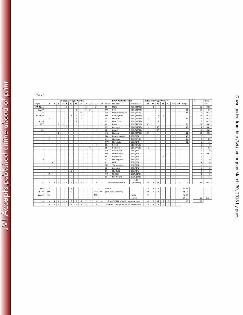
Table 1 shows the origin of samples and the samples contained within each DNA sequence type. 419
Origin of samples are described down the centre of the table, with the code on the left (for position 420
of these codes relating to UK geography, see Figures 1 and 2). Sequence type numbers are located 421
across the top (and correspond to those on the tree in Figures 1 and 2). The origin of each sample 422
within this group is located underneath in the column under the sequence type (for example 423
sequence type 3 has 4 samples from Wolverhampton, 1 from Birmingham etc). Single sequence 424
types are shown towards the ends of the rows. Total number of sequences from an area is shown on 425
the right (N) and the proportion of these which are 2a are shown on the far right. Samples of non-426
PDSA origin are shown at the bottom, with the total number of sequences per sequence type 427
underneath. The number of hospitals where a sequence type was isolated is at the very bottom of 428
the table. Sequence types which showed significant evidence of clustering in certain areas are 429
indicated with an *, though numbers were small (see text). 430
(Origin of non PDSA samples: HP= Hartlepool, CW = Crewe, LV = Liverpool, StH = St Helens, BL = 431
Blyth, WO = Worcester, CA = Cambridge, BA = Barrow, BP = Blackpool, SH= Shrewsbury) 432 on March 30, 2018 by guest
http://jvi.asm.org/
Dow
nloaded from
2b Sequence Type Number PDSA PetAid Hospital 2a Sequence Type Number N Ratio
single 2 3 5 6 9 12 13 15 18 23 27 29 Code Hospital prevalence 34 37 42 46 47 48 50 Single 2a
20, 30 , 1 1 2 3* 1 ST Stoke 79% (23/29) 1 11 0.09
11, 24 1 3* 3* BW Bow 51% (22/43) 39 10 0.1
16 4* WV Wolverhampton 52% (9/17) 31 6 0.17
14,17,26 1 2 1 1 BH Birmingham 71% (23/32) 1 1 1 12 0.25
2 2 1 LE Leicester 73% (11/15) 1 43 7 0.29
4, 28 2 1 NX New Cross 39% (11/28) 2 7 0.29
10,7 3 3 2 EV Everton 85% (49/57) 15* 33 26 0.62
3 CV Coventry 82% (14/17) 1 4* 45 9 0.67
21 1 CF Cardiff 76% (10/13) 4* 6 0.67
1 HU Huyton 82% (24/29) 10* 36 12 0.92
BM Bournemouth 33% (3/9) 1 40 2 1
1 GL Glasgow 45% (5/11) 44 2 0.5
NW Newcastle 50% (1/2) 49 1 1
1 BR Bristol 61% (8/13) 1 0
4* BS Basildon 63% (7/11) 1 5 0.2
1 GH Gateshead 66% (4/6) 1 0
1 MB Middlesboro 66% (4/6) 2 3 0.67
PL Plymouth 66% (2/3) 1 1 1
25 NT Nottingham 71% (5/7) 1 0
4* HL Hull 75% (6/8) 4 0
TM Thamesmead 75% (3/4) 1 1 1
1 CR Croydon 80% (4/5) 1 0
2 SF Sheffield 80% (4/5) 2 0
2 HD Hendon 100% (2/2) 2 0
SD Sunderland 100% (1/1) 1 1 1
16 7 4 4 3 4 3 7 6 7 7 6 3 Sub Total for PDSA
68%
(255/373) 28 1 11 2 2 2 2 9 134 0.43
19 HP 2 1 2 1 Others 2 1 1 32 BA
8 CW SH LV WO LV (non-PDSA samples) StH LV CA 38 CA
22. AS BL StH LV 41 BP
100%
(16/16) 35 StH 16 0.5
18 9 4 4 3 4 8 3 6 7 7 8 4 Grand TOTAL of each sequence type 30 2 12 2 2 2 2 13 150
9 1 2 1 2 5 2 3 4 2 6 3 Number of hospitals per sequence type 7 2 5 2 1 2 2
Table 1
on March 30, 2018 by guest
http://jvi.asm.org/
Dow
nloaded from
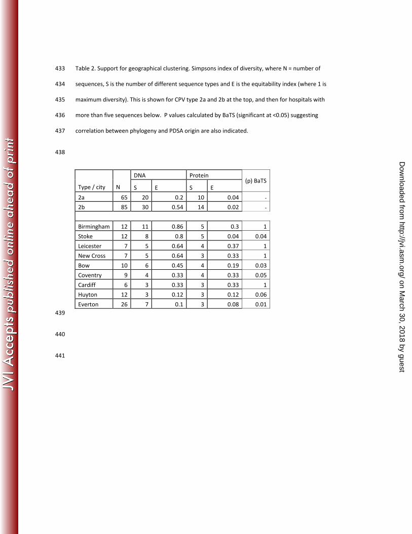
Table 2. Support for geographical clustering. Simpsons index of diversity, where N = number of 433
sequences, S is the number of different sequence types and E is the equitability index (where 1 is 434
maximum diversity). This is shown for CPV type 2a and 2b at the top, and then for hospitals with 435
more than five sequences below. P values calculated by BaTS (significant at <0.05) suggesting 436
correlation between phylogeny and PDSA origin are also indicated. 437
438
DNA Protein
Type / city N S E S E
(p) BaTS
2a 65 20 0.2 10 0.04 -
2b 85 30 0.54 14 0.02 -
Birmingham 12 11 0.86 5 0.3 1
Stoke 12 8 0.8 5 0.04 0.04
Leicester 7 5 0.64 4 0.37 1
New Cross 7 5 0.64 3 0.33 1
Bow 10 6 0.45 4 0.19 0.03
Coventry 9 4 0.33 4 0.33 0.05
Cardiff 6 3 0.33 3 0.33 1
Huyton 12 3 0.12 3 0.12 0.06
Everton 26 7 0.1 3 0.08 0.01
439
440
441
on March 30, 2018 by guest
http://jvi.asm.org/
Dow
nloaded from
442
443
444
445
446
447
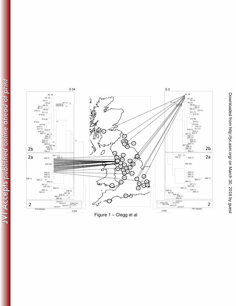
Figure 1 and 2. Phylogenetic analysis of the CPV VP2 DNA sequences in the UK. The phylogenetic 448
tree is based on maximum likelihood with bootstrap support for individual nodes indicated at 449
appropriate nodes (for clarity they are only included on the left-hand tree). In all trees, each 450
sequence type is labelled with its number followed by the number of identical sequences within that 451
group (e.g. S3 (4), indicating sequence type 3 contains 4 identical sequences). On the map, a circle 452
represents the approximate location in the UK of each individual hospital where a CPV positive 453
sample was obtained (for key to hospital origin codes see Table 1). Lines locating sequences to their 454
geographical origin link the map to the phylogeny. The classification of viruses as CPV type2, 2a and 455
2b, as indicated by the shading on each tree, are a classification based on key amino acid mutations 456
(see text). To save space, the same tree is shown in mirror image on either side of the map. 457
Highlighted in figure 1 are the geographical distribution of two different sequence types, S34 (left) 458
and S2 (right), and in figure 2, the genetic diversity of CPV in Everton on the left (lowest diversity as 459
measured by Simpsons index and BaTS) and Birmingham on the right (highest diversity as measured 460
by Simpsons index and BaTS). 461
462
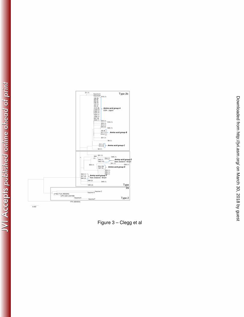
Fig 3. An neighbour-joining amino acid tree of the same samples as used in Figure 1. Samples are 463
labelled with the same sequence type number as is used in the DNA tree. Brackets are used to 464
on March 30, 2018 by guest
http://jvi.asm.org/
Dow
nloaded from
indicate large amino acid groups which are referred to in the text (marked A-F). For A, D and F, 465
where these sequences have been identified both in this country (this study) and in other countries 466
(based on Genbank data), the other countries are listed next to the taxa name. 467
468
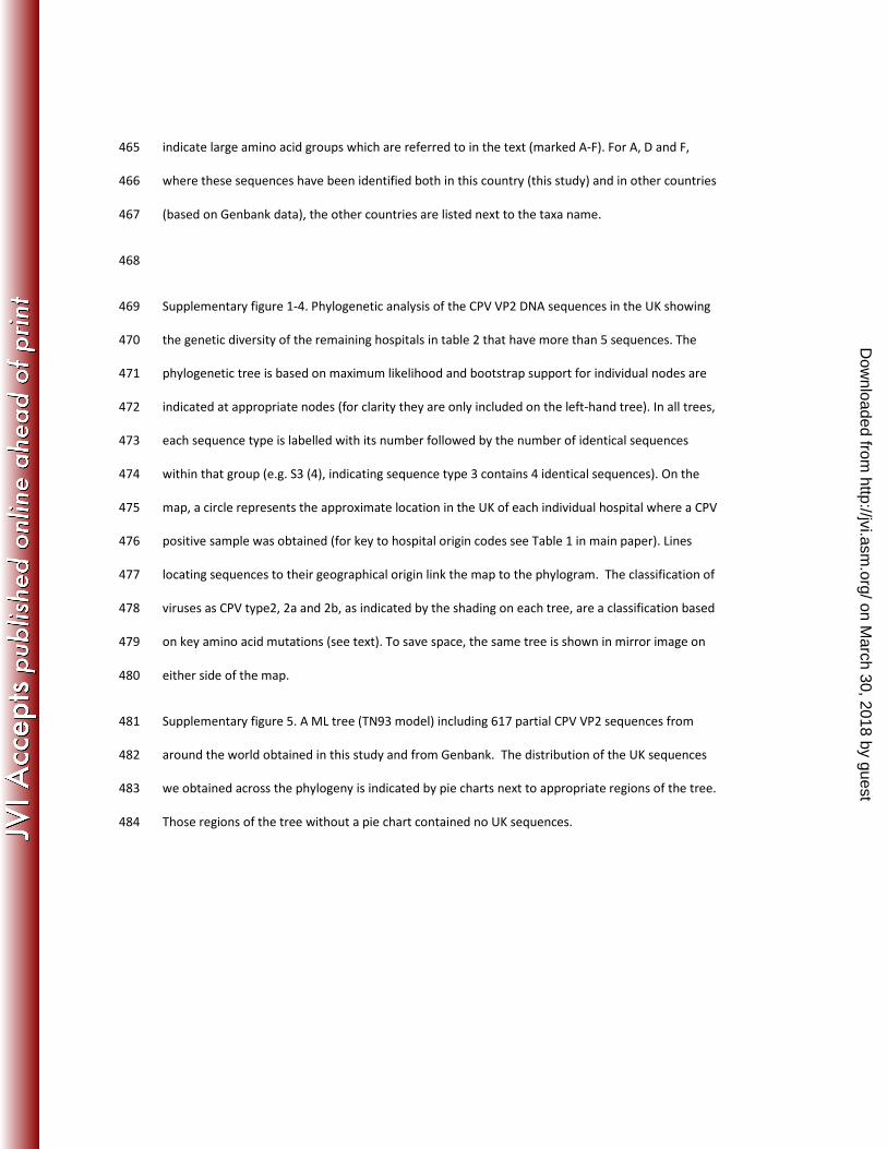
Supplementary figure 1-4. Phylogenetic analysis of the CPV VP2 DNA sequences in the UK showing 469
the genetic diversity of the remaining hospitals in table 2 that have more than 5 sequences. The 470
phylogenetic tree is based on maximum likelihood and bootstrap support for individual nodes are 471
indicated at appropriate nodes (for clarity they are only included on the left-hand tree). In all trees, 472
each sequence type is labelled with its number followed by the number of identical sequences 473
within that group (e.g. S3 (4), indicating sequence type 3 contains 4 identical sequences). On the 474
map, a circle represents the approximate location in the UK of each individual hospital where a CPV 475
positive sample was obtained (for key to hospital origin codes see Table 1 in main paper). Lines 476
locating sequences to their geographical origin link the map to the phylogram. The classification of 477
viruses as CPV type2, 2a and 2b, as indicated by the shading on each tree, are a classification based 478
on key amino acid mutations (see text). To save space, the same tree is shown in mirror image on 479
either side of the map. 480
Supplementary figure 5. A ML tree (TN93 model) including 617 partial CPV VP2 sequences from 481
around the world obtained in this study and from Genbank. The distribution of the UK sequences 482
we obtained across the phylogeny is indicated by pie charts next to appropriate regions of the tree. 483
Those regions of the tree without a pie chart contained no UK sequences. 484
on March 30, 2018 by guest
http://jvi.asm.org/
Dow
nloaded from
0.005FPV M24004
S46 (2)
S43 (1)
S31 * (1)
S2 (9)
S3 (4)
S4 (1)
S5 (4)
S6 (3)S7 (1)
S8 (1)
S10 (1)S11 (1)
S12 (3)
S13 (8)
S15 (6)
S16 (1)
S17 (1)
S19 (1) S20 (1)
S21 (1)S22 (1)S23 (7)
S24 (1)S25 (1)
S26 (1)
86
S28 (1)S27 (8)
S30 (1)
80
S32 (1)S33 (1)
S35 (1)S36 (1)
S37 (2)
S34 (30)
S38 (1)S39 (1)
S40 (1)
S41 (1) S42 (12)
S44 (1)
S45 (1)
98
S1 (1)vaccine Avaccine B
86
84
S47 (2)S48 (2)
S49 (1)87
S50 (2)94
97
vaccine Cvaccine D 2 New York
CPV USAvaccine Evaccine F 96
2b
2a
2
S9 (4)
S18 (7)
S29 (4)
S14(1)
WV
LE
NX
BH
BW
MB
ST
SF
SDNW
GHHP
SR
BL
CR
HD
HL
CF
BR
BM
BA
PL
StH
WO
SH
NT
BP
CR
GL
LV
HU
EV
0.005FPV M24004
S46 (2)
S43 (1)
S31 * (1)
S2 (9)
S3 (4)S4 (1)
S5 (4)
S6 (3)S7 (1)S8 (1)
S10 (1)S11 (1)
S12 (3)
S13 (8)
S15 (6)
S16 (1)
S17 (1)
S19 (1) S20 (1)
S21 (1)S22 (1)S23 (7)
S24 (1)S25 (1)
S26 (1)
S28 (1)S27 (8)
S30 (1)
S32 (1) S33 (1)
S35 (1)S36 (1)
S37 (2)
S34 (30)
S38 (1)S39 (1)
S40 (1)
S41 (1) S42 (12)
S44 (1)
S45 (1)
S1 (1)vaccine Avaccine B
S47 (2)S48 (2)
S49 (1)S50 (2)
vaccine Cvaccine D2 New York
CPV USAvaccine E
vaccine F
2b
2a
2
S9 (4)
S18 (7)
S29 (4)
S14(1)
CA
S 34 S 2
AS
Figure 1 – Clegg et al
CV
BS
TM
on March 30, 2018 by guest
http://jvi.asm.org/
Dow
nloaded from

0.005FPV M24004
S46 (2)
S43 (1)
S31 * (1)
S2 (9)
S3 (4)
S4 (1)
S5 (4)
S6 (3)S7 (1)
S8 (1)
S10 (1)S11 (1)
S12 (3)
S13 (8)
S15 (6)
S16 (1)
S17 (1)
S19 (1) S20 (1)
S21 (1)S22 (1)S23 (7)
S24 (1)S25 (1)
S26 (1)
86
S28 (1)S27 (8)
S30 (1)
80
S32 (1)S33 (1)
S35 (1)S36 (1)
S37 (2)
S34 (30)
S38 (1)S39 (1)
S40 (1)
S41 (1) S42 (12)
S44 (1)
S45 (1)
98
S1 (1)vaccine Avaccine B
86
84
S47 (2)S48 (2)
S49 (1)87
S50 (2)94
97
vaccine Cvaccine D 2 New York
CPV USAvaccine Evaccine F 96
2b
2a
2
S9 (4)
S18 (7)
S29 (4)
S14(1)
WV
LE
NX
BH
BW
MB
ST
SF
SDNW
GHHP
SR
Bl
CR
HD
HL
CF
BR
BM
BA
PL
StH
WO
SH
NT
BP
CR
GL
LV
HU
EV
0.005FPV M24004
S46 (2)
S43 (1)
S31 * (1)
S2 (9)
S3 (4)
S4 (1)
S5 (4)
S6 (3)S7 (1)S8 (1)
S10 (1)S11 (1)
S12 (3)
S13 (8)
S15 (6)
S16 (1)
S17 (1)
S19 (1) S20 (1)
S21 (1)S22 (1)S23 (7)
S24 (1)S25 (1)
S26 (1)
S28 (1)S27 (8)
S30 (1)
S32 (1) S33 (1)
S35 (1)S36 (1)
S37 (2)
S34 (30)
S38 (1)S39 (1)
S40 (1)
S41 (1) S42 (12)
S44 (1)
S45 (1)
S1 (1)vaccine Avaccine B
S47 (2)S48 (2)
S49 (1)S50 (2)
vaccine Cvaccine D2 New York
CPV USAvaccine E
vaccine F
2b
2a
2
S9 (4)
S18 (7)
S29 (4)
S14(1)
CA
Everton (EV) Birmingham (BH)
AS
Figure 2 – Clegg et al
CV
BS
TM
on March 30, 2018 by guest
http://jvi.asm.org/
Dow
nloaded from
S10 (1)
S13 (8)S18 (7)
S5 (4)
S9 (4)
S6 (3)S7 (1)
S2 (9)S3 (4)
S27 (8)
S25 (1)
S28 (1)S20 (1)
S19 (1)
S26 (1)
S14 (1)
S22 (1)S23 (7)
S30 (1) S29 (4)
S11 (1)S16 (1)
S5 (6)
S21 (1)
S24 (1)
S8 (1)
S12 (3)S11 (1)
S4 (1)
S31 (1)S43
(1) S45 (1)
S44 (1)
S42 (12)
S40 (1)
S36 (1)S39 (1)S32 (1)
S47 (2)
S46 (2)
S48 (2)S49 (1)
S50 (2)
S35 (1) S41 (1)
S38 (1) S34 (30)
Vaccine C Vaccine D
2 New York (M23255)
CPV USA (U22186)
Vaccine EVaccine F
S37 (2)
S33 (1)
FPV (M24004)
0.002
S1 (1) Vaccine A Vaccine B
Type 2
Type
2a
Type 2b
Amino acid group AUSA / Japan
Amino acid group B
Amino acid group C
Amino acid group DNew Zealand / Brazil
Amino acid group E
Amino acid group F
New Zealand / Brazil
Figure 3 – Clegg et al
on March 30, 2018 by guest
http://jvi.asm.org/
Dow
nloaded from