molecular detection of leishmania infantum in atypical ... · salvioni od, pereira j, sander mg,...
TRANSCRIPT

Journal of Dermatology and Clinical Research
Cite this article: Salvioni OD, Pereira J, Sander MG, Gómez CV (2017) Molecular Detection of Leishmania infantum in Atypical Cutaneous Lesions from Para-guayan Patients. J Dermatolog Clin Res 5(3): 1104.
Central
*Corresponding authorCeleste Vega Gómez, CEDIC, Manduvira 635 entre 15 de Agosto y O` Leary (Barrio La Encarnación), Asunción, Paraguay, Código Postal: 1255; Tel: 595-21-441513; Email:
Submitted: 29 April 2017
Accepted: 17 May 2017
Published: 25 May 2017
Copyright© 2017 Gómez et al.
OPEN ACCESS
Keywords•Cutaneous leishmaniasis•Real-time PCR•Immunocompetent
Research Article
Molecular Detection of Leishmania infantum in Atypical Cutaneous Lesions from Paraguayan PatientsOscar Daniel Salvioni1, JoséPereira1, Marco González Sander1, and CelesteVega Gómez1*1Center for the Development of Scientific Research, CEDIC, Paraguay2Center for Dermatological Specialties - National Leprosy Control Program, Paraguay
Abstract
Leishmaniasis is one of the most important vector-borne human diseases. Different parasite species are associated with the various forms of the disease. In Paraguay, visceral leishmaniasis is caused by L. infantum, whereas tegumentary leishmaniasis is due mainly to L. braziliensis. Recently, two cases of patients with atypical cutaneous lesions have been described in which the causative agent was determined to be L. infantum. Based on these findings, we aimed to determine the leishmania species of all cutaneous and mucosal biopsy lesions submitted to our research center during 2016 using real-time PCR. We found that 20% of all tegumentary leishmaniasis were due to L. infantum and most of these patients presented with atypical cutaneous lesions and no visceral involvement.
ABBREVIATIONS ACL: Atypical Cutaneous Leishmaniasis; CL: Cutaneous
Leishmaniasis; TL: Tegumentary Leishmaniasis; VIH: Human Immunodeficiency Virus; PCR: Polymerase Chain Reaction; WHO: World Health Organization
INTRODUCTIONThe WHO considers leishmaniasis one of the seven most
important tropical diseases. It has been estimated worldwide that 350 million people are at increased risk of contracting leishmaniasis, with a total incidence of 2 million cases per year. This infection is caused by an obligate protozoan parasite of the genus Leishmania, transmitted through the bite of dipterous insects belonging to the family Phlebotominae [1].
In Latin America, leishmaniasis is mainly caused by two subgenera, L. (Viannia) and L. (Donovani). The subgenus L. (Viannia) includes L. (V.) braziliensis, L. (V.) panamensis, L. (V.) guyanensis which are the causative agents of tegumentary leishmaniasis (TL). Species within the L. (Donovani) subgenus include L. (L.) infantum (synonym of L. (L.) chagasi) which is the causative agent of visceral leishmaniasis, L. (L.) mexicana and L. (L.) amazonensis, which are responsible for the cutaneous form of the disease [2].
In Paraguay, the phlebotomines and fly Lutzomyia longipalpis is the main vector of leishmaniasis, being L. infantum the causative agent of visceral leishmaniasis, L. braziliensis, and in some cases L. guyanensis, these of the majority of cases of mucocutaneous leishamaniasis [3].
A recent report has shown for the first time in Paraguay,
two cases of atypical cutaneous leishmaniasis (ACL), where the causative agent of the lesions was determined to be L. infantum. One of the reported cases was an HIV-immunocompromised adult patient [4]. The other reported case was an immunocompetent adult patient [5].
Based on these reported cases in Paraguay, we aimed to investigate this disease using real-time PCR to avoid false negatives, and to determine the species of all biopsy samples submitted to our laboratory that were positive for the genus Leishmania spp.
MATERIALS AND METHODSSamples
Cutaneous and mucosal lesion biopsies were obtained from a total of 54 patients with suspected leishmaniasis, submitted from all over the country to the Centro de Especialidades Dermatológicas (San Lorenzo, Paraguay) from January to December 2016. For each patient, the following data were collected: age, gender, pet ownership, and occupation.
The biopsies were obtained using a 4mm biopsy punch and sent in 70% ethanol to the Centro para el Desarrollo de la Investigación Científica (CEDIC), where they were stored at -20°C until DNA was extracted.
DNA extraction
Genomic DNA from cutaneous and mucosal lesions was extracted and purified with GeneJet Genomic DNA Purification Kit (#K0722 Thermo Scientific) according to the manufacturer´s instructions. The purity of the DNA samples was assessed with a
Gómez et al. (2017)Email:
J Dermatolog Clin Res 5(3): 1104 (2017) 2/3
Central
spectrophotometer (DeNovix DS-11FX+).
Real-Time PCR reactions and sequencing
For the detection of Leishmania spp DNA by real-time PCR, the following primers that amplify the ribosomal RNA internal transcribed spacer 1 (ITS-1) were used: LSGITS1F.5′CATTTTCCGATGATTACAC 3′ and LSGITS1R: 5′ CGTTATGTGAGCCGTTA 3′, described by Almeida et al. [6,7]. For amplification, Maxima Sybr Green qPCR (2X) (#K0251 Thermo Scientific®) was used.
For the determination of the L. (viannia) complex, the following pairs of primers that amplify a region of the flagellar actin gene were used: LVIAN36ACTF:
5 ˈ C A A G T G C G A C A T T G A T G A T G T G C G A 3 ˈ a n d LVIAN122ACTR:5ˈCGCTCCGGCAGATKTC 3ˈ. Determina-tion of L. (donovani) complex was performed using the fol-lowing pairs of primers that amplify the conserved hypo-thetical protein partial mRNA: 5ˈ CCGCGTGCCTGTCGT 3ˈ and 5ˈCCCACACAAGCGGGAACT3ˈ. Both pairs of primers were previ-ously described by Colombo et al. [8-10]. For amplification, Maxi-ma Probe qPCR (2X) Mix (#K0261 Thermo Scientific®) was used.
Reactions were performed using a Rotor Gene 6000 (Qiagen) thermocycler in a final reaction volume of 20µL. In each of the amplification reactions positive and negative controls were added.
To confirm the presence of L. infantum, a PCR reaction using pairs of primers that amplify the ribosomal RNA internal transcription spacer 2 (ITS-2) was performed, according to the conditions previously described by Almeida et al. [6]. The purified product was submitted to Macrogen (Korea) for sequencing and the results were analyzed by sequence alignment using the BioEdit Sequence Alignment Editor software.
Statistical analysis
The chi-square test (χ2) was used to assess whether patients, men and women working in high-risk occupations (farmers, carpenters, etc.), presented statistical significance (p 0,05) with respect to patients (men and women) working in risk-free áreas.
RESULTS AND DISCUSSION54 samples from patients of both sexes with suspected
leishmaniasis were analyzed, and 15 of them (28%) presented positivity for the genus Leishmaia spp, of whom 13 (87%) are men and 2 (13%) are women, with an average age of 42. 53%of the positive cases reported being pet owners.
Of the 54 patients, 15 (28%) are men working in high-risk occupations (farmers) and 18 (33%) are men with occupations that pose no apparent risk; 5 (9%) are women working in high-risk occupations (farmers), and 16 (30%) are women with occupations that pose no apparent risk. After analyzing the variables between gender and type of occupation, a chi-square value of 2,56 was obtained which led us to reject the hypothesis that the gender variable and risky occupation are independent, and that leishmaniasis has been historically regarded in America as a rural and sylvatic disease.
Of the 15 positive cases for the genus Leishmania spp, 12 (80%) corresponded to the L. viannia (L. braziliensis) complex
and 3 (20%) to the L. donovani (L. infantum) complex. The great majority of the positive patients were male (Table 1).
The three samples positive for the L. Donovani complex were submitted for sequencing. Once the obtained sequences were aligned, it was confirmed that all of them had a 99% similarity to L. infantum(GenBanK accession number KT438781.1.)
As for the geographical distribution, positive patients for the L. Viannia(L. braziliensis)complex live or work in rural areas of the country with agricultural activities, coinciding with a previous report by Velez ID et al. [7], and with a 2015 report by the Servicio Nacional de Erradicación delpaludismo (SENEPA, Paraguay) [11]. Whereas of the three cases positive for L. infantum, one patient resides in a rural area and the other two reside in urban areas of the Central Department. In 2015, SENEPA reported that the majority of the visceral leishmaniasis cases (61,3%) occur in the Central Department [12].
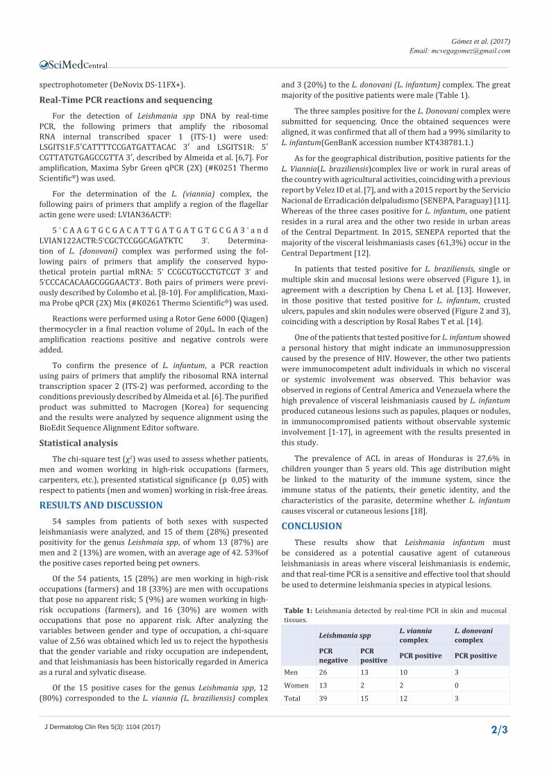
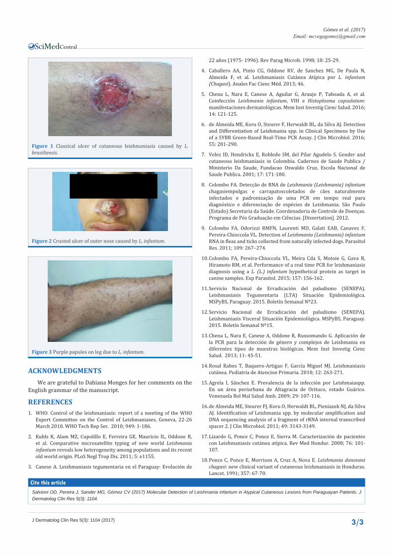
In patients that tested positive for L. braziliensis, single or multiple skin and mucosal lesions were observed (Figure 1), in agreement with a description by Chena L et al. [13]. However, in those positive that tested positive for L. infantum, crusted ulcers, papules and skin nodules were observed (Figure 2 and 3), coinciding with a description by Rosal Rabes T et al. [14].
One of the patients that tested positive for L. infantum showed a personal history that might indicate an immunosuppression caused by the presence of HIV. However, the other two patients were immunocompetent adult individuals in which no visceral or systemic involvement was observed. This behavior was observed in regions of Central America and Venezuela where the high prevalence of visceral leishmaniasis caused by L. infantum produced cutaneous lesions such as papules, plaques or nodules, in immunocompromised patients without observable systemic involvement [1-17], in agreement with the results presented in this study.
The prevalence of ACL in areas of Honduras is 27,6% in children younger than 5 years old. This age distribution might be linked to the maturity of the immune system, since the immune status of the patients, their genetic identity, and the characteristics of the parasite, determine whether L. infantum causes visceral or cutaneous lesions [18].
CONCLUSIONThese results show that Leishmania infantum must
be considered as a potential causative agent of cutaneous leishmaniasis in areas where visceral leishmaniasis is endemic, and that real-time PCR is a sensitive and effective tool that should be used to determine leishmania species in atypical lesions.
Table 1: Leishmania detected by real-time PCR in skin and mucosal tissues.
Leishmania spp L. viannia complex
L. donovani complex
PCR negative
PCR positive PCR positive PCR positive
Men 26 13 10 3
Women 13 2 2 0
Total 39 15 12 3
Gómez et al. (2017)Email:
J Dermatolog Clin Res 5(3): 1104 (2017) 3/3
Central
Salvioni OD, Pereira J, Sander MG, Gómez CV (2017) Molecular Detection of Leishmania infantum in Atypical Cutaneous Lesions from Paraguayan Patients. J Dermatolog Clin Res 5(3): 1104.
Cite this article
22 años (1975- 1996). Rev Parag Microb. 1998; 18: 25-29.
4. Caballero AA, Pinto CG, Oddone RV, de Sanchez MG, De Paula N, Almeida F, et al. Leishmaniasis Cutánea Atípica por L. infantum (Chagasi). Anales Fac Cienc Méd. 2013; 46.
5. Chena L, Nara E, Canese A, Aguilar G, Araujo P, Taboada A, et al. Coinfección Leishmania infantum, VIH e Histoplasma capsulatum: manifestaciones dermatológicas. Mem Inst Investig Cienc Salud. 2016; 14: 121-125.
6. de Almeida ME, Koru O, Steurer F, Herwaldt BL, da Silva AJ. Detection and Differentiation of Leishmania spp. in Clinical Specimens by Use of a SYBR Green-Based Real-Time PCR Assay. J Clin Microbiol. 2016; 55: 281-290.
7. Velez ID, Hendrickx E, Robledo SM, del Pilar Agudelo S. Gender and cutaneous leishmaniasis in Colombia. Cadernos de Saude Publica / Ministerio Da Saude, Fundacao Oswaldo Cruz, Escola Nacional de Saude Publica. 2001; 17: 171-180.
8. Colombo FA. Detecção de RNA de Leishmania (Leishmania) infantum chagasiempulgas e carrapatoscoletados de cães naturalmente infectados e padronização de uma PCR em tempo real para diagnóstico e diferenciação de espécies de Leishmania. São Paulo (Estado) Secretaria da Saúde. Coordenadoria de Controle de Doenças. Programa de Pós Graduação em Ciências. [Dissertation]. 2012.
9. Colombo FA, Odorizzi RMFN, Laurenti MD, Galati EAB, Canavez F, Pereira-Chioccola VL. Detection of Leishmania (Leishmania) infantum RNA in fleas and ticks collected from naturally infected dogs. Parasitol Res. 2011; 109: 267–274.
10. Colombo FA, Pereira-Chioccola VL, Meira Cda S, Motoie G, Gava R, Hiramoto RM, et al. Performance of a real time PCR for leishmaniasis diagnosis using a L. (L.) infantum hypothetical protein as target in canine samples. Exp Parasitol. 2015; 157: 156-162.
11. Servicio Nacional de Erradicación del paludismo (SENEPA). Leishmaniasis Tegumentaria (LTA) Situación Epidemiológica. MSPyBS, Paraguay. 2015. Boletín Semanal Nº23.
12. Servicio Nacional de Erradicación del paludismo (SENEPA). Leishmaniasis Visceral Situación Epidemiológica. MSPyBS, Paraguay. 2015. Boletín Semanal Nº15.
13. Chena L, Nara E, Canese A, Oddone R, Russomando G. Aplicación de la PCR para la detección de género y complejos de Leishmania en diferentes tipos de muestras biológicas. Mem Inst Investig Cienc Salud. 2013; 11: 45-51.
14. Rosal Rabes T, Baquero-Artigao F, García Miguel MJ. Leishmaniasis cutánea. Pediatria de Atencion Primaria. 2010; 12: 263-271.
15. Agrela I, Sánchez E. Prevalencia de la infección por Leishmaiaspp. En un área periurbana de Altagracia de Orituco, estado Guárico. Venezuela Bol Mal Salud Amb. 2009; 29: 107-116.
16. de Almeida ME, Steurer FJ, Koru O, Herwaldt BL, Pieniazek NJ, da Silva AJ. Identification of Leishmania spp. by molecular amplification and DNA sequencing analysis of a fragment of rRNA internal transcribed spacer 2. J Clin Microbiol. 2011; 49: 3143-3149.
17. Lizardo G, Ponce C, Ponce E, Sierra M. Caracterización de pacientes con Leishmaniasis cutánea atípica. Rev Med Hondur. 2008; 76: 101-107.
18. Ponce C, Ponce E, Morrison A, Cruz A, Nova E. Leishmania donovani chagasi: new clinical variant of cutaneous leishmaniasis in Honduras. Lancet. 1991; 357: 67-70.
Figure 1 Classical ulcer of cutaneous leishmaniasis caused by L. braziliensis.
Figure 2 Crusted ulcer of outer nose caused by L. infantum.
Figure 3 Purple papules on leg due to L. infantum.
ACKNOWLEDGMENTSWe are grateful to Dahiana Monges for her comments on the
English grammar of the manuscript.
REFERENCES1. WHO. Control of the leishmaniasis: report of a meeting of the WHO
Expert Committee on the Control of Leishmaniases, Geneva, 22-26 March 2010. WHO Tech Rep Ser. 2010; 949: 1-186.
2. Kuhls K, Alam MZ, Cupolillo E, Ferreira GE, Mauricio IL, Oddone R, et al. Comparative microsatellite typing of new world Leishmania infantum reveals low heterogeneity among populations and its recent old world origin. PLoS Negl Trop Dis. 2011; 5: e1155.
3. Canese A. Leishmaniasis tegumentaria en el Paraguay: Evolución de