molecular characterization of emabp, an apolipoprotein ai binding protein secreted by the
TRANSCRIPT
INFECTION AND IMMUNITY, Dec. 2009, p. 5564–5571 Vol. 77, No. 120019-9567/09/$12.00 doi:10.1128/IAI.00653-09Copyright © 2009, American Society for Microbiology. All Rights Reserved.
Molecular Characterization of EmABP, an Apolipoprotein A-I BindingProtein Secreted by the Echinococcus multilocularis Metacestode�
Peter Bernthaler,1 Kerstin Epping,1 Gerd Schmitz,2 Peter Deplazes,3 and Klaus Brehm1*Institute of Hygiene and Microbiology, University of Wurzburg, D-97080 Wurzburg, Germany1; Institute of Clinical Chemistry and
Laboratory Medicine, Regensburg University Medical Center, D-93042 Regensburg, Germany2; and Institute of Parasitology,University of Zurich, CH-8057 Zurich, Switzerland3
Received 9 June 2009/Returned for modification 13 July 2009/Accepted 18 September 2009
Cestodes are unable to synthesize de novo most of their own membrane lipids, including cholesterol, andhave to take them up from the host during an infection. The underlying molecular mechanisms are so farunknown. Here we report the identification and characterization of a novel gene, Emabp, which is expressed bylarval stages and adults of the fox tapeworm Echinococcus multilocularis. The encoded protein, EmABP, displayssignificant homologies to apolipoprotein A-I binding protein (AI-BP) of mammalian origin and to metazoanYjeF_N domain proteins. Like mammalian AI-BP, EmABP carries an export-directing signal sequence whichis absent in predicted AI-BP orthologs from the related flatworms Schistosoma japonicum and Schmidteamediterranea. Using a specific antibody and immunoprecipitation techniques, we demonstrate that EmABP issecreted into the extraparasitic environment and into the hydatid fluid of in vitro-cultivated metacestodevesicles. Furthermore, we show that apolipoprotein A-I (apoA-I), a major constituent of cholesterol-transport-ing high-density lipoproteins, is present in hydatid fluid. By pulldown experiments, we demonstrate thatrecombinantly expressed, purified EmABP interacts with purified human apoA-I and is able to precipitateapoA-I from human serum. On the basis of these features and the suggested function of AI-BP in cholesteroltransport in higher eukaryotes, we propose a role for EmABP in cholesterol and lipid uptake mechanisms oflarval E. multilocularis.
The metacestode larval stage of the fox tapeworm Echino-coccus multilocularis is the causative agent of alveolar echino-coccosis (AE) in humans, one of the most serious and life-threatening parasitoses of the Northern Hemisphere (16). TheE. multilocularis life cycle involves an adult stage which dwellsin the intestines of definitive hosts, such as foxes or dogs, andproduces infective eggs that contain the parasite’s oncospherelarva. Upon oral ingestion of the eggs by intermediate hosts(rodents and, occasionally, humans), the oncosphere is acti-vated, hatches, and penetrates the intestinal barrier. Withinthe liver of the intermediate host, the oncosphere undergoes ametamorphosis toward the bladder-like metacestode stagewhich grows infiltratively, like a malignant tumor, into thesurrounding host tissue. At a later stage of the infection, nu-merous protoscoleces are formed from the parasite’s germinaltissue, which are passed onto the definitive host when it takesthe prey (6–8, 16, 49). Human E. multilocularis infections arerelatively rare but pose serious problems to surgical and/orchemotherapeutic treatment (28). A very similar life cycle isdisplayed by the closely related dog tapeworm Echinococcusgranulosus, the causative agent of cystic echinococcosis (CE),with several modifications concerning the spectrum of hostspecies (domestic animals), metacestode morphology (uniloc-ular versus multilocular), and organ tropism (the lung, kidney,and brain in addition to the liver) (6, 16).
Although E. multilocularis and E. granulosus contain com-
plex mixtures of lipids, including cholesterol, in their mem-branes, they are unable to synthesize most of these moleculesde novo and share this trait with other cestodes (35). As aconsequence, they have to take up host-derived lipids duringan infection. Particularly in the case of cholesterol, Frayha (19,20) already demonstrated that this compound cannot be syn-thesized by both E. multilocularis and E. granulosus and that atleast E. granulosus incorporates radioactively labeled, host-derived cholesterol during experimental infection of mice. Al-though several Echinococcus proteins with fatty acid and hy-drophobic ligand binding properties have been reported (12,25), none of these displayed cholesterol binding activities norhas, as yet, any cestode molecule been identified that interactswith components of the host’s cholesterol transport machinery.
Mammalian cells acquire exogenous cholesterol mainly fromlow-density lipoprotein (LDL) particles via the LDL receptorpathway. During this process, the LDL receptor specificallyinteracts with the major protein component of LDL particles,apolipoprotein B-100 (apoB-100), resulting in the formation ofclathrin-coated vesicles which are processed via the classicalendocytic pathway. Upon fusion of the vesicles with lysosomes,the entire LDL particle is disassembled by enzymatic hydroly-sis, releasing cholesterol and lipids for cellular metabolism (9,36, 41). The majority of LDL receptors expressed in mammalsare on the surfaces of liver cells, although a certain level ofLDL receptor expression also occurs in the peripheral tissue(9). The LDL/LDL receptor lipid transport system appears tobe evolutionarily conserved, since apoB-100-like cholesterolbinding proteins (vitellogenins) have already been identified inyolk of invertebrates, such as Caenorhabditis elegans and Dro-sophila melanogaster (29, 34, 45). Furthermore, surface recep-
* Corresponding author. Mailing address: Institute of Hygiene andMicrobiology, University of Wurzburg, Josef-Schneider-Strasse 2,D-97080 Wurzburg, Germany. Phone: 49 931 20146168. Fax: 49 93120146445. E-mail: [email protected].
� Published ahead of print on 5 October 2009.
5564
Dow
nloa
ded
from
http
s://j
ourn
als.
asm
.org
/jour
nal/i
ai o
n 15
Nov
embe
r 20
21 b
y 92
.33.
234.
163.

tors of the LDL receptor family have been reported to beexpressed by invertebrates (34).
In addition to exogenous uptake of cholesterol, nearly allmammalian cells are able to also synthesize cholesterol denovo. In cells of peripheral tissues, excess cholesterol needs tobe removed and transported to the liver for reutilization andexcretion. The underlying mechanism of “reverse cholesteroltransport” is mediated by high-density lipoprotein (HDL) par-ticles, the major component of which is apolipoprotein A-I(apoA-I) (38). Lipid-free apoA-I is secreted predominantly bythe liver and intestine and acquires phospholipids and choles-terol via cellular efflux from peripheral tissue cells and macro-phages, giving rise to nascent HDL. Once mature, HDL particlesare transported to the liver, adrenal glands, and steroidogenictissue where they are recognized by the HDL receptor, scav-enger receptor type B class I, upon which the process of “se-lective lipid uptake” by the target cell is induced, which fun-damentally differs from receptor-mediated endocytosis (9, 36,38, 39). During “selective lipid uptake,” cholesterol and phos-pholipids are effectively transferred to target cells, releasingextracellular, lipid-depleted HDL particles which can reentercirculation. Although LDL particles are the major carriers ofcholesterol in human blood, sera from rodents and ungulatestypically contain much higher levels of HDL components thanLDL components (10). Another difference concerns the extra-cellular transfer of cholesteryl esters from HDL particles toother lipoproteins (e.g., LDL) for further transport, which canbe observed only in humans and not in rodents (9). Althoughas in the case of LDL receptors, the scavenger receptor type Bfamily appears to be evolutionarily conserved and occurs alsoin invertebrates (15), soluble apolipoproteins, such as apoA-Ior apoE, are probably deuterostome specific and may have firstappeared around 450 million years ago in an Ordovician ver-tebrate (27).
As yet, only two parasitic flatworm proteins which caninteract with the cholesterol and lipid transport machinery ofmammalian hosts have been reported and both derive fromtrematodes, an LDL receptor-like very low-density lipoproteinbinding protein from Schistosoma japonicum (17) and a CD36-like class B scavenger receptor from Schistosoma mansoni (15)which interacts with modified host LDL at the tegumentalsurface. As a first step toward the characterization of cestodemolecules that are involved in cholesterol uptake during infec-tions of the intermediate host, we herein report the identifica-tion and characterization of a secreted E. multilocularis pro-tein, EmABP, which interacts with human apoA-I, the majorcomponent of HDL particles for reverse cholesterol transport.The role of EmABP in Echinococcus cholesterol uptake mech-anisms is discussed in the background of what is known on thefunction of the homologous apolipoprotein A-I binding pro-tein (AI-BP) (26, 42, 44) in humans.
MATERIALS AND METHODS
Organisms and culture methods. All experiments were performed with thenatural Echinococcus multilocularis isolate H95 which was propagated in Mon-golian jirds or Mongolian gerbils (Meriones unguiculatus) as described previously(50). In vitro cultivation of metacestode vesicles under axenic conditions wasperformed according to an established protocol (47), and protoscoleces wereisolated from in vivo-cultivated parasite material as described previously (5).Pepsin activation of protoscoleces was performed as described previously (48) byincubation of the larvae in pepsin and low pH (0.5 mg/ml pepsin in Hanks’
solution, pH 2.0, 37°C) for 3 h. Egg-free, adult E. multilocularis worms wereisolated from experimentally infected dogs essentially as previously described(14) and kept on RNAlater RNA stabilization reagent (Qiagen) prior to RNAisolation.
RT-PCR analyses. For RNA isolation from in vitro-cultivated metacestodevesicles, protoscoleces, and adult worms, the RNeasy kit (Qiagen) was utilizedaccording to the manufacturer’s instructions. Three to five metacestode vesiclesof 5 mm in diameter or 30 to 50 protoscoleces were used per isolation step, andthe RNA was eluted in 30 �l diethyl pyrocarbonate-treated water. For cDNAsynthesis, total RNA was reverse transcribed using the Omniscript RT kit(Qiagen) according to a previously established protocol (5). Reverse transcrip-tion-PCR (RT-PCR) for the Emabp and elp genes (3) was performed on cDNApreparations from metacestode, invaginated protoscolex, and activated proto-scolex using the primers LA1BP-5�mod (5�-GTG CAG GTC GCT CCG TGCG-3�) and LABPUP2 (5�-GAG TGA GAT CAA GCA ATC TG-3�) as well asELP-DW and ELP-UP (5), respectively. PCR products were separated on a 1%agarose gel and stained with ethidium bromide.
Heterologous expression and purification of Emabp in Escherichia coli. Forheterologous expression in E. coli, the pBAD/TOPO-ThioFusion system (In-vitrogen) was used. The Emabp reading frame was PCR amplified from E.multilocularis cDNA using the primers APOBPDWY (5�-CTC AGT CAG GAGGAG GCG-3�) and LA1BP-3� (5�-CTT ATT GGG TGT CTG GAG G-3�), andthe resulting fragment was cloned into pBAD/Thio via TA cloning, yieldingplasmid pPB-ExABP. In pPB-ExABP, Emabp was translationally fused to anN-terminal thioredoxin moiety and carried the V5 antibody epitope (Invitrogen)as well as a hexahistidine tag at the C terminus (thio-EmABP; 43 kDa). Recom-binant protein expression was induced through the addition of arabinose to E.coli cultures as previously described (46), and purification of thio-EmABP undernative conditions was performed according to established protocols (22, 48). Fora control, we recombinantly expressed a fusion protein that consisted of only thethioredoxin moiety fused to the V5 epitope and the hexahistidine tags (thio-V5-His6; 16 kDa) from plasmid pBAD/Thio (Invitrogen) and purified it underidentical conditions as thio-EmABP.
Antibodies and Western blot analyses. For the detection of EmABP in culturesupernatant and hydatid fluid, a rabbit anti-human AI-BP polyclonal antiserum(42) was used. Detection of the heterologously expressed thio-EmABP andthio-V5-His6 was done using the monoclonal (mouse) anti-V5 antibody (Invitro-gen). For immunoprecipitation and detection of apoA-I, a goat anti-humanapoA-I antiserum (Acris Antibodies GmbH; catalogue no. R1029P) was used,and Echinococcus antigen B was detected by employing the monoclonal antibodyEB7 (23). For Western blot detection, lysates or immunoprecipitation complexeswere separated on 12% acrylamide gels and transferred to nitrocellulose mem-branes (Schleicher and Schuell GmbH). Detection was performed with theabove-mentioned antibodies using peroxidase-conjugated anti-mouse immuno-globulin G or M (IgG/M), anti-rabbit IgG (both Jackson Immuno ResearchLaboratories Inc.), or anti-goat/sheep IgG antibodies (Sigma) as secondary an-tibodies according to the manufacturer’s instructions.
Immunoprecipitation of ApoA-I and EmABP from hydatid fluid and medium.Thirty microliters of protein G-agarose (Upstate) was equilibrated in bindingbuffer (1% bovine serum albumin and 1% Triton X-100 in phosphate-bufferedsaline [PBS]). The anti-AI-BP (diluted 1:100) (41) or anti-apoA-I (1:100; Acris)antibodies were bound to the agarose beads overnight at 4°C in binding buffer.In vitro-cultivated metacestode vesicles (�1 cm in diameter) were washed withprewarmed PBS and transferred to an incubation tube. The vesicles werepinched with a syringe, and the cellular fraction was pelleted by centrifugation(1,000 rpm, 3 min) at 4°C. Hydatid fluid (supernatant) was transferred to a freshtube and kept on ice. After the protein G-antibody complexes were washed withbinding buffer, 2 ml of hydatid fluid was added and incubated under agitationovernight at 4°C. The protein G-antibody complexes were pelleted by centrifu-gation for 1 min at 14,000 rpm, and the supernatant was removed. The pellet wassubsequently washed three times with ice-cold washing buffer (1% Triton X-100in PBS). Finally, 30 �l of 2� sodium dodecyl sulfate sample buffer containing�-mercaptoethanol was added to the agarose beads, followed by boiling for 5 minprior to acrylamide gel electrophoresis and Western blot detection as describedabove.
Pulldown assays. To test the interaction between purified thio-EmABP andapoA-I, agarose G beads (Upstate) (30 �l) were equilibrated in binding buffer.The anti-ApoA-I antibody (1:100) and human apoA-I (15 �l) (1.32 mg/ml; Acris)were bound to the beads overnight at 4°C in binding buffer. After the proteinG-antibody/antigen complexes had been washed in washing buffer, 30 �l ofpurified thio-EmABP (110 �g/�l) was added and incubated for 2 h at 4°C. Theantibody/antigen complexes were pelleted by centrifugation for 1 min at 14,000rpm, and the pellet was washed three times with cold washing buffer prior to
VOL. 77, 2009 ECHINOCOCCUS APOLIPOPROTEIN A-I BINDING PROTEIN 5565
Dow
nloa
ded
from
http
s://j
ourn
als.
asm
.org
/jour
nal/i
ai o
n 15
Nov
embe
r 20
21 b
y 92
.33.
234.
163.
sample preparation for gel electrophoresis as described above. To precipitateapoA-I from human serum, 50 �l of protein A Sepharose (Sigma) was equili-brated in binding buffer. The anti-AI-BP antibody (1:100) (42) and purifiedEmABP (15 �l) (110 �g/�l) were bound to the beads overnight at 4°C in bindingbuffer. After the protein G-antibody/antigen complex was washed with washingbuffer, 1 ml of human serum was added. After overnight incubation at 4°C, theantibody/antigen complexes were further processed essentially as describedabove. Each assay was performed at least twice.
Computer-based analyses. Sequence alignments and comparisons were per-formed using the Basic Local Alignment Search Tool (BLAST) software on theSWISSPROT and nr-aa database collections available at http://blast.genome.jp,the Schmidtea mediterranea genome database (http://smedgd.neuro.utah.edu/),and the E. multilocularis genome project database (http://www.sanger.ac.uk/Projects/Echinococcus/). Predictions of export-directing signal sequences,transmembrane regions, and domains were done using the iPSORT algo-rithm, available at http://hc.ims.u-tokyo.ac.jp/iPSORT/ (2) and the SimpleModular Architecture ResearchTools (SMART), available at http://smart.embl-heidelberg.de/ (30). Secondary structure predictions were made usingthe Jpred 3 algorithm (http://www.compbio.dundee.ac.uk/www-jpred/index.html) as described previously (13).
Nucleotide sequence accession number. The complete Emabp cDNA se-quence reported in this paper was deposited in the GenBank database under theaccession number FM958505.
RESULTS
Characterization of the Emabp cDNA and genomic locus.During previous studies of the trans-splicing mechanism inEchinococcus multilocularis, we have established cDNA librar-ies for trans-spliced mRNAs, isolated from in vitro-cultivatedmetacestode vesicles and protoscoleces (4). Approximately 300cDNAs from each library were sequenced and subjected toBLAST sequence analyses using the SWISSPROT and nr-aadatabases. One of the characterized clones, designated Z2-45,which was derived from the metacestode cDNA library, en-coded a protein with significant homology to human AI-BP(42) and was further analyzed. Due to this homology and to the
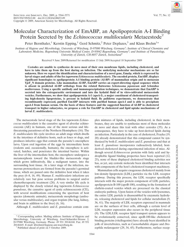
apoA-I binding activity of the encoded protein (see below),this gene was named Emabp (for Echinococcus multilocularisapoA-I binding protein), encoding the protein EmABP. Thefull-length Emabp cDNA comprised 950 nucleotides [exclud-ing the spliced leader (SL) and the poly(A) tail] and containeda putative polyadenylation signal (AATAAA), 17 nucleotidesupstream of the poly(A) tail. Immediately following the SL,there was an open reading frame which coded for a 273-amino-acid protein (theoretical molecular mass of 30 kDa) whichdisplayed significant homologies (up to 60% identical and 72%similar residues) to various mammalian AI-BP orthologs (Fig.1) and somewhat lower homologies to YjeF_N domain-con-taining proteins of invertebrates and bacteria (data not shown).Using SMART analyses, a YjeF_N domain was identified inEmABP between residues Y47 and C220, and all residues thatare known to be invariant or highly conserved in YjeF_Ndomains were also present in EmABP (Fig. 1). Furthermore,using the computer-based program iPSORT, an export-direct-ing signal sequence of 30 amino acids was identified at the Nterminus of EmABP (Fig. 1), leading to a mature protein of26.6 kDa after cleavage. Secondary structure predictions wereperformed using the Jpred 3 algorithm (13) for EmABP andhuman AI-BP and were compared with crystallographic datapreviously obtained for murine AI-BP (26). In all three cases,a very similar distribution of �-helices and �-strands was ob-served, indicating that all three proteins are capable of adopt-ing a Rossmann-like fold in the YjeF_N domain (data notshown). Homology searches in expressed sequence tag data-bases of the related parasitic tapeworm Schistosoma japonicum(31) and the free-living planarian Schmidtea mediterranea (43)revealed the presence of EmABP orthologs in both organisms,which displayed similarity values to the Echinococcus protein
FIG. 1. Structural features and sequence homologies of EmABP. Displayed is a CLUSTAL V alignment (MEGALIGN) between E. mul-tilocularis EmABP (this work), human AI-BP (Homo sapiens AI-BP [HsAI-BP]) (GenBank accession no. AJ315849) (42) as well as EmABPorthologs from S. japonicum (SjABP) (GenBank accession no. AY815871) (31) and S. mediterranea (SmedABP) (mk4.005551.03; http://smedgd.neuro.utah.edu/) (43). Amino acid positions are numbered at the right margin. Residues that are conserved in at least two of the sequences areprinted in white on black background. Homologies of the aligned proteins to EmABP are indicated at the end of the alignment (percent similarityand percent identity). A thick dashed overline indicates the YjeF_N domain, as predicted by the SMART algorithm (30). Arrows above and belowthe alignment indicate predicted signal peptidase cleavage sites in EmABP and human AI-BP (42), respectively. Asterisks above and below thealignment mark the positions of introns in the coding regions for EmABP and human AI-BP (44), respectively. Open circles below thealignment indicate invariant residues, and closed circles indicate highly conserved residues within YjeF_N domains (26). A circled “P” belowthe alignment indicates the serine residue which, in murine AI-BP, is phosphorylated by protein kinase A (26). Gaps introduced to maximizealignment are indicated by dashes in the sequences.
5566 BERNTHALER ET AL. INFECT. IMMUN.
Dow
nloa
ded
from
http
s://j
ourn
als.
asm
.org
/jour
nal/i
ai o
n 15
Nov
embe
r 20
21 b
y 92
.33.
234.
163.

in the range of values observed between EmABP and humanAI-BP. Interestingly, neither in the trematode nor in the pla-narian ortholog was a signal sequence present (Fig. 1). Be-tween the N-terminal signal sequence and the YjeF_N domain,mammalian AI-BP orthologs typically carry a stretch of 25amino acids (Fig. 1) which contains a serine residue that,during murine sperm capacitation, is phosphorylated by pro-tein kinase A (26). A similar sequence stretch was absent inEmABP and was also missing upstream of the YjeF_N domainin the Schistosoma and Schmidtea orthologs (Fig. 1).
Fernandez et al. (18) have previously generated SL- andoligonucleotide-capped cDNA libraries from E. granulosuswhich are available under ftp://ftp.sanger.ac.uk/pub/pathogens/Echinococcus/. Homology searches in these databases revealedthe presence of two full-length clones coding for proteins with98% (EGPSPsl-14g07.q1k) and 97% (EGPSPsl-10b12.q1k)identity to EmABP. Both derived from a SL library made fromprotoscolex mRNA and contained the E. granulosus SL at the5� end. Compared to EmABP, four amino acid exchanges wereobserved in the sequence from EGPSPsl-14g07.q1k (Q2L, P5L,N182K, and K198E) and two additional exchanges in the EGPSPsl-10b12.q1k sequence (K38R and Y47H).
The E. multilocularis genome is currently being se-quenced, and sequence information representing fourfoldcoverage is available under http://www.sanger.ac.uk/Projects/Echinococcus/. When the cDNA sequence of Emabp wascompared with the available data, all exons were identifiedon three adjacent contigs and the reading frame was shownto be interrupted by six introns (Fig. 1) which all containedcanonical GT and AG residues at the 5� and 3� ends, respec-tively. The human gene encoding AI-BP contains five introns(44) which all map to identical positions as the first five intronsof Emabp (Fig. 1), indicating that both genes share a commongenetic ancestor. On the genome sequence available at thistime, no further gene encoding a YjeF_N domain protein wasidentified. Hence, Emabp is most probably a single-copy gene.
Expression of Emabp in Echinococcus larval stages andadult worms. The expression of Emabp in larval stages wasanalyzed by semiquantitative RT-PCR. As shown in Fig. 2,Emabp transcripts were detected in metacestode vesicles, inresting protoscoleces, and to a somewhat lower extent, in theprotoscolex after activation through pepsin/low-pH treatment.We also isolated RNA from adult E. multilocularis worms (free
of eggs) and clearly identified Emabp in respective cDNApreparations by RT-PCR (data not shown). Taken together,these data indicated that Emabp is constitutively expressedthroughout the parasite’s life cycle.
Recombinant expression and purification of EmABP. TheEmabp reading frame, excluding the codons for the signalsequence, was translationally fused to thiredoxin (N-terminal)as well as a hexahistidine tag and the V5 antibody epitope(both C-terminal) using the pBAD/Thio-TOPO system (In-vitrogen). The induced fusion protein (thio-EmABP) was sub-sequently purified under native conditions (Fig. 3A). An anti-body against human AI-BP has previously been produced byimmunization of a rabbit with the peptide V263PPALEKKYQLNLPPYPDTE282 (42). Since EmABP contained a very simi-lar sequence (V236PPLLADKYNLCLPPYPGAS255 [identicalresidues underlined]), we tested whether the anti-AI-BP anti-body also detected EmABP, which was indeed the case (Fig.3B). On the other hand, the anti-AI-BP antibody did not in-teract with the thio-V5-His6 control protein (data not shown).
Secretion of EmABP and detection of apoA-I in hydatidfluid. Since an export-directing signal sequence was identifiedin the deduced amino acid sequence of EmABP, we investi-gated whether the protein is secreted into the extraparasiticenvironment by the E. multilocularis metacestode. In vitro-cultivated metacestode vesicles (47, 50) were taken out ofculture, thoroughly washed, and placed into minimal essentialmedium without protein supplements for 12 and 24 h. Westernblot analyses of the supernatant with the anti-AI-BP antibodydid not reveal bands, which was most probably due to a lowconcentration of secreted factors. We therefore employed theanti-AI-BP antibody in immunoprecipitation experiments and
FIG. 2. Emabp expression patterns. Total RNA has been isolatedfrom in vitro-cultivated metacestode vesicles (MC) as well as fromdormant (Pd) and low-pH/pepsin-activated (Pa) protoscoleces and wasreverse transcribed into cDNA. RT-PCR was performed using primersspecific for Emabp (abp) and the constitutively expressed control geneelp (3, 5). PCR products were separated on a 1% agarose gel andstained with ethidium bromide.
FIG. 3. Heterologous expression, purification, and detection ofEmABP. (A) Heterologous expression in E. coli and purification. TheEmabp reading frame was cloned in the pBAD/TOPO ThioFusionexpression system (Invitrogen) in E. coli. Lysates of arabinose-induced(lane 2) and noninduced (lane 1) E. coli were separated on an 12%acrylamide gel, transferred to a nitrocellulose membrane, and analyzedusing the anti-V5 antibody (Invitrogen). Heterologously expressedthio-EmABP was purified under native conditions, and samples ofelution steps 1 to 5 were applied to lanes 3 to 7, respectively. Separa-tion of the samples and protein detection was performed as describedabove. The positions of molecular size markers (in kilodaltons) areindicated to the left of the gel. (B) Detection of thio-EmABP using ananti-AI-BP antibody. Lysates of recombinant E. coli carrying plasmidsfor the thio-EmABP fusion protein were prepared after arabinoseinduction (lane 1) or without arabinose induction (lane 2), separatedon an 12% acrylamide gel, transferred to a nitrocellulose membrane,and detected by using an anti-AI-BP (human) antibody (42).
VOL. 77, 2009 ECHINOCOCCUS APOLIPOPROTEIN A-I BINDING PROTEIN 5567
Dow
nloa
ded
from
http
s://j
ourn
als.
asm
.org
/jour
nal/i
ai o
n 15
Nov
embe
r 20
21 b
y 92
.33.
234.
163.
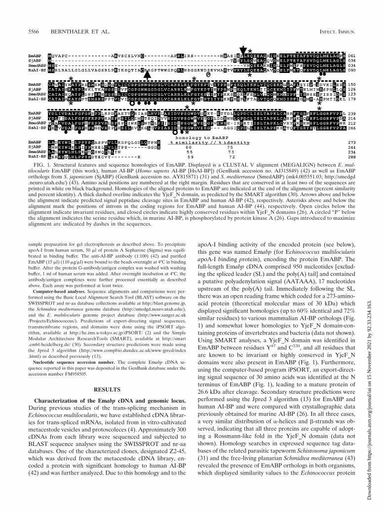
could successfully precipitate a single protein of the expectedsize (Fig. 4), indicating that EmABP is secreted by the E.multilocularis metacestode. For a control, we performed im-munoprecipitation with an antibody against antigen B, one ofthe most abundant proteins in hydatid fluid (33). However, wenever obtained bands despite the fact that antigen B was easilydetected in hydatid fluid using the same antibody. These re-sults showed that the metacestode vesicles in our assays werestructurally intact and that the supernatant did not containhydatid fluid contaminants.
Next, we investigated whether EmABP is also present inhydatid fluid. In Western blot analyses using the anti-AI-BPantibody on freshly isolated hydatid fluid, only a very faintband at the expected size was obtained (data not shown).Immunoprecipitation was therefore performed as describedabove with hydatid fluid from in vitro-cultured metacestodevesicles instead of supernatant, and as shown in Fig. 4, a pro-tein of the expected size was clearly present. Since we neverdetected mammalian AI-BP in the conditioned medium thatwas used to cultivate metacestode vesicles in vitro (data notshown), we concluded that the immunoreactive protein in hy-datid fluid was indeed EmABP and not host-derived AI-BPthat had been transported across the parasite’s laminated andgerminal layers. Using a commercial antibody against apoA-I,we also tried to immunoprecipitate the mammalian apoli-poprotein from hydatid fluid and were successful (Fig. 4).Since apoA-I orthologs are usually found only in higher meta-zoans (27) and since we could not find indications that theparasite’s genome contains a gene for such an ortholog, weconcluded that this immunoreactive protein indeed derivedfrom the host and not from E. multilocularis. Taken together,these data indicated that EmABP is secreted by the metaces-
tode both into the interior lumen (hydatid fluid) and into theexterior. Furthermore, similar to other host serum factors likealbumin or immunoglobulins (11), apoA-I is present in hydatidfluid.
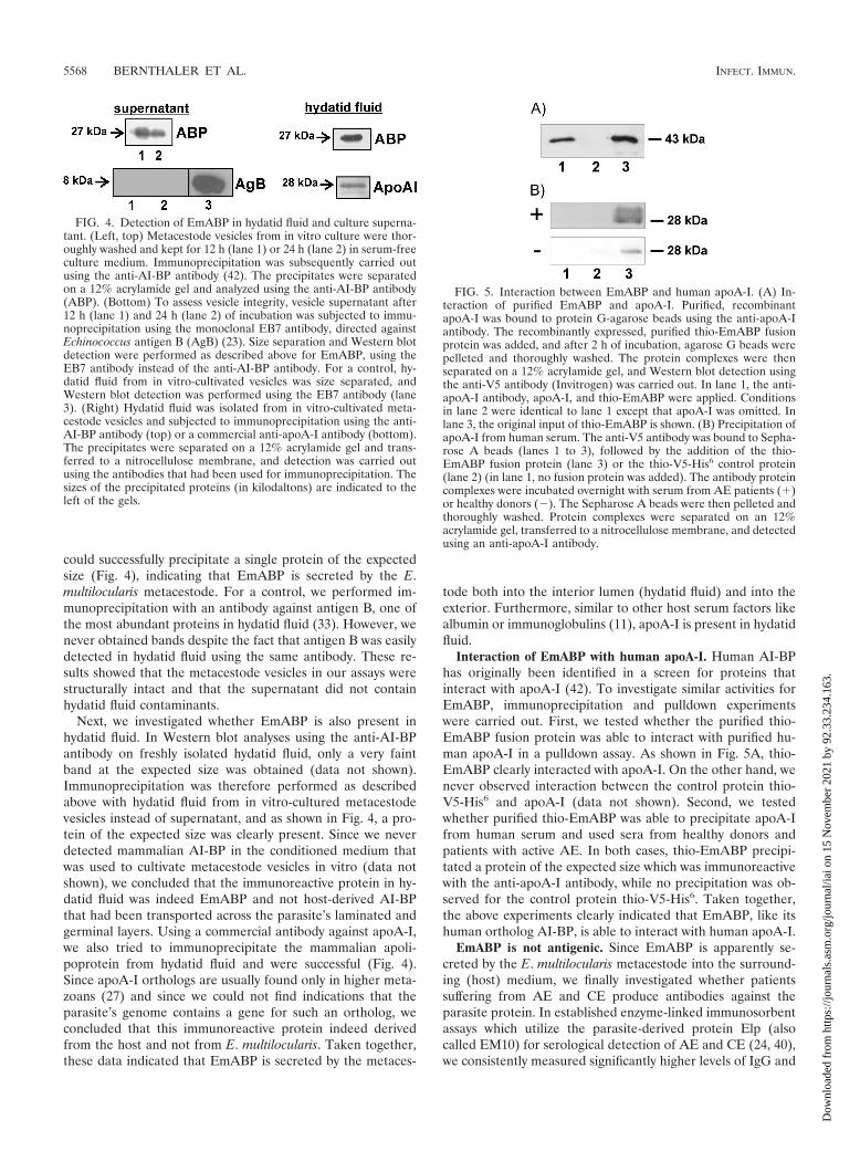
Interaction of EmABP with human apoA-I. Human AI-BPhas originally been identified in a screen for proteins thatinteract with apoA-I (42). To investigate similar activities forEmABP, immunoprecipitation and pulldown experimentswere carried out. First, we tested whether the purified thio-EmABP fusion protein was able to interact with purified hu-man apoA-I in a pulldown assay. As shown in Fig. 5A, thio-EmABP clearly interacted with apoA-I. On the other hand, wenever observed interaction between the control protein thio-V5-His6 and apoA-I (data not shown). Second, we testedwhether purified thio-EmABP was able to precipitate apoA-Ifrom human serum and used sera from healthy donors andpatients with active AE. In both cases, thio-EmABP precipi-tated a protein of the expected size which was immunoreactivewith the anti-apoA-I antibody, while no precipitation was ob-served for the control protein thio-V5-His6. Taken together,the above experiments clearly indicated that EmABP, like itshuman ortholog AI-BP, is able to interact with human apoA-I.
EmABP is not antigenic. Since EmABP is apparently se-creted by the E. multilocularis metacestode into the surround-ing (host) medium, we finally investigated whether patientssuffering from AE and CE produce antibodies against theparasite protein. In established enzyme-linked immunosorbentassays which utilize the parasite-derived protein Elp (alsocalled EM10) for serological detection of AE and CE (24, 40),we consistently measured significantly higher levels of IgG and
FIG. 4. Detection of EmABP in hydatid fluid and culture superna-tant. (Left, top) Metacestode vesicles from in vitro culture were thor-oughly washed and kept for 12 h (lane 1) or 24 h (lane 2) in serum-freeculture medium. Immunoprecipitation was subsequently carried outusing the anti-AI-BP antibody (42). The precipitates were separatedon a 12% acrylamide gel and analyzed using the anti-AI-BP antibody(ABP). (Bottom) To assess vesicle integrity, vesicle supernatant after12 h (lane 1) and 24 h (lane 2) of incubation was subjected to immu-noprecipitation using the monoclonal EB7 antibody, directed againstEchinococcus antigen B (AgB) (23). Size separation and Western blotdetection were performed as described above for EmABP, using theEB7 antibody instead of the anti-AI-BP antibody. For a control, hy-datid fluid from in vitro-cultivated vesicles was size separated, andWestern blot detection was performed using the EB7 antibody (lane3). (Right) Hydatid fluid was isolated from in vitro-cultivated meta-cestode vesicles and subjected to immunoprecipitation using the anti-AI-BP antibody (top) or a commercial anti-apoA-I antibody (bottom).The precipitates were separated on a 12% acrylamide gel and trans-ferred to a nitrocellulose membrane, and detection was carried outusing the antibodies that had been used for immunoprecipitation. Thesizes of the precipitated proteins (in kilodaltons) are indicated to theleft of the gels.
FIG. 5. Interaction between EmABP and human apoA-I. (A) In-teraction of purified EmABP and apoA-I. Purified, recombinantapoA-I was bound to protein G-agarose beads using the anti-apoA-Iantibody. The recombinantly expressed, purified thio-EmABP fusionprotein was added, and after 2 h of incubation, agarose G beads werepelleted and thoroughly washed. The protein complexes were thenseparated on a 12% acrylamide gel, and Western blot detection usingthe anti-V5 antibody (Invitrogen) was carried out. In lane 1, the anti-apoA-I antibody, apoA-I, and thio-EmABP were applied. Conditionsin lane 2 were identical to lane 1 except that apoA-I was omitted. Inlane 3, the original input of thio-EmABP is shown. (B) Precipitation ofapoA-I from human serum. The anti-V5 antibody was bound to Sepha-rose A beads (lanes 1 to 3), followed by the addition of the thio-EmABP fusion protein (lane 3) or the thio-V5-His6 control protein(lane 2) (in lane 1, no fusion protein was added). The antibody proteincomplexes were incubated overnight with serum from AE patients (�)or healthy donors (�). The Sepharose A beads were then pelleted andthoroughly washed. Protein complexes were separated on an 12%acrylamide gel, transferred to a nitrocellulose membrane, and detectedusing an anti-apoA-I antibody.
5568 BERNTHALER ET AL. INFECT. IMMUN.
Dow
nloa
ded
from
http
s://j
ourn
als.
asm
.org
/jour
nal/i
ai o
n 15
Nov
embe
r 20
21 b
y 92
.33.
234.
163.

IgE antibodies against Elp in sera from patients with con-firmed AE and CE compared to sera from healthy donors.When using thio-EmABP as an antigen, on the other hand, noelevated antibody levels were detected in patient sera. Further-more, anti-EmABP antibody levels in AE patients were con-sistently as low as anti-Elp antibody levels in healthy donors(data not shown). Taken together, we conclude from theseexperiments that EmABP is not acting as a parasite antigenduring active infections, which is most likely a result of itsoverall high homology to human AI-BP.
DISCUSSION
In the present study, we have identified a novel protein,EmABP, which is secreted by the Echinococcus multilocularismetacestode into the surrounding host medium (as well as intohydatid fluid; for a diagrammatic representation of the E. mul-tilocularis metacestode, please see reference 8) and interactswith host-derived apoA-I. Due to the crucial function ofapoA-I in mammalian lipid and cholesterol transport processesmediated through HDL particles, this feature suggests a po-tential role of EmABP in interfering with host lipid and cho-lesterol transport.
The fact that E. multilocularis and E. granulosus, like otherparasitic flatworms, cannot de novo synthesize cholesterol andthe majority of other lipid components, has already been firmlyestablished by previous studies (19, 20, 35). This is supportedby our own analyses of the first draft version of the E. mul-tilocularis genome, which showed that genes for the majority ofenzymes that are involved in cholesterol synthesis in otherorganisms (51) are absent in the cestode (data not shown). Inthe case of the E. granulosus metacestode, an uptake of radio-actively labeled, host-derived cholesterol during an infection oflaboratory animals has already been demonstrated (20), and itis reasonable to assume that E. multilocularis employs choles-terol uptake mechanisms similar to those of the closely relateddog tapeworm. Hence, in addition, or as an alternative, tointerfering with lipid/cholesterol transport of the host, EmABPmight be actively involved in cholesterol and lipid uptake bythe parasite.
Like its mammalian ortholog AI-BP, EmABP belongs to thewidespread YjeF_N protein family, members of which arepresent in eukaryotes, bacteria, and archaea (1). Unfortu-nately, neither the general biochemical properties nor the cel-lular functions of YjeF_N proteins are known at this time, theprotein family being in the top 10 list of highly attractivetargets for functional characterization (21). YjeF_N domainseither occur as single proteins, like in the case of EmABP andAI-BP, or as fusions with other domains and are commonlyassociated with enzymes (1). Structurally, the YjeF_N domaindisplays a Rossman-like fold, a structural motif found in pro-teins that bind nucleotides, especially the cofactor NAD (32),and it is generally thought that the domain exerts an unknownenzymatic activity, such as dephosphorylation, demethylation,and phosphoester or glycosyl bond hydrolysis (1). Although theprecise biochemical and cell biological functions of AI-BP areunknown at this time, several lines of evidence indicate thatthis protein plays a role in cellular cholesterol and lipid trans-port mechanisms in a variety of organs in higher eukaryotes.First, AI-BP is expressed in a wide variety of mammalian
tissues, and apart from its binding capacity to apoA-I, it local-izes in domains in sperm in which cholesterol is known to beconcentrated (26). Second, AI-BP is released into the mediumduring sperm capacitation, a process that is accompanied bycholesterol release (26). Third, immunohistochemical analysesof human testes and ovaries identified AI-BP in various celltypes coexpressed with ABCA1 (ATP binding cassette trans-porter A1), the main determinant for plasma HDL, which isinvolved in cellular lipid transport and steroid hormone syn-thesis and which also interacts with apoA-I (44). Apart from itsfunction in spermiogenesis and oogenesis, AI-BP seems to alsoplay an important role in hepatocyte metabolism, since it ishighly expressed in liver tissue as well as in HepG2 cells (42).Interestingly, although sera from healthy donors do not con-tain AI-BP, increased plasma levels were observed in patientssuffering from systemic inflammation and sepsis, which couldbe linked to general changes in plasma lipoprotein metabolismthat occur during inflammatory processes (35, 42). Hence,once released by the E. multilocularis metacestode at the pri-mary site of infection (the liver), EmABP could indeed affectboth the physiology of hepatocytes and the local immune re-sponse. It has to be kept in mind, however, that apoA-I andother lipid transporters are among the most abundant compo-nents of human serum (38) and that probably very largeamounts of EmABP would have to be secreted in order tosignificantly interfere with this equilibrium. Hence, as furtheroutlined below, we clearly favor a role for EmABP in choles-terol uptake mechanisms of the parasitic metacestode over afunction in altering the host’s immune response.
Apart from Emabp expression in the metacestode, we alsofound the gene transcribed in adult worms. Due to the absenceof apoA-I and apoA-I-dependent cholesterol transport activi-ties in the gut of the definitive host, we do not consider it likelythat the suggested role of the protein in cholesterol uptake isshared between metacestode vesicles and adult worms. SinceAI-BP orthologs are apparently also expressed by adult schis-tosomes and (free-living) planarians, they most probably exertadditional cellular functions that still have to be determined.Due to the location of human AI-BP in the testes and ovaries(42), flatworm EmABP orthologs might be specifically involvedin spermatogenesis and oogenesis, which does, however,clearly require additional experimentation.
One striking difference between mammalian AI-BP or-thologs and YjeF_N family members from bacteria, lower eu-karyotes, or invertebrates is the presence of an export-directingsignal sequence at the N termini of the mammalian proteins(42, 44). Interestingly, Rudolph et al. (44) identified two addi-tional YjeF_N proteins in humans which lack a signal se-quence, are not secreted, and fail to interact with apoA-I. Onthe basis of immunolocalization studies of testes and ovaries,these authors concluded that AI-BP and both additional mam-malian YjeF_N proteins are involved in mechanisms of cho-lesterol processing and steroid hormone metabolism and thatthe specific function of AI-BP is to link these functions to theHDL pathway (44). Since apoA-I and HDL transport mecha-nisms have arisen relatively late in animal evolution (27), it isreasonable to assume that this was accompanied by the acqui-sition of a signal sequence and thus, secretion of AI-BP intothe extracellular environment. In this context, it is interestingto note that EmABP contains a signal sequence which is absent
VOL. 77, 2009 ECHINOCOCCUS APOLIPOPROTEIN A-I BINDING PROTEIN 5569
Dow
nloa
ded
from
http
s://j
ourn
als.
asm
.org
/jour
nal/i
ai o
n 15
Nov
embe
r 20
21 b
y 92
.33.
234.
163.

in the AI-BP orthologs of the related flatworms S. japonicum(parasitic) and S. mediterranea (free-living) and which has ap-parently been acquired during cestode evolution throughgenomic rearrangements (as indicated by the presence of anintron at the respective position). The fact that this signalsequence is indeed active is clearly indicated by our immuno-precipitation experiments which detected EmABP in the ex-tralarval medium and in hydatid fluid. Hence, like in the caseof mammalian AI-BPs, EmABP might have acquired a signalsequence to specifically link the host’s HDL transport mecha-nisms to cholesterol and lipid uptake and metabolism by theparasite.
It is well established that host serum components, such asalbumin or immunoglobulins, are present in hydatid fluid (11),and in this study, we have identified apoA-I as another hostserum factor which is apparently transported from the sur-rounding host medium into the metacestode lumen. Unfortu-nately, the underlying transport mechanisms for any serumcomponent are completely unknown at present. Likewise, littleis known on protein secretion mechanisms in Echinococcus.Therefore, at this time, we cannot tell whether EmABP issimultaneously secreted by the metacestode into the hydatidlumen and into the exterior medium or whether the protein isfirst secreted into the surrounding medium and subsequentlytransported across the germinal layer into hydatid fluid. Thefact that we measured somewhat smaller amounts of secretedEmABP in two different samples of in vitro-cultured metaces-tode vesicles after 24 h compared to 12 h might indeed indicatethat the protein is first secreted and then transported into theparasite lumen once a critical external concentration is builtup. This is supported by results from our Western blot analysesshowing that EmABP is not one of the major constituents ofhydatid fluid but rather is present in smaller amounts. How-ever, although we have used comparable numbers of metaces-tode vesicles in both experimental settings, these might havehad slight physiological differences, so this hypothesis surelyawaits further experimentation. To this end, we are currentlytrying to use recombinantly expressed, purified EmABP to-gether with apoA-I and in vitro-reconstituted HDL particles(37) to study whether the parasite protein might be directlyinvolved in the transport host components into in vitro-culti-vated metacestode vesicles. In any case, the fact that bothapoA-I and EmABP are present in hydatid fluid indicates thatthe parasite protein plays a role in parasite-specific physiolog-ical processes. On the basis of the above outlined functionalproperties of the human ortholog AI-BP, we suggest that thisfunction involves cholesterol metabolism and uptake by E.multilocularis metacestode vesicles.
ACKNOWLEDGMENTS
This work was supported by grants SFB479 and IRTG 1522 (both toK.B.) from the Deutsche Forschungsgemeinschaft.
We are indebted to Gualberto Gonzalez-Sapienza (Montevideo,Uruguay) and Mara Rosenzvit (Buenos Aires, Argentina) for provid-ing the monoclonal EB7 antibody. We also thank Dirk Radloff andMonika Bergmann for excellent technical assistance. Special thanksare addressed to Peter D. Olson (Natural History Museum, London,United Kingdom) for critically reading the manuscript. Sequence datato screen the E. multilocularis genome for EmABP orthologs havebeen produced by the Parasite Sequencing Group at the Sanger Insti-tute and can be obtained from ftp.sanger.ac.uk/pub/pathogens/Echino-coccus.
REFERENCES
1. Anantharaman, V., and L. Aravind. 2004. Novel conserved domains in pro-teins with predicted roles in eukaryotic cell-cycle regulation, decapping andRNA stability. BMC Genomics 5:45.
2. Bannai, H., Y. Tamada, O. Marumyama, K. Nakai, and S. Miyano. 2002.Extensive feature detection of N-terminal protein sorting signals. Bioinfor-matics 18:298–305.
3. Brehm, K., K. Jensen, P. Frosch, and M. Frosch. 1999. Characterization ofthe genomic locus expressing the ERM-like protein of Echinococcus mul-tilocularis. Mol. Biochem. Parasitol. 100:147–152.
4. Brehm, K., K. Jensen, and M. Frosch. 2000. mRNA trans-splicing in thehuman parasitic cestode Echinococcus multilocularis. J. Biol. Chem. 275:38311–38318.
5. Brehm, K., M. Wolf, H. Beland, A. Kroner, and M. Frosch. 2003. Analysis ofdifferential gene expression in Echinococcus multilocularis larval stages bymeans of spliced leader differential display. Int. J. Parasitol. 33:1145–1159.
6. Brehm, K., M. Spiliotis, R. Zavala-Gongora, C. Konrad, and M. Frosch.2006. The molecular mechanisms of larval cestode development: first stepsinto an unknown world. Parasitol. Int. 55:S15–S21.
7. Brehm, K., and M. Spiliotis. 2008. The influence of host hormones andcytokines on Echinococcus multilocularis signalling and development. Para-site 15:286–290.
8. Brehm, K., and M. Spiliotis. 2008. Recent advances in the in vitro cultivationand genetic manipulation of Echinococcus multilocularis metacestodes andgerminal cells. Exp. Parasitol. 119:506–515.
9. Chang, T. Y., C. C. Y. Chang, N. Ohgami, and Y. Yamauchi. 2006. Choles-terol sensing, trafficking, and esterification. Annu. Rev. Cell Dev. Biol. 22:129–157.
10. Chapman, M. J. 1980. Animal lipoproteins: chemistry, structure, and com-parative aspects. J. Lipid Res. 21:789–853.
11. Chemale, G., A. J. van Rossum, J. R. Jefferies, J. Barrett, P. M. Brophy, H. B.Ferreira, and A. Zaha. 2003. Proteomic analysis of the larval stage of theparasite Echinococcus granulosus: causative agent of cystic hydatid disease.Proteomics 3:1633–1636.
12. Chemale, G., H. B. Ferreira, J. Barrett, P. M. Brophy, and A. Zaha. 2005.Echinococcus granulosus antigen B hydrophobic ligand binding proteins.Biochim. Biophys. Acta 1747:189–194.
13. Cole, C., J. D. Barber, and G. J. Barton. 2008. The Jpred 3 secondarystructure prediction server. Nucleic Acids Res. 36:W197–W201.
14. Deplazes, P., P. Alther, I. Tanner, R. C. Thompson, and J. Eckert. 1999.Echinococcus multilocularis coproantigen detection by enzyme-linked immu-nosorbent assay in fox, dog, and cat populations. J. Parasitol. 85:115–121.
15. Dinguirard, N., and T. P. Yoshino. 2006. Potential role of a CD36-like classB scavenger receptor in the binding of modified low-density lipoprotein(acLDL) to the tegumental surface of Schistosoma mansoni sporocysts. Mol.Biochem. Parasitol. 146:219–230.
16. Eckert, J., and P. Deplazes. 2004. Biological, epidemiological, and clinicalaspects of echinococcosis, a zoonosis of increasing concern. Clin. Microbiol.Rev. 17:107–135.
17. Fan, J., X. Gan, W. Yang, L. Shen, D. P. McManus, and P. J. Brindley. 2003.A Schistosoma japonicum very low-density lipoprotein-binding protein. Int.J. Biochem. Cell Biol. 35:1436–1451.
18. Fernandez, C., W. F. Gregory, P. Loke, and R. M. Maizels. 2002. Full-length-enriched cDNA libraries from Echinococcus granulosus contain separatepopulations of oligo-capped and trans-spliced transcripts and a high level ofpredicted signal sequences. Mol. Biochem. Parasitol. 122:171–180.
19. Frayha, G. J. 1968. A study on the synthesis and absorption of cholesterol inhydatid cysts (Echinococcus granulosus). Comp. Biochem. Physiol. 27:875–878.
20. Frayha, G. J. 1971. Comparative metabolism of acetate in the taeniid tape-worms Echinococcus granulosus, E. multilocularis and Taenia hydatigena.Comp. Biochem. Physiol. 39B:167–170.
21. Galperin, M. Y., and E. V. Koonin. 2004. Conserved hypothetical proteins:prioritization of targets for experimental study. Nucleic Acids Res. 32:5452–5463.
22. Gelmedin, V., R. Caballero-Gamiz, and K. Brehm. 2008. Characterizationand inhibition of a p38-like mitogen activated protein kinase (MAPK) fromEchinococcus multilocularis: antiparasitic activities of p38 MAPK inhibitors.Biochem. Pharmacol. 76:1068–1081.
23. Gonzalez-Sapienza, G., and R. E. Cachau. 2003. Identification of criticalresidues of an immunodominant region of Echinococcus granulosus antigenB. J. Biol. Chem. 278:20179–20184.
24. Helbig, M., P. Frosch, P. Kern, and M. Frosch. 1993. Serological differen-tiation between cystic and alveolar echinococcosis by use of recombinantlarval antigens. J. Clin. Microbiol. 31:3211–3215.
25. Jakobsson, E., G. Alvite, T. Bergfors, A. Esteves, and G. J. Kleywegt. 2003.The crystal structure of Echinococcus granulosus fatty-acid-binding protein 1.Biochim. Biophys. Acta 1649:40–50.
26. Jha, K. N., I. A. Shumilin, L. C. Digilio, O. Chertihin, H. Zheng, G. Schmitz,P. E. Visconti, C. J. Flickinger, W. Minor, and J. C. Herr. 2008. Biochemicaland structural characterization of apolipoprotein A-I binding protein, a
5570 BERNTHALER ET AL. INFECT. IMMUN.
Dow
nloa
ded
from
http
s://j
ourn
als.
asm
.org
/jour
nal/i
ai o
n 15
Nov
embe
r 20
21 b
y 92
.33.
234.
163.
novel phosphoprotein with a potential role in sperm capacitation. Endocri-nology 149:2108–2120.
27. Kasap, M., A. Sazci, G. Akpinar, and E. Ergul. 2008. Apolipoprotein Ephylogeny and evolution. Cell Biochem. Funct. 26:43–50.
28. Kern, P., H. Wen, N. Sato, D. A. Vuitton, B. Gruener, Y. Shao, E. Dala-brousse, W. Kratzer, and S. Bresson-Hadni. 2006. WHO classification ofalveolar echinococcosis: principles and application. Parasitol. Int. 55:S283–S287.
29. Kurzchalia, T. V., and S. Ward. 2003. Why do worms need cholesterol? Nat.Cell Biol. 5:684–688.
30. Letunic, I., T. Doerks, and P. Bork. 2009. SMART 6: recent updates and newdevelopments. Nucleic Acids Res. 37:D229–D232.
31. Liu, F., J. Lu, W. Hu, S. Y. Wang, S. J. Cui, M. Chi, Q. Yan, X. R. Wang,H. D. Song, N. X. Xu, et al. 2006. New perspectives on host-parasite interplayby comparative transcriptomic and proteomic analyses of Schistosoma ja-ponicum. PLoS Pathog. 2:e29.
32. Ma, B. G., L. Chen, H. F. Ji, Z. H. Chen, F. R. Yang, L. Wang, G. Qu, Y. Y.Jiang, C. Ji, and H. Y. Zhang. 2008. Characters of very ancient proteins.Biochem. Biophys. Res. Commun. 366:607–611.
33. Mamuti, W., Y. Sako, M. Nakao, N. Xiao, K. Nakaya, Y. Ishikawa, H.Yamasaki, M. W. Lightowlers, and A. Ito. 2006. Recent advances in charac-terization of Echinococcus antigen B. Parasitol. Int. 55:S57–S62.
34. Matyash, V., C. Geier, A. Henske, S. Mukherjee, D. Hirsch, C. Thiele, B.Grant, F. R. Maxfield, and T. V. Kurzchalia. 2001. Distribution and trans-port of cholesterol in Caenorhabditis elegans. Mol. Biol. Cell 12:1725–1736.
35. McManus, D. P., and C. Bryant. 1986. Biochemistry and physiology ofEchinococcus, p. 114–136. In R. C. A. Thompson (ed.), The biology ofEchinococcus and hydatid disease. George Allen and Unwin Ltd., London,United Kingdom.
36. Olofsson, S. O., O. Wiklund, and J. Boren. 2007. Apolipoprotein A-I and B:biosynthesis, role in the development of atherosclerosis and targets for in-tervention against cardiovascular disease. Vasc. Health Risk Manag. 3:491–502.
37. Pilon, A., O. Briand, S. Lestavel, C. Copin, Z. Majd, J. C. Fruchart, G.Castro, and V. Clavey. 2000. Apolipoprotein AII enrichment of HDL en-hances their affinity for class B type I scavenger receptor but inhibits specificcholesteryl ester uptake. Arterioscler. Thromb. Vasc. Biol. 20:1074–1081.
38. Pownall, H. J., and C. Ehnholm. 2006. The unique role of apolipoprotein A-Iin HDL remodelling and metabolism. Curr. Opin. Lipidol. 17:209–213.
39. Rader, D. J. 2006. Molecular regulation of HDL metabolism and function:implications for novel therapies. J. Clin. Investig. 116:3090–3100.
40. Reiter-Owona, I., B. Gruner, M. Frosch, A. Hoerauf, P. Kern, and D. Tappe.2009. Serological confirmatory testing of alveolar and cystic echinococcosis
in clinical practice: results of a comparative study with commercialized andin-house assays. Clin. Lab. 55:41–48.
41. Rhainds, D., and L. Brissette. 1999. Low density lipoprotein uptake: holo-particle and cholesteryl ester selective uptake. Int. J. Biochem. Cell Biol.31:915–931.
42. Ritter, M., C. Buechler, A. Boettcher, S. Barlage, A. Schmitz-Madry, E.Orso, S. M. Bared, G. Schmiedeknecht, C. H. Baehr, G. Fricker, and G.Schmitz. 2002. Cloning and characterization of a novel apolipoprotein A-Ibinding protein, AI-BP, secreted by cells of the kidney proximal tubules inresponse to HDL or apoA-I. Genomics 79:693–702.
43. Robb, S. M. C., E. Ross, and A. Sanchez-Alvarado. 2008. SmedGD: theSchmidtea mediterranea genome database. Nucleic Acids Res. 36:D599–D606.
44. Rudolph, C., A. Sigruener, A. Hartmann, E. Orso, M. Bals-Pratsch, W.Gronwald, B. Seifert, H. R. Kalbitzer, I. Verdorfer, C. M. Luetjens, O.Ortmann, S. R. Bornstein, and G. Schmitz. 2007. ApoA-I-binding protein(AI-BP) and its homologues hYjeF_N2 and hYjeF_N3 comprise the YjeF_Ndomain protein family in humans with a role in spermiogenesis and oogen-esis. Horm. Metab. Res. 39:322–335.
45. Smolenaars, M. M. W., O. Madsen, K. W. Rodenburg, and D. J. Van derHorst. 2007. Molecular diversity and evolution of the large lipid transferprotein superfamily. J. Lipid Res. 48:489–502.
46. Spiliotis, M., and K. Brehm. 2004. Echinococcus multilocularis: identificationand molecular characterization of a Ral-like small GTP-binding protein.Exp. Parasitol. 107:163–172.
47. Spiliotis, M., D. Tappe, L. Sesterhenn, and K. Brehm. 2004. Long-term invitro cultivation of Echinococcus multilocularis metacestodes under axenicconditions. Parasitol. Res. 92:430–432.
48. Spiliotis, M., C. Konrad, V. Gelmedin, D. Tappe, S. Bruckner, H. U. Mosch,and K. Brehm. 2006. Characterization of EmMPK1, an ERK-like MAPkinase from Echinococcus multilocularis which is activated in response tohuman epidermal growth factor. Int. J. Parasitol. 36:1097–1112.
49. Spiliotis, M., S. Lechner, D. Tappe, C. Scheller, G. Krohne, and K. Brehm.2008. Transient transfection of Echinococcus multilocularis primary cells andcomplete in vitro regeneration of metacestode vesicles. Int. J. Parasitol.38:1025–1039.
50. Spiliotis, M., and K. Brehm. 2009. Axenic in vitro cultivation of Echinoc-coccus multilocularis metacestode vesicles and the generation of primary cellcultures. Methods Mol. Biol. 470:245–262.
51. Vinci, G., X. Xia, and R. A. Veitia. 2008. Preservation of genes involved insterol metabolism in cholesterol auxotrophs: facts and hypotheses. PLoSOne 3:e2883.
Editor: J. F. Urban, Jr.
VOL. 77, 2009 ECHINOCOCCUS APOLIPOPROTEIN A-I BINDING PROTEIN 5571
Dow
nloa
ded
from
http
s://j
ourn
als.
asm
.org
/jour
nal/i
ai o
n 15
Nov
embe
r 20
21 b
y 92
.33.
234.
163.