mol cell proteomics-2013-koytiger-1204-13
TRANSCRIPT
Phosphotyrosine Signaling Proteins that DriveOncogenesis Tend to be HighlyInterconnected*□S
Grigoriy Koytiger‡§, Alexis Kaushansky§¶, Andrew Gordus§�, John Rush**,Peter K. Sorger‡ ‡‡, and Gavin MacBeath‡‡‡
Mutation and overexpression of receptor tyrosine kinasesor the proteins they regulate serve as oncogenic drivers indiverse cancers. To better understand receptor tyrosinekinase signaling and its link to oncogenesis, we usedprotein microarrays to systematically and quantitativelymeasure interactions between virtually every SH2 or PTBdomain encoded in the human genome and all knownsites of tyrosine phosphorylation on 40 receptor tyrosinekinases and on most of the SH2 and PTB domain-contain-ing adaptor proteins. We found that adaptor proteins, likeRTKs, have many high affinity bindings sites for otheradaptor proteins. In addition, proteins that drive cancer,including both receptors and adaptor proteins, tend to bemuch more highly interconnected via networks of SH2and PTB domain-mediated interactions than nononco-genic proteins. Our results suggest that network topolog-ical properties such as connectivity can be used to prior-itize new drug targets in this well-studied family ofsignaling proteins. Molecular & Cellular Proteomics 12:10.1074/mcp.M112.025858, 1204–1213, 2013.
Receptor Tyrosine Kinase (RTK)1 signaling networksevolved in Metazoans to process extracellular cues and elicitcellular responses such as differentiation, proliferation or mi-gration. Canonical RTK signaling is initiated when a ligandbinds to the extracellular domain of its cognate receptor,inducing receptor dimerization, activation of the intracellulartyrosine kinase domain, and auto-phosphorylation of intracel-lular tyrosine residues (1). These phosphotyrosine (pTyr) res-idues then serve as recruitment sites for one or more of the�120 SH2 domains (2) and 44 PTB domains (3) encoded inthe human genome. Upon recruitment, many adaptor proteinsbearing SH2 and PTB domains themselves become phospho-
rylated on tyrosine residues by active receptor or cytosolictyrosine kinases. This second set of phosphorylation eventsleads to the recruitment of additional SH2 and PTB domain-containing proteins (1). For example, the binding of GRB2 topTyr427 of SHC1 (or pTyr317 in the p52 SHC1 isoform) in-duces activation of the RAS/MAPK kinase cascade (4). Sim-ilarly, binding of phosphatidylinositol 3-kinase (PI3KR1, p85)to pTyr612 of IRS1 initiates signaling through the PI3K/AKTcascade in cells exposed to insulin or insulin-like growthfactors (5).
RTKs share a similar set of interactors and activate manyof the same response pathways, including RAS/MAPK andPI3K/AKT cascades (1). They do not, however, necessarilyelicit the same phenotypes. For example, both EGFR andNTRK1 induce MAPK signaling in PC12 cells, but EGFR trig-gers proliferation whereas NTRK1 promotes differentiation.Both of these phenotypes are dependent on ERK activity (6).This phenomenon is also observed clinically, where only asubset of RTKs have been shown to drive cancer despitesharing many downstream pathways (7). Current qualitativerepresentations of signaling networks as linear cascadesare inadequate to explain the diverse phenotypes that arisedownstream of different RTKs.
Aberrant signal processing by RTK networks has beencausally linked to cancer development, maintenance, andprogression in many human tissues. Well-studied examplesinclude overexpression of ERBB2 in breast cancer (8), KIT intesticular germ cell tumors (9), and MET in gastric cancer (10).Constitutive activating mutations of RTKs, such as those ob-served in the RET kinase (11, 12) in multiple endocrine neo-plasia type 2 or KIT in gastrointestinal stromal tumors (13) arealso capable of driving oncogenesis. Similarly, mutation oroverexpression of SH2 domain-containing cytosolic proteinssuch as ABL kinase (14) or the PI3K p85 regulatory subunit(15) can also drive cancer, in these cases by inducing consti-tutive enzymatic activity that is decoupled from upstreamsignaling events.
Recently, Barabasi and colleagues advanced a mathemat-ical argument that network driver nodes, the nodes that con-trol information flow in a network, should not be highly inter-connected (16). If this notion is extended to cancer, in which
From the ‡Department of Systems Biology, Harvard MedicalSchool, Boston, MA; §Department of Chemistry and Chemical Biol-ogy, Harvard University, Cambridge, MA; ¶Present address: SeattleBiomedical Research Institute, Seattle, WA; �Present address: TheRockefeller University, New York, NY; **Cell Signaling TechnologyInc., Danvers, MA
Received November 19, 2012Published, MCP Papers in Press, January 28, 2013, DOI 10.1074/
mcp.M112.0258581 The abbreviations used are: RTK, receptor tyrosine kinase.
Research© 2013 by The American Society for Biochemistry and Molecular Biology, Inc.This paper is available on line at http://www.mcponline.org
1204 Molecular & Cellular Proteomics 12.5
signaling networks are substantially altered or rewired, wewould expect that proteins driving oncogenesis would not behighly interconnected. We sought to determine experimentallyif there is indeed a link between network connectivity and thepropensity of a protein to drive cancer. In making this deter-mination, we cannot rely solely on literature-derived interac-tion networks (17, 18) as they are confounded by study bias(19, 20). Specifically, oncogenic proteins are more intensivelystudied than nononcogenic proteins, potentially resulting in abias in terms of number of binding partners. As a means tocollect systematic pTyr-mediated interaction data, in vivomethods like the yeast two-hybrid system are not suitable asthey do not allow for control over post-translational modifica-tion events (21). Systematic co-immunoprecipitation coupledwith mass spectrometry is also problematic, as many inter-actions mediated by tyrosine phosphorylation are transient,with half lives on the order of seconds, and any particular celltype expresses only a subset of the proteome (22). Previoussystematic research on the binding specificity of SH2/PTBdomains has therefore been performed using phosphorylatedpeptides and in vitro binding assays. For example, pioneeringstudies of this type involving peptide libraries uncovered con-sensus binding motifs for a variety of SH2 and PTB domains(23–25). It is now apparent, however, that these motifs aresimplified views of in vivo selectivity and that specificity is alsodefined by “anti-motifs” representing excluded contacts forSH2 domains (26), SH3 domains (27), and probably othermodular interaction domains as well. As a consequence, it isvery difficult to predict accurately whether an SH2 or PTBdomain will bind to a particular sequence known to be phos-phorylated in vivo.
To overcome these limitations, it is necessary to test inter-actions between binding domains and peptides bearing phys-iological sequences one at a time and in a noncompetitivesetting. To perform such assays in an unbiased, high-through-put, and quantitative manner, we used protein domain mi-croarrays (28). In brief, 134 purified recombinant SH2 and PTBdomains were printed as microarrays in individual wells of96-well microtiter plates. The arrays were then probed withfluorescently labeled phosphopeptides derived from knownsites of tyrosine phosphorylation on human proteins. By prob-ing the arrays with eight different concentrations of eachpeptide, full saturation binding curves were obtained, provid-ing an estimate of the equilibrium dissociation constant foreach biochemical interaction. Our previous work using thisassay focused on nine RTKs: FGFR1, IGF1R, MET, NTRK2,PDGFR� (29) and the four ErbB receptors (3, 30). In thesestudies, we found that the arrays correctly identify most pre-viously reported interactions (3). In addition, they invariablyhighlight new interactions, many of which we validated bio-logically: they occur in nonengineered cells and play impor-tant roles in signal transduction (3, 30), (31–33). We now usethis approach to systematically quantify, on a nearly pro-teome-wide level, interactions between SH2 or PTB domains
and known sites of tyrosine phosphorylation on both humanRTKs and the adaptor proteins themselves. We find a veryhigh degree of connectivity that challenges conventional, lin-ear views of receptor-adapter interaction. Moreover, RTKsand adaptor proteins that have been shown to play a causalrole in cancer tend to mediate substantially more interactionsthan those that do not. This suggests that these connectivityprofiles may provide insight into how networks are rewiredand may help prioritize new targets for anti-cancer drugdiscovery.
EXPERIMENTAL PROCEDURES
A detailed protocol for preparing protein domain microarrays hasbeen described previously (28).
Determining Interaction Affinity—Following peptide binding, arrayswere scanned at multiple PMT voltages on a Tecan LS400 microarrayscanner on both Cy5 and Cy3 channels. Spots with saturated pixelswere eliminated. The remaining spots were fit to a line that allows forthe conversion of all the Cy3 values measured at different PMTvoltages to the same scale. The fold-over-background value of atitration was determined by taking the trimmed mean of the Cy3values for each domain-peptide titration divided by the Cy3 values ofthe Thioredoxin control spots. The mean Cy3 value of the Thioredoxincontrol spots was then subtracted from the Cy3 value of the domainspot and the spots were normalized by the Cy5 value.
Each domain was printed in quadruplicate in each well and thearrays were probed using eight different concentrations of each pep-tide, ranging from 5 �M down to 10 nM. These 32 data points werethen fit to the following equation using MATLAB®’s robust fit functionwith bisquare weights:
Fobs � Fmax[Peptide]/(KD � [Peptide]) (Eq. 1)
where Fobs is the mean fluorescence of replicate spots, Fmax
is the fluorescence at saturation, [Peptide] is the total con-centration of phosphopeptide, and KD is the equilibrium dis-sociation constant. Robust fitting procedures are more resist-ant to the presence of outlier data resulting from missedspots, fluorescent debris or other aberrations (34). Only titra-tions with fold-over-background values in the top 10% of thedata and R2 values over 0.9 were kept. Replicate affinitieswere then averaged in log space. The MATLAB® code used toperform the analysis is supplied in supplementary information.
Determining Oncogene Status—Oncogene status for RTKs wasdetermined using the Sanger Institute Cancer Gene Census (7, 35).The Sanger list contains only those genes for which a strong causallink to cancer has been established. Because many SH2/PTB do-main-containing proteins have no catalytic activity of their own,they serve principally to amplify and propagate signals from up-stream proteins. For this reason, adaptors were assigned as havinga role in cancer by their presence in a union of the Sanger list andother cancer gene lists (36–38) compiled by the Bushman lab(http://microb230.med.upenn.edu/links/genelist).
Statistical Tests for Enrichment of Binding—To test against the nullhypothesis that the distribution of connectivity is equivalent for bothOncogene and non-Oncogene classes, we performed the nonpara-metric Mann-Whitney U test in MATLAB® R2011a (The MathWorks,Inc., Natick, MA). Only those interactions with dissociation constantsbelow 1 �M were considered.
RTK and Adaptor Expression in Tumor Samples—cBio CancerGenomics Data Server Matlab Toolbox v1.04 was used to retrieve
Oncogenes are Highly Interconnected
Molecular & Cellular Proteomics 12.5 1205
mRNA sequencing data corresponding to 1437 tumors from breastcancer, colon, and rectal adenocarcinoma (39) and clear cell kidneycancer collected by The Cancer Genome Atlas Network. We used a 1read per kilobase of exon model per million mapped reads (RPKM)threshold to separate genes which are expressed. This threshold hasbeen shown to correspond to 1 transcript per C2C12 cell (40) andoptimizes overlap with mass spectrometry results in HeLa cells (41).We consider this to be an upper bound estimate to the total numberof potential interaction partners.
RESULTS AND DISCUSSION
Data Collection—We started by compiling a list of knownsites of tyrosine phosphorylation on human RTKs and on allSH2 and PTB domain-containing proteins listed in the Phos-phoSitePlus database (42). We restricted our studies to ex-perimentally verified sites of tyrosine phosphorylation asnonphysiologically relevant sites, when artificially phosphoryl-ated, may also bind SH2/PTB domains (43) and thereby con-found any systems level conclusions drawn from the resultingdata. We focused on 40 of the 53 Uniprot-annotated RTKsthat had more than three phosphorylation sites to include onlythose receptors whose biology is sufficiently understood toenable systematic comparison. This resulted in a total of 729unique phosphopeptides that we successfully synthesized,fluorescently labeled, and purified using high performanceliquid chromatography (supplemental Table S1). We thenused each of the peptides to probe microarrays comprisingvirtually every human SH2 and PTB domain (supplementalTable S2). The arrays were probed at eight concentrations ofeach peptide, ranging from 10 nM to 5 �M (see, for example,Fig. 1A). By repeating this process for each phosphopeptidewe were able to generate a quantitative interaction map forthe receptor (Fig. 1B). This process was repeated for all of theremaining RTKs to generate, for the first time, a global, sys-tematic, and unbiased view of RTK recruitment (Fig. 1C).Larger versions of the connectivity diagrams for each RTKare available in supplemental Fig. S1. Only interactions withKD � 1 �M are included in these diagrams. SH2 domain-mediated interactions of lower affinity have been shown tobe physiologically relevant intramolecularly, but it is gener-ally accepted that most bona fide intermolecular interac-tions exhibit dissociation constants below 1 �M (44–46).
As with any assay, one invariably encounters false positivesand false negatives. With protein domain microarrays, themost frequent source of false positives is nonspecific bindingbetween peptide and domain, whereas false negatives likelyarise from low surface activity of the domains, presumablybecause some domains denature on the slide surface or arepreferentially immobilized in a way that blocks access to thebinding site. To estimate the stochastic false positive rate, weconservatively assumed that domains having the lowest fre-quency of binding were actually inactive in our assay andexhibited no true-positive interactions (any interactions iden-tified for these domains are by definition false positives). Weobserved that domains in the bottom 20% of the binding
frequency spectrum accounted for 20 interactions out of the37,490 titrations performed on the domains, corresponding toa stochastic false positive rate of 5.33 � 10�4. If we extendthis rate to all the titrations performed (195,546), we estimatethat 104 of the 2808 interactions we identified are liable to bestochastic false positives (false discovery rate of 3.72%). Asthere is no reason to believe that false positive errors areobserved preferentially with phosphopeptides derived fromcancer-causing proteins, the primary conclusions of thisstudy are not affected.
Another potential limitation of our approach is that thecurrent list of physiological sites of tyrosine phosphorylationin PhosphoSitePlus may be incomplete. This would be par-ticularly problematic if nononcogenic RTKs were less wellannotated than oncogenic ones. To investigate this possibil-ity, we used phosphotyrosine-directed mass spectrometry(30) to study six nononcogenic members of the Ephrin class ofRTKs - EphA2, EphA3, EphA4, EphB2, EphB3, and EphB4.We overexpressed each receptor in HEK293T cells, a proce-dure that induced receptor auto-activation and phosphoryla-tion in all six cases. Receptors were then immunoprecipitatedusing an anti-pTyr antibody and subjected to targeted anduntargeted �LC-MS/MS. Using this approach, we were ableto identify 32 out of the 38 known sites of intracellular tyrosinephosphorylation (84% sensitivity). Remarkably, we did notidentify any sites of tyrosine phosphorylation beyond thosealready reported in the PhosphoSitePlus database (supple-mental Table S4). This suggests that the many high through-put, pTyr-directed mass spectrometric studies that havebeen used to populate PhosphoSitePlus are not biasedagainst nononcogenic receptors and that the existing list oftyrosine phosphorylation events, at least on RTKs, is nearlycomplete.
Oncogenic RTKs are Highly Connected—By examining theconnectivity profile of the 40 RTKs at various affinity thresh-olds (Fig. 2A), we sought to identify whether a link existsbetween connectivity and oncogenicity. We determinedwhether or not an RTK is an oncogene based on its inclusionin the Sanger Institute’s Cancer Gene Census (7) (http://www.sanger.ac.uk/genetics/CGP/Census/), which seeks to deter-mine a strict causal (and not merely correlative) relationshipbetween cancer development and mutation and/or overex-pression of a given gene. Based on these assignments, wefound that oncogenic RTKs have a significantly higher medianconnectivity in our interaction data set than nononcogenicRTKs. At an affinity threshold of 1 �M, for example, the mediannumber of binding partners is 21 for nononcogenic RTKs and56 for oncogenic receptors, corresponding to a �2.5-folddifference in the number of interactions (Mann-Whitney U testp � 2.77 � 10�5). Nononcogenic RTKs also have a median ofsix phosphorylation sites, whereas oncogenic RTKs have ninephosphorylation sites (Mann-Whitney U test p � 0.007), cor-responding to a 50% increase. This indicates that the primaryreason for the increased connectivity of oncogenic RTKs is
Oncogenes are Highly Interconnected
1206 Molecular & Cellular Proteomics 12.5
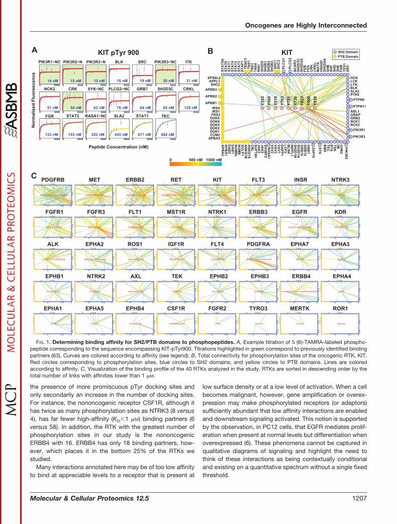
the presence of more promiscuous pTyr docking sites andonly secondarily an increase in the number of docking sites.For instance, the nononcogenic receptor CSF1R, although ithas twice as many phosphorylation sites as NTRK3 (8 versus4), has far fewer high-affinity (KD�1 �M) binding partners (6versus 58). In addition, the RTK with the greatest number ofphosphorylation sites in our study is the nononcogenicERBB4 with 16. ERBB4 has only 18 binding partners, how-ever, which places it in the bottom 25% of the RTKs westudied.
Many interactions annotated here may be of too low affinityto bind at appreciable levels to a receptor that is present at
low surface density or at a low level of activation. When a cellbecomes malignant, however, gene amplification or overex-pression may make phosphorylated receptors (or adaptors)sufficiently abundant that low affinity interactions are enabledand downstream signaling activated. This notion is supportedby the observation, in PC12 cells, that EGFR mediates prolif-eration when present at normal levels but differentiation whenoverexpressed (6). These phenomena cannot be captured inqualitative diagrams of signaling and highlight the need tothink of these interactions as being contextually conditionaland existing on a quantitative spectrum without a single fixedthreshold.
FIG. 1. Determining binding affinity for SH2/PTB domains to phosphopeptides. A, Example titration of 5 (6)-TAMRA-labeled phospho-peptide corresponding to the sequence encompassing KIT-pTyr900. Titrations highlighted in green correspond to previously identified bindingpartners (63). Curves are colored according to affinity (see legend). B, Total connectivity for phosphorylation sites of the oncogenic RTK, KIT.Red circles corresponding to phosphorylation sites, blue circles to SH2 domains, and yellow circles to PTB domains. Lines are coloredaccording to affinity. C, Visualization of the binding profile of the 40 RTKs analyzed in the study. RTKs are sorted in descending order by thetotal number of links with affinities lower than 1 �M.
Oncogenes are Highly Interconnected
Molecular & Cellular Proteomics 12.5 1207
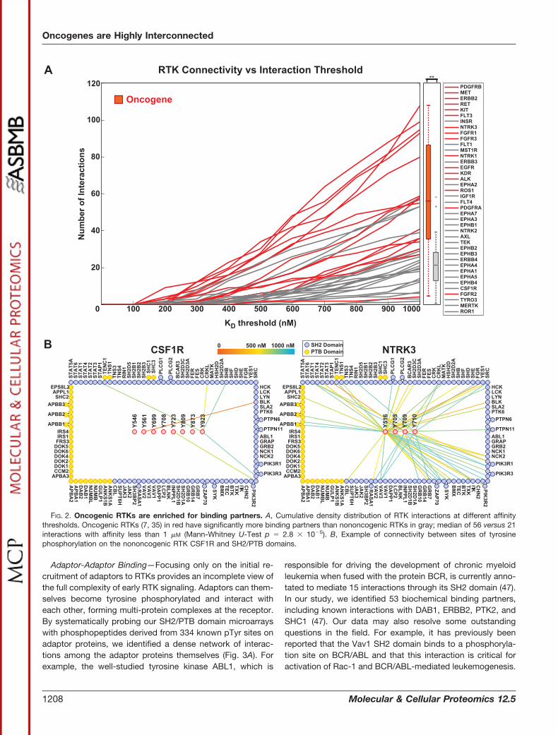
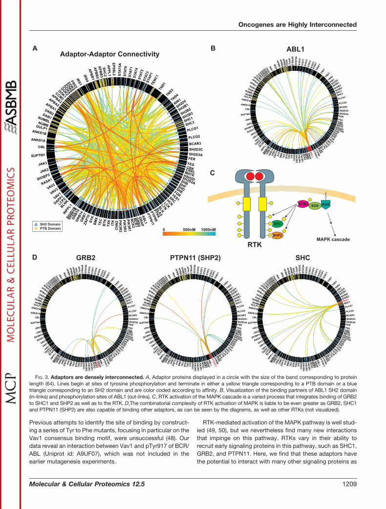
Adaptor-Adaptor Binding—Focusing only on the initial re-cruitment of adaptors to RTKs provides an incomplete view ofthe full complexity of early RTK signaling. Adaptors can them-selves become tyrosine phosphorylated and interact witheach other, forming multi-protein complexes at the receptor.By systematically probing our SH2/PTB domain microarrayswith phosphopeptides derived from 334 known pTyr sites onadaptor proteins, we identified a dense network of interac-tions among the adaptor proteins themselves (Fig. 3A). Forexample, the well-studied tyrosine kinase ABL1, which is
responsible for driving the development of chronic myeloidleukemia when fused with the protein BCR, is currently anno-tated to mediate 15 interactions through its SH2 domain (47).In our study, we identified 53 biochemical binding partners,including known interactions with DAB1, ERBB2, PTK2, andSHC1 (47). Our data may also resolve some outstandingquestions in the field. For example, it has previously beenreported that the Vav1 SH2 domain binds to a phosphoryla-tion site on BCR/ABL and that this interaction is critical foractivation of Rac-1 and BCR/ABL-mediated leukemogenesis.
FIG. 2. Oncogenic RTKs are enriched for binding partners. A, Cumulative density distribution of RTK interactions at different affinitythresholds. Oncogenic RTKs (7, 35) in red have significantly more binding partners than nononcogenic RTKs in gray; median of 56 versus 21interactions with affinity less than 1 �M (Mann-Whitney U-Test p � 2.8 � 10�5). B, Example of connectivity between sites of tyrosinephosphorylation on the nononcogenic RTK CSF1R and SH2/PTB domains.
Oncogenes are Highly Interconnected
1208 Molecular & Cellular Proteomics 12.5
Previous attempts to identify the site of binding by construct-ing a series of Tyr to Phe mutants, focusing in particular on theVav1 consensus binding motif, were unsuccessful (48). Ourdata reveal an interaction between Vav1 and pTyr917 of BCR/ABL (Uniprot id: A9UF07), which was not included in theearlier mutagenesis experiments.
RTK-mediated activation of the MAPK pathway is well stud-ied (49, 50), but we nevertheless find many new interactionsthat impinge on this pathway. RTKs vary in their ability torecruit early signaling proteins in this pathway, such as SHC1,GRB2, and PTPN11. Here, we find that these adaptors havethe potential to interact with many other signaling proteins as
FIG. 3. Adaptors are densely interconnected. A, Adaptor proteins displayed in a circle with the size of the band corresponding to proteinlength (64). Lines begin at sites of tyrosine phosphorylation and terminate in either a yellow triangle corresponding to a PTB domain or a bluetriangle corresponding to an SH2 domain and are color coded according to affinity. B, Visualization of the binding partners of ABL1 SH2 domain(in-links) and phosphorylation sites of ABL1 (out-links). C, RTK activation of the MAPK cascade is a varied process that integrates binding of GRB2to SHC1 and SHP2 as well as to the RTK. D,The combinatorial complexity of RTK activation of MAPK is liable to be even greater as GRB2, SHC1and PTPN11 (SHP2) are also capable of binding other adaptors, as can be seen by the diagrams, as well as other RTKs (not visualized).
Oncogenes are Highly Interconnected
Molecular & Cellular Proteomics 12.5 1209
well, increasing the complexity of MAPK pathway activation(Fig. 3D). This view aligns well with the idea that RTKs functionnot as discrete molecular machines, as typically depicted, butas pleomorphic ensembles that are highly complex and dy-namic with many rapidly interchanging states (22, 51).
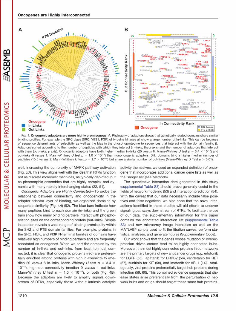
Oncogenic Adaptors are Highly Connected—To probe therelationship between connectivity and oncogenicity in theadaptor-adaptor layer of binding, we organized domains bysequence similarity (Fig. 4A) (52). The blue bars indicate howmany peptides bind to each domain (in-links) and the greenbars show how many binding partners interact with phospho-rylation sites on the corresponding protein (out-links). Simpleinspection reveals a wide range of binding promiscuity withinthe SH2 and PTB domain families. For example, proteins inthe SRC, HCK, and PI3K N-terminal families of domains haverelatively high numbers of binding partners and are frequentlyannotated as oncogenes. When we sort the domains by thenumber of in-links and out-links, from least to most con-nected, it is clear that oncogenic proteins (red) are preferen-tially enriched among proteins with high in-connectivity (me-dian 20 versus 8 in-links, Mann-Whitney U test p � 3.4 �
10�3), high out-connectivity (median 9 versus 1 out-links,Mann-Whitney U test p � 1.0 � 10�4), or both (Fig. 4B).Because the adaptors are likely to amplify signals down-stream of RTKs, especially those without intrinsic catalytic
activity themselves, we used an expanded definition of onco-gene that incorporates additional cancer gene lists as well asthe Sanger list (see Methods).
The quantitative interaction data generated in this study(supplemental Table S3) should prove generally useful in thefields of network modeling (53) and interaction prediction (54).With the caveat that our data necessarily include false posi-tives and false negatives, we also hope that the novel inter-actions identified in these studies will aid efforts to uncoversignaling pathways downstream of RTKs. To facilitate the useof our data, the supplementary information for this papercontains the annotated interaction list (supplemental TableS3) and raw microarray image intensities as well as theMATLAB� scripts used to fit the titration curves, perform sta-tistical analyses, and generate figures (Supplementary Code).
Our work shows that the genes whose mutation or overex-pression drives cancer tend to be highly connected hubs.Moreover, the most highly connected proteins in our networksare the primary targets of new anticancer drugs (e.g. erlotonibfor EGFR (55), lapatanib for ERBB2 (56), vandetanib for RET(57), sunitinib for KIT (58), and imatanib for ABL1 (14)). Anal-ogously, viral proteins preferentially target hub proteins duringinfection (59, 60). This combined evidence suggests that dis-ease states arise preferentially from the perturbation of net-work hubs and drugs should target these same hub proteins.
FIG. 4. Oncogenic adaptors are more highly promiscuous. A, Phylogeny of adaptors shows that genetically related domains share similarbinding profiles. For example the SRC class (SRC, YES1, FGR) of tyrosine kinases all show a large number of in-links. This can be becauseof sequence determinants of selectivity as well as the bias in the phosphoproteome to sequences that interact with the domain family. B,Adaptors sorted according to the number of peptides with which they interact (in-links; the x axis) and the number of adaptors that interactwith them (out-links; y axis). Oncogenic adaptors have both higher median in-links (20 versus 8, Mann-Whitney U test p � 3.4 � 10�3) andout-links (9 versus 1, Mann-Whitney U test p � 1.0 � 10�4) than nononcogenic adaptors. SH2 domains bind a higher median number ofpeptides (15.5 versus 2, Mann-Whitney U test p � 1.7 � 10�6) but share a similar number of out-links (Mann-Whitney U Test p � 0.01).
Oncogenes are Highly Interconnected
1210 Molecular & Cellular Proteomics 12.5
In conclusion, we have generated a systematic map cover-ing a substantial fraction of the potential interactions betweenSH2 or PTB domains and sites of tyrosine phosphorylation onRTKs and adaptor proteins. These interactions are very poorlyrepresented in existing unbiased human interactomes (61, 62)despite extensive evidence they play essential roles in signaltransduction. We observe a high degree of connectivityamong RTKs and adaptor proteins, and among adaptor pro-teins themselves. This is in contrast to the usual depiction ofreceptor-proximal signaling as a series of linear pathwaysconnecting sites of tyrosine phosphorylation on RTKs to a fewadaptor proteins and then to a few core signaling moleculessuch as ERK and AKT. Of course, the actual complexity of theresulting network in any particular cell type depends not onlyon the affinities of the interactions (as determined here) butalso on the relative abundance of RTKs and SH2/PTB-con-taining proteins. mRNA sequencing data of 1430 diverse tu-mor types collected by the TCGA consortium shows that theyexpress a median of 78% of the phosphotyrosine signalingproteins analyzed in this study. In fact, over half of theseproteins are expressed ubiquitously (in �95% of the tumorsamples; supplemental Fig. S2). Thus, it is highly likely thatreceptor and adaptor proteins combine to form a highly inter-connected mesh with the potential to perform complex signalprocessing functions.
Acknowledgments—We thank Jeffrey Knott, Susan Rogers, andColleen Hunter (Cell Signaling Technology Inc.) for peptide synthesis,Bogdan A. Budnick and William S. Lane (FAS Center for SystemsBiology) for mass spectrometry, Vijay Warrier for Ephrin receptorcloning, Bryan H. Chang for STAT protein cloning and expression,Ethan S. Karp for help with MATLAB® scripting, Zeba Wunderlich forhelpful discussions and Taran S. Gujral (Harvard Medical School) forcommenting on the manuscript.
* This work was supported by an award from the W. M. KeckFoundation and by grants from the National Institutes of Health (R33CA128726 and R01 GM072872). G.K. was the recipient of the Astra-Zeneca Graduate Fellowship and the Max Tishler Graduate Fellow-ship. A.K. was supported in part by the NIH Molecular, Cellular, andChemical Biology Training Grant (5 T32 GM07598).
□S This article contains supplemental Figs. S1 and S2 and TablesS1 to S4.
‡‡ To whom correspondence should be addressed: Department ofSystems Biology, Harvard Medical School, 200 Longwood Ave., Bos-ton, MA 02115. Tel.: 617-432-6902; Fax: 617-432-6990; E-mail:[email protected].
REFERENCES
1. Lemmon, M. A., and Schlessinger, J. (2010) Cell signaling by receptortyrosine kinases. Cell 141, 1117–1134
2. Liu, B. A., Jablonowski, K., Raina, M., Arce, M., Pawson, T., and Nash, P. D.(2006) The Human and Mouse Complement of SH2 Domain Proteins-Establishing the Boundaries of Phosphotyrosine Signaling. Mol. Cell 22,851–868
3. Jones, R. B., Gordus, A., Krall, J. A., and MacBeath, G. (2006) A quantitativeprotein interaction network for the ErbB receptors using protein microar-rays. Nature 439, 168–174
4. Salcini, A. E., McGlade, J., Pelicci, G., Nicoletti, I., Pawson, T., and Pelicci,P. G. (1994) Formation of Shc-Grb2 complexes is necessary to induceneoplastic transformation by overexpression of Shc proteins. Oncogene
9, 2827–28365. Esposito, D. L., Li, Y., Cama, A., and Quon, M. J. (2001) Tyr(612) and
Tyr(632) in human insulin receptor substrate-1 are important for fullactivation of insulin-stimulated phosphatidylinositol 3-kinase activity andtranslocation of GLUT4 in adipose cells. Endocrinology 142, 2833–2840
6. Marshall, C. J. (1995) Specificity of receptor tyrosine kinase signaling:Transient versus sustained extracellular signal-regulated kinase activa-tion. Cell 80, 179–185
7. Santarius, T., Shipley, J., Brewer, D., Stratton, M. R., and Cooper, C. S.(2010) A census of amplified and overexpressed human cancer genes.Nat. Rev. Cancer 10, 59–64
8. Perren, T. J. (1991) c-erbB-2 oncogene as a prognostic marker in breastcancer. Br. J. Cancer 63, 328–332
9. McIntyre, A., Summersgill, B., Grygalewicz, B., Gillis, A. J., Stoop, J., vanGurp, R. J., Dennis, N., Fisher, C., Huddart, R., Cooper, C., Clark, J.,Oosterhuis, J. W., Looijenga, L. H., and Shipley, J. (2005) Amplificationand overexpression of the KIT gene is associated with progression in theseminoma subtype of testicular germ cell tumors of adolescents andadults. Cancer Res. 65, 8085–8089
10. Comoglio, P. M., Giordano, S., and Trusolino, L. (2008) Drug developmentof MET inhibitors: targeting oncogene addiction and expedience. Nat.Rev. Drug Discovery 7, 504–516
11. Mulligan, L. M., Kwok, J. B., Healey, C. S., Elsdon, M. J., Eng, C., Gardner,E., Love, D. R., Mole, S. E., Moore, J. K., and Papi, L. (1993) Germ-linemutations of the RET proto-oncogene in multiple endocrine neoplasiatype 2A. Nature 363, 458–460
12. Hofstra, R. M., Landsvater, R. M., Ceccherini, I., Stulp, R. P., Stelwagen, T.,Luo, Y., Pasini, B., Hoppener, J. W., van Amstel, H. K., and Romeo, G.(1994) A mutation in the RET proto-oncogene associated with multipleendocrine neoplasia type 2B and sporadic medullary thyroid carcinoma.Nature 367, 375–376
13. Hirota, S., Isozaki, K., Moriyama, Y., Hashimoto, K., Nishida, T., Ishiguro,S., Kawano, K., Hanada, M., Kurata, A., Takeda, M., Tunio, G., Matsu-zawa, Y., Kanakura, Y., Shinomura, Y., and Kitamura, Y. (1998) Gain-of-function mutations of c-kit in human gastrointestinal stromal tumors.Science 279, 577–580
14. Druker, B. J., Sawyers, C. L., Kantarjian, H., Resta, D. J., Reese, S. F., Ford,J. M., Capdeville, R., and Talpaz, M. (2001) Activity of a specific inhibitorof the BCR-ABL tyrosine kinase in the blast crisis of chronic myeloidleukemia and acute lymphoblastic leukemia with the Philadelphia chro-mosome. N. Engl. J. Med. 344, 1038–1042
15. Cheung, L. W., Hennessy, B. T., Li, J., Yu, S., Myers, A. P., Djordjevic, B.,Lu, Y., Stemke-Hale, K., Dyer, M. D., Zhang, F., Ju, Z., Cantley, L. C.,Scherer, S. E., Liang, H., Lu, K. H., Broaddus, R. R., and Mills, G. B.(2011) High frequency of PIK3R1 and PIK3R2 mutations in endometrialcancer elucidates a novel mechanism for regulation of PTEN proteinstability. Cancer Discovery 1, 170–185
16. Liu, Y. Y., Slotine, J. J., and Barabasi, A. L. (2011) Controllability of complexnetworks. Nature 473, 167–173
17. Jonsson, P. F., and Bates, P. A. (2006) Global topological features of cancerproteins in the human interactome. Bioinformatics 22, 2291–2297
18. Wachi, S., Yoneda, K., and Wu, R. (2005) Interactome-transcriptome anal-ysis reveals the high centrality of genes differentially expressed in lungcancer tissues. Bioinformatics 21, 4205–4208
19. Hakes, L., Robertson, D. L., and Oliver, S. G. (2005) Effect of datasetselection on the topological interpretation of protein interaction net-works. BMC Genomics 6, 131
20. Hakes, L., Pinney, J. W., Robertson, D. L., and Lovell, S. C. (2008) Protein-protein interaction networks and biology–what’s the connection? Nat.Biotechnol. 26, 69–72
21. Phizicky, E., Bastiaens, P. I., Zhu, H., Snyder, M., and Fields, S. (2003)Protein analysis on a proteomic scale. Nature 422, 208–215
22. Mayer, B. J., Blinov, M. L., and Loew, L. M. (2009) Molecular machines orpleiomorphic ensembles: signaling complexes revisited. J. Biol. 8, 81
23. Huang, H., Li, L., Wu, C., Schibli, D., Colwill, K., Ma, S., Li, C., Roy, P., Ho,K., Songyang, Z., Pawson, T., Gao, Y., and Li, S. S.-C. (2008) Definingthe specificity space of the human src homology 2 domain. Mol. Cell.Proteomics 7, 768–784
24. Rodriguez, M., Li, S. S., Harper, J. W., and Songyang, Z. (2004) An orientedpeptide array library (OPAL) strategy to study protein-protein interac-tions. J. Biol. Chem. 279, 8802–8807
Oncogenes are Highly Interconnected
Molecular & Cellular Proteomics 12.5 1211
25. Zhou, S., Shoelson, S. E., Chaudhuri, M., Gish, G., Pawson, T., Haser,W. G., King, F., Roberts, T., Ratnofsky, S., Lechleider, R. J. (1993) SH2domains recognize specific phosphopeptide sequences. Cell 72,767–778
26. Liu, B. A., Jablonowski, K., Shah, E. E., Engelmann, B. W., Jones, R. B., andNash, P. D. (2010) SH2 domains recognize contextual peptide sequenceinformation to determine selectivity. Mol. Cell Proteomics 9, 2391–2404
27. Gorelik, M., Stanger, K., and Davidson, A. R. (2011) A Conserved residue inthe yeast Bem1p SH3 domain maintains the high level of binding spec-ificity required for function. J. Biol. Chem. 286, 19470–19477
28. Kaushansky, A., Allen, J. E., Gordus, A., Stiffler, M. A., Karp, E. S., Chang,B. H., and MacBeath, G. (2010) Quantifying protein-protein interactionsin high throughput using protein domain microarrays. Nat. Protoc. 5,773–790
29. Lemmon, M. A., and Schlessinger, J. (2010) Cell signaling by receptortyrosine kinases. Cell 141, 1117–1134
30. Kaushansky, A., Gordus, A., Budnik, B. A., Lane, W. S., Rush, J., andMacBeath, G. (2008) System-wide investigation of ErbB4 reveals 19 sitesof Tyr phosphorylation that are unusually selective in their recruitmentproperties. Chem. Biol. 15, 808–817
31. Jung, A. S., Kaushansky, A., Macbeath, G., and Kaushansky, K. (2011)Tensin2 is a novel mediator in thrombopoietin (TPO)-induced cellularproliferation by promoting Akt signaling. Cell Cycle 10, 1838–1844
32. Mehlitz, A., Banhart, S., Maurer, A. P., Kaushansky, A., Gordus, A. G.,Zielecki, J., Macbeath, G., and Meyer, T. F. (2010) Tarp regulates earlyChlamydia-induced host cell survival through interactions with the hu-man adaptor protein SHC1. J. Cell Biol. 190, 143–157
33. Boettcher, J. P., Kirchner, M., Churin, Y., Kaushansky, A., Pompaiah, M.,Thorn, H., Brinkmann, V., Macbeath, G., and Meyer, T. F. (2010) Ty-rosine-phosphorylated caveolin-1 blocks bacterial uptake by inducingVav2-RhoA-mediated cytoskeletal rearrangements. PLoS Biol. 8
34. Huber, P. J. (2003) Robust Statistics, John Wiley & Sons35. Futreal, P. A., Coin, L., Marshall, M., Down, T., Hubbard, T., Wooster, R.,
Rahman, N., and Stratton, M. R. (2004) A census of human cancer genes.Nature Reviews Cancer 4, 177–183
36. Huret, J. L., Minor, S. L., Dorkeld, F., Dessen, P., and Bernheim, A. (2000)Atlas of genetics and cytogenetics in oncology and haematology, aninteractive database. Nucleic Acids Res. 28, 349–351
37. Sjoblom, T., Jones, S., Wood, L. D., Parsons, D. W., Lin, J., Barber, T. D.,Mandelker, D., Leary, R. J., Ptak, J., Silliman, N., Szabo, S., Buckhaults,P., Farrell, C., Meeh, P., Markowitz, S. D., Willis, J., Dawson, D., Willson,J. K., Gazdar, A. F., Hartigan, J., Wu, L., Liu, C., Parmigiani, G., Park,B. H., Bachman, K. E., Papadopoulos, N., Vogelstein, B., Kinzler, K. W.,and Velculescu, V. E. (2006) The consensus coding sequences of humanbreast and colorectal cancers. Science 314, 268–274
38. Akagi, K., Suzuki, T., Stephens, R. M., Jenkins, N. A., and Copeland, N. G.(2004) RTCGD: retroviral tagged cancer gene database. Nucleic AcidsRes. 32, D523–527
39. Network, T. C. G. A. (2012) Comprehensive molecular characterization ofhuman colon and rectal cancer. Nature 487, 330–337
40. Mortazavi, A., Williams, B. A., McCue, K., Schaeffer, L., and Wold, B. (2008)Mapping and quantifying mammalian transcriptomes by RNA-Seq. Nat.Methods 5, 621–628
41. Nagaraj, N., Wisniewski, J. R., Geiger, T., Cox, J., Kircher, M., Kelso, J.,Paabo, S., and Mann, M. (2011) Deep proteome and transcriptomemapping of a human cancer cell line. Mol. Syst. Biol. 7, 548
42. Hornbeck, P. V., Kornhauser, J. M., Tkachev, S., Zhang, B., Skrzypek, E.,Murray, B., Latham, V., and Sullivan, M. (2011) PhosphoSitePlus: acomprehensive resource for investigating the structure and function ofexperimentally determined post-translational modifications in man andmouse. Nucleic Acids Research 40, D261-D270
43. Kaushansky, A., Gordus, A., Chang, B., Rush, J., and MacBeath, G. (2008)A quantitative study of the recruitment potential of all intracellular tyro-sine residues on EGFR, FGFR1 and IGF1R. Mol. Biosyst. 4, 643–653
44. Ladbury, J. E., Lemmon, M. A., Zhou, M., Green, J., Botfield, M. C., andSchlessinger, J. (1995) Measurement of the binding of tyrosyl phospho-peptides to SH2 domains: a reappraisal. Proc. Natl. Acad. Sci. U.S.A. 92,3199–3203
45. Piccione, E., Case, R. D., Domchek, S. M., Hu, P., Chaudhuri, M., Backer,J. M., Schlessinger, J., and Shoelson, S. E. (1993) Phosphatidylinositol3-kinase p85 SH2 domain specificity defined by direct phosphopeptide/
SH2 domain binding. Biochemistry 32, 3197–320246. Bibbins, K. B., Boeuf, H., and Varmus, H. E. (1993) Binding of the Src SH2
domain to phosphopeptides is determined by residues in both the SH2domain and the phosphopeptides. Mol. Cell. Biol. 13, 7278–7287
47. Colicelli, J. (2010) ABL tyrosine kinases: evolution of function, regulation,and specificity. Sci. Signal. 3, re6
48. Bassermann, F., Jahn, T., Miething, C., Seipel, P., Bai, R. Y., Coutinho, S.,Tybulewicz, V. L., Peschel, C., and Duyster, J. (2002) Association ofBcr-Abl with the Proto-oncogene Vav Is Implicated in Activation of theRac-1 Pathway. J. Biol. Chem. 277, 12437–12445
49. Tidyman, W. E., and Rauen, K. A. (2009) The RASopathies: Developmentalsyndromes of Ras/MAPK pathway dysregulation. Curr. Opin. Genet.Dev. 19, 230–236
50. McKay, M. M., and Morrison, D. K. (2007) Integrating signals from RTKs toERK/MAPK. Oncogene 26, 3113–3121
51. Kleiman, L. B., Maiwald, T., Conzelmann, H., Lauffenburger, D. A., andSorger, P. K. (2011) Rapid phospho-turnover by receptor tyrosine ki-nases impacts downstream signaling and drug binding. Mol. Cell 43,723–737
52. Letunic, I., and Bork, P. (2011) Interactive Tree Of Life v2: online annotationand display of phylogenetic trees made easy. Nucleic Acids Res. 39,W475-W478
53. Chen, W. W., Schoeberl, B., Jasper, P. J., Niepel, M., Nielsen, U. B.,Lauffenburger, D. A., and Sorger, P. K. (2009) Input-output behavior ofErbB signaling pathways as revealed by a mass action model trainedagainst dynamic data. Mol. Syst. Biol. 5, 239
54. Miller, M. L., Jensen, L. J., Diella, F., Jørgensen, C., Tinti, M., Li, L., Hsiung,M., Parker, S. A., Bordeaux, J., Sicheritz-Ponten, T., Olhovsky, M.,Pasculescu, A., Alexander, J., Knapp, S., Blom, N., Bork, P., Li, S.,Cesareni, G., Pawson, T., Turk, B. E., Yaffe, M. B., Brunak, S., andLinding, R. (2008) Linear motif atlas for phosphorylation-dependent sig-naling. Sci. Signal. 1, ra2
55. Raymond, E., Faivre, S., and Armand, J. P. (2000) Epidermal growth factorreceptor tyrosine kinase as a target for anticancer therapy. Drugs 60.15–23, discussion 41–42
56. Burris, H. A., 3rd, Hurwitz, H. I., Dees, E. C., Dowlati, A., Blackwell, K. L.,O’Neil, B., Marcom, P. K., Ellis, M. J., Overmoyer, B., Jones, S. F., Harris,J. L., Smith, D. A., Koch, K. M., Stead, A., Mangum, S., and Spector, N. L.(2005) Phase I safety, pharmacokinetics, and clinical activity study oflapatinib (GW572016), a reversible dual inhibitor of epidermal growthfactor receptor tyrosine kinases, in heavily pretreated patients with met-astatic carcinomas. J. Clin. Oncol. 23, 5305–5313
57. Degrauwe, N., Sosa, J. A., Roman, S., and Deshpande, H. A. (2012)Vandetanib for the treatment of metastatic medullary thyroid cancer.Clin. Med. Insights Oncol. 6, 243–252
58. Demetri, G. D., van Oosterom, A. T., Garrett, C. R., Blackstein, M. E., Shah,M. H., Verweij, J., McArthur, G., Judson, I. R., Heinrich, M. C., Morgan,J. A., Desai, J., Fletcher, C. D., George, S., Bello, C. L., Huang, X., Baum,C. M., and Casali, P. G. (14) Efficacy and safety of sunitinib in patientswith advanced gastrointestinal stromal tumour after failure of imatinib: arandomised controlled trial. Lancet 368, 1329–1338
59. Calderwood, M. A., Venkatesan, K., Xing, L., Chase, M. R., Vazquez, A.,Holthaus, A. M., Ewence, A. E., Li, N., Hirozane-Kishikawa, T., Hill, D. E.,Vidal, M., Kieff, E., and Johannsen, E. (2007) Epstein-Barr virus and virushuman protein interaction maps. Proc. Natl. Acad. Sci. U.S.A. 104,7606–7611
60. Shapira, S. D., Gat-Viks, I., Shum, B. O. V., Dricot, A., de Grace, M. M., Wu,L., Gupta, P. B., Hao, T., Silver, S. J., Root, D. E., Hill, D. E., Regev, A., andHacohen, N. (2009) A Physical and Regulatory Map of Host-InfluenzaInteractions Reveals Pathways in H1N1 Infection. Cell 139, 1255–1267
61. Rual, J. F., Venkatesan, K., Hao, T., Hirozane-Kishikawa, T., Dricot, A., Li,N., Berriz, G. F., Gibbons, F. D., Dreze, M., Ayivi-Guedehoussou, N.,Klitgord, N., Simon, C., Boxem, M., Milstein, S., Rosenberg, J., Gold-berg, D. S., Zhang, L. V., Wong, S. L., Franklin, G., Li, S., Albala, J. S.,Lim, J., Fraughton, C., Llamosas, E., Cevik, S., Bex, C., Lamesch, P.,Sikorski, R. S., Vandenhaute, J., Zoghbi, H. Y., Smolyar, A., Bosak, S.,Sequerra, R., Doucette-Stamm, L., Cusick, M. E., Hill, D. E., Roth, F. P.,and Vidal, M. (2005) Towards a proteome-scale map of the humanprotein-protein interaction network. Nature 437, 1173–1178
62. Venkatesan, K., Rual, J. F., Vazquez, A., Stelzl, U., Lemmens, I., Hirozane-Kishikawa, T., Hao, T., Zenkner, M., Xin, X., Goh, K.-I., Yildirim, M. A.,
Oncogenes are Highly Interconnected
1212 Molecular & Cellular Proteomics 12.5
Simonis, N., Heinzmann, K., Gebreab, F., Sahalie, J. M., Cevik, S.,Simon, C., Smet, A.-S. de, Dann, E., Smolyar, A., Vinayagam, A., Yu, H.,Szeto, D., Borick, H., Dricot, A., Klitgord, N., Murray, R. R., Lin, C.,Lalowski, M., Timm, J., Rau, K., Boone, C., Braun, P., Cusick, M. E.,Roth, F. P., Hill, D. E., Tavernier, J., Wanker, E. E., Barabasi, A. L., andVidal, M. (2009) An empirical framework for binary interactome mapping.Nature Methods 6, 83–90
63. Lennartsson, J., Wernstedt, C., Engstrom, U., Hellman, U., and Ronnstrand,L. (2003) Identification of Tyr900 in the kinase domain of c-Kit as aSrc-dependent phosphorylation site mediating interaction with c-Crk.Experimental Cell Research 288, 110–118
64. Krzywinski, M., Schein, J., Birol, I., Connors, J., Gascoyne, R., Horsman, D.,Jones, S. J., and Marra, M. A. (2009) Circos: An information aesthetic forcomparative genomics. Genome Research 19, 1639–1645
Oncogenes are Highly Interconnected
Molecular & Cellular Proteomics 12.5 1213