module b respiratory alterations nur 203 - · pdf filemodule b respiratory alterations ......
TRANSCRIPT

Page 1 of 28
Module B
Respiratory Alterations
NUR 203
Page 2 of 28
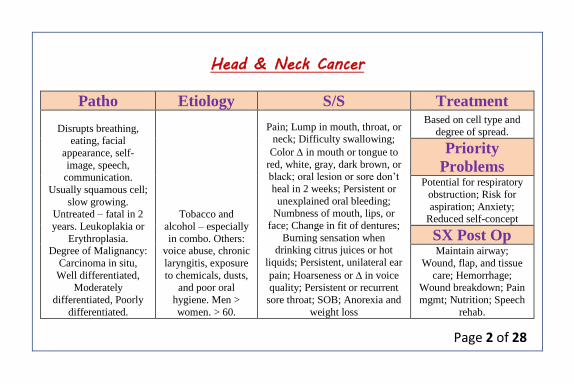
Head & Neck Cancer
Patho Etiology S/S Treatment
Disrupts breathing, eating, facial
appearance, self-
image, speech, communication.
Usually squamous cell;
slow growing.
Untreated – fatal in 2
years. Leukoplakia or
Erythroplasia. Degree of Malignancy:
Carcinoma in situ,
Well differentiated,
Moderately
differentiated, Poorly
differentiated.
Tobacco and
alcohol – especially
in combo. Others: voice abuse, chronic
laryngitis, exposure
to chemicals, dusts,
and poor oral
hygiene. Men >
women. > 60.
Pain; Lump in mouth, throat, or neck; Difficulty swallowing;
Color in mouth or tongue to
red, white, gray, dark brown, or black; oral lesion or sore don’t
heal in 2 weeks; Persistent or
unexplained oral bleeding;
Numbness of mouth, lips, or
face; Change in fit of dentures;
Burning sensation when drinking citrus juices or hot
liquids; Persistent, unilateral ear
pain; Hoarseness or in voice
quality; Persistent or recurrent
sore throat; SOB; Anorexia and
weight loss
Based on cell type and
degree of spread.
Priority
Problems Potential for respiratory
obstruction; Risk for
aspiration; Anxiety; Reduced self-concept
SX Post Op Maintain airway;
Wound, flap, and tissue
care; Hemorrhage;
Wound breakdown; Pain
mgmt; Nutrition; Speech
rehab.
Page 3 of 28
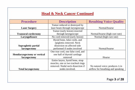
Head & Neck Cancer Continued
Procedure Description Resulting Voice Quality
Laser Surgery
Tumor reduced or destroyed by
laser beam through laryngoscope Normal/hoarse
Transoral cordectomy
Tumor (early lesion) resected through laryngoscope Normal/hoarse (high cure rate)
Laryngofissure No cord removed (early lesion) Normal (high cure rate)
Supraglottic partial
laryngectomy
Hyoid bone, false cords, and
epiglottis removed. Neck dissection on affected side
performed if nodes involved Normal/hoarse
Hemilaryngectomy or vertical
laryngectomy
One true cord, one false cord, and
one half of thyroid cartilage removed Hoarse
Total laryngectomy
Entire larynx, hyoid bone, strap
muscles, one or two tracheal rings removed. Nodal neck dissection if
nodes involved.
No natural voice; produces in
airflow for breathing and speaking

Page 4 of 28
Head & Neck Cancer Continued
Community Based Care After Laryngectomy
Assess respiratory rate.
Assess Condition of wound
Assess patient’s psychosocial status
Take patient’s temperature at each home care visit.
Assess the patient’s understanding of illness and adherence to
treatment.
Assess patient’s nutritional status.
Page 5 of 28
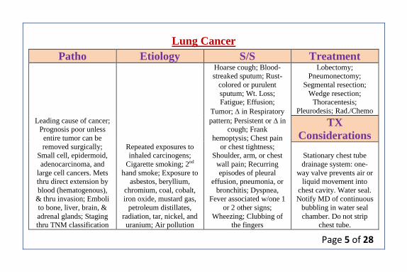
Lung Cancer
Patho Etiology S/S Treatment
Leading cause of cancer; Prognosis poor unless
entire tumor can be
removed surgically; Small cell, epidermoid,
adenocarcinoma, and
large cell cancers. Mets thru direct extension by
blood (hematogenous), & thru invasion; Emboli
to bone, liver, brain, &
adrenal glands; Staging thru TNM classification
Repeated exposures to inhaled carcinogens;
Cigarette smoking; 2nd
hand smoke; Exposure to asbestos, beryllium,
chromium, coal, cobalt, iron oxide, mustard gas,
petroleum distillates,
radiation, tar, nickel, and uranium; Air pollution
Hoarse cough; Blood-streaked sputum; Rust-
colored or purulent
sputum; Wt. Loss; Fatigue; Effusion;
Tumor; in Respiratory
pattern; Persistent or in cough; Frank
hemoptysis; Chest pain
or chest tightness; Shoulder, arm, or chest
wall pain; Recurring
episodes of pleural effusion, pneumonia, or
bronchitis; Dyspnea, Fever associated w/one 1
or 2 other signs;
Wheezing; Clubbing of the fingers
Lobectomy; Pneumonectomy;
Segmental resection;
Wedge resection; Thoracentesis;
Pleurodesis; Rad./Chemo
TX
Considerations
Stationary chest tube
drainage system: one-
way valve prevents air or liquid movement into
chest cavity. Water seal. Notify MD of continuous
bubbling in water seal
chamber. Do not strip chest tube.
Page 6 of 28
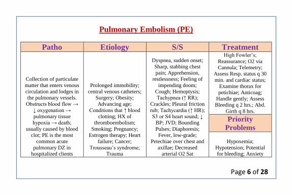
Pulmonary Embolism (PE)
Patho Etiology S/S Treatment
Collection of particulate
matter that enters venous
circulation and lodges in the pulmonary vessels.
Obstructs blood flow →
↓ oxygenation → pulmonary tissue
hypoxia → death;
usually caused by blood clot; PE is the most
common acute
pulmonary DZ in hospitalized clients
Prolonged immobility;
central venous catheters; Surgery; Obesity;
Advancing age;
Conditions that ↑ blood clotting; HX of
thromboembolism;
Smoking; Pregnancy; Estrogen therapy; Heart
failure; Cancer;
Trousseau’s syndrome; Trauma
Dyspnea, sudden onset;
Sharp, stabbing chest
pain; Apprehension, restlessness; Feeling of
impending doom;
Cough; Hemoptysis; Tachypnea (↑ RR);
Crackles; Pleural friction
rub; Tachycardia (↑ HR); S3 or S4 heart sound; ↓
BP; JVD; Bounding
Pulses; Diaphoresis; Fever, low-grade;
Petechiae over chest and
axillae; Decreased arterial O2 Sat
High Fowler’s;
Reassurance; O2 via Cannula; Telemetry;
Assess Resp. status q 30
min. and cardiac status; Examine thorax for
petichiae; Anticoag;
Handle gently; Assess
Bleeding q 2 hrs.; Abd.
Girth q 8 hrs.
Priority
Problems
Hypoxemia;
Hypotension; Potential for bleeding; Anxiety

Page 7 of 28
ARDS
Patho Etiology S/S Treatment Persistent Hypoxemia
despite 100% O2; ↓ pulmonary compliance;
Dyspnea; Non-cardiac
associated bilateral pulmonary edema;
Dense pulmonary
infiltrates on CXR
Indirect lung injury;
Direct lung injury; Inflammatory response;
Shock; Trauma; Serious
nervous system injury; Pancreatitis; Fat &
amniotic fluid emboli;
Pulmonary infections;
Sepsis; Inhalation of toxic
gases; Pulmonary aspiration; Drug
ingestion; Hemoolytic
disorders; Multiple blood transfusions;
Cardiopulmonary bypass;
Submersion in water w/water aspiration
1st Stage: Fluid in
interstitial space. Early dyspnea & tachypnea;
support/O2
2nd Stage: Fluid in alveoli – patchy
infiltrates; mech vent
3rd Stage: Day 2 – 10;
respond poorly to ↑ O2;
.↑ CO2 50; ↓ PaO2 60; ↓ PH 7.30 &
4th Stage: Starts after10
days; irreversible; late or chronic ARDS; prevent
sepsis, pneumonia,
MODS, wean from vent.
Corticosteroids –
prednisone, solu-medrol; Antibiotics; Turn patient
q 1 -2 hr.

Page 8 of 28
Endotracheal Intubation
During intubation, the nurse coordinates the response and continuously
monitors for changes in vital signs, signs of hypoxia, or hypoxemia,
dysrhythmias, and aspiration.
Ensure that each intubation attempt lasts no longer than 30 seconds
preferably less than 15 seconds. After 30 seconds, provide O2 by means
of a mask and manual resuscitation bag to prevent hypoxia and cardiac
arrest.
Check placement by end tidal carbon dioxide levels and by chest x-ray.
Check for breath sounds bilaterally, symmetric chest movement, and air
emerging from the ET tube.
Nursing Care – Neck flexion moves the tube away from the carina; neck
extension moves the tube closer to the carina.
Page 9 of 28
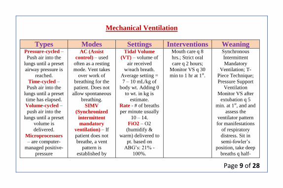
Mechanical Ventilation
Types Modes Settings Interventions Weaning Pressure-cycled –
Push air into the
lungs until a preset airway pressure is
reached.
Time-cycled – Push air into the
lungs until a preset
time has elapsed.
Volume-cycled –
push air into the
lungs until a preset volume is
delivered.
Microprocessors – are computer-
managed positive-
pressure
AC (Assist
control) – used
often as a resting mode. Vent takes
over work of
breathing for the patient. Does not
allow spontaneous
breathing.
SIMV
(Synchronized
intermittent
mandatory
ventilation) – If
patient does not
breathe, a vent
pattern is
established by
Tidal Volume
(VT) – volume of
air received w/each breath.
Average setting =
7 – 10 mL/kg of body wt. Adding 0
to wt. in kg is
estimate.
Rate - # of breaths
per minute usually
10 – 14. FiO2 – O2
(humidify &
warm) delivered to
pt. based on
ABG’s: 21% -
100%.
Mouth care q 8
hrs.; Strict oral
care q 2 hours; Monitor VS q 30
min to 1 hr at 1st.
Synchronous
Intermittent
Mandatory Ventilation; T-
Piece Technique;
Pressure Support Ventilation
Monitor VS after
extubation q 5
min. at 1st, and and
assess the
ventilator pattern for manifestations
of respiratory
distress. Sit in
semi-fowler’s
position, take deep
breaths q half-
Page 10 of 28
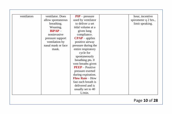
ventilators ventilator. Does
allow spontaneous breathing.
Weaning.
BiPAP – noninvasive
pressure support
ventilation by nasal mask or face
mask.
PIP – pressure
used by ventilator to deliver a set
tidal volume at a
given lung compliance.
CPAP – applies
positive airway pressure during the
entire respiratory
cycle for spontaneously
breathing pts. 0
vent breaths given PEEP – Positive
pressure exerted
during expiration. Flow Rate – How
fast each breath is
delivered and is usually set to 40
L/min.
hour, incentive
spirometer q 2 hrs., limit speaking.

Page 11 of 28
High-Pressure Alarm
Sounds when peak inspiratory pressure (PIP) reaches the set alarm limit (usually set 10-
20 mm Hg above the patient’s baseline PIP)
An ↑ amount of secretions or a mucus plug is in
the airways Suction as needed.
The patient coughs, gags, or bites on the oral
ET tube Insert oral airway to prevent biting the ET tube
The patient is anxious or fights the ventilator
Provide emotional support to ↓ anxiety; ↑ the
flow rate; Explain all procedures; sedation or
paralyzing agent per the physician’s
prescription.
Airway size ↓ related to wheezing or
bronchospasm Auscultate breath sounds
Pneumothorax occurs
Alert the physician or rapid response team for
management of bronchospasm; Auscultate
breath sounds; Alert the physician or Rapid
Response Team about a new onset of ↓ breath
sounds or unequal chest excursion, which may
be d/t pneumo
Page 12 of 28
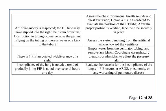
Artificial airway is displaced; the ET tube may
have slipped into the right mainstem bronchus
Assess the chest for unequal breath sounds and
chest excursion; Obtain a CXR as ordered to
evaluate the position of the ET tube; After the
proper postion is verified, tape the tube securely
in place
Obstruction in tubing occurs because the patient
is lying on the tubing or there is water or a kink
in the tubing
Assess the system, moving from the artificial
airway toward the ventilator
There is ↑ PIP associated w/deliverance of a
sight
Empty water from the ventilator tubing, and
remove any kinks; Coordinate w/respiratory
therapist or physician to adjust the pressure
alarm.
↓ compliance of the lung is noted; a trend of
gradually ↑’ing PIP is noted over several hours
or a day
Evaluate the reasons for the ↓ compliance of the
lungs; ↑ PIP occurs in ARDS, pneumonia, or
any worsening of pulmonary disease

Page 13 of 28
Low-Pressure Alarm
Low exhaled volume (Low-Pressure Alarm) sounds when there is a
disconnection or leak in the ventilator circuit or a leak in the patient’s artificial
airway cuff
A leak in the ventilator circuit prevents
breath from being delivered
Assess all connections and all ventilator
tubings for disconnection
The patient stops spontaneous breathing in
the SIMV or CPAP mode or on pressure
support ventilation
Evaluate the patient’s tolerance of the
mode
A cuff leak occurs in the ET or
tracheostomy tube
Evaluate the patient for a cuff leak. A cuff
leak is suspected when the patient can talk
(air escapes from the mouth) or when the
pilot balloon on the artificial airway is flat
Page 14 of 28
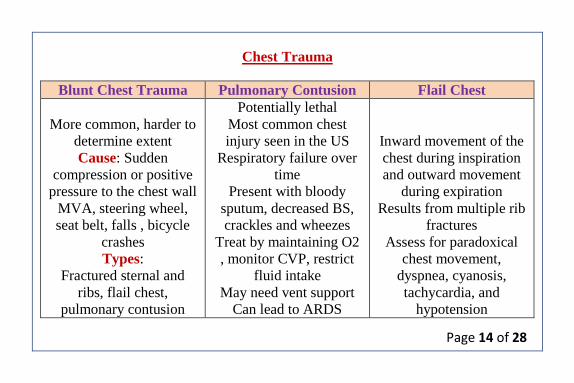
Chest Trauma
Blunt Chest Trauma Pulmonary Contusion Flail Chest
More common, harder to
determine extent
Cause: Sudden
compression or positive
pressure to the chest wall
MVA, steering wheel,
seat belt, falls , bicycle
crashes
Types:
Fractured sternal and
ribs, flail chest,
pulmonary contusion
Potentially lethal
Most common chest
injury seen in the US
Respiratory failure over
time
Present with bloody
sputum, decreased BS,
crackles and wheezes
Treat by maintaining O2
, monitor CVP, restrict
fluid intake
May need vent support
Can lead to ARDS
Inward movement of the
chest during inspiration
and outward movement
during expiration
Results from multiple rib
fractures
Assess for paradoxical
chest movement,
dyspnea, cyanosis,
tachycardia, and
hypotension

Page 15 of 28
Chest Trauma Continued
Pneumothorax Tension Pneumothorax Hemothorax
Chest injury that allows air to
enter the pleural space
Often seen with blunt chest
trauma
Can be open or closed
Assessment:
↓ BS, Hyperresonance,
Prominence of involved side,
Deviation of trachea,
Subcutaneous emphysema
Life-threatening complication
of blunt chest trauma
Assessment:
Asymmetry of thorax,
Tracheal deviation toward the
unaffected side, Respiratory
distress, Absence of BS on one
side, Distended neck veins,
Cyanosis,
Hypertympanic sound on
percussion on affected side
Treat with needle
decompression and CT
insertion
Simple—blood loss <1500 mL
into the chest
Massive—blood loss >1500
mL into the chest
Caused by blunt or
penetrating chest trauma
Assessment findings depend
on size of hemothorax
Treat with CT insertion or
open thoracotomy

Page 16 of 28
Pleural Effusion
Patho S/S Treatment
Collection of fluid in the
pleural space, usually
secondary to other disease
Causes:
Heart failure, TB, neoplastic
tumors, PE, connective tissue
diseases
Clear, bloody, or purulent
transudate vs. exudate
Dyspnea
Pleuritic chest pain
Decreased or absent breath
sounds
Confirm with CXR
Pleural biopsy with fluid
analysis
Nonsurgical:
Thoracentesis
Pleurodesis
CT insertions
Surgical:
Pleurectomy with catheter
insertion
Pleuroperitoneal shunt
Pain Management PCA pump
Thoracic epidural block Intercostal nerve block
Intermittent analgesics
Intrapleural administration of opioids
Page 17 of 28
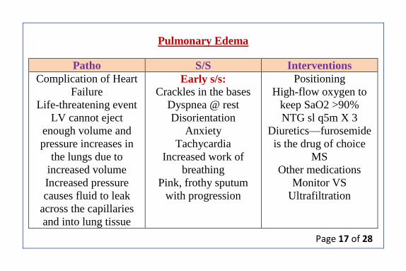
Pulmonary Edema
Patho S/S Interventions
Complication of Heart
Failure
Life-threatening event
LV cannot eject
enough volume and
pressure increases in
the lungs due to
increased volume
Increased pressure
causes fluid to leak
across the capillaries
and into lung tissue
Early s/s:
Crackles in the bases
Dyspnea @ rest
Disorientation
Anxiety
Tachycardia
Increased work of
breathing
Pink, frothy sputum
with progression
Positioning
High-flow oxygen to
keep SaO2 >90%
NTG sl q5m X 3
Diuretics—furosemide
is the drug of choice
MS
Other medications
Monitor VS
Ultrafiltration
Page 18 of 28
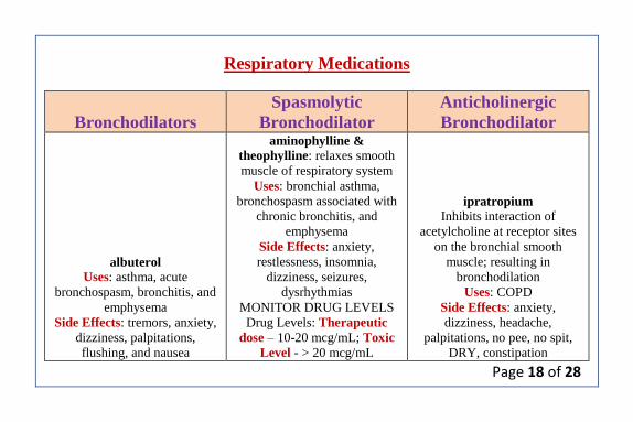
Respiratory Medications
Bronchodilators
Spasmolytic
Bronchodilator
Anticholinergic
Bronchodilator
albuterol
Uses: asthma, acute
bronchospasm, bronchitis, and
emphysema
Side Effects: tremors, anxiety,
dizziness, palpitations,
flushing, and nausea
aminophylline &
theophylline: relaxes smooth
muscle of respiratory system
Uses: bronchial asthma,
bronchospasm associated with
chronic bronchitis, and
emphysema
Side Effects: anxiety,
restlessness, insomnia,
dizziness, seizures,
dysrhythmias
MONITOR DRUG LEVELS
Drug Levels: Therapeutic
dose – 10-20 mcg/mL; Toxic
Level - > 20 mcg/mL
ipratropium
Inhibits interaction of
acetylcholine at receptor sites
on the bronchial smooth
muscle; resulting in
bronchodilation
Uses: COPD
Side Effects: anxiety,
dizziness, headache,
palpitations, no pee, no spit,
DRY, constipation

Page 19 of 28
Respiratory Medications Continued
Anti-
Inflammatories Diuretics Anticoagulants
Anticoagulant &
Antithrombotic prednisone
Uses: severe
inflammation,
immunosuppression, Side Effects: flushing,
hypertension,
thrombophlebitis,
embolism, GI
hemorrhage, increased
appetite
corticosteroids
Uses: prevention of
chronic asthma Side Effects: fever,
bronchospasm,
nervousness
furosemide
Loop Diuretic: inhibits
reabsorption of sodium
and chloride at proximal
and distal tubule and in
the Loop of Henle Uses: pulmonary edema,
edema in heart failure,
hypertension Side Effects: Circulatory
collapse, renal failure,
loss of hearing
warfarin
Uses: pulmonary emboli,
DVT, atrial fibrillation, valve replacement
Side Effects: hematuria,
hemorrhage
heparin
Uses: prevention of
DVT, pulmonary emboli,
MI, open heart surgery, atrial fibrillation
Side effects: hematuria,
hemorrhage

Page 20 of 28
Respiratory Labs PT & INR PTT D-Dimer
Used to monitor adequacy of
anticoagulation in pt. receiving
Coumadin Measures how long blood takes to
clot: Reflects how much of the
clotting factors II, V, VII, and X are present
Normal: 11.0 – 12.5 sec.
Therapy is considered appropriate when PT is prolonged by 1 ½ to 2
times the client's normal PT value INR: International Normalized
Ratio
Calculated by dividing the pt. PT by established standard PT
Normal: 0.7 – 1.8
Using INR to monitor Coumadin
therapy: goal is maintain the pt.
INR @ 2.0 and 3.0 regardless of
the actual PT
PTT- used to monitor
Heparin therapy
Used to assess the intrinsic
system and the common
pathway of clot formation;
evaluates factors I, II, V,
VIII, IX, X, XI, and XII
Normal: 25-35 seconds ;
anticoagulant therapy: 1.5-
2.5 times control value
Critical value: >70
seconds (if not on
anticoagulant therapy)
Normal finding: negative
Provides a simple and
confirmatory test for DIC
(disseminated intravascular
coagulation)
Levels of D-dimer also
increase with thrombotic
problems such as: DVT and
pulmonary embolism
Page 21 of 28
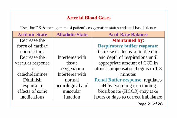
Arterial Blood Gases
Used for DX & management of patient’s oxygenation status and acid-base balance.
Acidotic State Alkalotic State Acid-Base Balance
Decrease the
force of cardiac
contractions
Decrease the
vascular response
to
catecholamines
Diminish
response to
effects of some
medications
Interferes with
tissue
oxygenation
Interferes with
normal
neurological and
muscular
function
Maintained by:
Respiratory buffer response:
increase or decrease in the rate
and depth of respirations until
appropriate amount of CO2 in
blood-compensation begins in 1-3
minutes
Renal Buffer response: regulates
pH by excreting or retaining
bicarbonate (HCO3)-may take
hours or days to correct imbalance
Page 22 of 28
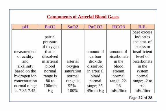
Components of Arterial Blood Gases
pH PaO2 SaO2 PaCO2 HCO3 B.E.
measurement
of acidity
and
alkalinity
based on the
hydrogen ion
concentration
normal range
is 7.35-7.45
partial
pressure
of oxygen
that is
dissolved
in arterial
blood
normal
range is
80 to
100mm
Hg
arterial
oxygen
saturation
normal
range is
95%-
100%
amount of
carbon
dioxide
dissolved
in arterial
blood
normal
range; 35-
45mm Hg
amount of
bicarbonate
in the
blood
stream
normal
range; 22-
26
mEq/liter
base excess
indicates
the amt. of
excess or
insufficient
level of
bicarbonate
in the
system
normal
range; -2 to
+2
mEq/liter
Page 23 of 28
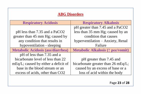
ABG Disorders
Respiratory Acidosis Respiratory Alkalosis
pH less than 7.35 and a PaCO2
greater than 45 mm Hg; caused by
any condition that results in
hypoventilation - sleeping
pH greater than 7.45 and a PaCO2
less than 35 mm Hg; caused by an
condition that causes
hyperventilation – Anxiety, Renal
Failure
Metabolic Acidosis (ass/diarrhea) Metabolic Alkalosis (↑ pee/vomit)
pH of less than 7.35 and a
bicarbonate level of less than 22
mEq/L; caused by either a deficit of
base in the blood stream or an
excess of acids, other than CO2
pH greater than 7.45 and
bicarbonate greater than 26 mEq/L;
caused by an excess of base or a
loss of acid within the body
Page 24 of 28

Page 25 of 28
ABG’s
ROME
Respiratory Opposite
Metabolic Equal
PH Normal = Fully
Compensated
All Values Abnormal =
Partially Compensated
Marching Band Suit
*Match PH w/Resp. or Metab.*
A B
PH 7.35 ———————— 7.45
B A
PcO2 35————————— 45 Resp
A B
HcO3 22————————— 26 Metab.

Page 26 of 28
Common Conversions
1 tsp = 5 mL
1 Tbsp = 3 tsp or 15 mL
1 oz = 30 mL
8 oz = 1 cup or 240 mL
1 pint = 1 lb or 16 oz
1 kg = 1000 g
1 g = 1000 mg
1 mg = 1000 mcg
1 L = 1000 mL

Page 27 of 28
Labs Normal Labs Normal
Na+ (Sodium) 135-145 K+ 3.5-5.0
Cl+ 98-106 Ca+ 9.0-10.5
Albumin (Liver) 3.5-5.0 Crea (Kidney) 0.7-1.3
BUN (Kidney) 8-25 Glucose 70-110
WBC 5,000-10,000 RBC (M)4.7-6.1 (F)4.2-5.4
Hgb (M)14-18(F)12-16 Hct (M)42-52(F)37-47
PLTS (ASA)
150,000-400,000
(↑Clot; ↓Bleed) Mag 1.6-2.6
PT (Heparin) 11-15 PTT (Heparin) 30-60
INR (Coumadin) 0.9-1.2 ALT (Liver) (M)10-40(F)7-35
ALT (Liver) (M)10-40(F)7-35 AST (Liver) 12-31
SG (Kidney)
1.005-1.03
(SIADH↑;DI↓) Amylase 25-151
Ammonia 10-80 T3 70-205
T4 4-12 TSH 0.3-5 (Hypo↑;Hyper↓)
Page 28 of 28
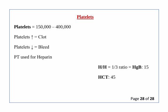
Platelets
Platelets = 150,000 – 400,000
Platelets ↑ = Clot
Platelets ↓ = Bleed
PT used for Heparin
H/H = 1/3 ratio = HgB: 15
HCT: 45