module # 8 – component # 4 protozoal diseases of wildlife · 2018-06-09 · – wildlifecampus...
TRANSCRIPT

– WildlifeCampus Wildlife Management Course
This course material is the copyrighted intellectual property of WildlifeCampus. It may not be copied, distributed or reproduced in any format whatsoever without the express written permission of WildlifeCampus
1
Protozoal Diseases of Wildlife © Copyright
Module # 8 – Component # 4
Protozoal Diseases of Wildlife
Objective
Identify the symptoms of important protozoal diseases of wildlife and to understand the control of these diseases.
Expected Outcome
List the most common symptoms of various important protozoal diseases Know how these diseases are transmitted Assist veterinarians in the control of these various diseases
Veterinarian kit

– WildlifeCampus Wildlife Management Course
This course material is the copyrighted intellectual property of WildlifeCampus. It may not be copied, distributed or reproduced in any format whatsoever without the express written permission of WildlifeCampus
2
Protozoal Diseases of Wildlife © Copyright
Introduction Classification of protozoa The word protozoa denotes unicellular animal. The word is from Greek origin, protos meaning first, and zoon an animal. There are more than 25 000 species of protozoa, of which 7000 are parasitic.
There are seven phyla, but only four are of importance in this context:
Phylum sarcomastigophora Phylum apicomplexa Phylum ciliophora Phylum microspora
Present-day protozoa are not necessarily primitive creatures. Many species are in fact highly specialized. The cells of multicellular animals have become part of various tissues and organs, each of which is specially developed for certain tasks. The unicellular animal has remained an ‘all-rounder’ and has developed organelles fulfilling essentially the same tasks as the organs of multicellular animals.
Roan antelope: Hippotragus equinus
– WildlifeCampus Wildlife Management Course
This course material is the copyrighted intellectual property of WildlifeCampus. It may not be copied, distributed or reproduced in any format whatsoever without the express written permission of WildlifeCampus
3
Protozoal Diseases of Wildlife © Copyright
Coccidia / Coccidiosis
This is a disease that occurs commonly in animals kept in captivity under less than ideal conditions.
Two genera of coccidia, Eimeria and Isospora, are present in almost all animal species’ gastro-intestinal tracts. They only cause clinical disease however when the animal’s resistance is poor for whatever reason. Oocysts (the spore stage of the protozoan life cycle), which are the infective stage are taken in per mouth and the protozoa then develop further in the mucous membranes of the animal’s gastro-intestinal tract.
Oocysts are then again released in the animal’s faeces but are only infective after sporulating outside the host in the environment – a process that takes at least two days. These spores can survive for long periods in cool moist conditions but are very susceptible to heat and desiccation.
Severe symptoms have been documented in impala, buffalo, springbok, kudu, eland and wild dogs.
Clinical symptoms are:
Diarrhoea that can often be bloody Chronic cases may show emaciation and/or dehydration
The diagnosis can be confirmed by means of large amounts of oocysts in the faeces.
Treatment consists of:
Sulpha antibiotics
Amprolium and monenzin are drugs that have preventative effects as well and can be included in the feed
Hygiene in terms of keeping bomas and camps clean and dry
The disease is host species and there is no human zoonosis.

– WildlifeCampus Wildlife Management Course
This course material is the copyrighted intellectual property of WildlifeCampus. It may not be copied, distributed or reproduced in any format whatsoever without the express written permission of WildlifeCampus
4
Protozoal Diseases of Wildlife © Copyright
Theileriosis / Cytauxzoonosis / Tileria
This tick-transmitted, deadly disease of cattle is important to the game rancher for the following reason:
Buffalo harbour the parasite without showing any clinical symptoms and act as a
source of infection for ticks that can then transmit the disease to cattle
The disease is caused by Theileria parva lawrencei and can be transmitted by the following two tick species:
Brown ear tick (Rhipicephalus appendiculatus) Zambesi brown ear tick (Rhipicephalus zambeziensis)
After being bitten by a tick the parasite starts to multiply within the host’s white blood cells.
It is this stage of the infection that causes the following clinical symptoms in cattle:
Fever Enlarged lymph nodes Emaciation Diarrhoea Watery nose secretions Weakness Muscle spasms Coughing Death. Almost all cattle die – extremely high mortality rate.
In the next stage of the parasite’s life cycle they enter the animal’s red blood cells. This stage does however only occur in buffalo as cattle die before this stage. When a tick now feeds on such a buffalo it becomes infected and capable of spreading the disease.

– WildlifeCampus Wildlife Management Course
This course material is the copyrighted intellectual property of WildlifeCampus. It may not be copied, distributed or reproduced in any format whatsoever without the express written permission of WildlifeCampus
5
Protozoal Diseases of Wildlife © Copyright
Various drugs are available that is effective in treating the disease in cattle but are not permitted in South Africa for the following reasons:
The drugs are effective against the first stage of the parasite but do not kill all the
parasites. The bovine will thus survive but so will a few of the parasites and they will then complete their lifecycles in the hosts red blood cells. This bovine will then become a carrier and capable of infecting ticks and thus spreading the disease to other cattle.
Tick larvae and nymphs that have fed on infected buffalo can remain alive for a
long period of time in the veld. This has the implication that even if buffalo moved through an area a while ago, cattle can still become infected when bitten by a tick on this veld.
Buffalo from the Addo-elephant National Park are free from this disease since the two most important tick-vector species do not occur there. Should these buffalo be moved to an area where the infected ticks are present they will become infected too and act as carriers.
Control to prevent spread of the disease is therefore aimed at:
Prevent contact between cattle and infected buffalo Prevent cattle from becoming carriers by not treating them Prevent ‘clean’ buffalo populations from becoming infected
Certain areas in South Africa are proclaimed as controlled areas where the above-mentioned control measures are strictly enforced in conjunction with the
following:
All cattle are to be dipped or treated with an appropriate registered drug to protect them against the ticks
No animal may be treated for corridor disease without written permission from the Directorate Animal Health.
All property with buffalo on must be registered with the Directorate Animal Health Such a property must be fenced according to the local nature conservation
authority’s regulations pertaining to buffalo. If this property is in a controlled area the fence must be electrified as well.
Buffalo are only allowed to be moved with an appropriate permit from the relevant state veterinarian

– WildlifeCampus Wildlife Management Course
This course material is the copyrighted intellectual property of WildlifeCampus. It may not be copied, distributed or reproduced in any format whatsoever without the express written permission of WildlifeCampus
6
Protozoal Diseases of Wildlife © Copyright
Buffalo can also act as carriers of East Coast Fever (Cytauxzoonosis) caused by Theileria parva parva. This tick-borne disease of cattle was luckily eradicated from
South Africa in the 1950’s.
The genus Cytauxzoon closely resembles Theileria and is also transmitted by ticks. It occurs in a variety of wildlife species but only causes clinical disease in animals with decreased resistance. This disease is an important killer of tsessebe calves, but has also been documented to occur in kudu, sable, roan, giraffe and grey duiker.
Clinically ill animals show the following symptoms:
Fever Weak Emaciated Anaemic Enlarged lymph nodes
Control of this parasite in not practical due to its widespread occurrence.
Healthy Sable Antelope: Hippotragus niger

– WildlifeCampus Wildlife Management Course
This course material is the copyrighted intellectual property of WildlifeCampus. It may not be copied, distributed or reproduced in any format whatsoever without the express written permission of WildlifeCampus
7
Protozoal Diseases of Wildlife © Copyright
Heartwater
This disease of ruminants is not caused by a protozoon but by the rickettsia Cowdria ruminantium. The Rickettsiae comprise a group of non-motile, non-sporeforming small bacteria-like organisms with typical cell walls that are commonly found in the tissues of arthropods, such as fleas, lice, mites and ticks.
Heartwater is restricted to the sub- and tropical regions and is transmitted by the ‘bont tick’ Amblyomma hebraeum. Springbok, eland and various exotic wildlife species are susceptible. Infection in blesbok and black wildebeest is transitory. Blue wildebeest, impala, buffalo, kudu, giraffe and warthogs are susceptible but do not show any clinical signs. In general, wildlife appears to be more resistant to heartwater than domesticated livestock.
Clinical symptoms include:
In peracute cases in livestock the animals die without showing any clinical signs
In acute cases the following can be seen: Increased rectal temperature Hypersensitivity Rapid, shallow breathing Abnormal behaviour Nervous symptoms such as: - Gritting on teeth - Backward arching of neck - Abnormal eye movements - Continuous muscle spasms
Large amounts of fluid are found in the animal’s lungs and thorax at post mortem examination.
Control measures should include:
Vaccination of all susceptible wildlife before relocating them to areas where the disease occurs
The use of certain antibiotics such as doxycycline is very effective but needs further research
Tick control to decrease incidence of the disease Veld management as the ticks require bushveld as a habitat. The disease does not
occur in animals restricted to grassveld only

– WildlifeCampus Wildlife Management Course
This course material is the copyrighted intellectual property of WildlifeCampus. It may not be copied, distributed or reproduced in any format whatsoever without the express written permission of WildlifeCampus
8
Protozoal Diseases of Wildlife © Copyright
Ecological Effect of Heartwater This disease is also very significant in terms of the distribution of game animals. Looking at the many reserves in South Africa will show that while many species occur in all habitats, certain species are restricted by habitat. This is mostly a function of the desert and semi-desert animals preferring this habitat, and even when unrestricted in movement, do not migrate to temperate climates.
One specific species that early wildlife managers attempted to introduce from semi-desert to bushveld was the Springbok. This is our national animal after all, and it would seem only right that tourists should be able to view in all our National Park, and particularly the Kruger National Park, the flag-ship of our National Park board. Small herds of Springbok were captured from the dry western parts of the country and translocated to the Kruger, where they promptly died within a few weeks. Most deaths were not due to either predation or capture myopathy, but still the whole population of translocated Springbok died.
The reason for this was the Heartwater tick and its associated disease. Springbok are extremely susceptible to this disease and when exposed, quickly contract it and die. Death is partly due to an accumulation of fluid in the pericardial sac surrounding the heart – thus heartwater disease. The reason why Springbok thrive in the arid western region of South Africa is because the heartwater tick cannot survive in these dry conditions. Therefore, we very seldom see impala and springbok together. These antelope are ecological equivalents, but due to the disease cannot live together. Well not quite, the Pilanesberg National Park is one of the very few areas where both species can be seen side by side. This is primarily since this area falls between the dry West and the Eastern savanna (KNP).
Springbok: Antidorcas marsupialis

– WildlifeCampus Wildlife Management Course
This course material is the copyrighted intellectual property of WildlifeCampus. It may not be copied, distributed or reproduced in any format whatsoever without the express written permission of WildlifeCampus
9
Protozoal Diseases of Wildlife © Copyright
Babesiosis
This disease is caused by protozoa from the genus Babesia and is transmitted by several tick species. In general, a specific animal species will be parasitized by its corresponding Babesia protozoa only. The protozoa that causes ‘redwater’ in cattle for instance does not affect wild antelope.
Babesia protozoa parasites infect the host’s red blood cells and causes the destruction of these cells.
This causes the following clinical signs:
Red discolouration of the urine (not always seen in wildlife) Animals become anaemic Feverish
The disease is mostly seen in individuals that were not exposed to the protozoa from early on in their life. This is especially so in animals raised in zoos and then released back into the wild. This was the case with several sable imported from Europe, that died from babesiosis caused by Babesia irvinesmithi. Wild dogs also seem to be susceptible to Babesia canis from time to time, whilst black-backed jackal might act as carriers of this disease.
Other animal species infected with Babesia protozoa but not always showing clinical signs thereof include:
Lion Leopard Cheetah Warthogs and bushpigs - These two species may carry Babesia trautmanni, that
can be transmitted by the tick Rhipicephalus simus to domesticated pigs.

– WildlifeCampus Wildlife Management Course
This course material is the copyrighted intellectual property of WildlifeCampus. It may not be copied, distributed or reproduced in any format whatsoever without the express written permission of WildlifeCampus
10
Protozoal Diseases of Wildlife © Copyright
Sarcocystosis These protozoa have what is called a ‘obligatory heteroxenic’ life cycle. This means that the protozoa require two hosts, one a herbivore and the other a carnivore, to complete its life cycle. The protozoa are very host specific. The herbivore becomes infected orally when sporocysts are ingested. The protozoa then form sarcocysts in the herbivorous host’s muscles. When the carnivorous host consumes meat infected with sarcocysts it then becomes infected and shed sporocysts in its faeces.
The clinical symptoms in the carnivorous host is usually very mild and slight gastro-intestinal upsets might be seen. People acting as the carnivorous host can suffer from abdominal pain and diarrhoea.
Lion: Panthera leo
– WildlifeCampus Wildlife Management Course
This course material is the copyrighted intellectual property of WildlifeCampus. It may not be copied, distributed or reproduced in any format whatsoever without the express written permission of WildlifeCampus
11
Protozoal Diseases of Wildlife © Copyright
Toxoplasmosis / Neosporosis Toxoplasma is a coccidian that has a typical gastro-intestinal cycle in cats, but forms tissue cysts in a wide range of mammals and birds.
Toxoplasmosis is an important disease of humans. Acquired infections are usually asymptomatic but abortions and congenital disease may occur.
Neospora caninum closely resembles Toxoplasma, but dogs rather than cats play a role in its life cycle. It is not a very significant disease for wildlife.
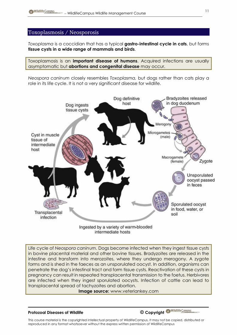
Life cycle of Neospora caninum. Dogs become infected when they ingest tissue cysts in bovine placental material and other bovine tissues. Bradyzoites are released in the intestine and transform into merozoites, where they undergo merogony. A zygote forms and is shed in the faeces as an unsporulated oocyst. In addition, organisms can penetrate the dog’s intestinal tract and form tissue cysts. Reactivation of these cysts in pregnancy can result in repeated transplacental transmission to the foetus. Herbivores are infected when they ingest sporulated oocysts. Infection of cattle can lead to transplacental spread of tachyzoites and abortion.
Image source: www.veteriankey.com

– WildlifeCampus Wildlife Management Course
This course material is the copyrighted intellectual property of WildlifeCampus. It may not be copied, distributed or reproduced in any format whatsoever without the express written permission of WildlifeCampus
12
Protozoal Diseases of Wildlife © Copyright
Anaplasmosis This disease is known as ‘gallsickness’ in cattle. Wildlife species such as blesbok, blue- and black wildebeest, grey duiker and impala can be carriers but their role as reservoir hosts is unknown.
Anaplasmosis can be transmitted by ticks feeding on animals as well as mechanically by biting flies.
Blue Wildebeest: Connochaetes taurinus

– WildlifeCampus Wildlife Management Course
This course material is the copyrighted intellectual property of WildlifeCampus. It may not be copied, distributed or reproduced in any format whatsoever without the express written permission of WildlifeCampus
13
Protozoal Diseases of Wildlife © Copyright
Besnoitiosis
The protozoan Besnoitia besnoiti causes this mild or severe but usually non-fatal disease of cattle, known as ‘elephant skin disease’. Biting flies probably transmits the disease. Similar protozoa are found in impala and blue wildebeest but do not cause clinical disease. Cattle can also be infected by these protozoa, but do not become clinically ill either. Instead they become immune against Besnoitia besnoiti.
In fact, the vaccine against cattle ‘elephant skin disease’ was manufactured from protozoa of blue wildebeest origin. The significance for wildlife is the potential for its spread from wildlife to domestic animals, where they share the same habitat.
Blue Wildebeest: Connochaetes taurinus

– WildlifeCampus Wildlife Management Course
This course material is the copyrighted intellectual property of WildlifeCampus. It may not be copied, distributed or reproduced in any format whatsoever without the express written permission of WildlifeCampus
14
Protozoal Diseases of Wildlife © Copyright
Nagana This disease of domesticated stock is caused by a Trypanosoma species and is transmitted by tsetse flies. Various species of wildlife can carry the disease without showing clinical signs. Tsetse flies become infected when feeding on carrier animals.
Cattle show the following clinical symptoms:
Undulating fever Lethargy Anaemia Emaciation Enlarged lymph nodes
Previously large numbers of game were killed in the KwaZulu / Natal province to control the carrier wildlife populations of nagana. This was not effective, and the disease was only controlled in 1950’s when tsetse flies were killed by poison sprayed from aircraft. Recently, cases of nagana were reported from the KwaZulu / Natal province but no control measures involving game are in place.
Source: www.financialgazette.co.zw