modulatory role of emblica officinalis fruit extract against arsenic induced oxidative stress in...
TRANSCRIPT
Mo
AC
a
ARRAA
KEASGHO
1
nBltspllwaaelDsmpp
0d
Chemico-Biological Interactions 180 (2009) 20–30
Contents lists available at ScienceDirect
Chemico-Biological Interactions
journa l homepage: www.e lsev ier .com/ locate /chembio int
odulatory role of Emblica officinalis fruit extract against arsenic inducedxidative stress in Swiss albino mice
mbika Sharma, Mukesh Kumar Sharma, Madhu Kumar ∗
ell and Molecular Biology Laboratory, Department of Zoology, University of Rajasthan, Jaipur 302004, India
r t i c l e i n f o
rticle history:eceived 19 November 2008eceived in revised form 22 January 2009ccepted 23 January 2009vailable online 3 February 2009
eywords:mblica officinalis
a b s t r a c t
Arsenic, an important human toxin, is naturally occurring in groundwater and its accumulation in plantsand animals have assumed a menacing proportion in a large part of the world, particularly Asia. Epi-demiological studies have shown a strong association between chronic arsenic exposure and variousadverse health effects, including cardiovascular diseases, neurological defects and cancer of lung, skin,bladder, liver and kidney. The protective role of the fruits of Emblica officinalis (500 mg/kg b.wt.) wasstudied in adult Swiss albino mice against arsenic induced hepatopathy. Arsenic treated group (NaAsO2,4 mg/kg b.wt.) had a significant increase in serum transaminases and lipid peroxidation (LPO) content in
rsenicerum transaminaseslutathione-S-transferaseistopathologyxidative stress
liver, whereas significant decrease was recorded in hepatic superoxide dismutase (SOD), catalase (CAT),glutathione-S-transferase (GST) and serum alkaline phosphatase activity. Combined treatment of Emblicaand arsenic (pre and post) declined the serum transaminases and LPO content in liver whereas significantincrease was noticed in SOD, CAT, GST and serum alkaline phosphatase activities. Liver histopathologyshowed that Emblica fruit extract had reduced karyolysis, karyorrhexis, necrosis and cytoplasmic vac-uolization induced by NaAsO2 intoxication. Thus it can be concluded that pre- and post-supplementationof E. officinalis fruit extract significantly reduced arsenic induced oxidative stress in liver.
. Introduction
Arsenicals are widespread in the environment as a result ofatural or anthropogenic activities. Nearly 50 million people inangladesh and parts of West Bengal in India are drinking toxic
evel of arsenic daily knowingly or unknowingly [1,2]. Arsenic ishe first metalloid to be identified as a human carcinogen. Expo-ure to arsenic contaminated drinking water causes several healthroblems [3], Blackfoot disease [4], hypertension [5], diabetes mel-
itus [6], disturbances in nervous system [7], cancers of liver, kidney,ung and bladder in humans [8]. Arsenic forms strong complexes
ith various sulfhydryl groups [9] and exert its toxicity by gener-ting reactive oxygen species (ROS) such as superoxide, hydroxylnd peroxyl radicals during its metabolism in cells [10]. Arsenicxposure was shown to depress the antioxidant defense system [11]eading to oxidative damage to cellular macromolecules includingNA, proteins, lipids [12], wreak havoc in biological system by tis-
ue damage, altering biochemical compounds and corroding cellembranes [13]. As the oxidative stress plays a central role in liver
athogenesis and progression, the use of antioxidants has beenroposed as therapeutic agents as well as drug coadjuvants to coun-
∗ Corresponding author. Tel.: +91 141 2395524.E-mail address: [email protected] (M. Kumar).
009-2797/$ – see front matter © 2009 Elsevier Ireland Ltd. All rights reserved.oi:10.1016/j.cbi.2009.01.012
© 2009 Elsevier Ireland Ltd. All rights reserved.
teract liver damages [14] and to protect the cellular machinery fromperoxidative injury inflicted by ROS [15]. Our earlier reports showedthe protective efficacies of Mentha piperita [11], Spirulina fusiformis[16,17] and Oscimum sanctum [18] against sodium arsenite and mer-curic chloride induced oxidative stress.
Emblica officinalis Gaertn (Commonly known in India as Amla,Syn. Phyllanthus emblica L.; Family: Euphorbiaceae) is regarded as“one of the best rejuvenating herbs” in the Ayurveda: an Indiantraditional medicinal science. Pozharitskaya et al. [19] demon-strated that E. officinalis extract contains several antioxidants suchas emblicanin A and B, gallic acid, ellagic acid, ascorbic acid thatpossesses strong antioxidative activity. A recent study also showsthat Emblica extract has potent antioxidant activity in term of freeradical scavenging properties [20].
Amla is an important dietary source of minerals, amino acids,tannin and sugar. The fruit extract has many pharmacologicalactivities for the treatment of a number of diseases [21] andis a constituent of many hepatoprotective formulations [22,23].It protect against radiation [24]; possess antidiabetic activity[25]; inhibits clastogenicity of benzopyrene and cyclophosphamide
[26]; gastroprotective [27]; cytoprotective and immunomodulatory[28]. New pharmacological activities viz. cytoprotective activ-ity against chromium [29]; protects against oxidative stress inischemic-reperfusion injury [30]; shows antivenom capacity [31];ameliorates hyperthyroidism and hepatic lipid peroxidation [32];
A. Sharma et al. / Chemico-Biological
dcawa
ma
2
2
mI((pdLwiJ
2
4Mt
2
2is2bda
2
hRUd
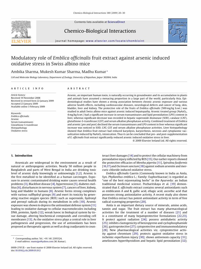
Fig. 1. Determination of LD50/30 of sodium arsenite.
isplays antiproliferative activity on MCF7 and MDA-MB-231 breastancer cell lines [33]; shows antitussive activity [34] and inducespoptosis in Dalton’s Lymphoma Ascites and CeHa cell lines [35]ere also reported. It act as antibacterial [36]; anti-inflammatory
gent [37] and modifies metal induced clastogenic effects [38].In the present study, the objective is to elucidate the biochemical
echanism associated with hepatoprotective role of E. officinalis onrsenic-induced liver damages and oxidative stress in mice.
. Materials and methods
.1. Test system
Adult male Swiss albino mice (6–8 weeks old, weighing 25 ± 2 g)aintained in the animal house as inbred colony (Procured from
VRI, Izatnagar, India) under controlled conditions of temperature25 ± 2 ◦C), relative humidity (50 ± 15%) and normal photoperiod12 h light and 12 h dark). The animals were housed in standardolypropylene laboratory cages containing 5 cm deep layer of saw-ust bedding. Mice were given standard mice feed (Hindustan Levertd., India) and tap water ad libitum. Once in a fortnight tetracyclineater was given as a preventive measure against infection. The eth-
cal committee of Department of Zoology, University of Rajasthan,aipur (India) has approved to carry out the experimental protocol.
.2. Test chemical
Arsenic in the form of NaAsO2 (mol. wt. 129.9, CAS no. 7784-65) was obtained from standard commercial suppliers [Himedia,umbai, India Ltd., Batch no. 3-1621 RM 1847]. Chemical used for
he present study was of analytical grade.
.2.1. Optimum dose selection of arsenicArsenic (NaAsO2) was administered at various dose levels, i.e.
.5, 5, 10, 20, 40 mg/kg body weight (b.wt.) intraperitoneally (i.p.)n 0.9% NaCl. The animals were observed for 30 days for any sign ofickness and mortality and the acute toxicity was observed within4 h. The LD50/30 of NaAsO2 was 22.98 mg/kg b.wt. (Fig. 1). On theasis of mortality data, the dose 4.0 mg/kg b.wt. (one tenth of lethalose) was selected for the investigation. The dose selected wasdministered i.p. at once.
.3. Plant material
Fresh fruits of E. officinalis were collected from the market at thearvest time. The plant was identified and a voucher specimen (No.UBL 20131) was stored in the herbarium of Department of Botany,niversity of Rajasthan. The fruits were sliced, dried in shade, pow-ered and extraction was carried out with DDW (double distilled
Interactions 180 (2009) 20–30 21
water) in soxhlet apparatus for 36 h. The extract was filtered andthen vacuum evaporated to get powdered form.
2.3.1. E. officinalis drug tolerance study and optimum doseselection
The animals were administered Emblica fruit extract dissolved inDDW orally up to 30 days (100, 300, 500, 700 and 900 mg/kg. b.wt.)and LPO and GSH contents were measured in the liver. The opti-mum dose selection of Emblica fruit extract was done on the basisof maximum GSH and minimum LPO content. Among the doses,500 mg/kg. b.wt. (or 0.5 ml/kg. b.wt.) was selected for the study(Fig. 2(A and B)).
2.3.2. Determination of radical scavenging activity of E. officinalisusing DPPH* assay
The radical scavenger activity of the aqueous Emblica fruitextract was measured spectrophotometrically using DPPH (1,1-diphenyl-2-picryl hydrayzyl) radical [39]. Aqueous fruit extract(0.1 ml) of different concentrations (2, 4, 8, 16 and 32 �g/ml) wereadded to 3 ml of 0.001 M DPPH* solution in methanol. The solu-tion was shaken and incubated at 37 ◦C for 30 min in the dark. Thedecrease in absorbance of DPPH* was measured against a blankat 517 nm. Percent (%) inhibition was calculated by comparing theabsorbance values with and without extract as:
%Inhibition = Ao − AeAo
× 100
Ao = Absorbance without extract. Ae = Absorbance with extract.In the present investigation, 50% Inhibitory concentration (IC50)
of Emblica fruit extract was found to be 8.43 �g/ml and 97.07%as DPPH* percent inhibition activity was also observed (Fig. 2(C)).Thus, E. officinalis possess significant radical scavenging activity.
2.4. Experimental design
Mice selected from inbred colony were divided into 4 groups.Groups Number of
animalsTreatment
I Control 6 Only vehicle DDW for 30consecutive days
II NaAsO2 treatment 6 Arsenic at 4 mg/kg b.wt. in0.9%NaCl i.p. at once only
III Emblica officinalis treatment 6 Emblica fruit extract 500 mg/kgb.wt. orally in DDW for 30consecutive days
IV Emblica + NaAsO2 + Emblica(Combination)
6 Emblica fruit extract (orally500 mg/kg b.wt.) was administered10 days before NaAsO2 (4 mg/kgb.wt.) and continued up to 30 daysafter arsenic treatment
The animals from all the groups were sacrificed on 1, 3, 7, 15and 30 days. Liver were removed, rinsed in cold saline, blotted,weighed, processed for histological and various biochemical assays.Fresh unhaemolysed serum was used for transaminases and alka-line phosphatase (ALP) activities.
2.5. Antioxidants assays
2.5.1. Serum glutamate oxaloacetate transaminase (SGOT)(2.6.1.1)
The SGOT activity in the serum was estimated by Reitman andFrankel [40] method. The oxaloacetate formed in the reaction is
coupled with 2,4-dinitrophenyl hydrazine (DNPH) to give the corre-sponding hydrazone, which give brown colour in alkaline mediumand this is measured colorimetrically at 505 nm. A standard curveobtained using different amounts of pyruvate and enzyme activitywas expressed as unit/ml.
22 A. Sharma et al. / Chemico-Biological Interactions 180 (2009) 20–30
Fig. 2. (A) Variation in liver reduced in glutathione (GSH) content of different doses of Emblica fruit extract. (B) Variation in liver lipid peroxidation (LPO) level of doses ofEmblica fruit extract. (C) Emblica officinalis fruit extract DPPH* radical scavenging assay.

ogical
2
aiitow
F(pc
A. Sharma et al. / Chemico-Biol
.5.2. Serum glutamate pyruvate transaminase (SGPT) (2.6.1.2)SGPT activity was also estimated by the method of Reitman
nd Frankel [40] using DNPH as colour reagent. Pyruvate formed
n the reaction is coupled with 2,4-DNPH to give the correspond-ng hydrazone, which give brown colour in alkaline medium andhis is measured colorimetrically at 505 nm. A standard curve wasbtained using different amounts of pyruvate and enzyme activityas expressed as unit/ml.ig. 3. (A) Variation in Serum glutamate oxaloacetate transaminase (SGOT) activity in diffeSGPT) activity in different experimental groups. (C) Variation in Serum alkaline phospheroxidation (LPO) content in different experimental groups. (E) Variation in liver superoxatalase (CAT) activity in different experimental groups. (G) Variation in liver glutathione
Interactions 180 (2009) 20–30 23
2.5.3. Serum alkaline phosphataseSerum alkaline phosphatase activity was measured by the
method of Kind and King [41] using commercially accessible kits
from Span diagnostic limited, Surat, India. Alkaline phosphatasefrom serum converts phenyl phosphate to inorganic phosphate andphenol. Phenol reacts in alkaline medium with 4 amino antipyrinein the presence of an oxidizing agent, potassium ferricyanide andforms an orange-red coloured complex which can be measuredrent experimental groups. (B) Variation in Serum glutamate pyruvate transaminaseatase activity (KAU) in different experimental groups. (D) Variation in liver lipidide dismutase (SOD) activity in different experimental groups. (F) Variation in liver-S-transferase (GST) activity in different experimental groups.

24 A. Sharma et al. / Chemico-Biological Interactions 180 (2009) 20–30
(Cont
ct(
2
ms
Fig. 3.
olorimetrically at 510 nm. The colour intensity is proportionalo the enzyme activity was expressed as King Armstrong UnitKAU).
.5.4. Lipid peroxidation (LPO)Lipid peroxidation level in the liver was measured by the
ethod of Ohkawa et al. [42] as thiobarbituric acid reactiveubstances (TBARS). The concentration of TBARS was expressed
inued )
as n moles of malondialdehyde per mg of tissue using 1,1,3,3-tetramethoxypropane (TMP) as standard. A standard curve wasprepared by using tetramethoxy propane (mol. wt. 164.20 pur-
chased from Lancaster, England). 1–12 nmol of TMP was taken incorresponding no. of test tubes with all the reagents (minus sam-ple) were added. Standard curve was plotted by taking absorbanceagainst TMP concentration on abscissa. The absorbance was read at532 nm using UV–vis systronic spectrophotometer.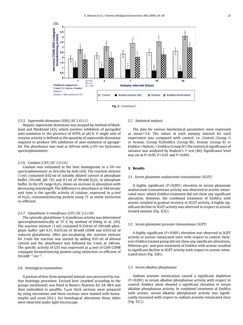
A. Sharma et al. / Chemico-Biological Interactions 180 (2009) 20–30 25
(Conti
2
laerls
2
s(bbduoc
2
sTprfcTc9
2
tgtbtw
Fig. 3.
.5.5. Superoxide dismutase (SOD) (EC 1.15.1.1)Hepatic superoxide dismutase was assayed by method of Mark-
und and Marklund [43], which involves inhibition of pyrogalloluto-oxidation in the presence of EDTA at pH 8. A single unit ofnzyme activity is defined as the quantity of superoxide dismutaseequired to produce 50% inhibition of auto-oxidation of pyrogal-ol. The absorbance was read at 420 nm with a UV–vis Systronicspectrophotometer.
.5.6. Catalase (CAT) (EC 1.11.1.6)Catalase was estimated in the liver homogenate in a UV–vis
pectrophotometer as describe by Aebi [44]. The reaction mixture1 ml) contained 0.02 ml of suitably diluted cytosol in phosphateuffer (50 mM, pH 7.0) and 0.1 ml of 30 mM H2O2 in phosphateuffer. In the UV range H2O2 shows an increase in absorption withecreasing wavelength. The difference in absorbance at 240 nm pernit time is the specific activity of catalase, expressed in � molf H2O2 consumed/min/mg protein using 71 as molar extinctiono-efficient.
.5.7. Glutathione-S-transferase (GST) (EC 2.5.1.18)The cytosolic glutathione-S-transferase activity was determined
pectrophotometrically at 37 ◦C by method of Habig et al. [45].he reaction mixture (1 ml) contained 0.334 ml of 100 mM phos-hate buffer (pH 6.5), 0.033 ml of 30 mM CDNB and 0.033 ml ofeduced glutathione. After pre-incubating the reaction mixtureor 2 min the reaction was started by adding 0.01 ml of dilutedytosol and the absorbance was followed for 3 min at 340 nm.he specific activity of GST was expressed as � mol of GSH-CDNBonjugate formed/min/mg protein using extinction co-efficient of.6 mM−1 cm−1.
.6. Histological examination
A portion of liver from autopsied animals was processed by rou-ine histology procedure. Excised liver (marked according to theroups mentioned) was fixed in Bouin’s fixatives for 24–48 h andhen embedded in paraffin. 5 �m thick sections were preparedy using microtome and these sections were stained with hema-oxylin and eosin [H.E.]. For histological alterations these slidesere observed under light microscope.
nued ).
2.7. Statistical analysis
The data for various biochemical parameters were expressedas mean + S.E. The values at each autopsy interval for eachexperiment was compared with control, i.e. Control (Group I)vs Arsenic (Group II)/Emblica (Group III); Arsenic (Group II) vsEmblica + NaAsO2 + Emblica (Group IV).The statistical significance ofvariance was analyzed by Student’s ‘t’ test [46]. Significance levelwas set at P < 0.05, P < 0.01 and P < 0.001.
3. Results
3.1. Serum glutamate oxaloacetate transaminase (SGOT)
A highly significant (P < 0.001) elevation in serum glutamateoxaloacetate transaminase activity was observed in arsenic intoxi-cated mice, where Emblica treatment did not show any significantalteration. However, the combined treatment of Emblica witharsenic resulted in gradual recovery in SGOT activity. A highly sig-nificant decline in SGOT activity was observed in respect to arsenictreated animals (Fig. 3(A)).
3.2. Serum glutamate pyruvate transaminase (SGPT)
A highly significant (P < 0.001) elevation was observed in SGPTactivity in arsenic intoxicated mice with respect to control. How-ever Emblica treated group did not show any significant alterations.Whereas pre- and post-treatment of Emblica with arsenic resultedin significant decline in SGPT activity with respect to arsenic intox-icated mice (Fig. 3(B)).
3.3. Serum alkaline phosphatase
Sodium arsenite intoxication caused a significant depletion(P < 0.001) in serum alkaline phosphatase activity with respect to
control. Emblica alone showed a significant elevation in serumalkaline phosphatase activity. In combined treatment of Emblicawith arsenic, serum alkaline phosphatase activity was signifi-cantly increased with respect to sodium arsenite intoxicated mice(Fig. 3(C)).
26 A. Sharma et al. / Chemico-Biological Interactions 180 (2009) 20–30
F uclearm
3
iad(
3
NlEWSa
Fg
ig. 4. [Day-1]: (a) NaAsO2 group [Group-II]: hepatocytoplasmic vacuolization, naintained cytoplasmic granulation, enucleation at few places (H.E. 400×).
.4. Lipid peroxidation (LPO)
A highly significant elevation in liver LPO level in arsenic intox-cated mice was found. Wherein Emblica alone treatment showed
significant decline. In combined treatment a highly significantepletion was recorded with respect to arsenic intoxicated miceFig. 3(D)).
.5. Superoxide dismutase (SOD)
SOD catalyzes the conversion of superoxide anion into H2O2.aAsO2 intoxication caused a significant depletion (P < 0.001) in
iver superoxide dismutase (SOD) activity with respect to control.mblica alone showed a significant elevation in liver SOD activity.hereas in combined treatment of Emblica with sodium arsenite
OD activity showed a highly significant elevation as compared torsenic treated animals (Fig. 3(E)).
ig. 5. [Day-3]: (a) NaAsO2 group [Group-II]: more cytoplasmic vacuolization, occasional eranular cytoplasm and normal hepatocytes (H.E. 400×).
changes karyorrhexis, occluded central vein. (b) Combination group [Group-IV]:
3.6. Catalase (CAT)
The primary role of catalase is to scavenge H2O2 that has beengenerated by free radical or by SOD in removal of superoxideanions. Sodium arsenite intoxication caused a significant deple-tion (P < 0.001) in liver CAT activity with respect to control. Emblicaalone showed a significant elevation in liver CAT activity and incombined treatment of Emblica with arsenic a significant increasewas observed with respect to sodium arsenite intoxicated mice(Fig. 3(F)).
3.7. Glutathione-S-transferase (GST)
GSTs are a multigene family of isozymes that catalyze theconjugation of cellular antioxidants to a variety of electrophiliccompounds and thereby exert a critical role in cytoprotectionagainst reactive oxygen species. Sodium arsenite administration
nucleation alongwith karyorrhexis. (b) Combination group [Group-IV]: maintained
A. Sharma et al. / Chemico-Biological Interactions 180 (2009) 20–30 27
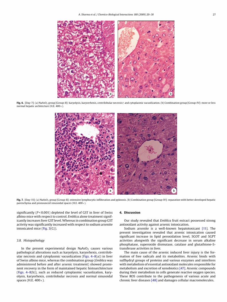
Fig. 6. [Day-7]: (a) NaAsO2 group [Group-II]: karyolysis, karyorrhexis, centrilobular necrosis↑ and cytoplasmic vacuolization. (b) Combination group [Group-IV]: more or lessnormal hepatic architecture (H.E. 400×).
F d pykp
saiai
3
puoan(os
ig. 7. [Day-15]: (a) NaAsO2 group [Group-II]: extensive lymphocytic infilteration anarenchyma and pronounced sinusoidal spaces (H.E. 400×).
ignificantly (P < 0.001) depleted the level of GST in liver of Swisslbino mice with respect to control. Emblica alone treatment signif-cantly increases liver GST level. Whereas in combination group GSTctivity was significantly increased with respect to sodium arsenitentoxicated mice (Fig. 3(G)).
.8. Histopathology
In the present experimental design NaAsO2 causes variousathological alterations such as karyolysis, karyorhexis, centrilob-lar necrosis and cytoplasmic vacuolization (Figs. 4–8(a)) in liverf Swiss albino mice, whereas the combination group (Emblica was
dministered before and after arsenic treatment) showed promi-ent recovery in the form of maintained hepatic histoarchitectureFigs. 4–8(b)), such as reduced cytoplasmic vacuolization, kary-lysis, karyorhexis, centrilobular necrosis and normal sinusoidalpaces (H.E. 400×).nosis. (b) Combination group [Group-IV]: reparation with better developed hepatic
4. Discussion
Our study revealed that Emblica fruit extract possessed strongantioxidant activity against arsenic intoxication.
Sodium arsenite is a well-known hepatotoxicant [11]. Thepresent investigation revealed that arsenic intoxication causedsignificant increase in lipid peroxidation level, SGOT and SGPTactivities alongwith the significant decrease in serum alkalinephosphatase, superoxide dismutase, catalase and glutathione-S-transferase activities in liver.
The main cause of the arsenic induced liver injury is the for-mation of free radicals and its metabolites. Arsenic binds withsulfhydryl groups of proteins and various enzymes and interferes
with metabolism of essential antioxidant molecules responsible formetabolism and excretion of xenobiotics [47]. Arsenic compoundsduring their metabolism in cells generate reactive oxygen species.These ROS contribute to the pathogenesis of various acute andchronic liver diseases [48] and damages cellular macromolecules.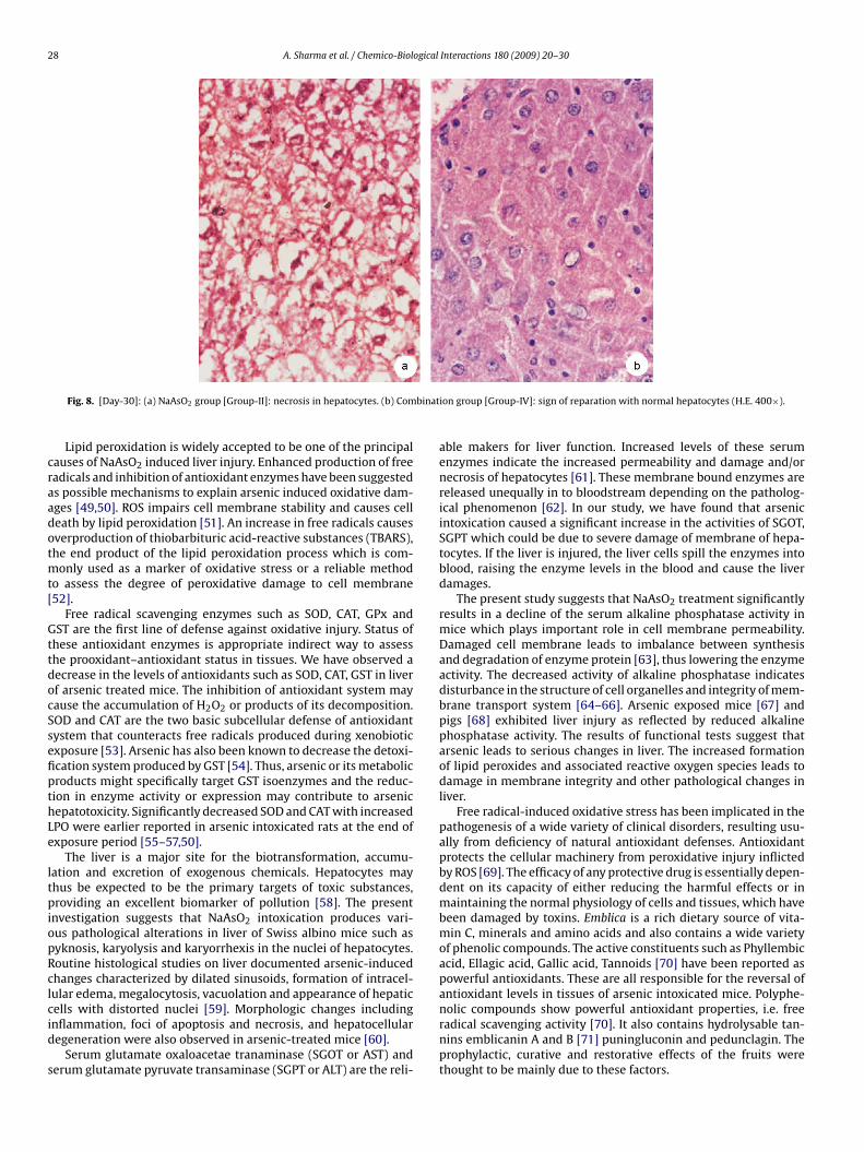
28 A. Sharma et al. / Chemico-Biological Interactions 180 (2009) 20–30
binat
craadotmt[
GttdocSsefipthLe
ltpiopRclcid
s
Fig. 8. [Day-30]: (a) NaAsO2 group [Group-II]: necrosis in hepatocytes. (b) Com
Lipid peroxidation is widely accepted to be one of the principalauses of NaAsO2 induced liver injury. Enhanced production of freeadicals and inhibition of antioxidant enzymes have been suggesteds possible mechanisms to explain arsenic induced oxidative dam-ges [49,50]. ROS impairs cell membrane stability and causes celleath by lipid peroxidation [51]. An increase in free radicals causesverproduction of thiobarbituric acid-reactive substances (TBARS),he end product of the lipid peroxidation process which is com-
only used as a marker of oxidative stress or a reliable methodo assess the degree of peroxidative damage to cell membrane52].
Free radical scavenging enzymes such as SOD, CAT, GPx andST are the first line of defense against oxidative injury. Status of
hese antioxidant enzymes is appropriate indirect way to assesshe prooxidant–antioxidant status in tissues. We have observed aecrease in the levels of antioxidants such as SOD, CAT, GST in liverf arsenic treated mice. The inhibition of antioxidant system mayause the accumulation of H2O2 or products of its decomposition.OD and CAT are the two basic subcellular defense of antioxidantystem that counteracts free radicals produced during xenobioticxposure [53]. Arsenic has also been known to decrease the detoxi-cation system produced by GST [54]. Thus, arsenic or its metabolicroducts might specifically target GST isoenzymes and the reduc-ion in enzyme activity or expression may contribute to arsenicepatotoxicity. Significantly decreased SOD and CAT with increasedPO were earlier reported in arsenic intoxicated rats at the end ofxposure period [55–57,50].
The liver is a major site for the biotransformation, accumu-ation and excretion of exogenous chemicals. Hepatocytes mayhus be expected to be the primary targets of toxic substances,roviding an excellent biomarker of pollution [58]. The present
nvestigation suggests that NaAsO2 intoxication produces vari-us pathological alterations in liver of Swiss albino mice such asyknosis, karyolysis and karyorrhexis in the nuclei of hepatocytes.outine histological studies on liver documented arsenic-inducedhanges characterized by dilated sinusoids, formation of intracel-ular edema, megalocytosis, vacuolation and appearance of hepatic
ells with distorted nuclei [59]. Morphologic changes includingnflammation, foci of apoptosis and necrosis, and hepatocellularegeneration were also observed in arsenic-treated mice [60].Serum glutamate oxaloacetae tranaminase (SGOT or AST) anderum glutamate pyruvate transaminase (SGPT or ALT) are the reli-
ion group [Group-IV]: sign of reparation with normal hepatocytes (H.E. 400×).
able makers for liver function. Increased levels of these serumenzymes indicate the increased permeability and damage and/ornecrosis of hepatocytes [61]. These membrane bound enzymes arereleased unequally in to bloodstream depending on the patholog-ical phenomenon [62]. In our study, we have found that arsenicintoxication caused a significant increase in the activities of SGOT,SGPT which could be due to severe damage of membrane of hepa-tocytes. If the liver is injured, the liver cells spill the enzymes intoblood, raising the enzyme levels in the blood and cause the liverdamages.
The present study suggests that NaAsO2 treatment significantlyresults in a decline of the serum alkaline phosphatase activity inmice which plays important role in cell membrane permeability.Damaged cell membrane leads to imbalance between synthesisand degradation of enzyme protein [63], thus lowering the enzymeactivity. The decreased activity of alkaline phosphatase indicatesdisturbance in the structure of cell organelles and integrity of mem-brane transport system [64–66]. Arsenic exposed mice [67] andpigs [68] exhibited liver injury as reflected by reduced alkalinephosphatase activity. The results of functional tests suggest thatarsenic leads to serious changes in liver. The increased formationof lipid peroxides and associated reactive oxygen species leads todamage in membrane integrity and other pathological changes inliver.
Free radical-induced oxidative stress has been implicated in thepathogenesis of a wide variety of clinical disorders, resulting usu-ally from deficiency of natural antioxidant defenses. Antioxidantprotects the cellular machinery from peroxidative injury inflictedby ROS [69]. The efficacy of any protective drug is essentially depen-dent on its capacity of either reducing the harmful effects or inmaintaining the normal physiology of cells and tissues, which havebeen damaged by toxins. Emblica is a rich dietary source of vita-min C, minerals and amino acids and also contains a wide varietyof phenolic compounds. The active constituents such as Phyllembicacid, Ellagic acid, Gallic acid, Tannoids [70] have been reported aspowerful antioxidants. These are all responsible for the reversal ofantioxidant levels in tissues of arsenic intoxicated mice. Polyphe-
nolic compounds show powerful antioxidant properties, i.e. freeradical scavenging activity [70]. It also contains hydrolysable tan-nins emblicanin A and B [71] puningluconin and pedunclagin. Theprophylactic, curative and restorative effects of the fruits werethought to be mainly due to these factors.
ogical
leEoha
ea
asatt
apmtmflgfa
rsp
C
A
pf
R
[
[
[
[
[
[
[
[
[
[
[
[
[
[
[
[
[
[
[
[
[
[
[
[
[
[
[
[
[
[
[
A. Sharma et al. / Chemico-Biol
Administration of Emblica significantly decreased the level ofipid peroxidation when compared with arsenic treated mice. Non-nzymatic antioxidant such as vitamin C, the richest amount inmblica, plays an excellent role in protecting the cell from lipid per-xidation. Our results obviously indicate that Emblica fruit extractas radical scavenging activity, inhibit the lipid peroxidation dam-ge and shows hepatoprotective potential against arsenic toxicity.
The antioxidants found in Emblica fruit extract maintains thendogenous antioxidants, thus reduces oxidative stress and allevi-te the pathological changes caused by arsenic in liver.
E. officinalis is a constituent of various liver tonics used againstcute viral hepatitis and other liver disorders [22,72]. Antioxidantsuch as ellagic acid [73] have been reported to protect liver injurynd fibrosis induced by hepatotoxins. E. officinalis fruit extract neu-ralize the oxidizing potentials of reactive oxygen species generatedhereby maintaining cell membrane integrity and viability.
E. officinalis extract shows efficacy as a good inhibitor againstrsenic-induced oxidative stress as observed by decrease in lipideroxidation and increase in SOD, CAT and GST activity and alsoaintained the structural integrity of the hepatocellular archi-
ecture. The protective action of fruit extract of the E. officinalisay be due to the presence of the known anti inflammatory
avanoid, luteolin (5,7,3%,4%-tetrahydroxyflavone) and its 3-O-lycoside derivatives, which provides maximum conjugation withree radical species, thus reducing the number of free radicals avail-ble and the extent of cellular damages.
In conclusion, the enhanced levels of antioxidant enzymes andeduced amount of lipid peroxides and serum transaminases areuggested to be the major mechanisms of Emblica fruit extract inreventing the liver damages induced by arsenic.
onflict of interest statement
None.
cknowledgement
Authors are gratefully acknowledge to the CSIR-New Delhi forroviding financial assistance to Ambika Sharma as Senior research
ellow, Letter No.: 09/149/(0489)/2008/EMR-I.
eferences
[1] F. McLellan, Arsenic contamination affects millions in Bangladesh, Lancet 359(2002) 1127.
[2] R. MacDonald, Providing clean water: lessons from Bangladesh, Br. Med. J. 322(2001) 626–627.
[3] D.N. Guha Mazumder, Chronic arsenic toxicity & human health, Indian J. Med.Res. 128 (2008) 436–447.
[4] C.H. Tseng, C.K. Chong, C.J. Chen, T.Y. Tai, Dose–response relationship betweenperipheral vascular disease and ingested inorganic arsenic among residentsin Blackfoot disease endemic villages in Taiwan, Atherosclerosis 120 (1996)125–133.
[5] M.S. Lai, Y.M. Hsueh, C.H. Chen, M.P. Shyu, S.Y. Chen, T.L. Kuo, M.M. Wu, T.Y. Tai,Ingested inorganic arsenic and prevalence of diabetes mellitus, Am. J. Epidemiol.139 (1994) 484–492.
[6] C.J. Chen, Y.M. Hsueh, M.S. Lai, M.P. Shyu, S.Y. Chen, M.M. Wu, T.L. Kuo, T.Y. Tai,Increased prevalence of hypertension and long term arsenic exposure, Hyper-tension 25 (1995) 53–60.
[7] E. García-Chávez, I. Jiménez, B. Segura, L.M. Del Razo, Lipid oxidative damageand distribution of inorganic arsenic and its metabolites in the rat nervoussystem after arsenite exposure: influence of alpha tocopherol supplementation,Neurotoxicology 27 (2006) 1024–1031.
[8] T.K. Hei, M. Filipic, Role of oxidative damage in the genotoxicity of arsenic, FreeRadic. Biol. Med. 37 (2004) 574–581.
[9] H.V. Aposhian, Biochemical toxicology of arsenic, Rev. Biochem. Toxicol. 10(1989) 265–299.
10] J. Liu, M. Kaduska, Y. Liu, W. Qu, R.P. Mason, M.P. Walker, Acute arsenic inducedfree radical production and oxidative stress related gene expression in mice,Toxicologists 54 (2000) 280–281.
11] A. Sharma, M.K. Sharma, M. Kumar, Protective effect of Mentha piperita againstarsenic-induced toxicity in liver of Swiss albino mice, Basic Clin. Pharmacol.Toxicol. 100 (2007) 249–257.
[
[
Interactions 180 (2009) 20–30 29
12] H. Shi, X. Shi, K.J. Liu, Oxidative mechanism of arsenic toxicity and carcinogen-esis, Mol. Cell Biochem. (2004) 25567–25578 (Review).
13] H. Wiseman, B. Halliwell, Damage to DNA by reactive oxygen and nitrogenspecies: role in inflammatory disease and progression to cancer, Biochem. J.313 (1996) 17–29.
14] P. Vitaglione, F. Morisco, N. Caporaso, V. Fogliano, Dietary antioxidant com-pounds and liver health, Crit. Rev. Food Sci. Nutr. 44 (2004) 575–586.
15] B. Halliwell, O.I. Arouma, DNA and Free Radicals, Ellis Horwood Ltd., Chichester,UK, 1993.
16] M.K. Sharma, A. Sharma, A. Kumar, M. Kumar, Spirulina fusiformis provides pro-tection against mercuric chloride induced oxidative stress in Swiss albino mice,Food Chem. Toxicol. 45 (2007) 2412–2419.
17] M. Kumar, M.K. Sharma, A. Kumar, Spirulina fusiformis: a food supplementagainst mercury induced hepatic toxicity, J. Health Sci. 51 (2005) 424–430.
18] M.K. Sharma, M. Kumar, A. Kumar, Ocimum sanctum aqueous leaf extract pro-vides protection against mercury induced toxicity in Swiss albino mice, IndianJ. Exp. Biol. 40 (2002) 1079–1082.
19] O.N. Pozharitskaya, S.A. Ivanova, A.N. Shikov, V.G. Makarov, Separation and eval-uation of free radical-scavenging activity of phenol components of Emblicaofficinalis extract by using an HPTLC-DPPH* method, J. Sep. Sci. 30 (2007)1250–1254.
20] P.K. Jain, V. Ravichandran, S. Sharma, R.K. Agarwal, The antioxidant activity ofsome medicinal plants, Turk. J. Biol. 32 (2008) 197–202.
21] R.N. Chopra, I.C. Handa, I.D. Kapur, Indigenous Drugs of India, second ed., U.N.Dhar and Sons, Calcutta, 1958.
22] D.S. Antarkar, B.V. Ashok, J.C. Doshi, A.V. Athavale, K.S. Vinchoo, M.R. Natekar, P.S.Thathed, V. Ramesh, N. Kale, Double-bind clinical trial of Arogyawardhani—anayurvedic drug in acute viral hepatitis, Indian J. Med. Res. 72 (1980) 588–593.
23] S. De, B. Ravishankar, G.C. Bhavsar, Plants with hepatoprotective activity—areview, Indian Drugs 30 (1993) 355–363.
24] P. Scartezzini, E. Speroni, Review on some plants of Indian traditional medicinewith antioxidant activity, J. Ethnopharmacol. 71 (2000) 23–43.
25] M.C. Sabu, R. Kuttan, Anti-diabetic activity of medicinal plants and its rela-tionship with their antioxidant property, J. Ethnopharmacol. 81 (2002) 155–160.
26] N. Sharma, P. Trikha, M. Athar, S. Raisuddin, Inhibitory effect of Emblica officinalson the in vivo clastogenicity of benzo pyrene and cyclophosphamide in mice,Hum. Exp. Toxicol. 19 (2000) 377–384.
27] A.J. Al-Rehaily, T.A. Al-Howiriny, M.O. Al-Sohaibani, S. Rafatullah, Gastropro-tective effects of ‘Amla’ Emblica officinalis on in vivo test models in rats,Phytomedicine 9 (2002) 515–522.
28] M. Sai Ram, D. Neetu, B. Yogesh, B. Anju, P. Dipti, T. Pauline, S.K. Sharma,S.K. Sarada, G. Ilavazhagan, D. Kumar, W. Selvamurthy, Cyto-protective andimmunomodulating properties of Amla (Emblica officinalis) on lymphocytes:an in-vitro study, J. Ethnopharmacol. 81 (2002) 5–10.
29] M. Sai Ram, D. Neetu, P. Deepti, M. Vandana, G. Ilavazhagan, D. Kumar, W. Selva-murthy, Cytoprotective activity of Amla (Emblica officinalis) against chromium(VI) induced oxidative injury in murine macrophages, Phytother. Res. 17 (2003)430–433.
30] S. Rajak, S.K. Banerjee, S. Sood, A.K. Dinda, Y.K. Gupta, S.K. Gupta, S.K. Maulik,Emblica officinalis causes myocardial adaptation and protects against oxidativestress in ischemic-reperfusion injury in rats, Phytother. Res. 18 (2004) 54–60.
31] M.I. Alam, A. Gomes, Snake venom neutralization by Indian medicinal plants(Vitex negundo and Emblica officinalis) root extracts, J. Ethnopharmacol. 86(2003) 75–80.
32] S. Panda, A. Kar, Fruit extract of Emblica officinalis ameliorates hyperthyroidismand hepatic lipid peroxidation in mice, Pharmazie 58 (2003) 753–755.
33] E. Lambertini, R. Piva, M.T. Khan, I. Lampronti, N. Bianchi, M. Borgatti, R.Gambari, Effects of extracts from Bangladeshi medicinal plants on in vitro pro-liferation of human breast cancer cell lines and expression of estrogen receptoralpha gene, Int. J. Oncol. 24 (2003) 419–423.
34] G. Nosal’ova, J. Mokry, K.M. Hassan, Antitussive activity of the fruit extractof Emblica officinalis Gaertn. (Euphorbiaceae), Phytomedicine 10 (2003) 583–589.
35] N.V. Rajeshkumar, M.R. Pillai, R. Kuttan, Induction of apoptosis in mouse andhuman carcinoma cell lines by Emblica officinalis polyphenols and its effect onchemical carcinogenesis, J. Exp. Clin. Cancer Res. 22 (2003) 201–212.
36] S.H. Godbole, G.S. Pendse, Antibacterial property of some plants, Indian J.Pharm. 22 (1960) 39–42.
37] M.Z. Asmawi, H. Kankaanranta, E. Moilanen, H. Vapaatalo, Anti-inflammatoryactivities of Emblica officinalis Gaertn. leaf extracts, J. Pharm. Pharmacol. 45(1993) 581–584.
38] H. Dhir, R.K. Roy, A. Sharma, G. Talukdar, Modification of clastogenicity of leadand aluminium in mouse bone marrow cells by dietary ingestion of Phyllanthusemblica fruit extract, Mutat. Res. 241 (1990) 305–312.
39] M. Gulluce, F. Sahin, M. Sokmen, H. Ozer, D. Daferera, A. Sokmen, M. Polissiou,A. Adiguzel, H. Ozkan, Antimicrobial and antioxidant properties of the essentialoils and methanol extract from Mentha longifolia L. ssp. longifolia, Food Chem.103 (2007) 1449–1456.
40] S. Reitman, S. Frankel, A colorimetric method for the determination of serum
glutamic oxalacetic and glutamic pyruvic transaminases, Am. J. Clin. Pathol. 28(1957) 56–63.41] P.R.N. Kind, E.J. King, Estimation of plasma phosphatase by determination ofhydrolysed phenol with anti-pyrine, J. Clin. Pathol. 7 (1954) 322–326.
42] H. Ohkawa, N. Ohishi, K. Yagi, Assay for lipid peroxides in animal tissues bythiobarbituric acid reaction, Anal. Biochem. 95 (1979) 351–358.

3 ogical
[
[[
[
[
[
[
[
[
[
[
[
[
[
[
[
[
[
[
[[
[
[
[
[
[
[
[
[Part 1—the chemistry and antioxidant effects of two new hydrolysable tannins,Emblicanin A and B, Indian J. Chem. 35B (1996) 941–948.
0 A. Sharma et al. / Chemico-Biol
43] S. Marklund, G. Marklund, Involvement of the superoxide anion radical in theautoxidation of pyrogallol and a convenient assay for superoxide dismutase,Eur. J. Biochem. 47 (1974) 469–474.
44] H. Aebi, Catalase in vitro, Methods Enzymol. 105 (1984) 121–126.45] W.H. Habig, M.J. Pabst, W.B. Jakoby, Glutathione-S-transferases. The first enzy-
matic step in mercapturic acid formation, J. Biol. Chem. 249 (1974) 7130–7139.46] G.J. Bourke, L.E. Daly, J.C. Mc Gilvary, In Interpretation and Uses of Medical
Statistics, third ed., Blackwell Scientific Publication, Oxford, 1985.47] B. Hultberg, A. Andersson, A. Isaksson, Interactions of metals and thiols in cell
damage and glutathione distribution: potential of mercury toxicity by dithio-threitol, Toxicology 56 (2001) 93–100.
48] D.B. Zorov, M. Juhaszova, S.J. Sollott, Mitochondrial ROS-induced ROS release:an update and review, Biochim. Biophys. Acta 1757 (2006) 509–517.
49] S.X. Liu, M. Athar, I. Lippai, C. Waldren, T.K. Hei, Induction of oxyradicals byarsenic: implication for mechanism of genotoxicity, Proc. Natl. Acad. Sci. U.S.A.98 (2001) 1643–1648.
50] O. Ramos, L. Carrizales, L. Yanez, J. Mejia, L. Batres, D. Ortiz, F. Barriga, Arsenicincreased lipid peroxidation in rat tissues by a mechanism independent ofglutathione levels, Environ. Health Perspect. 103 (1995) 85–88.
51] E.J. Morcillo, J. Estrela, J. Cortijo, Oxidative stress and pulmonary inflammation:pharmacological intervention with antioxidants, Pharmacol. Res. 40 (1999)393–404.
52] M. Cini, R.Y. Fariello, A. Bianchetti, A. Moretti, Studies on lipid peroxidation inthe rat brain, Neurochem. Res. 19 (1994) 283–288.
53] B. Halliwell, Free radicals, antioxidants, and human disease: curiosity, cause, orconsequence? Lancet 344 (1994) 721–724.
54] B. Halliwell, J.M.C. Gutteridge, Free Radicals in Biology and Medicine, OxfordUniversity Press, 2000, pp. 148–149.
55] S. Maiti, A.K. Chatterjee, Differential response of cellular antioxidant mecha-nism of liver and kidney to arsenic exposure and its relation to dietary proteindeficiency, Environ. Toxicol. Pharmacol. 8 (2000) 227–235.
56] S.J. Flora, Arsenic induced oxidative stress and its reversibility following com-bined administration of N-acetyl cysteine and meso-2,3 dimercaptosuccinicacid in rats, Clin. Exp. Pharmacol. Physiol. 26 (1999) 865–869.
57] K. Ramanathan, B.S. Balakumar, C. Panneerselvam, Effects of ascorbic acid andalpha tocopherol on arsenic induced oxidative stress, Human Exp. Toxicol. 21
(2002) 675–680.58] T. Braunbeck, A. Völkl, Toxicant induced cytological alterations in fish liver asbiomarkers of environmental pollution a case-study on hepatocellular effectsof dinitro-o-cresol in golden ide (Leuciscus idus melanotus), in: T. Braunbeck,W. Hanke, H. Segner (Eds.), Fish in Ecotoxicology and Ecophysiology, VerlagChemie, Weinheim, 1993, pp. 55–80.
[
[
Interactions 180 (2009) 20–30
59] S. Datta, D.R. Saha, D. Ghosh, T. Majumdar, S. Bhattacharya, S. Mazumder, Sub-lethal concentration of arsenic interferes with the proliferation of hepatocytesand induces in vivo apoptosis in Clarias batrachus L., Comp. Biochem. Physiol. CToxicol. Pharmacol. 145 (2007) 339–349.
60] Y. Xie, K.J. Trouba, J. Liu, M.P. Waalkes, D.R. Germolec, Biokinetics and subchronictoxic effects of oral arsenite arsenate, monomethylarsonic acid and dimethy-larsinic acid in v-Ha-ras transgenic (Tg. AC) mice, Environ. Health Perspect. 112(2004) 1255–1263.
61] D.M. Goldberg, C. Watts, Serum enzyme changes as evidence of liver reactionto oral alcohol, Gastroenterology 49 (1965) 256–261.
62] P. Sillanaukee, Laboratory markers of alcohol abuse, Alcohol 31 (1996) 613–616.63] M.J. Hardonk, J. Koudstaal, Enzyme histochemistry as a link between biochem-
istry and morphology, Prog. Histochem. Cytochem. 8 (1976) 1–68.64] M. Humtsoe, R. Davoodi, B.G. Kulkarni, B. Chavan, Effect of arsenic on the
enzymes of the Rohu carp, Labeo Rohita (Hamilton 1822), Raffle Bull. Zool. 14(2007) 17–19.
65] S. Karatas, M. Kala, Accumulation of the lead in the gill, liver, kidney and braintissues of Tilapia Zilli, Turk. J. Vet. Anim. Sci. 26 (2002) 471–477.
66] S.S. Roy, Some toxicological aspects of Chlorpyrifos to the intertidal fishBoleopthalamus Dussumieri, Ph.D. Thesis, University of Mumbai, India, 2002,pp. 52–71.
67] R. Gupta, D.K. Dubey, G.M. Kannan, S.J. Flora, Concomitant administration ofMoringa oleifera seed powder in the remediation of arsenic-induced oxidativestress in mouse, Cell Biol. Int. 31 (2007) 44–56.
68] G.M. Kannan, S.J. Flora, Combined administration of N-acetylcysteine andmonoisoamyl DMSA on tissue oxidative stress during arsenic chelation therapy,Biol. Trace Elem. Res. 110 (2006) 43–59.
69] B. Halliwell, O.I. Arouma, DNA and Free Radicals, Ellis Harwood, New York, 1993,pp. 211–222.
70] M. Monagas, B. Hernandez-Ledesma, C. Gomez-Cordoves, B. Bartalome, Com-mercial dietary ingredients from Vitis vinifera L. leaves and grape skins:antioxidant and chemical characterization, J. Agric. Food Chem. 54 (2006)319–327.
71] S. Ghosal, V.K. Tripathi, S. Chauhan, Active constituents of Emblica officinalis:
72] S.S. Handa, A. Sharma, K.K. Chakraborti, Natural products and plants as liverprotecting drugs, Fitoterapia 57 (1986) 307–321.
73] K.C. Thresiamma, R. Kuttan, Inhibition of liver fibrosis by ellagic acid, Indian J.Physiol. Pharmacol. 404 (1996) 363–366.