modulation of cognition: the role of gnidia glauca
TRANSCRIPT

Research ArticleModulation of Cognition: The Role of Gnidia glauca onSpatial Learning and Memory Retention in High-Fat Diet-InducedObese Rats
Wycliffe Makori Arika ,1 Cromwell Mwiti Kibiti,2 Joan Murugi Njagi,3
and Mathew Piero Ngugi1
1Department of Biochemistry, Microbiology and Biotechnology, School of Pure and Applied Sciences, Kenyatta University,P.O. Box 43844-00100, Nairobi, Kenya2Department of Pure and Applied Sciences, Technical University of Mombasa, P.O. Box 90420-80100, Mombasa, Kenya3Department of Environmental and Occupational Health, School of Environmental Sciences, Kenyatta University, P.O. Box 43844-00100, Nairobi, Kenya
Correspondence should be addressed to Wycliffe Makori Arika; [email protected]
Received 27 April 2019; Revised 22 July 2019; Accepted 13 August 2019; Published 3 September 2019
Guest Editor: Ahmmed Ally
Copyright © 2019 Wycliffe Makori Arika et al. This is an open access article distributed under the Creative Commons AttributionLicense, which permits unrestricted use, distribution, and reproduction in any medium, provided the original work isproperly cited.
Chronic exposures to high-fat diets are linked to neuropathological changes that culminate in obesity-related cognitive dysfunctionand brain alteration. Learning, memory performance, and executive function are the main domains affected by an obesogenic diet.There are limited effective therapies for addressing cognitive deficits. Thus, it is important to identify additional and alternativetherapies. In African traditional medicine, Gnidia glauca has putative efficacy in the management of obesity and associatedcomplications. The use of Gnidia glauca is largely based on its long-term traditional use. Its therapeutic application has not beenaccompanied by sufficient scientific evaluation to validate its use. Therefore, the current study sought to explore the modulatoryeffects of dichloromethane leaf extracts of Gnidia glauca on cognitive function in the high-fat diet- (HFD-) induced obese rats.Obesity was induced by feeding the rats with prepared HFD and water ad libitum for 6 weeks. The in vivo antiobesity effectswere determined by oral administration of G. glauca at dosage levels of 200, 250, and 300mg/kg body weight in HFD-inducedobese rats from the 6th to the 12th weeks. The Lee obesity index was used as a diagnostic criterion of obesity. The Morris watermaze was employed to test spatial learning and memory retention in rats. The results indicated that Gnidia glauca showedpotent antiobesity effects as indicated in the reduction of body weight and obesity index in extract-treated rats. Moreover,Gnidia glauca exhibited cognitive-enhancing effects in obese rats. The positive influences on cognitive functions might beattributed to the extracts’ phytochemicals that have been suggested to confer protection against obesity-induced oxidativedamage, reduction of central inflammation, and increased neurogenesis. The therapeutic effects observed suggest that Gnidiaglauca might be an alternative to current medications for the symptomatic complications of obesity, such as learning andmemory loss. Further studies are therefore needed to establish its toxicity profiles.
1. Introduction
Obesity is a significant modern health concern with wide-spread implications for individual health and well-being,family, and society [1]. The negative systemic effects of obe-sity on cardiovascular disease, metabolic physiology, andneuropsychological sequelae have attracted increased atten-
tion [2]. In rats, chronic exposure to a high-fat diet resultsin an obesogenic state that is coupled with positive impair-ment of the energy balance equation [3]. In particular, ahigh-fat diet linked with neuropathological changes culmi-nates in obesity-related cognitive dysfunction and brainalterations [4, 5]. The cognitive domains mostly affected byan obesogenic diet include learning, memory performance,
HindawiNeural PlasticityVolume 2019, Article ID 2867058, 16 pageshttps://doi.org/10.1155/2019/2867058

and the executive function [6–10]. These cognitive behaviorsare mainly subserved by the hippocampus and the prefrontalcortex of the brain [11, 12].
The hippocampus is part of the limbic system bilater-ally located in the medial temporal lobes of the brain crit-ical for learning and memory processes [13]. It is highlysusceptible to any endo- or exogenous insults [8]. Alterationsin hippocampal morphology and function have implica-tions for diverse behaviors such as cognitive flexibility, eatingbehaviors, working memory, emotional regulation, episodicmemory, stress reactivity, spatial learning, and memoryretention [13, 14].
Numerous preclinical studies have shown that chronicconsumption of a high-fat diet is associated with cognitivedecline, poorer cognitive performance, and an increased riskof depression, dementia, and Alzheimer’s disease [4, 15, 16].For instance, rats fed a 25% high-fat diet for 3 months demon-strated cognitive deficits in locating a hidden escape platformin an open-field water maze test [17]. Reduced cognitivefaculties in the form of short-term memory and executivefunctioning are the main consequences of obesity [18].The potential mechanisms underlying obesity-induced cog-nitive impairment include oxidative stress, central inflam-mation, brain atrophy, and breakdown of the blood-brainbarrier [19].
Despite the remarkable progress in the management ofobesity-induced cognitive impairment by synthetic drugs,there has been a renewed interest in medicinal plants. Medic-inal plants are readily available, affordable, and biologicallycompatible, unlike the chemically synthesized drugs whichhave been associated with adverse effects and numeroushealth hazards [20]. Hereby, the identification and evalua-tion of active principles from herbal prescriptions havebecome the prime focus in the validation of their folkloreuse and drug discovery programs. The presence of phyto-chemicals in herbal medicines has been shown to enhancecognition through the activation of neurotransmitters onneurons [21], inhibition of beta-amyloid plaque formation[22, 23], and protection from neurotoxicity of amyloid β-peptide [24]. These phytobiotics exhibit free radical scaveng-ing activities and sequester the insoluble protein aggregatesin the brain [25]. They also confer anti-inflammatory effectsand enhance cerebral blood flow [20, 25].
In traditional African medicine, Gnidia glauca has beentherapeutically applied against many diseases such as sorethroat, abdominal pain, wounds, burns, and snake bites [26].It has also demonstrated significantly superior efficacy in themanagement of obesity and associated symptomatic compli-cations such as anxiety, panic attacks, and cognitive impair-ments. The rationale for its utilization has rested largely onits long-term clinical experience. However, the continuedupsurge in its use has not been accompanied by scientific evi-dence to support a traditional practitioner’s claims. There-
fore, the determination of cognitive-enhancing effects ofdichloromethane (DCM) leaf extract of G. glauca in high-fat diet-induced obese rats is of prime focus in the validationof its folklore. Besides, since herbal medicines are viewed bythe pharmaceutical industry as a source of “qualified leads”in the synthesis of modern drugs, the findings of this studywill form a basis for the recruitment of Gnidia glauca as acandidate for drug design against cognitive deficits.
2. Materials and Methods
2.1. Collection of Medicinal Plant. Fresh leaves of Gnidiaglauca were collected from their natural habitat at Makun-guru Village, Nthawa Location, Siakago Division, MbeereNorth Subcounty, Embu County, Kenya. The botanical iden-tity of the plant was authenticated by a taxonomist and avoucher specimen deposited at the National Museums ofKenya Herbarium for future reference. The specimen wasassigned a voucher number of WAM-V1. Coordinates forthe locations of collection points were taken using a hand-held GPS machine model type Garmin etrex-H and recordedas shown in Table 1. The study was undertaken in the ani-mal handling and experimental laboratory at the Depart-ment of Biochemistry, Microbiology and Biotechnology ofKenyatta University.
2.2. Processing and Extraction of the Plant Material. Freshleaves of G. glauca were dried on a shade at room tempera-ture for 21 days. The dried leaves of G. glauca were groundinto fine powder by use of an electric mill. The milled plantsample was kept at room temperature free from direct sun-light in a dry airtight plastic container before extraction. Inone liter of dichloromethane (DCM), 500 grams of the pow-dered sample of G. glauca was added and soaked for forty-eight hours. The dissolved compounds were decanted andfiltered using a muslin cloth into a dry clean conical flask.The filtrate was concentrated under reduced pressure byuse of a rotary evaporator at 40°C to attain a semisolid resi-due [27]. The yield of the plant extract was determined andsubsequently refrigerated at -20°C before analysis.
2.3. Preparation of Appropriate Doses for Bioassays. Theappropriate bioassay doses of DCM leaf extract of G. glaucafor 5 animals per group were prepared by dissolving 0.23 gin 2.5ml of 1% DMSO (200mg/kg body weight), 0.29 g in2.5ml of 1% DMSO (250mg/kg body weight), and 0.35 g in2.5ml of 1% DMSO (300mg/kg body weight). Similarly,the dose of the reference drug, Orlistat, was prepared by dis-solving 0.035 g in 2.5ml of 1% DMSO (30mg/kg bodyweight). The 1% DMSO was prepared by mixing 99ml ofPBS with 1ml of 10% DMSO solution. In the entire dosingperiod, each experimental animal received a daily single-dose oral administration of 0.5ml of treatments at 0800hr.
Table 1: Coordinates of the site of collection of the plant sample.
Plant species UTM eastings UTM northings Latitude DMS Longitude DMS
G. glauca 348,712.48 9,936,131.99 0o34′39.61″S 37o38′25.72″EThe coordinates of the location where the Gnidia glauca was collected.
2 Neural Plasticity

The choice for the oral route of drug administration wasbased on the fact that it mimics the commonly prescribedroute of administration of G. glauca by herbalists. After thepilot study, the optimal activity of the extract was indicatedat the doses of 200, 250, and 300mg/kg body weight. Allthe treatment solutions were stored at -20°C until being usedfor bioassay.
2.4. Experimental Animals. Thirty female white albinoWistarrats of about eight to ten weeks weighing 120 ± 10 g wereordered from Kenya Medical Research Institute, Nairobi,Kenya. The rats were gonadally intact. Before the initiationof the experiment, the animals were randomly housed ingroups of five in standard polypropylene cages which weremaintained under controlled room temperature (23 ± 2°C),bench level lighting of 360 lux, and humidity (55 ± 5%) witha 12hr light and 12 hr dark cycle for one week for acclimati-zation. The lights were turned on at 0700 and off at 1900 hr.During this period, the rats were fed a standard laboratorydiet, in the form of rodent pellets from Unga Feeds Limited,Nairobi, Kenya, and water ad libitum. The rats were moni-tored thrice every day for health status. When no adverseevents were indicated, the animals were weighed again beforethe initiation of the experiment. All procedures were carriedout per the Public Health Service (PHS) Policy on HumaneCare and Use of Laboratory Animals.
2.5. Obesity Induction. Obesity was induced by feeding theexperimental animals with a high-fat diet and water ad libi-tum for twelve weeks. The composition of a high-fat diet
was as provided by Levin and Dunn-Meynell [28] as indi-cated in Table 2. All ingredients of a high-fat diet were thor-oughly mixed and baked in the oven at 65°C overnight. Thenormal rat chow diet was referred to as the control diet andgiven to negative control rats.
During the entire experimental period, the body weightof each rat was assessed in grams after every seven days usinga digital Mettler PJ 3000 weighing balance.
The obesity index was defined by the Lee index. The Leeindex was calculated according to the formula described byLee [29].
Lee index % = Body weight gNose to anus length cm
3 × 1000 1
Rats with a Lee obesity index value (equivalent to BMI≥ 30 in humans) of 310 and above were considered obese[29] and used in the study. Following exposure to HFD(except for the normal control group) for 6 weeks, all the ratsin the HFD group, HFD+Orlistat, and HFD+G. glaucaextract-treated groups attained the target diagnostic valueof obesity, indicating the end to the obesity induction phase.The obesity induction phase lasted for the first 6 weeks of thestudy while the treatment phase took the preceding 6 weeks.The naso-anal length (NAL) (cm) of rats was measured by anonextensible thread and readings taken using a ruler withan accuracy of 0.1 cm.
2.6. Experimental Design. Thirty female rats were randomlygrouped into 6 different sets of 5 animals each. Group 1 isthe control (normal chow). Group 2 is the HFD alone. Group3 is HFD+Orlistat. Groups 4-6 are HFD+G. glauca extract at3 doses. All rats received water ad libitum throughout thestudy period.
2.7. Morris Water Maze. Spatial learning and memory reten-tion (cognitive function) were determined using the Morriswater maze (MWM) experiment [30, 31] to ascertain theeffect of a six-week oral administration of G. glauca.
2.8. The Water Maze Apparatus. The water maze appara-tus consists of a cylindrical metal barrel drum (pool)(diameter = 130 cm, height = 35 cm) (Figure 1). The poolwas filled with water (22°C ± 2°C) and was made opaque bythe addition of 1 kg of skim milk powder to ensure camou-flage of the escape platform. A plexiglas cylinder with astripped top (diameter = 9 cm, height = 20 cm) was used asthe escape platform in the maze (Figure 1). The cylindricalescape platform was filled with water to weigh it down inthe pool. The level of the water in the pool was adjusted to1 cm below the surface of the striped top of the platform, thuscreating a visible escape platform and to 1 cm above thestriped top of the platform, thus creating an invisible escapeplatform. The pool was divided into four quadrants: north-west, northeast, southwest, and southeast, in the center ofwhich a mark was made to ensure proper placement of theescape platform (branded 1-4) (Figure 2). Boundaries ofthese quadrants were marked on the edges of the pool withmasking tape and labeled: north (N), south (S), east (E),
Table 2: Composition of high-fat diet and normal rat chow diet.
High fat diet
Nutrients %/100 g
Carbohydrate 43
Protein 17
Fat 40
Ingredients g/100 g
Powdered rat feed 68.0
Maize oil 6.0
Ghee 6.0
Milk powder 20.0
Total energy (kcal/100 g) 514.0
Normal rat chow diet
Nutrients %/100 g
Carbohydrate 48.8
Protein 21
Fat 3
Calcium 0.8
Phosphorus 0.4
Fiber 5
Powdered rat feed 68
Ash 8
Total energy (kcal/100 g) 356.2
Nutrients and ingredients of high-fat diet and normal rat chow diet.
3Neural Plasticity

and west (W) (Figure 2). Visible cues were mounted on thewalls of the pool for orientation (Figure 1). Experimental ses-sions were captured by a video camera placed above themaze. The recorded trials were then fed to a detection system(HVS Image), which allowed tracking of the navigation pathsand quantification of several parameters (Figure 1).
2.9. Procedure. The water maze test consisted of an acqui-sition phase, a reversal phase, and a probe trial phase thatlasted ten days. A total of four trials were conducted foreach replicate in each of the experimental group. The first
day was an acquisition training with a visible platformfollowed by another acquisition training with an invisiblesubmerged platform, for the 3 proceeding consecutive days.On day five, an acquisition probe trial was conducted witha no escape platform in the maze. On day six, reverse trialswere conducted using the visible platform. Days 7–9 werereversal training days, again with an invisible platform. Onthe tenth day, a reverse probe trial was conducted with noescape platform.
2.10. Acquisition (Platform in Northwest Quadrant for Days1, 2, 3, 4, and 5). During acquisition training, the water levelwas adjusted appropriately such that the escape platform wassubmerged by 1 cm of water (invisible platform). The plat-form was positioned on the mark in the center of the north-west quadrant. Each animal received four trials of 60 seconds(max) per day. The starting positions of the animals werepredetermined, which prevented any sequence of trials tobe repeated by the same animal during any other day. Theselected possible start positions were at the boundaries ofthe quadrants (such as west, north, east, or south). For eachtrial, the rats were placed in the water, facing the wall of thetank, in one of the four start locations. The rats were thenpermitted to explore the pool and to search for the hiddenescape platform for 60 seconds. If the rats found the platform,the timer was stopped and the animal was permitted toremain on the platform for 15 sec. If the animal could notfind the platform during the allotted time, the animal wasguided onto the platform and allowed to remain on the plat-form for 15 sec to visually explore their surroundings. After
Computer
Distal visual cues
Bulb Camera
Escape platform
Experimental animal
Morris water maze
Figure 1: Morris water maze/navigation task.
21
3 4
N
W
S
E
Figure 2: An illustration of the four quadrants of the water maze.
4 Neural Plasticity

each training session, the rats were dried with a towel andreturned to their holding cage.
2.11. Reversal (Platform in Southeast Quadrant for Days 6, 7,8, 9, and 10). The invisible escape platform was moved to theopposite quadrant (Southeast quadrant), and rats were againassigned to appropriate start positions. The same proceduresadapted in acquisition training were replicated during theentire period of reversal training.
2.12. Probe Trial (No Platform, Day 5 and Day 10). To assessspatial memory retention ability, the animals were subjectedto the 60 sec probe trial following the last training session ofeach phase (acquisition phase and reversal phase) upon theremoval of the escape platform from the maze. The frequencyof occupancy in the target quadrant was recorded.
2.13. Visible Platform (Visible Platform in NorthwestQuadrant for Day 1 and Southeast Quadrant for Day 6).To obtain a visible platform, the water level was adjustedappropriately such that the platform emerged 1 cm abovethe water surface. The visible platform was stationed in thenorthwest quadrant of the pool on day 1 in the acquisitionphase while on day 6, the visible platform was in the south-east quadrant during the reversal phase. The same proce-dures as described above in both acquisition and reversaltrainings were followed.
2.14. Running of Experimental Animals. During the entireexperimental period, the animals were held in a cage linedwith a paper towel to allow rats to dry. The paper towels werereplaced when they become completely wet. Rats were thenrun sequentially per group with 5 minutes between each trialfor each rat.
2.15. Gas Chromatography-Mass Spectrometry Analysis.Sample analysis was determined using GC-MS (7890/5975Agilent Technologies Inc., Beijing, China) consisting of agas chromatograph integrated into a mass spectrometerinstrument. The GC-MS was equipped with a HP-5 MS(5% phenyl methyl siloxane) low bleed capillary column of0.25 μm film thickness, 0.25mm diameter, and length of30m. An electron ionization system with ionization energyof 70 eV was used in GC-MS detection. A helium (99.99%)gas carrier was used at a consistent flow rate (1.25ml/min)in a split mode. The mass transfer line and injector tempera-ture were set at 200°C and 250°C, respectively. One microliterwas used as an injection volume. Oven temperature was pro-grammed from thirty-five degrees Celsius for five minutes,with an elevation of ten degrees Celsius per minute to twohundred and eighty degrees Celsius for 10.5 minutes,followed by fifty degrees Celsius per minute to two hundredand eighty-five degrees Celsius for 29.9minutes with seventyminutes run time. The mass spectrometry operating condi-tions were as follows: ionization energy of 70 eV, ion sourcetemperature of 230°C, relative detector gain mode scan speedof 1666μ/sec, solvent cut time of 3.3min, interface tempera-ture of 250°C, and scan range of 40-550m/z.
2.16. Data Management and Statistical Analysis. The data ontime latency to reach the escape platform, swimming speed,navigation distance, and quadrant frequency was computedby the WatermazeBeta, Actimetrics Software, configured onan IBM PC-compatible computer. The data for each naviga-tion variable was exported to Microsoft® Excel spreadsheet,where it was organized and later transferred to Minitab soft-ware version 17.1 for analysis. The data was found to con-form to the assumptions of parametric data. One-wayANOVA was used to test the significance among the normalchow, HFD, HFD+Orlistat, and HFD+G. glauca extract-treated groups at a 99% confidence level. The data was fur-ther subjected to Tukey’s post hoc for pairwise comparisonand separation of means. The results were expressed as themean ± standard deviation (SD) and presented in tables andgraphs. Phytocompound identities were proposed based ontheir general fragmentation pattern using reference spectrapublished by the library-mass spectral databases [NationalInstitute of Standards and Technology (NIST) library version(2005), software, Turbomas 5.2].
3. Results
3.1. Effects of Oral Administration of DCM Leaf Extract ofGnidia glauca on Body Weight of HFD-Induced Obese Rats.As depicted in Figure 3(a), the HFD group recorded a persis-tent increase in body weight throughout the study period. Incontrast, the HFD+Orlistat and HFD+G. glauca extract-treated groups showed a reduction in body weight gain inthe same period (Figure 3(a)). The analysis of the percentagechange of the body weights of rats revealed that the HFDgroup and the normal chow group indicated a positive per-centage change in body weight gain per week (Figure 3(b)).However, the increase in body weights of rats in the HFDgroup was higher than those of rats in the normal chowgroup. The HFD+Orlistat and HFD+G. glauca extract-treated groups showed a negative weekly percentage changein body weights throughout the study period (Figure 3(b)).Moreover, the HFD+G. glauca extract-treated groups indi-cated a higher rate of decrease in weight than the Orlistat-treated group of rats (Figure 3(b)).
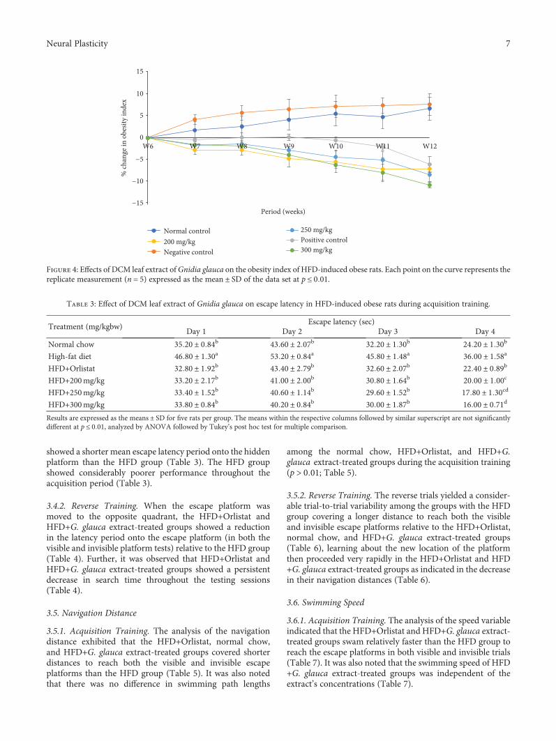
3.2. Effects of Oral Administration of DCM Leaf Extract ofGnidia glauca on Obesity Index in HFD-Induced ObeseExperimental Rats. Results showed that chronic exposuresof rats to high-fat diets caused an increase in the obesityindex in the HFD group of rats throughout the study period(Figure 4). On the other hand, the treatment of rats with thereference drug, Orlistat, and the three doses of the plantextract caused a persistent decrease in the obesity index fromthe 6th to the 12th weeks of the study (Figure 4).
3.3. Analysis of the Navigation Behavior. The rats adopted thecharacteristic adult swimming posture of forepaws tuckedunder the head and hind legs used to propel it forward. Thehead largely remained above the water surface except forbrief moments. Initially, the rats were thigmotaxic, swim-ming around the perimeter of the pool near or against thesidewalls, making occasional efforts to escape by forepaw
5Neural Plasticity

climbing movements against the side. Later, they swam outinto open water, crossing the pool several times during eachof the initial habituation sessions. The rats readily climbedonto the escape platforms when they encountered them orwere guided to locate the platform when they failed to locateit within the experimental period. Regularly, the rats wouldthen rear and/or turn around for a few seconds before mak-ing vigorous “wet-dog” shakes followed by grooming andoccasional face-washing. There was no sign of the animalstreating the “underwater” platform any differently from the“above-water” platform as a refuge from the water. All rats
exhibited similar navigation behavior independent of treat-ment or exposure to diet.
3.4. Latency Period
3.4.1. Acquisition Training. On the first day of the visibleplatform tests, the HFD group exhibited a longer latencyperiod to escape onto the visible platform relative to theHFD+Orlistat and HFD+G. glauca extract-treated groups(Table 3). In the hidden platform tests (2nd-4th days), theHFD+Orlistat and HFD+G. glauca extract-treated groups
−40
−30
−20
−10
0
10
20
30
40
50
W6 W7 W8 W9 W10 W11 W12
% ch
ange
in b
ody
wei
ght
Period (weeks)
Normal control
Negative controlPositive control200 mg/kg250 mg/kg
300 mg/kg
(a)
−20
−15
−10
−5
0
5
10
15
20
25
30
% �훥
in b
ody
wei
ght (
wee
k)
Experimental groups
B
A
C
DD DN
orm
al co
ntro
l
Neg
ativ
e con
trol
Posit
ive c
ontr
ol
HFD
+200
mg/
kg
HFD
+250
mg/
kg
HFD
+300
mg/
kg
(b)
Figure 3: (a) Effects of DCM leaf extract of Gnidia glauca on body weights of HFD-induced obese laboratory rats per week. Each point on thecurve represents the replicate measurement (n = 5) expressed as the mean ± SD of the data set at p ≤ 0 01. (b) Effects of DCM leaf extract ofGnidia glauca on the rate of change in body weights of rats per week. Each bar graph in the respective experimental group represents thereplicate measurement (n = 5) expressed as the mean ± SD of the data set. The means with different letters across the experimental groupsare statistically significant at p ≤ 0 01.
6 Neural Plasticity
showed a shorter mean escape latency period onto the hiddenplatform than the HFD group (Table 3). The HFD groupshowed considerably poorer performance throughout theacquisition period (Table 3).
3.4.2. Reverse Training. When the escape platform wasmoved to the opposite quadrant, the HFD+Orlistat andHFD+G. glauca extract-treated groups showed a reductionin the latency period onto the escape platform (in both thevisible and invisible platform tests) relative to the HFD group(Table 4). Further, it was observed that HFD+Orlistat andHFD+G. glauca extract-treated groups showed a persistentdecrease in search time throughout the testing sessions(Table 4).
3.5. Navigation Distance
3.5.1. Acquisition Training. The analysis of the navigationdistance exhibited that the HFD+Orlistat, normal chow,and HFD+G. glauca extract-treated groups covered shorterdistances to reach both the visible and invisible escapeplatforms than the HFD group (Table 5). It was also notedthat there was no difference in swimming path lengths
among the normal chow, HFD+Orlistat, and HFD+G.glauca extract-treated groups during the acquisition training(p > 0 01; Table 5).
3.5.2. Reverse Training. The reverse trials yielded a consider-able trial-to-trial variability among the groups with the HFDgroup covering a longer distance to reach both the visibleand invisible escape platforms relative to the HFD+Orlistat,normal chow, and HFD+G. glauca extract-treated groups(Table 6), learning about the new location of the platformthen proceeded very rapidly in the HFD+Orlistat and HFD+G. glauca extract-treated groups as indicated in the decreasein their navigation distances (Table 6).
3.6. Swimming Speed
3.6.1. Acquisition Training. The analysis of the speed variableindicated that the HFD+Orlistat and HFD+G. glauca extract-treated groups swam relatively faster than the HFD group toreach the escape platforms in both visible and invisible trials(Table 7). It was also noted that the swimming speed of HFD+G. glauca extract-treated groups was independent of theextract’s concentrations (Table 7).
−15
−10
−5
0
5
10
15
W6 W7 W8 W9 W10 W11 W12
% ch
ange
in o
besit
y in
dex
Period (weeks)
Normal control
Negative controlPositive control200 mg/kg250 mg/kg
300 mg/kg
Figure 4: Effects of DCM leaf extract ofGnidia glauca on the obesity index of HFD-induced obese rats. Each point on the curve represents thereplicate measurement (n = 5) expressed as the mean ± SD of the data set at p ≤ 0 01.
Table 3: Effect of DCM leaf extract of Gnidia glauca on escape latency in HFD-induced obese rats during acquisition training.
Treatment (mg/kgbw)Escape latency (sec)
Day 1 Day 2 Day 3 Day 4
Normal chow 35 20 ± 0 84b 43 60 ± 2 07b 32 20 ± 1 30b 24 20 ± 1 30b
High-fat diet 46 80 ± 1 30a 53 20 ± 0 84a 45 80 ± 1 48a 36 00 ± 1 58a
HFD+Orlistat 32 80 ± 1 92b 43 40 ± 2 79b 32 60 ± 2 07b 22 40 ± 0 89b
HFD+200mg/kg 33 20 ± 2 17b 41 00 ± 2 00b 30 80 ± 1 64b 20 00 ± 1 00c
HFD+250mg/kg 33 40 ± 1 52b 40 60 ± 1 14b 29 60 ± 1 52b 17 80 ± 1 30cd
HFD+300mg/kg 33 80 ± 0 84b 40 20 ± 0 84b 30 00 ± 1 87b 16 00 ± 0 71d
Results are expressed as the means ± SD for five rats per group. The means within the respective columns followed by similar superscript are not significantlydifferent at p ≤ 0 01, analyzed by ANOVA followed by Tukey’s post hoc test for multiple comparison.
7Neural Plasticity

3.6.2. Reverse Training. The set of analyses that were per-formed on these sessions yielded a similar pattern of resultsas those of the acquisition test (Table 8). The HFD+Orlistat
and HFD+G. glauca extract-treated groups swam faster thanthe HFD group (Table 8). The analysis also yielded a sessioneffect which indicated that swimming speed increased over
Table 4: Effect of DCM leaf extract of Gnidia glauca on escape latency in HFD-induced obese rats during reverse training.
Treatment (mg/kgbw)Escape latency (sec)
Day 6 Day 7 Day 8 Day 9
Normal chow 40 80 ± 0 84b 48 60 ± 0 55b 38 80 ± 1 30b 28 80 ± 0 84b
High-fat diet 50 60 ± 0 55a 57 60 ± 1 14a 46 80 ± 1 92a 38 60 ± 1 14a
HFD+Orlistat 37 40 ± 1 14c 46 20 ± 1 92bc 35 60 ± 1 14c 26 60 ± 1 14bc
HFD+200mg/kg 37 40 ± 1 52c 45 80 ± 2 59bc 35 80 ± 0 84c 24 60 ± 1 34c
HFD+250mg/kg 38 20 ± 0 84c 44 80 ± 0 84c 34 60 ± 1 52cd 21 40 ± 1 34d
HFD+300mg/kg 38 60 ± 1 14c 45 00 ± 1 00c 32 00 ± 1 58d 19 60 ± 0 89d
Results are expressed as the means ± SD for five rats per group. The means within the respective columns followed by similar superscript are not significantlydifferent at p ≤ 0 01, analyzed by ANOVA followed by Tukey’s post hoc test for multiple comparison.
Table 5: Effect of DCM leaf extract of Gnidia glauca on navigation distance in HFD-induced obese rats during acquisition training.
Treatment (mg/kgbw)Navigation distance/path length (cm)
Day 1 Day 2 Day 3 Day 4
Normal chow 230 02 ± 20 04b 315 14 ± 20 75b 269 66 ± 9 08b 204 72 ± 9 01b
High-fat diet 390 72 ± 20 07a 481 10 ± 14 83a 448 34 ± 16 90a 389 02 ± 13 84a
HFD+Orlistat 207 70 ± 23 20b 305 90 ± 28 70b 257 32 ± 21 17b 208 32 ± 21 17b
HFD+200mg/kg 220 30 ± 25 60b 299 30 ± 25 40b 252 40 ± 23 80b 205 70 ± 24 60b
HFD+250mg/kg 218 92 ± 18 61b 305 96 ± 21 32b 258 30 ± 23 30b 193 50 ± 27 70b
HFD+300mg/kg 209 80 ± 16 46b 284 90 ± 31 60b 233 96 ± 17 51b 178 30 ± 23 20b
Results are expressed as the means ± SD for five rats per group. The means within the respective columns followed by similar superscript are not significantlydifferent at p ≤ 0 01, analyzed by ANOVA followed by Tukey’s post hoc test for multiple comparison.
Table 6: Effect of DCM leaf extract of Gnidia glauca on navigation distance in HFD-induced obese rats during reverse training.
Treatment (mg/kgbw)Navigation distance/path length (cm)
Day 6 Day 7 Day 8 Day 9
Normal chow 253 62 ± 17 44b 342 32 ± 8 59b 271 60 ± 39 6b 235 32 ± 16 85b
High-fat diet 431 02 ± 17 19a 501 52 ± 15 33a 464 64 ± 8 14a 447 90 ± 24 70a
HFD+Orlistat 249 74 ± 20 12b 326 40 ± 16 14b 274 78 ± 14 87b 230 58 ± 20 87b
HFD+200mg/kg 253 18 ± 21 45b 329 92 ± 18 70b 284 32 ± 20 11b 237 68 ± 17 93b
HFD+250mg/kg 253 10 ± 24 40b 333 78 ± 20 16b 278 40 ± 24 90b 224 70 ± 20 44b
HFD+300mg/kg 232 70 ± 6 90b 331 14 ± 6 83b 260 92 ± 21 29b 215 80 ± 21 22b
Results are expressed as the means ± SD for five rats per group. The means within the respective columns followed by similar superscript are not significantlydifferent at p ≤ 0 01, analyzed by ANOVA followed by Tukey’s post hoc test for multiple comparison.
Table 7: Effect of DCM leaf extract of Gnidia glauca on swimming speed in HFD-induced obese rats during acquisition training.
Treatment (mg/kgbw)Swimming speed (cm/s)
Day 1 Day 2 Day 3 Day 4
Normal chow 12 22 ± 0 46a 11 86 ± 1 07a 14 61 ± 0 73a 16 77 ± 1 16d
High-fat diet 6 21 ± 0 32b 7 16 ± 0 35b 7 62 ± 0 53b 8 05 ± 0 71e
HFD+Orlistat 12 47 ± 1 12a 11 69 ± 0 88a 14 09 ± 1 41a 18 26 ± 1 39cd
HFD+200mg/kg 12 73 ± 1 54a 12 19 ± 0 56a 14 74 ± 1 37a 20 31 ± 1 25bc
HFD+250mg/kg 12 57 ± 0 84a 12 46 ± 0 42a 15 53 ± 1 34a 22 24 ± 2 74ab
HFD+300mg/kg 12 13 ± 0 61a 12 56 ± 0 24a 14 53 ± 1 39a 23 67 ± 1 57a
Results are expressed as the means ± SD for five rats per group. The means within the respective columns followed by similar superscript are not significantlydifferent at p ≤ 0 01, analyzed by ANOVA followed by Tukey’s post hoc test for multiple comparison.
8 Neural Plasticity

the course of the trials thereby decreasing the swim latencyperiod in the HFD+Orlistat and HFD+G. glauca extract-treated groups (Table 8).
3.7. Spatial Memory Retention
3.7.1. Acquisition. The analysis performed on the probe teston the 5th day in which the platform was withdrawn fromthe water indicated that the HFD+Orlistat and HFD+G.glauca extract-treated groups exhibited preference for thenorthwest quadrant (which was the correct location of theescape platform during the acquisition training) than theHFD group (Figure 5(a)). Besides, the HFD+G. glaucaextract-treated group exhibited high frequency for the cor-rect quadrant in a dose-dependent manner. The quadrantfrequency in the HFD+G. glauca extract-treated group washigher than that observed in both the HFD+Orlistat and nor-mal chow groups (Figure 5(a)). The HFD+Orlistat groupshowed no much difference in spatial bias towards the target
quadrant as was observed in the normal chow (p ≤ 0 01;Figure 5(a)).
3.7.2. Reverse Training. During the reverse trial (on the 10th
day), it was observed that the HFD+Orlistat and HFD+G.glauca extract-treated groups showed a higher number ofentries into the correct quadrant (southeast quadrant) relativeto the HFD group (Figure 5(b)). The increase in the numberof entries into the correct quadrant was in a concentration-related manner in the HFD+G. glauca extract-treated group.Besides, results showed that rats in the HFD+G. glaucaextract-treated group concentrated their search in the quad-rant where the platform was previously located during thetraining sessions more than rats in the normal chow group(Figure 5(b)).
3.7.3. Relative Abundance of Bioactive Compounds in DCMLeaf Extract of G. glauca. The gas chromatography-massspectrometry analysis (GC-MS) of the DCM leaf extract of
Table 8: Effect of DCM leaf extract of Gnidia glauca on swimming speed in HFD-induced obese rats during reverse training.
Treatment (mg/kgbw)Swimming speed (cm/s)
Day 6 Day 7 Day 8 Day 9
Normal chow 11 12 ± 0 51a 11 16 ± 0 12a 12 68 ± 0 41b 15 12 ± 0 82d
High-fat diet 6 54 ± 0 28b 6 98 ± 0 35b 8 13 ± 1 07c 9 02 ± 0 69e
HFD+Orlistat 12 03 ± 0 61a 11 41 ± 0 46a 13 35 ± 0 74ab 16 21 ± 1 10cd
HFD+200mg/kg 12 15 ± 1 06a 11 59 ± 0 51a 13 53 ± 0 64ab 17 81 ± 0 40bc
HFD+250mg/kg 11 87 ± 0 71a 11 92 ± 0 49a 13 86 ± 1 15ab 19 93 ± 1 91ab
HFD+300mg/kg 11 22 ± 0 44a 11 81 ± 0 30a 14 41 ± 0 33a 21 26 ± 1 65a
Results are expressed as the means ± SD for five rats per group. The means within the respective columns followed by similar superscript are not significantlydifferent at p ≤ 0 01, analyzed by ANOVA followed by Tukey’s post hoc test for multiple comparison.
Treatments
ControlsHFD+Gnidia glauca
0
5
10
15
Nor
mal
chow
Qua
dran
t fre
quen
cy
HFD
HFD
+Orli
stat
HFD
+200
mg/
kg
HFD
+250
mg/
kg
HFD
+300
mg/
kg
(a)
Treatments
Nor
mal
chow
HFD
HFD
+Orli
stat
HFD
+200
mg/
kg
HFD
+250
mg/
kg
HFD
+300
mg/
kg
0
2
4
6
Qua
dran
t fre
quen
cy
ControlsHFD+Gnidia glauca
(b)
Figure 5: (a) Effect of DCM leaf extract of G. glauca on spatial memory retention in HFD-induced obese rats during acquisition training. Thecriterion for significance was set at p ≤ 0 01. (b) Effect of DCM leaf extract of G. glauca on spatial memory retention in HFD-induced obeserats during reverse training. The criterion for significance was set at p ≤ 0 01.
9Neural Plasticity

G. glauca indicated the presence of 17 compounds (Table 9).Based on the analysis, oleic acid (21 05 ± 2 34%) was themost abundant followed by γ-sitosterol (18 84 ± 1 04%), cur-cumin (16 91 ± 2 30%), quercetin (15 74 ± 1 01%), stilbenes(13 39 ± 4 06%), vitamin E (12 25 ± 1 67%), octadecanoicacid (stearic acid) (10 73 ± 1 55%), and others (Table 9).
4. Discussion
The Morris water maze is a versatile behavioral tool used totest cognitive processes such as spatial learning and memoryretention in laboratory rodent models [14]. The experimentalanimals rely on extramaze cues to navigate around an openswimming arena from identified start points to locate a sub-merged invisible escape platform [32]. To assess spatiallearning, repeated trials are practiced while reference mem-ory is determined by a probe test when the escape platformis absent in the target quadrant [33].
High-fat diet-induced obesity increases the risk ofdevelopment of cognitive impairment in the form of short-term memory, executive function deficits, dementia, andAlzheimer’s disease [7, 34]. Short-term exposure to anobesogenic diet even for only 72 hours is sufficient toimpair hippocampal-dependent memory [35, 36]. Thehippocampal-dependent spatial learning examines the abilityof an experimental animal to acquire spatial information bymeasuring various variables such as escape latency, swim-ming speed, and navigation distance [32]. The spatial mem-
ory retention examines the ability of an animal to recall thecorrect location of the escape platform in the maze [32].
The current study is aimed at evaluating the effect of G.glauca on memory and learning processes in HFD-inducedobese rats. The results showed that chronic exposures tohigh-fat diets resulted in obesogenic states as evidencedby the increased body weight and Lee obesity index. Theincrease in body weight could be due to a high rate of acyla-tion of saturated fatty acids into triglycerides that are subse-quently stored in the adipose tissues [37]. The obesity indexhas been established to be the best predictor of intra-abdominal fat in rats and, therefore, of central obesity [38].The Lee index (∛weight/naso − anal length × 1000) has beenshown to correlate with fat mass. Even though the naso-anallength is a relatively weak predictor of fat-free mass in rats,the Lee index is currently used as a fast and accurate wayto access obesity in rodents subjected to a weight gainmethod [39].
The results also showed that chronic exposures to high-fat diets directly influences the spatial learning and memoryfunction of rats in the maze. It was observed that the HFDgroup indicated longer latency to escape onto the hiddenplatform, swam slower, and covered larger distances to reachthe submerged platform in both acquisition training andreversal trials. Also, rats in the HFD group searched theescape platform rather randomly and spent more time innontarget quadrants in the maze, an indication of their infe-rior memory of the learning task. Similar studies have dem-onstrated that rodents fed high-fat diets portray markedimpairments in cognitive functions as determined by latencyto find the hidden escape platform [40], their swimmingspeed, navigation distance, and probe trials in the Morriswater maze [41, 42].
The cognitive variables such as escape latency and navi-gation distance reflect the integrity of the hippocampus tolocate the hidden escape platform and ought to translate inshorter time latency and path distances [43]. The navigationspeed is influenced by the weight of an animal and, therefore,reflects the perceptive or motor capacities of the experimen-tal animals [43]. Spatial learning and memory retention asdetermined by the probe test demonstrate whether naviga-tion behavior towards the escape platform depends on allo-centric cues [43].
Chronic exposure to HFD is often associated withhyperglycemia due to insulin insensitivity [44]. One of themetabolic complications associated with persistent highblood sugar levels is retinopathy, which renders poor vision[45]. The dismal performance of rats in the HFD groupcould be attributed to poor vision, which renders them toinappropriately perceive the surrounding external cues thatare necessary to locate the escape platform. These observa-tions corroborate with findings of Bélanger et al. [43],whose 8-week untreated diabetic ZDF rats developed cata-racts, which consequently resulted in poor performance inthe maze.
The observation that the HFD group swam slower indi-cates impaired motor functions. The altered motor capaci-ties of these obese rats could be due to their overweightcondition. A study by Frisbee and Stepp [46] showed that
Table 9: Quantity of phytochemical compounds in DCM leafextract of Gnidia glauca.
RT Compound nameRelative
abundance (%)
21.53 Pyridine-3-carboxamide 10 15 ± 1 5823.06 Oleic acid 21 05 ± 2 3424.73 3,5,4′-Trihydroxy-trans-stilbene 13 39 ± 4 0624.92 Catechins 9 27 ± 2 0525.44 Octadecanoic acid (stearic acid) 10 73 ± 1 5525.96 Naringenin chalcone 7 71 ± 1 63
26.359,12,15-Octadecatrienoic acid,(Z,Z,Z)-(α-linolenic acid)
9 74 ± 2 85
26.98 Luteolin 9 77 ± 2 6227.90 Eicosapentaenoic acid 7 62 ± 0 8928.48 Docosahexaenoic acid 7 94 ± 0 4429.22 Curcumin 16 91 ± 2 3030.07 Phytol 11 04 ± 1 1830.24 Quercetin 15 74 ± 1 0130.79 γ-Sitosterol 18 84 ± 1 0434.92 Cholecalciferol (vitamin D) 9 95 ± 1 4235.41 Vitamin E 12 25 ± 1 6735.48 Stigmasterol 7 75 ± 2 23Summary of compounds identified in Gnidia glauca extract with theirrelative abundance. Results are expressed as the means ± SD for replicatemeasurement (n = 3). RT is the retention time.
10 Neural Plasticity
the muscular tissues of ZDF rats deteriorate faster in com-parison to those of normal control rats. Several studies onRosmarinus officinalis (Rosemary) [47], Bacopa monnieri[48], and Centella asiatica [49] have also demonstratedan increase in ambulatory activities, enhancement of cog-nition, and positive modulation of mood, anxiety, anddepression disorders.
Chronic exposure to calorically dense foods has beenimplicated to contribute to cognitive impairment, dementia,and Alzheimer’s disease [50]. The potential mediators under-lying obesity-induced cognitive decline include brain atro-phy, breakdown of blood brain barrier (BBB), systemic andcentral inflammation, and oxidative stress [50]. Increasedadiposity has shown a positive correlation with reduced hip-pocampal volume and cognitive decline [51]. A high dietaryfat was shown to induce hippocampal-hypothalamic neuro-nal apoptosis resulting in the reduction in hippocampalweight [52, 53]. Besides, high-fat diets decrease the levels ofthe hippocampal brain-derived neurotrophic factor (BDNF),which mediates neuronal changes involved in learning,memory, neurogenesis, and synaptic plasticity [54]. The met-abolic and dietary consequences of a high-fat diet intakeinfluence the brain function by disrupting the integrity ofthe BBB [55]. The obesity-induced BBB dysfunction, neuro-nal impairment, and memory loss are the frequent conse-quences of plasma accumulation of amyloid proteins (Aβ)that pathologically affects the cerebrovasculature [56].
Systemic and central inflammation have been shown tocontribute to cognitive decline via cytokine-mediated pro-duction [57]. The synthesis and release of proinflammatorycytokines such as IL-1β, IL-6, and TNF-α contribute to cog-nitive impairment by affecting cognitive processes such assynaptic plasticity, neurogenesis, neuromodulation, mem-ory consolidation, and long-term potentiation (LTP) [58].Chronic consumption of a high-fat diet exacerbates oxidativedamage in the hippocampus due to attenuated antioxidantdefenses and facilitates the production of proinflammatorycytokines, chemokines and brain’s resident immune cells,the microglia, and astrocytes [59, 60]. These events, there-fore, culminate in impaired neurogenesis, synaptic remodel-ing, and reduction in neuronal spine density as well asneuronal apoptosis, deregulated HPA axis, neurodegenera-tion, and brain atrophy [61, 62].
Treatment of rats with the standard drug, Orlistat, andthe three doses of G. glauca caused a decrease in body weightand Lee obesity index. Orlistat is a saturated derivative of lip-statin that inhibits the activity of pancreatic lipase resultingin reduced dietary fat absorption [63]. The antiobesity effectsof the extract might also be attributed to the reduction of tri-glyceride absorption through the inhibition of the action ofpancreatic lipase. The extract might have also led to anincrease in energy expenditure, inhibited the differentiationand proliferation of preadipocytes, and stimulated satiety sig-nals (such as leptin) thereby resulting in the reduction ofbody and obesity index [64].
The results also showed that the HFD+Orlistat and HFD+G. glauca extract-treated groups recorded shorter latency toescape onto the hidden platform, swam faster, and coveredshorter distances to reach the submerged platform in the
maze. In the probe test, the same group of rats showed anovel behavioral strategy of concentrating their search inthe target quadrant, where the platform was located in theprevious training sessions. These behaviors suggest thatthe treatment of rats with the standard drug, Orlistat, andthe three extract doses improves obesity-induced memoryimpairments. Consistent with this study, it was reported thatthe high-fat and high-fructose diet-induced obese micetreated with Camellia sinensis (green tea) showed lowerlatency and escape distance than the high-fat and high-fructose diet-induced obese untreated mice on each testday. Besides, the treated mice spent a long time in the targetquadrant and had greater numbers of platform crossingsthan the untreated obese mice [65]. Since navigation towardsthe escape platform is based on allocentric cues, Orlistat andthe plant extract might play a role in the improvement ofvision through amelioration of metabolic complications ofobesity-induced type 2 diabetes.
The probable mechanisms attributed to the positiveinfluences on cognitive functions by G. glauca among othersinclude the normalization of antioxidant mechanisms [66],reduction of inflammation [67, 68], increment in the expres-sion of hippocampal neurotrophic factors (BDNF), andenhancement of neurogenesis which facilitates neural plastic-ity in the hippocampus [69, 70].
The cognitive-enhancing ability of G. glauca may be dueto the presence of phytochemicals which have been impli-cated in the improvement of learning and memory [38].The GC-MS analysis revealed the presence of various bioac-tive compounds such as polyphenols (luteolin, catechins,curcumin, quercetin, anthocyanins, and naringenin), alka-loids, long-chain polyunsaturated fatty acids, and VitaminE. The normal chow pellet also contains appreciable amountsof these phytochemicals such as catechins, flavanols, querce-tin, and vitamins (B, D, and E). These phytocompounds con-fer multiple physiological effects that not only serve toprotect the brain from the pathogenic but also reverse theunderlying disease process [38]. The normalization of cogni-tive effects in extract-treated groups as that observed in thenormal chow group could be attributed to the synergisticand/or additive effects of these biocompounds.
Luteolin has been shown to ameliorate obesity-inducedcognitive impairments [71]. The administration of luteolin tohigh-fat diet-induced obesemice decreased the circulating levelsof serum adipocytokines, alleviated neuroinflammation, andreduced neuronal insulin resistance [71]. Luteolin increasedthe levels of brain-derived neurotrophic factor (BDNF) andimproved oxidative stress-mediated cognitive decline [71].Luteolin was also shown to enhance the action of synapsin I(SYP-1) and postsynaptic density protein 95 (PSDP-95) in thehippocampus and medulla cortex of the brain [71].
Catechins such as epigallocatechin-3-gallate have beenimplicated to have the potential to alleviate high-fat andhigh-fructose-induced insulin resistance [71]. It has beenshown to improve cognitive impairment in mice [71]. Thetreatment of high-fat and high-fructose-induced mice withcatechins significantly increased the average time spent inthe target quadrant [65]. The treated mice also recorded agreater number, in platform crossings, than their obese-
11Neural Plasticity

untreated counterparts, an indication of improved memory[65, 71]. Catechins exhibit their protective effects by pre-venting Aβ-induced neuronal injury through scavengingof ROS [72]. Specifically, catechins decrease the levels of mal-onyldialdehyde (MDA) and caspase, thereby resulting indecreased ROS [72]. Catechins inhibit fibrillogenesis of Aβthrough their direct binding to the unfolded polypeptidesAβ, thereby converting them to unstructured, nontoxic Aβ-oligomers instead of β-sheet-rich aggregates [73].
Stilbene such as resveratrol is a polyphenolic compoundwhich has been reported to reduce the accumulation of amy-loid plaques (Aβ peptide) in Tg2576 neuron cultures [74].Amyloid plaques trigger microglial activation by interactingwith toll-like receptors (TLR) such as TLR4. Activatedmicroglia induce neuronal inflammation and cell death[74]. Therefore, the anti-inflammatory activities of stilbenesprotect microglia against Aβ-induced inflammation [75].
Quercetin is a flavonoid exhibiting antioxidant, antia-poptotic, and anti-inflammatory properties [76]. Quercetinwas reported to confer cognitive-enhancing effects throughreduction of β-amyloid plaque aggregation [76]. Quercetintreatment of 3xTg Alzheimer’s disease mice decreased IL-1β/COX-2/iNOS proinflammatory signaling in the hippo-campal CA1 region [77].
Curcumin is a natural polyphenol whose supplementa-tion in the diet has been reported to ameliorate HFD-induced cognitive deficits [78]. Curcumin modulates cogni-tion by improving synaptic plasticity through alterations ofthe N-methyl-D-aspartate receptor (NMDAR) and calcium/-calmodulin-dependent kinase II (CaMKII) [79]. Curcuminreduces oxidative stress and promotes the synthesis of doco-sahexaenoic acid (DHA) from its precursor, α-linolenic acid,by stimulating the activity of enzymes involved in the synthe-sis of DHA such as elongase and fatty acid desaturase-2(FADS-2) in the brain [80, 81].
Anthocyanins (ANTs) have been implicated in the reduc-tion of Aβ-induced neurotoxicity by reducing ROS formationupon exposure of Aβ1-40 and Aβ25-35 to neuro-2A cells[82]. Anthocyanins also reduce Aβ-induced neurotoxicitythrough perturbation of calcium balance and inhibition ofmetabolism of apolipoprotein E (ApoE) [82]. Anthocyaninspromote the formation of nontoxic forms of Aβ aggregatesinstead of the toxic amyloid fibrils by direct binding to Aβ mol-ecules, thereby suppressing amyloid fibril formation [83].
Naringenin chalcone neuroprotective effects are wellcharacterized. It enhances learning and memory ability inmice by reducing senile plaque formation and increasing glu-cose uptake in the brain [84]. Naringenin enhances cognitionthrough inhibition of GSK3β activity and mitigation of mito-chondrial dysfunction mediated oxidative stress [85]. It alsoenhances cognition by stimulating the activity of CaMKIIand suppression of acetylcholinesterase activity [85, 86].
Alkaloids isolated from Huperzia serrata were shown tobe a potent, reversible, and selective inhibitor of acetylcholin-esterase (AChE) [87]. Alkaloids exhibit memory-enhancingefficacy due to their ability to penetrate the blood-brainbarrier and inhibit acetylcholinesterase action [87].
Previous studies have reported that vitamin E is animportant component of the body antioxidant systems. Its
antioxidant activity and anti-inflammatory properties con-tribute to its neuroprotective effects [88]. Vitamin E inhibitsAβ accumulation in the brain. Moreover, the reduction ofoxidative stress by vitamin E protects against phosphoryla-tion of Aβ-induced tau through the inhibition of the activa-tion of p38-MAPK [89].
Long-chain polyunsaturated fatty acids such as omega-3fatty acids (such as alpha-linolenic acid, docosahexaenoicacid, and eicosapentaenoic acid) and omega-9 fatty acid(oleic acid) have been implicated in the improvement of cog-nitive deficits [90]. Omega-3 and omega-9 fatty acids are pre-dominantly found in the brain [90]. A low dietary ratio ofomega-3/omega-6 is linked with cognitive impairments anddementia [91, 92]. The dietary omega-3/omega-6 ratio isinversely correlated with cognitive decline and hippocampalinflammation [93]. Omega-3 supplementation increasesmolecular markers involved in neuronal plasticity such asBDNF and tropomyosin receptor kinase B (TrkB) [94]. Micefeeding with a high-omega-3/omega-6 ratio diet showed lowmRNA expression levels of hippocampal inflammatorymarkers such as TNF-α and IL-1β [95]. Therefore, PUFAcontributes to the prevention of neuroinflammatory pro-cesses [93].
5. Conclusion
The present study focused on the determination of cognitive-enhancing effects of the DCM leaf extract of G. glauca inHFD-induced obese rats. Results showed that the DCM leafextract of G. glauca ameliorates learning and memory deficitsin HFD-induced obese rats. The hippocampal-dependentspatial learning examines the ability of the experimental ani-mal to acquire spatial information by measuring various var-iables including escape latency, swim speed, and navigationdistance. Spatial memory retention is assessed using theprobe test in the Water maze. The improvement of cognitivevariables (escape latency, swim speed, and navigation dis-tance as well as probe trial) in the DCM leaf extract of G.glauca-treated rats relative to the HFD group obese rats areindicative of extracts’ potential to treat cognitive deficits.The positive influences on cognitive functions could relateto the antiobesity effects of the extract. The cognitive-enhancing ability of the extract might be attributed to itscapacity to confer protection against obesity-induced oxida-tive damage, restoration of redox homeostatic status, andreduction of central inflammation. Also, the extract mighthave enhanced the gene expression of hippocampal neuro-trophic factor (BDNF) and increased neurogenesis, therebyfacilitating neuronal plasticity in the hippocampus. The ther-apeutic effects of the extract are mainly attributed to the phy-tochemicals present. The learning and cognitive-enhancingeffects of G. glauca leaf extract might also have a positiveimplication in the management of dementia and Alzheimer’sdisease. This study, therefore, provides a basis for furtherresearch on G. glauca as a plant-derived source of a drugagainst symptomatic complications of obesity such as learn-ing and memory loss. However, there is a need to conductcomprehensive toxicity studies to establish the safety profileof this plant.
12 Neural Plasticity

Data Availability
No data was used to support this study.
Ethical Approval
The experimental protocols and procedures used in this studywere approved by the Ethics Committee for the Care and Useof Laboratory Animals of Kenyatta University, Kenya.
Conflicts of Interest
The authors declare no conflict of interest.
Authors’ Contributions
Wycliffe Makori Arika carried out the study and wrote themanuscript. Cromwell Mwiti Kibiti, Joan Murugi Njagi,and Mathew Piero Ngugi contributed to the conception ofthe review and supervised the manuscript writing. Allauthors have read and approved the final manuscript.
Acknowledgments
The authors wish to acknowledge the Department of Bio-chemistry, Microbiology and Biotechnology of KenyattaUniversity for allowing them to use the departmental animalhouse facility for rat breeding and performing the efficacystudies. The authors also acknowledge Mr. Joshua Muleleand Ms. Sheila Cheptoo of the Department of Physical Sci-ence (Biochemistry Section) of Chuka University as well asMr. Shadrack Njagi and Mr. Daniel Gitonga Mwaniki ofthe Department of Biochemistry, Microbiology and Biotech-nology of Kenyatta University for the technical support.
References
[1] G. H. Norris, C. M. Porter, C. Jiang, C. L. Millar, and C. N.Blesso, “Dietary sphingomyelin attenuates hepatic steatosisand adipose tissue inflammation in high-fat-diet-inducedobese mice,” The Journal of Nutritional Biochemistry, vol. 40,pp. 36–43, 2017.
[2] K. S. Sellbom and J. Gunstad, “Cognitive function and declinein obesity,” Journal of Alzheimer's Disease, vol. 30, no. s2,pp. S89–S95, 2012.
[3] R. R. Lennox, C. Moffett, D. W. Porter, N. Irwin, V. A. Gault,and P. R. Flatt, “Effects of glucose-dependent insulinotropicpolypeptide receptor knockout and a high-fat diet on cognitivefunction and hippocampal gene expression in mice,” Molecu-lar Medicine Reports, vol. 12, no. 1, pp. 1544–1548, 2015.
[4] K. W. Cho, B. F. Zamarron, L. A. Muir et al., “Adipose tissuedendritic cells are independent contributors to obesity-induced inflammation and insulin resistance,” The Journal ofImmunology, vol. 197, no. 9, pp. 3650–3661, 2016.
[5] N. Medic, H. Ziauddeen, K. D. Ersche et al., “Increased bodymass index is associated with specific regional alterations inbrain structure,” International Journal of Obesity, vol. 40,no. 7, pp. 1177–1182, 2016.
[6] R. Molteni, R. J. Barnard, Z. Ying, C. K. Roberts, andF. Gómez-Pinilla, “A high-fat, refined sugar diet reduces hip-pocampal brain-derived neurotrophic factor, neuronal plastic-
ity, and learning,” Neuroscience, vol. 112, no. 4, pp. 803–814,2002.
[7] A. J. Murray, N. S. Knight, L. E. Cochlin et al., “Deteriorationof physical performance and cognitive function in rats withshort-term high-fat feeding,” The FASEB Journal, vol. 23,no. 12, pp. 4353–4360, 2009.
[8] S. E. Kanoski and T. L. Davidson, “Western diet consumptionand cognitive impairment: links to hippocampal dysfunctionand obesity,” Physiology & Behavior, vol. 103, no. 1, pp. 59–68, 2011.
[9] S. Kosari, E. Badoer, J. C. D. Nguyen, A. S. Killcross, and T. A.Jenkins, “Effect of Western and high fat diets on memory andcholinergic measures in the rat,” Behavioural Brain Research,vol. 235, no. 1, pp. 98–103, 2012.
[10] A. D. McNeilly, R. Williamson, C. Sutherland, D. J. K. Balfour,and C. A. Stewart, “High fat feeding promotes simultaneousdecline in insulin sensitivity and cognitive performance in adelayed matching and non-matching to position task,” Behav-ioural Brain Research, vol. 217, no. 1, pp. 134–141, 2011.
[11] B. Park, J. L. Plass, and R. Brünken, “Cognitive and affectiveprocesses in multimedia learning,” Learning and Instruction,vol. 29, pp. 125–127, 2014.
[12] T. W. Kim, H. H. Choi, and Y. R. Chung, “Treadmill exercisealleviates impairment of cognitive function by enhancing hip-pocampal neuroplasticity in the high-fat diet-induced obesemice,” Journal of Exercise Rehabilitation, vol. 12, no. 3,pp. 156–162, 2016.
[13] G. Neves, S. F. Cooke, and T. V. P. Bliss, “Synaptic plasticity,memory and the hippocampus: a neural network approachto causality,” Nature Reviews Neuroscience, vol. 9, no. 1,pp. 65–75, 2008.
[14] P. J. Lucassen, E. F. G. Naninck, J. B. van Goudoever,C. Fitzsimons, M. Joels, and A. Korosi, “Perinatal program-ming of adult hippocampal structure and function; emergingroles of stress, nutrition and epigenetics,” Trends in Neurosci-ences, vol. 36, no. 11, pp. 621–631, 2013.
[15] C. R. Balistreri, C. Caruso, and G. Candore, “The role of adi-pose tissue and adipokines in obesity-related inflammatorydiseases,” Mediators of Inflammation, vol. 2010, Article ID802078, 19 pages, 2010.
[16] A. A. Miller and S. J. Spencer, “Obesity and neuroinflamma-tion: a pathway to cognitive impairment,” Brain, Behavior,and Immunity, vol. 42, pp. 10–21, 2014.
[17] K. H. Alzoubi, O. F. Khabour, H. A. Salah, and Z. Hasan,“Vitamin E prevents high-fat high-carbohydrates diet-induced memory impairment: the role of oxidative stress,”Physiology & Behavior, vol. 119, pp. 72–78, 2013.
[18] I. T. Lott and M. Dierssen, “Cognitive deficits and associ-ated neurological complications in individuals with Down’ssyndrome,” The Lancet Neurology, vol. 9, no. 6, pp. 623–633, 2010.
[19] J. Lucke and B. Partridge, “Towards a smart population: a pub-lic health framework for cognitive enhancement,” Neuroethics,vol. 6, no. 2, pp. 419–427, 2013.
[20] B. H. May, M. Lit, C. C. L. Xue et al., “Herbal medicine fordementia: a systematic review,” Phytotherapy Research,vol. 23, no. 4, pp. 447–459, 2009.
[21] R. J. Williams and J. P. E. Spencer, “Flavonoids, cognition, anddementia: actions, mechanisms, and potential therapeutic util-ity for Alzheimer disease,” Free Radical Biology & Medicine,vol. 52, no. 1, pp. 35–45, 2012.
13Neural Plasticity

[22] K. Ono, M. M. Condron, L. Ho et al., “Effects of grape seed-derived polyphenols on amyloid β-protein self-assembly andcytotoxicity,” Journal of Biological Chemistry, vol. 283,no. 47, pp. 32176–32187, 2008.
[23] J. Wang, L. Ho, W. Zhao et al., “Grape-derived polyphenolicsprevent Aβ oligomerization and attenuate cognitive deteriora-tion in a mouse model of Alzheimer’s disease,” The Journal ofNeuroscience, vol. 28, no. 25, pp. 6388–6392, 2008.
[24] M. A. Ansari, H. M. Abdul, G. Joshi, W. O. Opii, and D. A.Butterfield, “Protective effect of quercetin in primary neuronsagainst Aβ(1–42): relevance to Alzheimer’s disease,” The Jour-nal of Nutritional Biochemistry, vol. 20, no. 4, pp. 269–275,2009.
[25] S. M. Poulose, M. G. Miller, and B. Shukitt-Hale, “Role of wal-nuts in maintaining brain health with age,” The Journal ofNutrition, vol. 144, no. 4, pp. 561S–566S, 2014.
[26] B. W. R. C. Amarajeewa, A. P. Mudalige, and V. Kumar,“Chemistry and mosquito larvicidal activity of G. glauca,” inProceedings of the Peradeniya University Research Sessions,pp. 101-102, Sri Lanka, 2007.
[27] W. C. Evans, D. Evans, and G. E. Trease, Trease and EvansPharmacognosy, Elsevier, New York, NY, USA, 2009.
[28] B. E. Levin and A. A. Dunn-Meynell, “Defense of body weightdepends on dietary composition and palatability in rats withdiet-induced obesity,” American Journal of Physiology-Regula-tory, Integrative and Comparative Physiology, vol. 282, no. 1,pp. R46–R54, 2002.
[29] M. O. Lee, “Determination of the surface area of the white ratwith its application to the expression of metabolic results,”American Journal of Physiology-Legacy Content, vol. 89,no. 1, pp. 24–33, 1929.
[30] R. Morris, “Developments of a water-maze procedure forstudying spatial learning in the rat,” Journal of NeuroscienceMethods, vol. 11, no. 1, pp. 47–60, 1984.
[31] A. N. Sharma, K. M. Elased, T. L. Garrett, and J. B. Lucot,“Neurobehavioral deficits in db/db diabetic mice,” Physiology& Behavior, vol. 101, no. 3, pp. 381–388, 2010.
[32] V. K. Sharma, “Morris water maze–a versatile cognitive tool,”Memory, vol. 8, no. 9, 2009.
[33] C. V. Vorhees and M. T. Williams, “Morris water maze: proce-dures for assessing spatial and related forms of learning andmemory,” Nature Protocols, vol. 1, no. 2, pp. 848–858, 2006.
[34] J. Woo, K. Shin, S. Park, K. Jang, and S. Kang, “Effects of exer-cise and diet change on cognition function and synaptic plas-ticity in high fat diet induced obese rats,” Lipids in Healthand Disease, vol. 12, no. 1, p. 144, 2013.
[35] J. E. Beilharz, J. Maniam, and M. J. Morris, “Short exposure toa diet rich in both fat and sugar or sugar alone impairs place,but not object recognition memory in rats,” Brain, Behavior,and Immunity, vol. 37, pp. 134–141, 2014.
[36] T. M. Hsu and S. E. Kanoski, “Blood-brain barrier disruption:mechanistic links between Western diet consumption anddementia,” Frontiers in Aging Neuroscience, vol. 6, p. 88, 2014.
[37] L. H. Storlien, X. F. Huang, S. Lin, X. Xin, H. Q. Wang, andP. L. Else, “Dietary fat subtypes and obesity,” in Fatty Acidsand Lipids-New Findings, vol. 88, pp. 148–154, Karger Pub-lishers, 2001.
[38] M.-J. R. Howes and P. J. Houghton, “Plants used in Chineseand Indian traditional medicine for improvement of memoryand cognitive function,” Pharmacology Biochemistry andBehavior, vol. 75, no. 3, pp. 513–527, 2003.
[39] A. B. Malafaia, P. A. N. Nassif, C. A. P. M. Ribas, B. L. Ariede,K. N. Sue, and M. A. Cruz, “Obesity induction with high fatsucrose in rats,” Arquivos Brasileiros de Cirurgia Digestiva,vol. 26, Supplement 1, pp. 17–21, 2013.
[40] H. Kuang, M. Sun, J. Lv et al., “Hippocampal apoptosisinvolved in learning deficits in the offspring exposed to mater-nal high sucrose diets,” The Journal of Nutritional Biochemis-try, vol. 25, no. 9, pp. 985–990, 2014.
[41] S. J. Kim, J. S. Kim, H. S. Cho et al., “Carnosol, a component ofrosemary (Rosmarinus officinalis L.) protects nigral dopami-nergic neuronal cells,” NeuroReport, vol. 17, no. 16, pp. 1729–1733, 2006.
[42] E. Soares, R. D. Prediger, S. Nunes et al., “Spatial memoryimpairments in a prediabetic rat model,” Neuroscience,vol. 250, pp. 565–577, 2013.
[43] A. Belanger, N. Lavoie, F. Trudeau, G. Massicotte, andS. Gagnon, “Preserved LTP and water maze learning in hyper-glycaemic–hyperinsulinemic ZDF rats,” Physiology & Behav-ior, vol. 83, no. 3, pp. 483–494, 2004.
[44] S. E. Shoelson, L. Herrero, and A. Naaz, “Obesity, inflamma-tion, and insulin resistance,” Gastroenterology, vol. 132, no. 6,pp. 2169–2180, 2007.
[45] M. N. Piero, J. M. Njagi, C. M. Kibiti, J. J. Ngeranwa, and E. N.Njagi, “Metabolic complications of diabetes mellitus: areview,” South Asian Journal of Biological Sciences, vol. 2,pp. 37–49, 2012.
[46] J. C. Frisbee and D. W. Stepp, “Impaired NO-dependent dila-tion of skeletal muscle arterioles in hypertensive diabetic obeseZucker rats,” American Journal of Physiology-Heart and Circu-latory Physiology, vol. 281, no. 3, pp. H1304–H1311, 2001.
[47] M.Moss, J. Cook, K.Wesnes, and P. Duckett, “Aromas of rose-mary and lavender essential oils differentially affect cognitionand mood in healthy adults,” International Journal of Neuro-science, vol. 113, no. 1, pp. 15–38, 2003.
[48] C. Calabrese, W. L. Gregory, M. Leo, D. Kraemer, K. Bone, andB. Oken, “Effects of a standardized Bacopa monnieri extract oncognitive performance, anxiety, and depression in the elderly:a randomized, double-blind, placebo-controlled trial,” TheJournal of Alternative and Complementary Medicine, vol. 14,no. 6, pp. 707–713, 2008.
[49] J. Wattanathorn, L. Mator, S. Muchimapura et al., “Positivemodulation of cognition and mood in the healthy elderly vol-unteer following the administration of Centella asiatica,” Jour-nal of Ethnopharmacology, vol. 116, no. 2, pp. 325–332, 2008.
[50] G. Shefer, Y. Marcus, and N. Stern, “Is obesity a brain dis-ease?,” Neuroscience & Biobehavioral Reviews, vol. 37, no. 10,pp. 2489–2503, 2013.
[51] S. Debette, S. Seshadri, A. Beiser et al., “Midlife vascular riskfactor exposure accelerates structural brain aging and cogni-tive decline,” Neurology, vol. 77, no. 5, pp. 461–468, 2011.
[52] P. Rivera, L. Bindila, A. Pastor et al., “Pharmacological block-ade of the fatty acid amide hydrolase (FAAH) alters neuralproliferation, apoptosis and gliosis in the rat Hippocampus,hypothalamus and striatum in a negative energy context,”Frontiers in Cellular Neuroscience, vol. 9, p. 98, 2015.
[53] E. Calvo-Ochoa, K. Hernández-Ortega, P. Ferrera,S. Morimoto, and C. Arias, “Short-term high-fat-and-fructosefeeding produces insulin signaling alterations accompanied byneurite and synaptic reduction and astroglial activation in therat hippocampus,” Journal of Cerebral Blood Flow & Metabo-lism, vol. 34, no. 6, pp. 1001–1008, 2014.
14 Neural Plasticity

[54] H. Francis and R. Stevenson, “The longer-term impacts ofWestern diet on human cognition and the brain,” Appetite,vol. 63, pp. 119–128, 2013.
[55] T. L. Davidson, A. Monnot, A. U. Neal, A. A. Martin, J. J.Horton, and W. Zheng, “The effects of a high-energy dieton hippocampal-dependent discrimination performance andblood–brain barrier integrity differ for diet-induced obeseand diet-resistant rats,” Physiology & Behavior, vol. 107,no. 1, pp. 26–33, 2012.
[56] A. Jahangiri, P. G. Wilson, T. Hou, A. Brown, V. L. King,and L. R. Tannock, “Serum amyloid A is found on ApoB-containing lipoproteins in obese humans with diabetes,” Obe-sity, vol. 21, no. 5, pp. 993–996, 2013.
[57] J. McAfoose and B. T. Baune, “Evidence for a cytokine modelof cognitive function,” Neuroscience & Biobehavioral Reviews,vol. 33, no. 3, pp. 355–366, 2009.
[58] J. C. D. Nguyen, A. S. Killcross, and T. A. Jenkins, “Obesity andcognitive decline: role of inflammation and vascular changes,”Frontiers in Neuroscience, vol. 8, p. 375, 2014.
[59] D. J. Loane and K. R. Byrnes, “Role of microglia in Neuro-trauma,” Neurotherapeutics, vol. 7, no. 4, pp. 366–377, 2010.
[60] P. J. Pistell, C. D. Morrison, S. Gupta et al., “Cognitive impair-ment following high fat diet consumption is associated withbrain inflammation,” Journal of Neuroimmunology, vol. 219,no. 1-2, pp. 25–32, 2010.
[61] C. D. Morrison, P. J. Pistell, D. K. Ingram et al., “High fat dietincreases hippocampal oxidative stress and cognitive impair-ment in aged mice implications for decreased Nrf2 signaling,”Journal of Neurochemistry, vol. 114, no. 6, pp. 1581–1589,2010.
[62] L. R. Freeman, L. Zhang, A. Nair et al., “Obesity increases cer-ebrocortical reactive oxygen species and impairs brain func-tion,” Free Radical Biology & Medicine, vol. 56, pp. 226–233,2013.
[63] R. M. Viner, Y. Hsia, T. Tomsic, and I. C. Wong, “Efficacy andsafety of anti-obesity drugs in children and adolescents: sys-tematic review and meta-analysis,” Obesity Reviews, vol. 11,no. 8, pp. 593–602, 2010.
[64] S. Rayalam, M. Dellafera, and C. Baile, “Phytochemicals andregulation of the adipocyte life cycle,” The Journal of Nutri-tional Biochemistry, vol. 19, no. 11, pp. 717–726, 2008.
[65] Y. Liu, X. Fu, N. Lan et al., “Luteolin protects against high fatdiet-induced cognitive deficits in obesity mice,” BehaviouralBrain Research, vol. 267, pp. 178–188, 2014.
[66] L. J. P. Altmann, E. Stegemöller, A. A. Hazamy et al., “Aerobicexercise improves mood, cognition, and language function inParkinson’s disease: results of a controlled study,” Journal ofthe International Neuropsychological Society, vol. 22, no. 9,pp. 878–889, 2016.
[67] Y. J. Sim, “Treadmill exercise alleviates impairment of spatiallearning ability through enhancing cell proliferation in thestreptozotocin-induced Alzheimer’s disease rats,” Journal ofExercise Rehabilitation, vol. 10, no. 2, pp. 81–88, 2014.
[68] S. S. Baek, “Role of exercise on the brain,” Journal of ExerciseRehabilitation, vol. 12, no. 5, pp. 380–385, 2016.
[69] C. L. Ma, X. T. Ma, J. J. Wang, H. Liu, Y. F. Chen, and Y. Yang,“Physical exercise induces hippocampal neurogenesis and pre-vents cognitive decline,” Behavioural Brain Research, vol. 317,pp. 332–339, 2017.
[70] T. B. Seo, T. W. Kim, M. S. Shin et al., “Aerobic exercise allevi-ates ischemia-induced memory impairment by enhancing cell
proliferation and suppressing neuronal apoptosis in Hippo-campus,” International Neurourology Journal, vol. 18, no. 4,pp. 187–197, 2014.
[71] X. Liu, L. Zhu, J. Tan et al., “Glucosidase inhibitory activity andantioxidant activity of flavonoid compound and triterpenoidcompound from Agrimonia pilosa Ledeb,” BMC Complemen-tary and Alternative Medicine, vol. 14, no. 1, 2014.
[72] Y. T. Choi, C. H. Jung, S. R. Lee et al., “The green tea polyphe-nol (−)-epigallocatechin gallate attenuates β-amyloid-inducedneurotoxicity in cultured hippocampal neurons,” Life Sciences,vol. 70, no. 5, pp. 603–614, 2001.
[73] D. E. Ehrnhoefer, J. Bieschke, A. Boeddrich et al., “EGCGredirects amyloidogenic polypeptides into unstructured, off-pathway oligomers,” Nature Structural & Molecular Biology,vol. 15, no. 6, pp. 558–566, 2008.
[74] V. Vingtdeux, U. Dreses-Werringloer, H. Zhao, P. Davies,and P. Marambaud, “Therapeutic potential of resveratrol inAlzheimer’s disease,” BMC Neuroscience, vol. 9, Supplement2, p. S6, 2008.
[75] H. Capiralla, V. Vingtdeux, H. Zhao et al., “Resveratrol miti-gates lipopolysaccharide- and Aβ-mediated microglial inflam-mation by inhibiting the TLR4/NF-κB/STAT signalingcascade,” Journal of Neurochemistry, vol. 120, no. 3, pp. 461–472, 2012.
[76] R. J. Nijveldt, E. van Nood, D. E. C. van Hoorn, P. G. Boelens,K. van Norren, and P. A. M. van Leeuwen, “Flavonoids: areview of probable mechanisms of action and potential appli-cations,” The American Journal of Clinical Nutrition, vol. 74,no. 4, pp. 418–425, 2001.
[77] F. Vargas-Restrepo, A. M. Sabogal-Guáqueta, and G. P.Cardona-Gómez, “Quercetin ameliorates inflammation inCA1 hippocampal region in aged triple transgenic Alzheimer’sdisease mice model,” Biomédica, vol. 38, pp. 69–76, 2018.
[78] S. Y. Yu, M. Zhang, J. Luo, L. Zhang, Y. Shao, and G. Li, “Curcu-min ameliorates memory deficits via neuronal nitric oxide syn-thase in aged mice,” Progress in Neuro-Psychopharmacologyand Biological Psychiatry, vol. 45, pp. 47–53, 2013.
[79] C. Y. Sun, S. S. Qi, P. Zhou et al., “Neurobiological and phar-macological validity of curcumin in ameliorating memory per-formance of senescence-accelerated mice,” PharmacologyBiochemistry and Behavior, vol. 105, pp. 76–82, 2013.
[80] A. Ataie, M. Sabetkasaei, A. Haghparast, A. H. Moghaddam,and B. Kazeminejad, “Neuroprotective effects of the polyphe-nolic antioxidant agent, curcumin, against homocysteine-induced cognitive impairment and oxidative stress in therat,” Pharmacology Biochemistry and Behavior, vol. 96, no. 4,pp. 378–385, 2010.
[81] A. Wu, E. E. Noble, E. Tyagi, Z. Ying, Y. Zhuang, andF. Gomez-Pinilla, “Curcumin boosts DHA in the brain: impli-cations for the prevention of anxiety disorders,” Biochimica etBiophysica Acta (BBA) - Molecular Basis of Disease, vol. 1852,no. 5, pp. 951–961, 2015.
[82] P. H. Shih, C. H. Wu, C. T. Yeh, and G. C. Yen, “Protectiveeffects of anthocyanins against amyloid β-peptide-induceddamage in neuro-2A cells,” Journal of Agricultural and FoodChemistry, vol. 59, no. 5, pp. 1683–1689, 2011.
[83] M. Y. Yamakawa, K. Uchino, Y. Watanabe et al., “Anthocy-anin suppresses the toxicity of Aβ deposits through diver-sion of molecular forms in in vitro and in vivo models ofAlzheimer’s disease,” Nutritional Neuroscience, vol. 19, no. 1,pp. 32–42, 2016.
15Neural Plasticity

[84] D. Wang, K. Gao, X. Li et al., “Long-term naringin consump-tion reverses a glucose uptake defect and improves cognitivedeficits in a mouse model of Alzheimer’s disease,” Pharmacol-ogy Biochemistry and Behavior, vol. 102, no. 1, pp. 13–20, 2012.
[85] D. M. Wang, Y. J. Yang, L. Zhang, X. Zhang, F. F. Guan,and L. F. Zhang, “Naringin enhances CaMKII activity andimproves long-termmemory in a mouse model of Alzheimer’sdisease,” International Journal of Molecular Sciences, vol. 14,no. 3, pp. 5576–5586, 2013.
[86] A. K. Sachdeva, A. Kuhad, and K. Chopra, “Naringin amelio-rates memory deficits in experimental paradigm of Alzhei-mer’s disease by attenuating mitochondrial dysfunction,”Pharmacology Biochemistry and Behavior, vol. 127, pp. 101–110, 2014.
[87] X. C. Tang and Y. F. Han, “Pharmacological profile of Huper-zine A, a novel acetylcholinesterase inhibitor from Chineseherb,” CNS Drug Reviews, vol. 5, no. 3, pp. 281–300, 1999.
[88] Y. Nishida, S. Ito, S. Ohtsuki et al., “Depletion of vitamin Eincreases amyloid β accumulation by decreasing its clearancesfrom brain and blood in a mouse model of Alzheimer disease,”Journal of Biological Chemistry, vol. 284, no. 48, pp. 33400–33408, 2009.
[89] E. Giraldo, A. Lloret, T. Fuchsberger, and J. Viña, “Aβ and tautoxicities in Alzheimer’s are linked via oxidative stress-inducedp38 activation: protective role of vitamin E,” Redox Biology,vol. 2, pp. 873–877, 2014.
[90] K. A. Youdim, A. Martin, and J. A. Joseph, “Essential fattyacids and the brain: possible health implications,” Interna-tional Journal of Developmental Neuroscience, vol. 18, no. 4-5, pp. 383–399, 2000.
[91] B. Heude, P. Ducimetière, and C. Berr, “Cognitive decline andfatty acid composition of erythrocyte membranes—the EVAstudy,” The American Journal of Clinical Nutrition, vol. 77,no. 4, pp. 803–808, 2003.
[92] P. Barberger-Gateau, C. Raffaitin, L. Letenneur et al., “Dietarypatterns and risk of dementia: the Three-City Cohort Study,”Neurology, vol. 69, no. 20, pp. 1921–1930, 2007.
[93] T. Grundy, C. Toben, E. J. Jaehne, F. Corrigan, and B. T.Baune, “Long-term omega-3 supplementation modulatesbehavior, hippocampal fatty acid concentration, neuronal pro-genitor proliferation and central TNF-α expression in 7 monthold unchallenged mice,” Frontiers in Cellular Neuroscience,vol. 8, p. 399, 2014.
[94] F. Gomez-Pinilla, “Collaborative effects of diet and exercise oncognitive enhancement,” Nutrition and Health, vol. 20, no. 3-4, pp. 165–169, 2011.
[95] V. F. Labrousse, A. Nadjar, C. Joffre et al., “Short-term longchain omega3 diet protects from neuroinflammatory processesand memory impairment in aged mice,” PLoS One, vol. 7,no. 5, article e36861, 2012.
16 Neural Plasticity

Hindawiwww.hindawi.com Volume 2018
Research and TreatmentAutismDepression Research
and TreatmentHindawiwww.hindawi.com Volume 2018
Neurology Research International
Hindawiwww.hindawi.com Volume 2018
Alzheimer’s DiseaseHindawiwww.hindawi.com Volume 2018
International Journal of
Hindawiwww.hindawi.com Volume 2018
BioMed Research International
Hindawiwww.hindawi.com Volume 2018
Research and TreatmentSchizophrenia
Hindawi Publishing Corporation http://www.hindawi.com Volume 2013Hindawiwww.hindawi.com
The Scientific World Journal
Volume 2018Hindawiwww.hindawi.com Volume 2018
Neural PlasticityScienti�caHindawiwww.hindawi.com Volume 2018
Hindawiwww.hindawi.com Volume 2018
Parkinson’s Disease
Sleep DisordersHindawiwww.hindawi.com Volume 2018
Hindawiwww.hindawi.com Volume 2018
Neuroscience Journal
MedicineAdvances in
Hindawiwww.hindawi.com Volume 2018
Hindawiwww.hindawi.com Volume 2018
Psychiatry Journal
Hindawiwww.hindawi.com Volume 2018
Computational and Mathematical Methods in Medicine
Multiple Sclerosis InternationalHindawiwww.hindawi.com Volume 2018
StrokeResearch and TreatmentHindawiwww.hindawi.com Volume 2018
Hindawiwww.hindawi.com Volume 2018
Behavioural Neurology
Hindawiwww.hindawi.com Volume 2018
Case Reports in Neurological Medicine
Submit your manuscripts atwww.hindawi.com