modular training training or confocal microscopy and diagnosis of skin cancerf from basic to expert...
TRANSCRIPT

MODULARTRAINING FOR CONFOCAL MICROSCOPY AND DIAGNOSIS OF SKIN CANCER
FROM BASIC TO EXPERTDirector: Prof. Giovanni Pellacani University of Modena and Reggio Emilia - Italy
Scientific Secretary: Caterina Longo, MD, Prof.
Organizative Secretary: Silvana Ciardo, BS

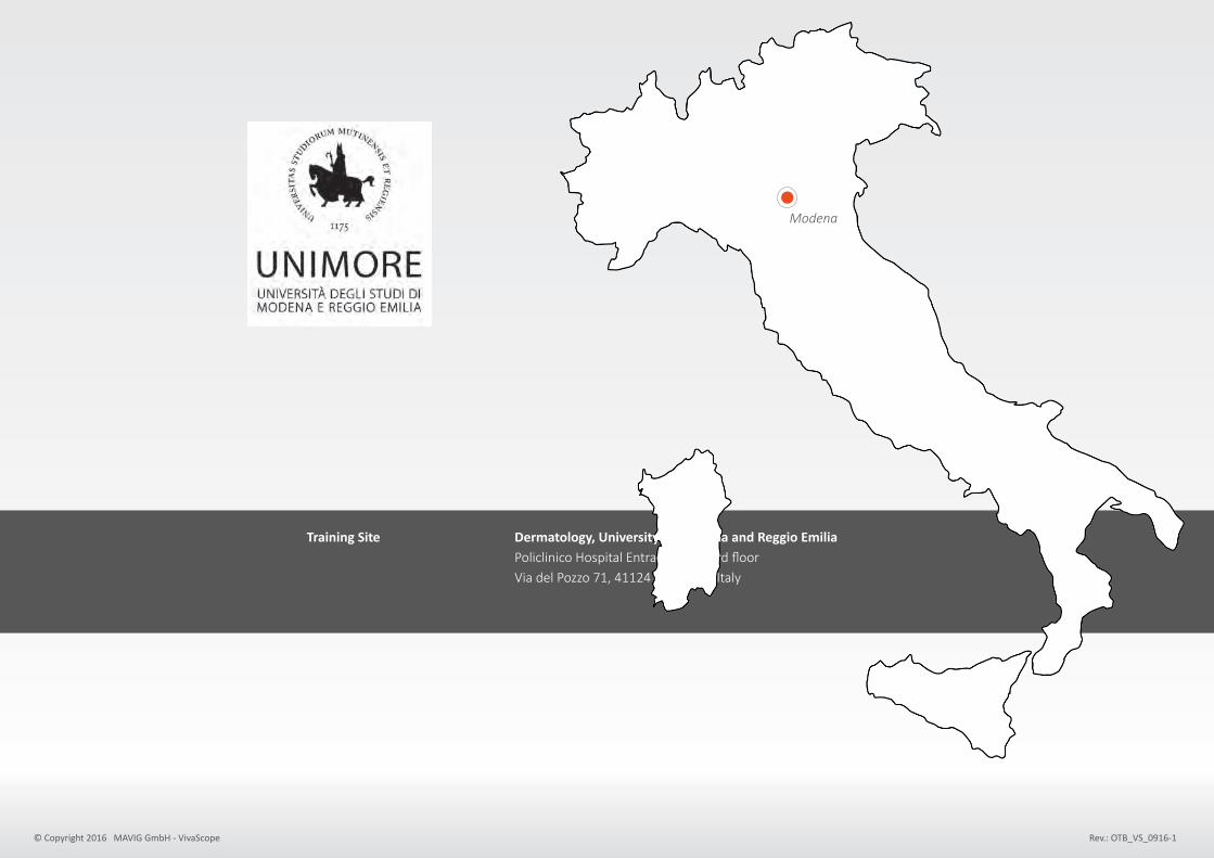
Training Site Dermatology, University of Modena and Reggio Emilia Policlinico Hospital Entrance n. 3, 3rd floor Via del Pozzo 71, 41124 Modena – Italy
Email-Contacts [email protected]
On-Site Secretary / Contacts E-mail [email protected] Tel. +39 (0) 59 422 28 75 Fax +39 (0) 59 422 42 71
2
Modena – Palazzo Comunale e Duomo

PRESENTATION OF THE TRAINING PROGRAM
This training program is designed specifically for physicians and residents who are interested in becoming confident and independent in skin cancer detection and management, using reflectance confocal microscopy (RCM).
The program is divided into three modules, a basic, a practical and an advanced course aiming at a transfer of complete knowledge and skills for the clinical use of confocal microscopy.The courses we propose are coordinated by leading dermatologists in the field of dermoscopy and RCM, and take place at the University of Modena in the Department of Dermatology.
They include didactic theoretical instructions, self-study and practical experience. The topics covered in the course are the main aspects of melanocytic and non-melanocytic skin lesions, the diagnostic clues for their correct identification, the application of the technology in inflammatory skin diseases, pathophysiology of the skin and cosmetology.
The participants will benefit from the expertise of medical doctors and researchers with long and proven experience in the field of confocal microscopy.
The full training consists of 3 modules. Thedurationofthebasic and the practical module is 3 days, each. The advanced module takes 2 days to be completed.
The aim of the courses is to build a high level of competence and confidence in order to ensure the appropriate approach and management of pigmented as
well as non-pigmented skin lesions.
3

BACKGROUND / STATE OF THE ART
Skintumorsarethemostfrequentcancerinthegeneralpopulationwithanupwardtrend.InEuropeand theUSanincidenceof150per100,000inhabitantsisestimatedforBasalCellCarcinomaandarangebetween 6-20 for Squamous Cell Carcinoma and Melanoma.
Nakedeyediagnosishasalowsensitivityandpoorspecificityforsmallandearlytumorsandcanbesupplemented bydermoscopy,whichenablesamoreaccuratediagnosis. Toexciseonemelanoma,however,onaverage30benignlesionsareunnecessarilyexcised,withtheexceptionofhighly specializedcenters.Confocalmicroscopyisanoveltechniqueenablinginvivoexaminationoftheskinatacellularlevel. Thismethodprovidesexcellentdiagnosticaccuracy,specificityimprovementfrom30%to70%inequivocallesions, andhelpstodifferentiatecosmeticallydelicatefaciallesions.Reducingthenumberofunnecessarybiopsiesand excisionscandramaticallyhelpreducingreferralwaittimesandprovidesignificantcostsavings/aversionsforthehealthcare system.
SystematicapplicationofReflectanceConfocalMicroscopy(RCM)inthetriageandmanagementofskincancerpatients maypotentiallyimprovediagnosticaccuracyandconsistentlyreducethenumberofunnecessarybiopsiesandexcisions. Clinicians who learn the specific semiology, build up confidence and competence in using these modalities will help achieving these goals.Therefore,wehaveestablishedthesededicatedtheoreticalandpracticalinstructionmoduleson confocalmicroscopy.ProvidingaspecificeducationinRCMwillgenerateskilledPhysiciansintheclinicalassessment andmanagementofpigmentedaswellasnon-pigmentedlesions.
Physicianswillbeabletoincreasetheirdiagnosticability,reducethenumberofunnecessarysurgicalexcisions ofbenignlesionsandthus,patientsatisfactionwillbeimproved.The intensive training program provides an organized education in the main aspects of dermatologic diagnosis, focusing on RCM. Theprogramincludesbothdidactic theoreticalinstructionandpracticalexperience.
4
CONTENT OF THE TRAINING PROGRAM
The program aims to develop the following specific competencies:
ToconfidentlyrecognizethemainRCMaspectsofskincancer Tointerpretthepatternsofalesioninthecontextofthepatientandoftheclinicalsituation Tolearnasystematicandeffectivepatient-tailoredapproachandmanagement Todevelophighlevelskillsindifferentiatingmelanocyticfromnon-melanocyticskinlesions, and malignant from benign skin lesions Toachieveearlydiagnosisofmelanomaandotherskinmalignancy
The courses will be focused on RCM, image interpretation and on difficult clinical situations where confocal microscopy can be applied. Experts in diagnostics, clinicians and researchers will give all the lectures. During the practical courses the trainees will consolidate knowledge through assisted revision of numerous cases and clinical situations.To acquire a strong practical ability and diagnostic confidence the trainees will take part in daily clinical activities of the confocal microscopy clinics. Collaborative learning represents an important part of the practical training. For this reason, participants will be asked to review and discuss a large number of cases together in the presence of tutors. The most difficult or interesting cases encountered during clinical practice and a case-review session will be supervised by the experts.
Feat
ure
s a
nd
Fa
cult
y FRONTAL LESSONS / COURSES (Faculty:G.Pellacani,C.Longo,F.Farnetani,S.Bassoli,A.Witkowski,M.Ulrich,M.Ardigò,S.Ciardo)
Courseswillprovideageneralviewonthediagnosticcluesdetectablebytheuseofthedifferenttechniques, consideredintheclinicalcontest,alongwithexpertsuggestionsforpracticalmanagement.
TUTORED CLINICAL ACTIVITY (Tutors:F.Farnetani,F.Giusti,S.Bassoli,A.Casari)
DailyattendancetotheclinicalactivitiesoftheMelanomaandSkinCancerClinicoftheUniversityofModena. Traineeswillparticipateinpatientsevaluationandmanagement,assistedbyskilleddermatologists from8:30amto1:00pm.SelectedinterestingcaseswillbeexaminedwithRCMinthepresenceofexpertConfocalists.
TUTOR-ASSISTED CASE REVIEW (Tutors:F.Farnetani,S.Bassoli,A.Casari,A.Witkowski)
Theafternoonswillbededicatedtodailypeerreviewofinterestingdermoscopiccases,alsoincludingconfocalimages.
5

MODULE 2
REFLECTANCE CONFOCAL MICROSCOPY – PRACTICAL ACTIVITIESFacultyandtutors:G.Pellacani,F.Farnetani,S.Bassoli,A.Casari,A.Witkowski,S.Ciardo
MONDAY TUESDAY WEDNESDAY
9:00-11:00 Tutoredclinicactivity Tutoredclinicactivity Tutoredclinicactivity 11:00-13:00 Tutoredclinicactivity Tutoredclinicactivity Tutoredclinicactivity 14:00-16:00 Self-studycasereview Self-studycasereview - 16:00-17:00 Tutoredcasereview Tutoredcasereview -
Thu
Thu
MODULES IN DETAILMODULE 1
REFLECTANCE CONFOCAL MICROSCOPY – BASIC COURSEFaculty:G.Pellacani,C.Longo,M.Ulrich,M.Ardigò,S.Ciardo
The course will provide a general view on the main confocal patterns and clues, along with expert suggestions for practical management of different clinical situations. HOW TO USE THE TECHNOLOGY will provide the skills to correctly use and integrate the technology as well as develop practical skills needed for an appropriate lesion documentation.
ThURSDAY FRIDAY SATURDAY
9:00-11:00 - RCMBasicCourse RCMBasicCourse 11:00-13:00 Howtousethetechnology RCM Basic Course RCM Basic Course 14:00-16:00 RCMBasicCourse RCMBasicCourse RCMBasicCourse 16:00-17:00 RCMBasicCourse RCMBasicCourse -
Fri
Sat
HOWTOUSE THE TECHNOLOGY 11:00 - 11:30 The hardware and software of digital dermoscopy and confocal microscopy 11:30 - 12:30 Hands-on: the use of the technologies with simulated patients 12:30 - 13:00 Storage and data management. Image evaluation
BASIC REFLECTANCE CONFOCAL MICROSCOPY 14:00 - 14:30 Why to use reflectance confocal microscopy 14:30 - 15:30 Correlation of RCM with dermoscopy and histopathology and RCM terminology 15:30 - 17:00 Melanocytic nevi: confocal correlates and interpretation
9:00 - 10:30 Melanoma: different faces of the disease 10:45 - 11:30 Benign non-melanocytic tumors 11:30 - 13:00 Basal cell carcinoma and malignant non-melanocitic tumors – AK. and SCC 14:00 - 17:00 Cases and situations: interactive session
9:00 - 9:30 Normal skin, dry skin and hydration, skin aging 9:30 - 10:30 Pattern analysis for inflammatory skin diseases 10.45 - 11.30 Spongiotic Hyperkeratotic and Interface dermatitis 11:30 - 13.00 Granuloumatous disorders, follicle/gland diseases, hair and adnexia 14:00 - 14:30 Pigment disorders (Vitiligo, Melasma, Post-inflammatory pigmentation) 14:30 - 15:00 Bullous diseases 14:30 - 16:00 Practical use of confocal findings in inflammatory diseases
TUTORED CLINICAL ACTIVITY Daily attendance to the clinical activities of the Melanoma and Skin Cancer Clinic of the University of Modena. Trainees will participate in patient evaluation and management, assisted by skilled dermatologists. Selected interesting cases will be evaluated with RCM in the presence of expert Confocalists.
SELF-STUDYANDTUTOR-ASSISTEDCASEREVIEW The afternoons will be dedicated to review of interesting cases, followed by discussions with experts focussing on clinical issues.
6

MODULE 3
REFLECTANCE CONFOCAL MICROSCOPY – ADVANCED COURSEFaculty:G.Pellacani,C.Longo,A.Witkowski
Difficult clinical situations, in which confocal can be particularly useful, will be presented along with expert tricks and interactive case discussions.
FRIDAY SATURDAY
9:00-11:00 AdvancedRCMCourse AdvancedRCMCourse 11:00-13:00 AdvancedRCMCourse AdvancedRCMCourse 14:00-16:00 AdvancedRCMCourse - 16:00-17:00 AdvancedRCMCourse -
Fri
Sat
TABLE OF COSTS (Exclusive Travel Expenditures)
MODULE 1 1,500.00 € +VATREFLECTANCE CONFOCAL MICROSCOPY – BASIC (Maximumof20Participants,Language:English)
MODULE 2 500.00 € +VATTUTORED CLINICAL ACTIVITY (Maximumof10Participants,Language:English)
MODULE 3 800.00 € +VATREFLECTANCE CONFOCAL MICROSCOPY – ADVANCED (Maximumof20Participants,Language:English)
ADVANCED REFLECTANCE CONFOCAL MICROSCOPY 9:00 - 11:00 Differential diagnosis: difficult pigmented lesions, atypical nevi and early melanoma 11:00 - 13:00 Amelanotic-hypopigmented tumors. The diagnostic challenge 14:00 - 16:00 Lesions on the face and sun-damaged skin 16:00 - 17:00 Papules and nodules: where the trap is hidden
9:00 - 10:00 Confocal microscopy in dark lesions 10:00 - 11:00 Lesions on special sites and use of the hand-held probe 11:00 - 11:30 Telediagnosis platform: how it works and what are the limitations 11:30 - 13:00 Cases on a telediagnosis setting (confidence and how to avoid pitfalls)
7
Training Site Dermatology, University of Modena and Reggio Emilia Policlinico Hospital Entrance n. 3, 3rd floor Via del Pozzo 71, 41124 Modena – Italy
Modena
© Copyright 2016 MAVIG GmbH - VivaScope Rev.: OTB_VS_0916-1