modification of the plant retinoblastoma-related...
TRANSCRIPT

Modification of the Plant Retinoblastoma-Related Protein
(RBR1) by SUMO: Structural and Functional Studies
Edward Businge
Umeå Plant Science Centre Department of Forest Genetics and Plant Physiology, SLU, Umeå
Master thesis, 30 hp
Master programme in Plant and Forest Biotechnology 2009

SLU, Sveriges lantbruksuniversitet Faculty of Forest Sciences, Umeå Plant Science Centre, Department of Forest Genetics and Plant Physiology, Umeå Author: Edward Businge Title: SUMO-modifiering av det retinoblastom-relaterade proteinet RBR1; Strukturella och funktionella studier. Modification of the Plant Retinoblastoma-Related Protein (RBR1) by SUMO: Structural and Functional Studies. Key words: SUMO, retinoblastoma, plant protein, Arabidopsis Supervisor: Dr Laszlo Bako, Umeå Plant Science Centre, Department of Plant Physiology, Umeå University, SE-901 87 Sweden Examiner: Dr Karin Ljung, Umeå Plant Science Centre, Department of Forest Genetics and Plant Physiology, SLU, SE-901 83 Sweden
Course title: Master thesis in Biology at the dept of Forest Genetics and Plant Physiology, 30 hp; Examensarbete i Biologi vid inst för skoglig genetik och växtfysiologi, 30 hp
Master programme: Plant and Forest Biotechnology
Course code: EX0490 Level: Master E Umeå, 2009

����
�
TABLE OF CONTENTS
TABLE OF CONTENTS .................................................................................................. III
ACKNOWLEDGEMENTS ............................................................................................... V
LIST OF ABBREVIATIONS .......................................................................................... VII
ABSTRACT ................................................................................................................... VIII
CHAPTER ONE ............................................................................................................... 1
INTRODUCTION......................................................................................................... 1
1.0 Background ........................................................................................................... 1
1.1 Objectives of the study.......................................................................................... 2
CHAPTER TWO .............................................................................................................. 3
LITERATURE REVIEW ............................................................................................ 3
2.1 The Plant Cell-cycle ............................................................................................. 3
2.2 Regulation of the cell cycle.................................................................................. 3
2.2.1 Cyclins and cyclin dependent Kinases (CDKs) ................................................. 3
2.2.2 Rb/E2F/DP Pathway ......................................................................................... 5
2.2.2.1 Regulation of the Rb/E2F/DP Pathway ........................................................ 6
2.3 Posttranslational modification of proteins ......................................................... 8
2.3.1 SUMO ................................................................................................................ 9
2.3.2 SUMO conjugation pathway........................................................................... 10
2.3.2.1 SUMO activating enzyme E1 ...................................................................... 10
2.3.2.2 SUMO conjugating enzyme E2 .................................................................... 11
2.4 Regulation of SUMO conjugation ...................................................................... 12
2.5 Biological effects of SUMO ............................................................................. 13
2.5.1 Modulation of transcriptional activity ............................................................. 14
2.5.2 Sub-cellular localization of proteins ................................................................ 14
2.5.3 DNA repair....................................................................................................... 15
2.5.4 Cell cycle regulation ........................................................................................ 15
2.5.5 A biotic stress response .................................................................................... 16
2.5.6 Abscisic acid (ABA) signaling ........................................................................ 16
CHAPTER THREE ........................................................................................................ 17
MATERIALS AND METHODS ............................................................................... 17
���
�
3.1 Construct .......................................................................................................... 17
3.1.1 Mutation of SUMOylation site ........................................................................ 17
3.2 PCR-site directed mutagenesis of SUMO conjugation site ............................... 17
3.3 Cloning RBR-mtSUMO..................................................................................... 18
3.3.1 Restriction and ligation ................................................................................... 19
3.3.1.1 Transformation of Escherichia coli .............................................................. 19
3.4 Transient expression of GFP-RBRmtSUMO in suspension culture cells .......... 20
3.4.1 Transfection of protoplasts .............................................................................. 20
3.5 Sub cloning RBR-mtSUMO into vector pGEX-5X-1 ........................................ 22
3.6 Protein Expression and purification .................................................................... 22
3.6.1 Expression of RBR-mtSUMO ......................................................................... 22
3.6.2 Purification ....................................................................................................... 23
3.6.3 Electrophoresis of protein fractions ................................................................. 24
3.7 In-vitro SUMOylation ......................................................................................... 25
3.7.1 Electrophoresis and western blot analysis ....................................................... 25
CHAPTER FOUR ........................................................................................................... 27
RESULTS .................................................................................................................... 27
4.1 Mutation of Arabidopsis RBR1 SUMO modification site .................................. 27
4.2 Effect of SUMO modification on RBR1 localization ......................................... 28
4.3 Expression and Purification of protein RBR1-mtSUMO ................................... 29
4.4 Mutation of Lysine 1007 prevents modification of RBR1 by SUMO ................ 30
CHAPTER FIVE ............................................................................................................ 32
DISCUSSION .............................................................................................................. 32
5.1 Discussion ........................................................................................................... 32
5.2 Conclusions ......................................................................................................... 34
5.3 Future Direction .................................................................................................. 34
REFERENCES ................................................................................................................ 35
�

��
�
ACKNOWLEDGEMENTS
�
I am greatly indebted to my supervisor Dr. Laszlo Bako and co-supervisor Dr. Karin
Ljung. Thanks Laszlo for the supervision, guidance, advice and for giving me the
opportunity to do my exam work in your lab.
I wish to express my appreciation to Dr. Miskolczi Pál, Dr. Ötvös Krisztina for the
priceless advice and help during the course of the project.
Am grateful to Dr. J.M.Kabirizi, Dr. M.K.Magunda, MegBauer and Mika-Whirtmash for
all the support.
I appreciate the help offered by my classmates, masters’ program coordinators and Maria
Sterner at the SLU international office.
Thanks Kristi, Danna-L.A, and Karen-L.A for the timely “rendezvous pitch maneuvers”.
Lastly but not the least, am grateful for the financial support from the Swedish Institute
(SI) throughout the course of the Masters program.
�
�
�
�

���
�
�
�
�
�
�
�
��
�
�
������������ ������������������� ����������� ��������������������������� �
�
�
��
�
�
�
�
�
�
�
�
�
�

����
�
LIST OF ABBREVIATIONS
ATP: Adenosine Tri phosphate.
CDK: Cyclin-dependent kinase.
CycD: Cyclin D.
DNA: Deoxyribonucleic acid.
DP: DNA-binding heterodimerization partner protein.
E2F: Adenovirus E2 promoter-binding factor.
GFP: Green fluorescent protein.
GST: Glutathione-S-transferase.
HDAC: Histone deacetylase
LB: Luria-Bertani medium
PCR: Polymerase chain reaction.
PTM: Post translational modification
Rb: Retinoblastoma.
RBR1: Retinoblastoma-related protein.
SENPs: SUMO-specific proteases.
SUMO: Small ubiquitin-related modifier.
�
�

�����
�
ABSTRACT
The retinoblastoma protein (Rb) is a transcription regulator and key component of the
Rb/E2F/DP pathway which regulates progression of the cell cycle in plants and animals.
Within the pathway, Rb blocks E2F transcriptional activity consequently ensuring
restricted cell proliferation. Of great importance too, is a family of posttranslational
modifiers referred to as small ubiquitin-related modifiers (SUMO), whose modification
consequences include; sub cellular localization of proteins, alteration of protein to protein
interaction and regulation of transcriptional activity.
In order to study and depict the plant retinoblastoma related protein (RBR1) as a SUMO
substrate; its modification site was mutated to address the effect of the mutation on
protein localization. Additionally, an in-vitro assay was used to further illustrate the
consequences of the mutation. In protoplasts transfected with wild type RBR1 the protein
was solely present in the nucleus while those transfected with mutated RBR1, the protein
was seen in both the nucleus and the cytosol. From the in vitro SUMOylation assay it was
evident that while wild type RBR1 could be modified by SUMO, its mutated version
could not undergo modification.
�
The results from this study don’t only show RBR1 as a SUMO substrate; they also
suggest that modification by SUMO could be needed for its sub-cellular localization.
�

��
�
CHAPTER ONE
INTRODUCTION
1.0 Background
The growth, development and division of cells within multi-cellular organisms such as
plants depends on a sequence of synchronized events which are spatially and temporarily
tightly regulated within individual cells (Desvoyes et al., 2006). Furthermore, organ
development in plants is just about a post embryonic process; as a result organogenesis
depends on constant ability of a given set of cells to grow prior to undergoing specific
differentiation programs (Desvoyes et al., 2006). Consequently the post-embryonic
nature of organogenesis, multi-cellularity of plants requires precise linking of cell
proliferation, differentiation and arrest of the cell cycle so that all processes are
coordinated with the overall development program (Desvoyes et al., 2006).
Unlike animals, development of new plant organs such as roots, leaves and flowers
repeatedly occurs over the plants life span which can at times extend over thousands of
years. As a result of continuous organogenesis, regulation of the cell cycle is of
fundamental importance for plant growth and development (Wildwater et al. 2005; Inze
et al., 2006).
Gutierrez and co workers (2002) acknowledged that in addition to hormonal signals, a
key regulator of the plant cell cycle is the Rb/E2F/DP pathway which consists of the
retinoblastoma protein (Rb) and the E2F transcription factor. Rb functions by binding the
E2F transcription factor consequently blocking transcription of cell related cycle genes;
this in turn prevents uncontrolled proliferation of cells (Brehm et al., 1999).
��
�
Like most proteins, Rb is subject to posttranslational modification by processes such as
phosphorylation, glucosylation and acetylation resulting in chemical alteration of the
amino acids within the protein (Verger et al., 2003). Alternatively the protein can also be
modified by addition of other polypeptides such as ubiquitin and the small ubiquitin
related modifiers also referred to as SUMO (Verger et al., 2003; Gill, 2004). The
consequences of these modifications include sub-cellular localization of proteins,
alteration of protein to protein interactions and degradation of proteins (Richards, 2008).
1.1 Objectives of the study
Conjugation of SUMO to substrate proteins has been implicated in regulation of a
number of cellular processes ranging from nuclear transport, cell cycle control;
transcription regulation and DNA repair (Gill, 2004). Earlier in-vitro experimentation in
the Bako lab (UPSC) had shown that the plant retinoblastoma-related protein RBR1,
whose function is crucial for plant cell division and development, is modified by SUMO.
The site(s) of modification and functional consequence of SUMOylation on RBR1
function were however yet to be determined. Hence the objectives of this study were;
1. To indentify a site(s) on Arabidopsis RBR1 protein modified by SUMO followed
by; PCR-directed mutagenesis of predicted conjugation site(s), as well as analysis
of mutant proteins by in vitro SUMOylation assays.
2. Functional studies of RBR1 carrying mutation(s) of SUMO conjugation site(s)
through; Transient expression of RBR1 mutant fused to GFP in plant cells to
investigate the effect of mutation on intracellular localization using confocal
microscopy.

��
�
CHAPTER TWO
LITERATURE REVIEW
2.1 The Plant Cell-cycle
The cell cycle also referred to as the cell-division cycle involves a sequence of four
coordinated events culminating in replication of the cells genetic material. The four
events include; Gap phases (G1 and G2) which separate DNA replication (S phase) and
the M (mitosis) phase respectively (Ferreira et al., 1994). An additional role of gap
phases within the cycle is to serve as check points’ ensuring each phase is successfully
completed before the next is initiated (Ferreira et al., 1994; Dewitte et al., 2003).
Under the influence of hormonal signals and metabolic changes during G1 Phase, cells
start preparing for the impending division (Dewitte et al., 2003). At a certain point, the
cell moves into S-phase during which genetic material is replicated and doubled resulting
in chromosomes with two sister chromatids. The cell subsequently moves into G2 phase
during which it continues to grow while assembling cytoplasmic material required for
eventual division. The final phase of the cycle is M phase which involves nuclear
division accompanied by cytoplasmic division (cytokinesis) resulting into two daughter
cells (Dewitte et al., 2003).
2.2 Regulation of the cell cycle
2.2.1 Cyclins and cyclin dependent Kinases (CDKs) �
Regulation of the cell cycle is essential to ensure; division of cells never occurs until all
DNA has been replicated and repaired in case of damages (Inze et al., 2006). Hence cycle
regulation occurs throughout the cell division machinery with a unique characteristic of

��
�
all regulation points being under the control of cyclin dependent kinases (CDKs) (Pines,
1999).
Cyclins are a multi group of proteins characterized by a poorly conserved region essential
for interaction with partner CDKs. The conserved cyclin region is 250 amino acids long
comprising of two folds of five helices (Dewitte et al., 2003). Using sequence based
classification; five types of cyclins (A, B, C, D and H) have been indentified in plants
with significant roles at different stages of the cell cycle (Renaudin et al., 1996;
Vandepoele et al., 2002). “A-type cyclins appear at the beginning of the S-phase and play
a role in its progression, B-type cyclins are involved in G2 /M transition while D-type
cyclins control progression through G1 and S phase” (Dewitte et al., 2003).
Pines (1999), noted that progression of the cell cycle is dependent on activation and
deactivation of cyclin dependent kinases (CDKs) which belong to a conserved
serine/theorine protein kinase family. CDKs remain inactive until their partner cyclins
bind them to form Cyc-CDK complexes; these are then activated by CDK-activating
kinases (CAKs) by phosphorylating threonine residues contained in CDK T-loops (Pines,
1999). Consequently, CDK induced phosphorylation is responsible for onset of DNA
replication and mitosis during S and M phases respectively (Pines, 1999; Inze et al.,
2006).
Four CDKs namely CDKA1, CDKA2 and CDKB1, CDKB2 have been indentified in
Arabidopsis thaliana belonging to CDKA and CDKB families respectively. CDK related
studies in Antirrhinum cells revealed that CDKA activity is up regulated during G1 and S

��
�
phase while CDKB activity is intense during S, G2 and M phases (Fobert et al., 1994;
Joubes et al., 2000; Vandepoele et al 2002).
Once activated, Cyc-CDKs covalently add phosphate groups to serine or threonine
residues within substrate proteins consequently altering their properties. One notable
substrate of CDKs is the retinoblastoma protein (Rb) whose phosphorylation is essential
for progression of the cell cycle (Dewitte et al., 2003).
2.2.2 Rb/E2F/DP Pathway
The Rb/E2F/DP pathway controls transition from G1 to S phase of the plant cell cycle, it
consists of the retinoblastoma protein (Rb) a transcription regulator and the E2F
transcription factor (Gutierrez et al., 2002; Inze et al., 2006). According to Weinberg
(1995), involvement of the Rb protein in regulation of cell division was originally
exclusively associated with animals. However this view changed as a result of the
discovery that Rb related proteins and components of the Rb/E2F/DP� pathway do
actually exist in plants too (Huntley et al., 1998)
De Jager et al., (1999) referred to the Rb protein “as being part of a conserved pathway
controlling the activation of cell division in animals”. It contains a number of functional
domains two of which denoted A and B are conserved in humans and plants (Harbour et
al., 2000). Furthermore, interaction of the two domains results in formation of a central
pocket which enables Rb to interact with partner proteins such as cyclin D, histone
deacetylases via their LxCxE motifs (L-leucine, C-cysteine, E-glutamic acid and x being
any amino acid) (Harbour, 1998).
Rb is a nuclear protein characterized by a number of potential binding sites for CDKs.
Furthermore, the protein is crucial in regulation of the cell cycle, cell differentiation

��
�
during which it functions by blocking E2F transcription activity consequently preventing
uncontrolled cell proliferation (Dewitte et al., 2003). The importance of Rb in cell cycle
regulation was elucidated by inhibiting its function through virus-induced gene silencing
which resulted in prolonged cell proliferation and delayed differentiation of Nicotiana
benthamiana leaf and stem cells (Park et al., 2005). Additionally, the plants showed
retarded flower formation highlighting a possible role for Rb in flower development
(Park et al., 2005).
The second component of the cell cycle regulating pathway is a family of transcription
factors referred to as E2Fs. E2Fs contain a DP heterodimerization domain enabling them
to combine with DP proteins resulting in an active E2F/DP transcription complex which
induces expression of genes involved in cell cycle progression (Desvoyes et al., 2006).
Furthermore, E2Fs also contain binding domains to which the Rb protein binds to block
transcription activity. Six E2Fs (E2Fa, E2Fb, E2Fc, DEL1, DEL2, and DEL3), two DP
proteins (DPa and DPb) and a single Rb homolog (RBR1) have been indentified in
Arabidopsis thaliana (Desvoyes et al., 2006; Hirano et al., 2008).
2.2.2.1 Regulation of the Rb/E2F/DP Pathway
Rb functions by binding E2F transcription factors (Figure 1A) consequently blocking
transcription of cell cycle related genes (Brehm et al., 1999). Repression of E2F activity
is due to the Retinoblastoma protein recruiting histone deacetylase (HDAC) enzymes
which are co-repressors of transcription. HDACs remove acetyl groups from histones
within DNA resulting in chromatin modification, condensation of DNA and inhibition of
transcription (Brehm et al., 1999).

��
�
�
Figure 1 Transition from G1 to S phase of the cell cycle in plants. (A) The retinoblastoma protein binds to the E2F/DP transcription factor blocking expression of cell cycle related genes. (B) Expression of D type cyclins in response to metabolite and hormonal signals leads to activation of cyclin dependent kinases. Activated CDKs go on to phosphorylated Rb, once Phosphorylated Rb activity is terminated leading to progression of the cell cycle (Modified from De Jager et al., 1999).
However, in response to signals induced by metabolites (sucrose) and hormones (auxin
and cytokinins), D-type cyclins are expressed (Figure 1B). CycDs subsequently bind their
partner CDKs forming CycD-CDK complexes which are activated by CDK-activating
kinases (CAKs). Active Cyc-CDKs are capeable of phosphorylating target proteins
resulting in altered protein properties (Dewitte et al., 2003).
The Retinoblastoma protein being a CDK target is phosphorylated leading to termination
of its repression activity on the E2F transcription factor (De Jager et al., 1999). In
absence of Rb induced repression (Figure 1B), E2Fs are eventually capeable of inducing
the expression of genes involved in G1/S transition of the cell cycle (De Jager et al.,
1999; Dewitte et al., 2003).

�
�
Xie and co workers (1995) reported that interaction between viral proteins and Rb
induces direct entry of the cell cycle into the S phase. This is attributed to ability of viral
proteins to mutate the LxCxE motif on the HDACs terminating their co-repression
activity (Park et al., 2005).
2.3 Posttranslational modification of proteins
Posttranslational modification (PTM) is the chemical alteration of proteins after
biosynthesis; it may involve a change in the chemical nature of amino acids through
processes such as phosphorylation, acetylation, hydroxylation, glycosylation, alkylation
and methylation. Alternatively, proteins can also be modified by addition of other
polypeptides (Verger et al., 2003; Gill, 2004). The consequences of these modifications
include localization and degradation of proteins (Richards, 2008).
The most common polypeptide involved in PTM is the 76 amino acid (Ubiquitin) present
in most eukaryotic species whose biological function is marking proteins for eventual
degradation by the 26S proteasome (Kurepa et al., 2002). It is covalently attached to
lysine residues within substrate proteins via an isopeptide bond formed between the
lysine residues and a poly ubiquitin chain (Kurepa et al., 2002). The ubiquitination
pathway is ATP dependent and relies on an activating enzyme (E1), conjugating enzyme
(E2) and a ligase (E3) which enable poly-ubiquitination of target proteins (Kurepa et al.,
2002).
In an ATP dependent reaction, E1 activates the ubiquitin precursor via a thiol ester bond
formed between a glycine in ubiquitin and the cysteine end in the E1 enzyme (Smalle et
al., 2004). Activated ubiquitin is then transferred to E2 or ubiquitin conjugating enzyme,
eventually, ubiquitin is conjugated on to target proteins via an isopeptide bond between

�
�
the C-terminal glycine of ubiquitin and the lysine residue of the substrate protein with the
help of an E3 ligase enzyme (Smalle et al., 2004).
2.3.1 SUMO
In addition to ubiquitin, families of ubiquitin like proteins (Ubls) have over the years
been discovered and found to be involved in post translational of proteins in animals and
plants. Key among Ubls is a family referred to as SUMO with functions spread across a
variety of biological processes (Johnson, 2004).
SUMO is an acronym for small ubiquitin-related modifier, a family of conserved proteins
present in all eukaryotes. SUMOs are approximately 11 KDa in molecular weight, 25
amino acids longer than ubiquitin but with a 20% sequence identity with ubiquitin
(Johnson, 2004). Initial identification of SUMO was in 1997 after unearthing its
conjugation to the GTPase activating protein RanGAP1, a protein involved in nuclear
transport and the cell cycle in animals (Matunis et al., 1998; Verger et al., 2003; Marx,
2005). As is the case with ubiquitination, SUMOylation also involves addition of SUMO
to lysine residues within substrate proteins. “SUMO modifies a number of proteins which
participate in diverse cellular processes such as; transcriptional regulation, nuclear
transport, maintenance of genome integrity, and signal transduction” (Verger et al., 2003;
Johnson, 2004; Bossis et al., 2006).
Three SUMOs have so far been indentified in animals namely; SUMO-1, SUMO-2 and
SUMO-3 while a total of 9 genes (SUM1 to SUM9) with strong resemblance to animal
and fungal SUMOs have been identified in Arabidopsis thaliana (Kim et al., 2002;
Novatchkova et al., 2004). Although referred to as ubiquitin like proteins, there are
notable differences between SUMO and ubiquitin; for instance SUMO-1contains a long

���
�
and flexible N-terminal which is absent in ubiquitin (Bayer et al., 1998). Secondly, unlike
ubiquitin, SUMO-1 lacks Lysine-48 hence its inability to form multiple SUMOylation
chains as is the case with ubiquitin (Bayer et al., 1998).
Additionally, while ubiquitination is associated with marking proteins for degradation,
SUMOylation has been implicated in influencing protein to protein interactions,
regulating protein stability and sub-cellular localization of substrate proteins (Bossis et
al., 2006). In plants, animals SUMO modification occurs at all development stages and
their respective tissues. This is made possible by Specific SUMO proteases which de-
conjugate SUMO precursor from substrate proteins making it available to be reused,
although the long term fate of sumoylated proteins remains altered even after the
modifier has been de-conjugated (Verger et al., 2003; Gill, 2004; Hay, 2005; Bossis et
al., 2006).
2.3.2 SUMO conjugation pathway
SUMOylation (Figure 2) occurs in the nucleus and cytoplasm in a pathway relying on
enzymes similar to those in ubiquitination but specific to SUMO. The pathway makes use
of an activating enzyme (E1), conjugating enzyme (E2) and a ligase (E3) (Johnson,
2004).
2.3.2.1 SUMO activating enzyme E1
The activating enzyme consists of two un-identical protein sub units which must combine
to form an active heterodimer. In Arabidopsis thaliana the enzyme is denoted SAE
(SUMO activating enzyme) with two subunits SAE1a and SAE1b whose gene

���
�
annotations are At4g24940 and At5g50580 respectively (Johnson et al., 1997;
Novatchkova et al., 2004).
2.3.2.2 SUMO conjugating enzyme E2
Contrary to ubiqutination in which E3 ligases ensure substrate specificity, previous in
vitro studies have highlighted the SUMO cojugating enzyme as being able to attach
SUMO on to substrates without the E3 ligase (Rodriguez et al. 1999 and 2001). A single
E2 enzyme exists in Arabidopsis thaliana denoted SCE or SUMO conjugating enzyme
(Novatchkova et al., 2004).
Melchior et al., (2003) and Johnson (2006) attributed SUMO specificity to a short precise
sequence which the conjugating and ligase enzymes recognize in all substrate proteins.
“The sequence denoted �KXE or �-Lys-X-Glu; where � is a large hydrophobic amino
acid; K the lysine residue which is modified; X is any residue; and E/Glu is glutamic acid
(Johnson, 2006).

���
�
Figure 2 The SUMO conjugation pathway. SUMO precursor is cleaved by SUMO proteases to expose the glycine at its C-terminal end. Cleaved SUMO is then energetically activated by the E1-activating enzyme and transferred to the conjugating enzyme (E2). The final step of the pathway involves attaching SUMO on to the substrate protein in the presence of an E3 ligase (Modified from Marx, 2005). Prior to activation (Figure 2), the SUMO precussor is processed by SUMO-specific
proteases (SENPs) to expose the glycine at its C-terminal end. Then, in an ATP
dependent reaction a thioester bond is formed between the exposed glycine and the
catalytic cysteine of the activating enzyme (E1). Next, activated SUMO is transferred to
the cysteine end of the E2 cojugating enzyme. The final step involves attachment of
SUMO to lysine residues on target proteins a reaction that is aided by SUMO-E3-ligases
(Johnson, 2004; Gill, 2004; Bossis et al., 2006).
2.4 Regulation of SUMO conjugation
Reversibility of the SUMOylation pathway is ensured by SUMO-specific proteases or
isopeptidases which remove SUMOs from modified proteins making it available for re-

���
�
use as a precursor (Johnson, 2006). Though effects of SUMOylation have clearly been
established, not much is known about how the pathway is regulated. However, Johnson
(2004) suggested that regulation of the pathway could be occurring during conjugation or
de-conjugation of SUMO leading to change in amount of modified proteins.
Studies in animals have however revealed a role for Gam1 protein in regulating SUMO
conjugation and de-conjugation (Bossis et al., 2006). Gam1 is a viral protein residing in
CELO (Chicken embryo lethal orphan virus); it is involved in transcriptional activation of
cellular and viral genes that inactivate HDACs (Boggio et al., 2005).
In an earlier study, Boggio and co workers (2004) showed that Gam1 targets SUMO E1
and E2 enzymes. Consistent with this finding, cellular expression of Gam1 resulted in
decreased levels of SUMO conjugates, implying that the protein could be targeting the
respective SUMO enzymes for proteasome degradation (Boggio et al., 2005).
Furthermore, abiotic factors such as heat shock, osmotic and oxidative stress have also
been implicated in regulating the SUMOylation pathway in animals (Bossis et al., 2006).
2.5 Biological effects of SUMO
Various nuclear proteins with roles related to transcriptional regulation, DNA repair and
signaling have been identified as targets of post translational modification by SUMOs
resulting in sub cellular localization of the proteins and alteration of protein-protein
interaction (Gill, 2004). SUMOylation also affects processes including DNA repair,
hormonal signaling and stress response.

���
�
2.5.1 Modulation of transcriptional activity
The majority of proteins modified by SUMO include signaling proteins, enzymes and
transcription factors or regulators (Johnson, 2004). SUMOylation of the latter proteins
results in increased repression of transcription activity, an assertion supported by studies
in which mutation of SUMO acceptor lysines resulted in increased transcription activity.
Additionally over expression of de-conjugating enzymes has also shown increased
transcription activity in the previously affected transcription factors (Gill, 2005).
According to Johnson (2004), inhibition of transcription activity could be due to SUMO
induced interactions with proteins that co-repress transcription. Furthermore, some
transcription factors could require post translation modification by processes such as
phosphorylation and ubiquitination. However SUMOs have the added ability of
preventing such modifications by blocking lysine residues where they would normally
occur (Johnson, 2005).
2.5.2 Sub-cellular localization of proteins
Studies involving the first indentified SUMO substrate protein RanGAP1 have revealed a
role of SUMOylation in sub cellular localization of proteins. RanGAP1 is a GTPase
activating protein with a significant role in protein transportation into the nucleus via the
nuclear pore complex (Melchior et al., 1993; Gill, 2004). In animals unmodified
RanGAP1 is located in the cytoplasm, however subsequent to SUMOylation the protein
is present in the nuclear pore. Additionally, the modified protein shows increased
interaction with the nuclear protein RANBP2 (RAN binding protein2) which could imply
structural alterations within sumoylated RanGAP1 resulting in elevated levels of
interaction with RANBP2 (Matunis et al., 1996). Consistent with this suggestion, protein

���
�
sequencing showed that interaction between RanGAP1 and RANBP2 required prior
modification of RanGAP1 by SUMO (Matunis et al., 1996 and 1998)
2.5.3 DNA repair
Involvement of SUMOs in DNA repair was highlighted in experimentation using
thymine DNA glycosylate (TDG). TDG is a DNA repair enzyme which removes uracil
and thymine from U-G or T-G mismatched base pairs respectively, resulting in DNA
devoid of a pyrimidine site (s) which is then repaired by downstream enzymes
(Hardeland et al., 2002).
In vitro studies with unmodified TDG show that the enzyme could only perform a single
round of base removal as a result of being tightly bound to the reaction products
(Johnson, 2004). Contrary to this, sumoylated TDG did perform multiple base removal
reactions an indication that modified TDG was not being impeded by the end products
(Johnson, 2004).
2.5.4 Cell cycle regulation
In addition to being involved in DNA repair, sub-cellular localization of proteins and
modulation of transcription activity; SUMO modification has also been implicated in
regulating progression of the cell cycle (Johnson et al., 1997). Saccharomyces cerevisiae
SUMO- E1 and SUMO- E2 mutants have cell cycle defects and arrest at the G2 to M
boundary (Johnson et al., 1997). Similarly, mutation of SUMO- E1 and SUMO- E2 in
Schizosaccharomyces pombe resulted in cells with stern impaired growth and mitotic
defects, leading to suggestions that SUMOylation is crucial for cell cycle progression
(Hay, 2005).

���
�
2.5.5 A biotic stress response
Saitoh et al., (2000) reported that in animals SUMO2/3 is involved in cellular response to
environmental stress. Similarly SUMOs do indeed play an equivalent role in plant abiotic
stress response. Exposure of Arabidopsis cells to ethanol and hydrogen peroxide (H2O2),
caused a striking increase in SUMO conjugates while withdrawal of the two stress
factors resulted in a reduction of SUMO conjugate levels (Kurepa et al., 2003). This
observation could be an indication of involvement of SUMOs in plant stress response.
2.5.6 Abscisic acid (ABA) signaling
In addition to the aforementioned role in stress response, biological consequences of
SUMOylation in plants extend to hormonal signaling in particular with the stress
hormone ABA (Lois et al., 2003). Arabidopsis SUM1 over expressers have superior
insensitivity towards ABA induced root growth inhibition (Lois et al., 2003). However,
when SCE1 (sumo conjugating enzyme) activity is suppressed, the plants succumb to the
ABA effect resulting in a stunted root growth phenotype (Lois et al., 2003).

���
�
CHAPTER THREE
MATERIALS AND METHODS
�
The study was conducted within the Department of Plant Physiology at Umea University
(UmU).
3.1 Construct
The construct pRT104-GFP-RBR (7.2 kb) was used as the raw template for this study. It
consisted of; vector pRT104, green fluorescent protein (GFP) and the full length RBR1
protein from Arabidopsis thaliana.
3.1.1 Mutation of SUMOylation site
In order to generate RBR1 with a mutated SUMOylation site, nucleotides coding for the
lysine at the SUMOylation site (�KXE) in the wild type protein were substituted for
alanine using Silent Site Selector web tool (http://rana.lbl.gov/SSS/). A forward and
reverse primer pair was designed and synthesized to introduce the desired mutation by
PCR- site directed mutagenesis
3.2 PCR-site directed mutagenesis of SUMO conjugation site
A 720bp RBR1 fragment denoted “RBR1-mtSUMO” with a mutated SUMOylation site
was amplified by polymerase chain reaction using the primer pair; R: 5'-
ATAGGTACCCTATGAATCTGTTGGCTCGGTCGCGAGGGGTGCGGCACCAC-3'
and F: 5'-TTCAGTCGACACATTGACCAGATCATTCTCTGTTGCTTCTACGGAGT
GC-3'.

��
�
PCR amplification was performed in 200µL thermal tubes using a MJ mini thermo cycler
(Bio-Rad, CA, USA), with each tube containing a 50µL reaction mixture containing;
0.5µL of DNA template (15ng/µL) mixed with 2.5µL of each 0.5mM primer, 5µL of 10X
PCR buffer (Invitrogen, Oxon, UK), 2.5µL of 2.5mM Invitrogen MgCl2, 1.25µL of
0.25mM dNTPs, 0.75µL of Taq DNA polymerase (Invitrogen) and 35µL of water. The
mixture was subjected to an amplification program with an initial denaturation step at
95oC for 4 minutes followed by 30 cycles consisting of; annealing of primers with
template at 55 oC for 1 minute, extension of primers at 72 oC for 1 minute and final
elongation at 72 oC for 5 minutes.
After PCR, 10µL of amplified product were resolved by gel electrophoresis on a 1%
agarose gel containing 0.25µg/uL ethidium bromide while making use of a 1Kb DNA
ladder (Invitrogen) to determine fragment size. After electrophoresis, fragment size was
viewed using a UV documentation camera (Techtum Lab, Umea, Sweden).
Next, 40uL of the remaining PCR product were treated with proteinase K (Fermentas,
Vilnius, Lithuania) and purified by phenol/chloroform extraction (15:24:1, Sigma,
Germany), precipitated, washed with 70% ethanol and finally resuspended in 15µL of
water. Treatment with proteinase K was to remove tightly bound Taq polymerase that
would interfere with restriction digestion.
3.3 Cloning RBR-mtSUMO
The construct pRT104-GFP-RBR was double digested with appropriate restriction
enzymes to rid it of the C-terminal part of RBR1 protein which was subsequently
replaced with amplified RBR1 containing a mutated SUMOylation site.

��
�
3.3.1 Restriction and ligation
3µg of the cloning vector were digested with enzyme SalI (Fermentas) for 2hrs at 37˚C
after which enzyme activity was terminated with 65˚C incubation on a heating block.
After 15 minutes the vector was purified by phenol/chloroform extraction, precipitated,
washed with 70% ethanol and finally resuspended in 15µL of water. It was re-digested
with KpnI (Fermentas) for 2hrs at 37˚C after which enzyme activity was terminated by
incubating at 80˚C on a heating block.
Next, the vector was dephosrylated in a reaction containing; 25µL of vector, 2.9µL 10X
dephosphorylation buffer and 1µL of shrimp alkaline phosphate (Fermentas). The
reaction was performed at 37˚C for 30minutes followed by termination of SAP activity at
70˚C for 15 minutes. The DNA was resolved on a 1% agarose gel and isolated using the
NA45 membrane (Schleicher and Schuell Bioscience, Dassel, Germany).
Prior to ligation, restriction of the PCR product was done with the same enzymes and
conditions used for the vector. The vector and insert were then ligated together in a 10uL
reaction; 5uL of insert, 3uL of vector, 1uL 5X ligase buffer and 1uL T4DNA ligase
(Invitrogen). Ligation was done over night at 12˚C in a thermo cycler (Bio-Rad).
3.3.1.1 Transformation of Escherichia coli
100µL of thawed XLI blue E.coli competent cells in a 1.5mL Eppendorf�tube were mixed
with 10µL of ligation mix and left on ice, after 30 minutes the cells were heat shocked in
a water bath at 42˚C for 90 seconds. 900uL of SOC medium (5X SOB media, glucose
and water) were immediately added to the heat shocked cells to aid cell recovery. Next,
the transformed cells were placed on a rotary shaker at 37˚C for 1hour after which 200µL

���
�
of the cells were spread on LB plates and grown overnight at 37˚C. From the overnight
LB plates, a single colony was used to inoculate 5ml of LB media (containing 50mg/L
carbenicillin) in a 15mL falcon tube overnight at 37˚C.
Mini-preparation was performed using a QIAprep spin mini-prep kit (Qiagen, Solna,
Sweden) following the manufacturer’s guidelines. To detect presence of the mutation,
mini-preps were double digested in a single reaction with SalI and NruI (Fermentas). The
NruI restriction site had been integrated into the reverse primer used to induce the
mutation. Furthermore, the minipreps were sequenced using a 35S terminal antisense and
a pRT104 35S primer.
3.4 Transient expression of GFP-RBRmtSUMO in suspension culture cells
3.4.1 Transfection of protoplasts
4 day old Arabidopsis thaliana root suspension cells were collected in a 50mL falcon
tube by centrifugation at 1200rpm, 5 minutes in a ZK380 centrifuge (Hermle, Wehingen,
Germany), after which the supernatant was discarded. In order to rid the cells of their cell
walls, they were resuspended in 25mL of enzyme solution which was topped up with
25mL of B5-0.34M glucose mannitol (Table 1). Re-suspended cells were then transferred
to petri-dishes and subjected to vigorous shaking on a rotary shaker.
After 4hrs, the cell suspension was centrifuged, supernatant discarded and the pellet
resuspended in 25mL of B5-0.34M glucose mannitol followed by further centrifugation.
Viable cells were collected by re-suspension in 5mL of B5-0.28M sucrose in which they
floated.

���
�
Table 1: Composition of solutions used in transfection of Arabidopsis protoplasts.
�
Prior to transformation of protoplasts, a single positively sequenced colony was used to
inoculate 2mL of fresh LB medium for a scale up culture. After 2.5hrs, 2mL of scale up
culture were added to 50mL of fresh LB medium (100µg/mL carbenicillin) and grown
overnight at 37˚C with shaking. Plasmid DNA was prepared using a plasmid mid kit
(Qiagen) as described by manufacturer.
3-5�g of plasmid DNA (GFP-RBRmtSUMO) were gently mixed with 50µL of
protoplasts followed by 150µL of PEG solution, the transformation mix was left in the
dark at room temperature for 30 minutes. Protoplasts were twice washed with 0.275M
Ca(NO3)2 to clear them of PEG solution after which they were collected by centrifugation
and mixed with 0.5mL of B5-0.34M glucose mannitol. The cells were placed in a
microtiter plate� and� incubated overnight in the dark; localization of GFP in the
transfected cells was performed by confocal laser scanning microscopy

���
�
3.5 Sub cloning RBR-mtSUMO into vector pGEX-5X-1
Fragment RBR-mtSUMO was digested with SalI (Fermentas) while expression vector
pGEX-5X-1 was first digested with XhoI (Fermentas), next the ends were filled with
Klenow polymerase (Takara, Saint-Germain-en-Laye, France) to clear the vector of
protruding 3' overhangs produced by XhoI. The Klenow filling reaction contained 20uL
of vector, 3uL 10X Klenow buffer, 0.5µL 2.5mM dNTP, 0.5µL Klenow polymerase and
26uL of water. The mixture was incubated at room temperature for 10 minutes followed
by incubation at 70˚C on a heating block to terminate Klenow activity.
Next the vector was digested with SalI, dephosphorylated with shrimp alkaline phosphate
and purified from a 0.8% agarose gel using the NA45 membrane (Schleicher and
Schuell). The purified vector was ligated with the insert (RBR-mtSUMO) after which the
ligation was used to transform E. coli XL1blue competent cells.
Mini-preparation was done using a QIAprep spin mini-prep kit (Qiagen). To verify
presence of the insert, mini-preps were digested with NruI (Fermentas), SalI (Fermentas)
and resolved by electrophoresis on a 0.8% agarose gel. Positive mini-prep samples were
further sequenced with the sequencing primers GEX lower (5'-CCGGGAGCTGCATGG
TCAGAGG-3') and GEX upper (5'-GGGCTGGCAAGCCACGTTTGGTG-3').
3.6 Protein Expression and purification
3.6.1 Expression of RBR-mtSUMO �
0.5uL of mini-prep DNA (pGEX-5X-1-RBR-mtSUMO) were used to transform 100uL of
E.coli BL21 cells by heat shock, the cells were spread on LB plates containing
Carbenicillin (100µg/mL) and grown overnight at 37˚C. A single colony was used to

���
�
inoculate 5ml of LB medium (100µg/mL carbenicillin) overnight at 37˚C. Next, a scale
up culture containing 3mL of overnight culture and 200ml fresh LB medium was grown
without induction.
After 2.5hrs, 200mL of scale up culture were added to 600mL of fresh LB medium
(100µg/mL carbenicillin); the overall culture was grown at 37˚C until its OD600 reached
0.7. 80uL of isopropyl-b-D-1-thiogalactopyranoside (IPTG) were then added to the
culture to a final concentration of 0.1mM after which growth was continued at 28˚C for
2.5hrs. The cells were eventually harvested by centrifugation at 3000g for 20minutes in a
J-20xp centrifuge (Beckman coulter, CA, USA); the resulting pellet was retained while
the supernatant was discarded.
3.6.2 Purification
Each gram of pelleted cells was re-suspended in 5mL of lysis buffer (25mM Tris-HCl
pH7.8, 100mM NaCl, 2mM EDTA, 1mM DDT, 0.5mM PMSF and 1mM Benzamidin)
followed by 0.5mg/mL of lysozyme and 0.2% Triton X-100 with gentle stirring at 4˚C.
The cell suspension was agitated for 12 minutes with a Branson sonifier (Kebo Lab,
Sweden), before adding 0.1mg/mL of DNase and MgCl2 to a final concentration of
10mM.
Next the cell lysate was centrifuged at 12000g for 30 minutes; the resulting supernatant
was filtered through a 0.45� cartridge (Sarstedt,�Nümbrecht, Germany) after which 5M
NaCl was added to the filtrate culminating in a final salt concentration of 200mM. In
order to bind the protein, 0.5mL of Glutathione-Sepharose resin equilibritaed in lysis
buffer were next added to the filtrate with gentle stirring at 4˚C.
���
�
After 45 minutes the resin solution was thrice washed with lysis buffer, 10 protein
fractions were then eluted each with 1mL of 10X elution buffer ( 50mM Tris-HCl pH8.0,
200mM NaCl, 1mM DDT, 0.02% Triton X-100 and 10mM reduced glutathione). The
protein fractions were left in dialysis buffer (25mMTris-HCl ph 7.8, 100mM NaCl, 1mM
EDTA, 1mM DDT, 10% glycerol, 0.2mM PMSF and 0.5mM Benzamidine) overnight at
4˚C. After dialysis proteins were concentrated using micron YM-30 filters (Millipore),
protein concentration was measured and aliquots were flash frozen in liquid Nitrogen
before storage at -80˚C for later use.
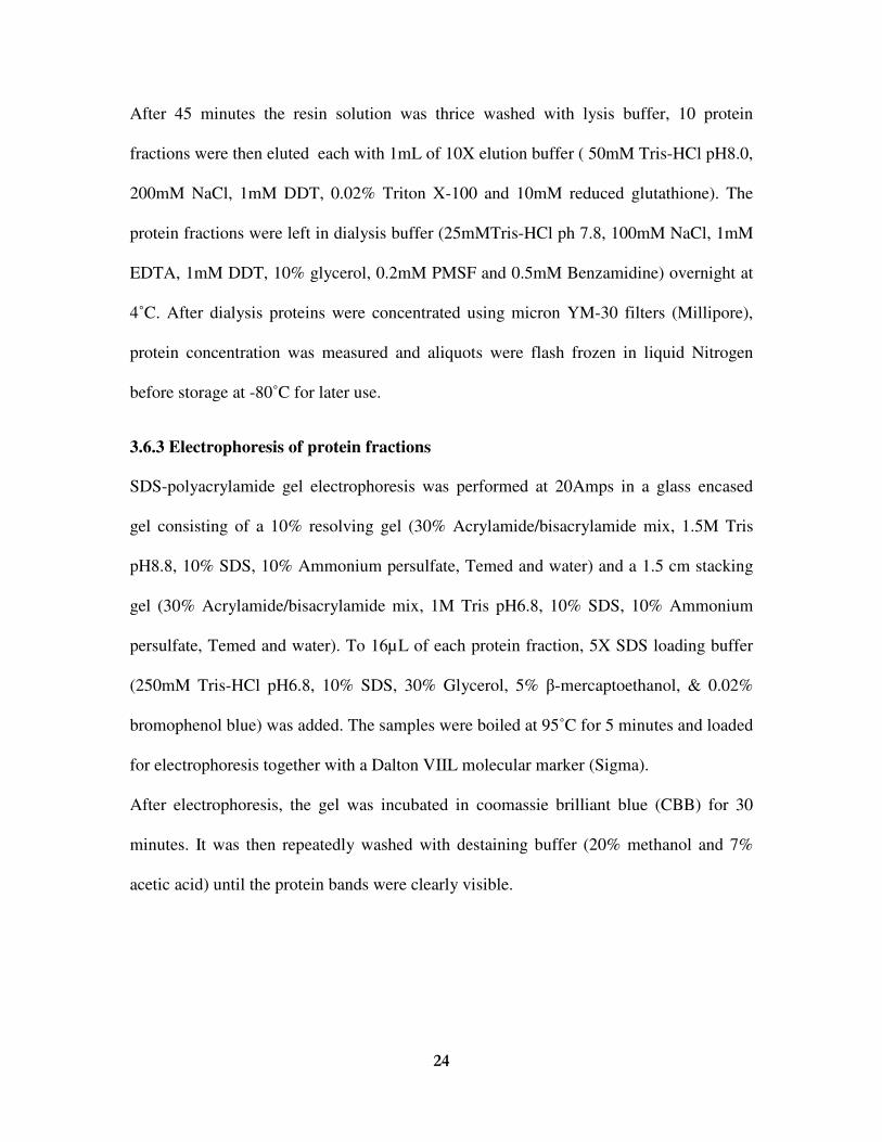
3.6.3 Electrophoresis of protein fractions
SDS-polyacrylamide gel electrophoresis was performed at 20Amps in a glass encased
gel consisting of a 10% resolving gel (30% Acrylamide/bisacrylamide mix, 1.5M Tris
pH8.8, 10% SDS, 10% Ammonium persulfate, Temed and water) and a 1.5 cm stacking
gel (30% Acrylamide/bisacrylamide mix, 1M Tris pH6.8, 10% SDS, 10% Ammonium
persulfate, Temed and water). To 16µL of each protein fraction, 5X SDS loading buffer
(250mM Tris-HCl pH6.8, 10% SDS, 30% Glycerol, 5% �-mercaptoethanol, & 0.02%
bromophenol blue) was added. The samples were boiled at 95˚C for 5 minutes and loaded
for electrophoresis together with a Dalton VIIL molecular marker (Sigma).
After electrophoresis, the gel was incubated in coomassie brilliant blue (CBB) for 30
minutes. It was then repeatedly washed with destaining buffer (20% methanol and 7%
acetic acid) until the protein bands were clearly visible.

���
�
3.7 In-vitro SUMOylation
The in-vitro assay was performed using a SUMOylation kit (Enzo, NY, USA). Four
separate reactions were set up in 1.5mL Eppendorf tubes as shown in table 2. The
reaction tubes were then incubated on a heating block at 30˚C, after 1hr the reaction was
stopped by adding 60µL of stop buffer (1X PBS, 25mMEDTA and 0.2mg/mL BSA) to
each tube. 10µL of GST-magnetic beads were then added to each tube to bind the
proteins, the reaction was conducted at 4˚C with gentle shaking. Next the magnetic beads
in each tube were thrice washed with 150µL of wash buffer (1X PBS and 0.1mg/mL
BSA) followed by protein elution with 35µL of 1X SDS sample buffer. The samples
were boiled for 5 minutes at 95˚C.
Table 2: Composition of reactions used for the in-vitro SUMOylation assay
�
3.7.1 Electrophoresis and western blot analysis
SDS-polyacrylamide gel electrophoresis was performed in a glass encased gel consisting
of an 8% resolving gel and a 1.5 cm stacking gel. 15uL of each sample were loaded into
the gel wells together with 5uL of protein molecular marker (All blue).
���
�
After electrophoresis, proteins were electrophoretically transferred to a 6.5 by 9 cm
PVDF membrane (Millipore, MA, USA) in transfer buffer (50mM Trisbase and 50mM
Boric acid) overnight at 4˚C in a trans-blot apparatus (Bio-Rad).
Subsequent to overnight protein transfer, the PVDF membrane was stained with Ponceau
S solution for 1 minute to detect protein bands after which it was destained using 1%
acetic acid. Next the membrane was incubated in blocking solution at room temperature,
after 2hrs it was rinsed with 1X TBST (50mM Tris-pH8.0, 150mM NaCl and 0.05%
Tween) and re-incubated in 4mL of blocking solution containing an anti-RBR antibody
for 2hrs. This was followed by re-incubation with a peroxidase-conjugated rabbit anti-
chicken (IgG) for 1hr.
After antibody incubation, the membrane was washed with 25mL of TBST; 25mL
blocking solution diluted with TBST, 25mL of TBST and lastly with Milli-Q water each
wash lasting 10 minutes. Finally, the immunoreactions present on the PVDF membrane
were developed onto x-ray film using enhanced chemiluminescence (ECL, Bio-Rad).
�
�

���
�
CHAPTER FOUR
RESULTS
4.1 Mutation of Arabidopsis RBR1 SUMO modification site
Modification by SUMO occurs at the �KXE sequence motif which is present in almost
all substrate proteins. The motif is made up of a large hydrophobic amino acid (�), a
Lysine (K) which is modified, an amino acid residue (X) and a glutamic acid denoted E
(Johnson, 2006). Hence in order to study the effect of SUMOylation on the
Retinoblastoma related protein (RBR1), a mutation was introduced in the nucleotides
coding for the Lysine amino acid at the SUMOylation site.
Alignment of C-terminal sequences (Fig. 3a) of RBR proteins from different plants
shows the presence of the SUMOylation motif (�KXE). The Arabidopsis RBR1 protein
carrying a mutation at the SUMOylation site was amplified by PCR-site directed
mutagenesis resulting in a 720bp fragment (Fig. 3b, Lane 2).
Figure 3 (a) Alignment of C-terminal sequences of different plant RBR proteins showing the presence of a SUMOylation motif (�KXE). (b) 720bp RBR1 fragment (Lane 2) with a mutated SUMOylation site amplified by PCR, in lane 1 is the 1Kb DNA ladder.

��
�
4.2 Effect of SUMO modification on RBR1 localization
In order to assess the effect of SUMO modification on RBR1 localization, Arabidopsis
protoplasts were transfected with plasmid DNA coding for wild type RBR1 and RBR1
carrying a mutated SUMOylation site. Both constructs contained the green fluorescent
protein to aid in protein localization.
In protoplasts transfected with wild type DNA, the protein was exclusively localized in
the nucleus (Fig.4; a-c). However, protoplasts transfected with the protein carrying a
mutated SUMOylation site, localization was not exclusive to the nucleus but also in the
cytosol (Fig.4; d-f).
�
Figure 4 Effect of SUMO on RBR1 localization in Arabidopsis protoplasts, 14hrs after transformation. (a-c) In protoplasts transformed with the wild type protein (wtRBR1-GFP), the green fluorescent protein is exclusively localized in the nucleus. (d-f) Transformants with RBR1-mtSUMO containing a mutated SUMOylation site however show that the GFP is not exclusive to the nucleus but also in the cytosol. �

��
�
4.3 Expression and Purification of protein RBR1-mtSUMO
To assess the effect of mutating Lysine 1007 on RBR1 SUMOylation, the amplified PCR
product (RBR1-mtSUMO) carrying a mutated SUMO modification site was sub cloned
into a glutathione-s-transferase (GST) gene fusion vector (pGEX5X-1) for protein
expression. Cell pellets were obtained from LB media before and after induction of
protein expression. After induction of protein expression with IPTG, RBR1-mtSUMO
(52KDa) was clearly expressed (Figure 5a, Lanes 3 and 5). The protein was subsequently
purified using glutathione Sepharose resins (Figure 5b, Lanes 3 and 4).
Figure 5 Protein expression and purification. (a) Expression of the protein RBR-mtSUMO; Lanes 2 and 4 are from samples before induction of protein expression while lanes 3 and 5 are samples taken after induction showing the expressed protein ~52KDa. (b) Protein purification; Lanes 3 and 4 show the purified protein. In both figures, lane 1 contains the molecular weight marker.

���
�
4.4 Mutation of Lysine 1007 prevents modification of RBR1 by SUMO
The purified protein was tested using an in vitro SUMOylation assay kit to determine if it
could be modified by SUMO. The assay contained RBR1-mtSUMO or RBR1-wt, SUMO
precursor, E1 enzyme, E2 enzyme and Mg-ATP. Figure 6a, shows protein transfer on to
the PVDF membrane after staining in Ponceau S solution for 1 minute. When the wild
type protein was incubated with all the components of the SUMOylation assay (Fig 6b,
Lane 2), a SUMO modified protein was observed slightly less than 75KDa. On the
contrary, the wild type protein which was incubated in the assay lacking the E1-activating
and E2-conjugating enzymes was not modified (Figure 6b, Lane 1). Even in the presence
of all assay components, the protein carrying a mutated site could not be SUMOylated
(Figure 6b, Lane 4). The same result was observed when the mutated protein was
incubated in the assay lacking the activating and conjugating enzymes (Figure 6b, Lane
3).
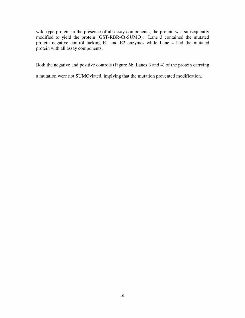
Figure 6 Ponceau staining and in-vitro SUMOylation. (a) Ponceau staining of the PVDF membrane showing protein transfers (b) In-vitro SUMOylation; Lane 1 contained the wild type protein (wt-RBR1) in the absence of the E1 and E2 enzymes. Lane 2 had the
���
�
wild type protein in the presence of all assay components; the protein was subsequently modified to yield the protein (GST-RBR-Ct-SUMO). Lane 3 contained the mutated protein negative control lacking E1 and E2 enzymes while Lane 4 had the mutated protein with all assay components. Both the negative and positive controls (Figure 6b, Lanes 3 and 4) of the protein carrying
a mutation were not SUMOylated, implying that the mutation prevented modification.

���
�
CHAPTER FIVE
DISCUSSION
5.1 Discussion
Before 1997, ubiquitin was perhaps the most known polypeptide to be involved in post
translational modification of proteins (Marx, 2005). However a number of other
polypeptides have since been indentified SUMO being among them (Kurepa et al., 2002).
Substrate proteins contain a characteristic �KXE sequence motif specific for
SUMOylation; mutation of the lysine within the motif has been shown to affect
modification and localization of RanGAP1 a nuclear transport protein in animals
(Matunis et al., 1998).
Alignment of the Arabidopsis retinoblastoma related protein (RBR1) sequence with other
plant sequences, revealed the presence of the unique SUMOylation motif in all
sequences. Hence in this study Lysine 1007 located within the motif was mutated in order
to address the effect of SUMO modification on RBR1. The outcome of the mutation was
highlighted by restricted localization of the wild type protein in the protoplast nucleus
while the protein carrying a mutation was seen to be present in both the cytosol and the
nucleus.
Johnson (2004) noted that in addition to sub cellular localization of proteins, modification
by SUMO alters protein to protein interaction and protein interaction with other
substrates. Hence localization of the wild type protein in the nucleus observed in this
study could most likely be a post SUMO modification effect caused by; interactions
between SUMO (the modifier) and nuclear proteins resulting in sub cellular localization
of the modified wild type protein.

���
�
Additionally as noted by Gill (2005), modification of transcription factors by SUMO
enhances their interactions with proteins that would normally have less or no interaction
with the unmodified protein. Hence there is still a likely possibility that after modification
of RBR1, there is an onset of interactions between the modified protein and nuclear
proteins resulting in the eventual sub-cellular localization of the protein.
Alternatively it is also possible that once sumoylated, the wild type protein undergoes
conformation changes that could expose or conceal certain binding sites. These changes
in protein conformation could play a major role in facilitating eventual localization of the
protein in the nucleus. Unlike the wild type protein, the absence of these SUMO induced
effects on the mutated protein could be the cause of its presence in the cytosol.
It is however important to highlight the fact that, a fatal error was noticed during the
course of the study. The delivered forward primer which was used for site directed
mutagenesis deviated from the native sequence as it contained a point mutation in form of
a missing T nucleotide. Putting that in consideration it is likely that the construct GFP-
RBR1-mtSUMO had an additional mutation instead of the required one at the
modification site. This without doubt results in a truncated protein, unable to be
sumoylated and hence ending up in the cytosol giving the impression that it’s the
unmodified full length protein.
Having rectified the primer problem prior to protein expression, the results from the in
vitro SUMOylation assay did clearly show that the wild type protein was SUMOylated.
However in the absence of an acceptor lysine, the protein RBR1-mtSUMO could not be

���
�
SUMOylated. This result is similar and in agreement with results obtained from a related
study by Matunis et al., (1998) where they showed that, substituting the acceptor lysine
for arginine in RanGAP1 prevented modification of the protein by SUMO.
5.2 Conclusions
In conclusion, transfection results from this study do not provide undisputed confirmation
that modification by SUMO affects sub-cellular localization of RBR1. However, the in-
vitro study did certainly confirm that, the plant retinoblastoma-related protein can’t be
modified by SUMO in the absence of the acceptor lysine contained in its modification
site.
Additionally it can also be concluded that failure of the mutated protein to undergo
modification, means that the C-terminal of RBR1 contains a single SUMOylation site.
5.3 Future Direction
Though protein localization seen in this study is in agreement with results from related
mutational studies, the first course of action would be to re-transform protoplasts with a
corrected construct in order to confirm that the mutated protein is indeed localized in both
the nucleus and the cytosol unlike the wild type protein.
In order to confirm the presence of the transiently expressed GFP-tagged protein, it can
be extracted from the transfected cells and subjected to immuno blot analysis with GFP
and RBR1 specific antibodies.
It would also be interesting to do a structural study to get a clear picture of the structures
of the wild type protein, its SUMO modified form and the mutated protein using studies
such as nuclear magnetic resonance (NMR) or X-ray crystallography. This would shade
more light on the occurrence of SUMO induced conformational changes.

���
�
REFERENCES
Bayer P, Arndt A, Metzger S, Mahajan R, Melchior F, Jaenicke R and Becker J
(1998). Structure determination o f small ubiquitin related modifier SUMO-1. J. Mol.
Biol 280(2):275-286.
Boggio R and Chiocca S (2005). Gam1 and the SUMO pathway. Cell cycle 4(4): 533-
535.
Boggio R, Colombo R, Hay R.T, Draetta G.F and Chiocca S (2004). A mechanism for
inhibiting the SUMO pathway. Mol Cell 16:549-561.
Bossis G and Melchior (2006). SUMO: regulating the regulator. Cell 1:1-13.
Brehm A and Kouzarides T (1999). Retinoblastoma protein meets chromatin. Trends
Biochem. Sci 12:2245-2262.
Dahiya A., Gavin M.R., Luo R.X. and Dean D.C (2000). Role of the LXCXE Binding
Site in Rb Function. Mol Cell Biol 20(18): 6799–6805.
De Jager S.M and Murray J.A.H (1999). Retinoblastoma proteins in plants. Plant Mol.
Bio. 41:295-299.
Desvoyes B, Elena R.P, Xie Q, Chua N.H, and Gutierrez C (2006). Cell type-specific
role of the retinoblastoma/E2F pathway during Arabidopsis leaf development. Plant
Physiol 140:67-80.
Ferreira P, Hemerly A, Van Montagu M and Inze D (1994). Control of cell
proliferation during plant development. Plant Mol. Bio 26:1289-1303.
Fobert P.R, Gaudin V, Lunness P, Coen E.S, Doonan J.H (1996). Distinct classes of
cdc2-related genes are differentially expressed during the cell division cycle in plants.
Plant Cell 8:1465-76.
���
�
Gill G (2004). SUMO and ubiquitin in the nucleus: different functions, similar
mechanisms? Genes & Dev 18:2046-2059.
Gutierrez C, Ramirez-Parra E, Castellano M.M, Del Pozo J.C (2002). G (1) to S
transition: more than a cell cycle engine switch. Curr. Opin. Plant Biol. 5: 480–486.
Harbour J.W and Dean D.C (2000). The Rb/E2F pathway: expanding roles and
emerging paradigms. Genes Dev 14:2393-2409.
Hardeland U, Steinacher R, Jiricny J and Schar P (2002). Modification of the human
thymine-DNA glycosylase by ubiquitin-like proteins facilitates enzymatic turnover.
EMBO J 21:1456–64.
Hirano H, Harashima H and Shinmyo (2008). Arabidopsis RETINOBLASTOMA-
RELATED PROTEIN 1 is involved in G1 phase cell cycle arrest caused by sucrose
starvation. Plant Mol. Biol. 66:259-275.
Hunter T (1993). Breaking the cell cycle. Cell 75:839-841.
Huntley R, Healy S, Freeman D, Lavender P, De Jager S, Greenwood J, Makkerh J,
Walker E, Jackman M, Xie Q, Bannister A.J, Kouzarides T, Gutierrez C, Doonan
J.H and Murray J.A.H (1998). The maize retinoblastoma protein homologue ZmRb-1 is
regulated during maize leaf development and displays conserved interactions with G1/S
regulators and plant cyclin D (CycD) proteins. Plant Mol. Biol. 37:155-169.
Inze D and DeVeylder L (2006). Cell cycle regulation in plant development. Annu. Rev.
Gen. 40:77-105.
Johnson E.S and Blobel G (1997). Ubc9p is the conjugation enzyme for the ubiquitin-
like protein Smt3p. J Biol Chem 272(43):26799-26802.
Johnson E.S (2004). Protein modification by SUMO. Annu. Rev. Biochem 73:355-382.

���
�
Joubes J, Chevalier C, Dudits D, Herberle-Bors E, Inze D, Umeda M and Renaundin
J.P (2000). CDK-related protein kinases in plants. Plant Mol. Biol. 43:607-20.
Kim K.I, Baek S.H and Chung C.H (2002). Versatile Protein Tag, SUMO: Its
enzymology and biological function. J.Cell. Physiol. 191:257-268.
Kurepa J., Walker J.M., Smalle J., Gosink M.M., Davis S.J., Durham T.L., Sung D-
Y., and Vierstra R.D (2002). The Small Ubiquitin-like Modifier (SUMO) Protein
Modification System in Arabidopsis. Accumulation of SUMO1 and -2 conjugates is
increased by stress. J Biol. Chem. 278: 6862–6872.
Lois L.M, Lima C.D and Chua N-H (2003). Small ubiquitin-like modifier modulates
abscisic acid signaling in Arabidopsis. Plant Cell 15:1–13.
Matunis M.J, Coutavas E. and Blobel G (1996). A novel ubiquitin-like modification
modulates the partitioning of the Ran-GTPase-activating protein RanGAP1 between the
cytosol and the nuclear pore complex. J. Cell Biol. 135:1457-1470.
Matunis M.J., Wu J and Blobel G (1998). SUMO-1 modification and its role in
targeting the Ran GTPase-activating protein, Ran GAP1, to the nuclear pore complex. J
Cell Biol 140(3):499-509.
Melchior F, Schergaut M and Pichler A (2003). SUMO: ligases, isopeptidases and
nuclear pores. Trends in Biochem. Sci. Vol.28 No.11.
Melchior F, Paschal B, Evans J and Gerace L (1993). Inhibition of nuclear protein
import by non-hydrolysable analogues of GTP and identification of the small GTPase
Ran/TC4 as an essential transport factor. J Cell Biol 123(6):1649-1659.
Novatchkova M, Budhiraja R, Coupland G, Eisenhaber F and Bachmair M (2004).
SUMO conjugation in plants. Planta 220:1-8.

��
�
Park J.A, Ahn J.W, Kim Y.K, Kim S.J, Kim W.T and Pa S.H (2005). Retinoblastoma
protein regulates cell proliferation, differentiation, and endoreduplication in plants. The
Plant Journal 42:153–163.
Pines J (1999). Four dimensional control of the cell cycle. Nature Cell Biol 1, E73 - E79.
Renaudin J.P, Doonan J.H, Freeman D, Hashimoto J, Hirt H, Inze D, Jacobs T,
Kouchi T, Rouze T, Sauter M, Savoure A, Sorrell A.D, Sundaresan V and Murray
J.A.H (1996). Plant cyclins: a unified nomenclature for plant A-, B- and D-type cyclins
based on sequence organisation. Plant Mol. Bio 32:1003-18.
Richards N.G.J (2008). Shining a light on post-translational modification. HFSP
Journal DOI: 10.2976/1.2889161.
Rodriguez M.S, Desterro J.M, Lain S, Midgley CA, Lane D.P and Hay R.T (1999).
SUMO-1 modification activates the transcriptional response of p53. EMBO J
18(22):6455-6461.
Rodriguez M.S, Dargemont C and Hay R.T (2001). SUMO-1 conjugation in vivo
requires both a consensus modification and nuclear targeting. J. Biol Chem
276(16):12654-12659.
Saitoh H and Hinchey J (2000). Functional Heterogeneity of Small Ubiquitin-related
Protein Modifiers SUMO-1 versus SUMO-2/3. J Biol. Chem. 275: 6252–6258.
Shen W.H (2007). G1/S Transition and the Rb-E2F Pathway. Springer-Verlag Berlin
Heidelberg 2007.
Smalle J and Vierstra R.D (2004). The Ubiquitin 26S proteasome proteolytic pathway.
Annu. Rev. Plant. Biol. 55:555–90.

��
�
Vandepoele K, Raes J, DeVeylder L, Rouze P, Rombauts S, Inze D (2002).Genome-
wide analysis of core cell cycle genes in Arabidopsis. Plant Cell 14:903-16.
Verger A, Perdomo J and Crossley M (2003). Modification with SUMO. EMBO
reports Vol 4 No 2.
Weinberg R.A (1995). The retinoblastoma protein and cell cycle control. Cell 81:323-
330.
Wildwaters M, Campiho A, Perez-Perez J.M, Heidstra R, Blilou I, Korthout H,
Chatterjee J, Mariconti L, Gruissem W and Scheres B (2005). The
RETINOBLASTOMA- RELATED gene regulates stem cell maintenance in Arabidopsis
roots. Cell 123:1337-1349.
Xie Q, Suarez-Lopez P and Gutierrez C (1995). Identification and analysis of
Retinoblastoma binding motif in the replication protein of a plant virus: requirement for
efficient viral DNA replication. EMBO J. 14:4073-4082.