moderate otitis externa and aural hematoma …...moderate otitis externa and aural hematoma surgical...
TRANSCRIPT

Moderate otitis externa and aural hematoma
Surgical and non-surgical options
Gary W Ellison DVM MS Diplomate ACVS
Professor and Service Chief
University of Florida 32610
The term otitis externa does not refer to a specific disease process but literally to an inflammation
of the external ear canal. The prevalence in the dog has been reported to be between 10 and 20
percent, although in more tropical climates the prevalence is 50% or more. The incidence in the
cat is less and reported to be approximately 2 to 10 percent. Otitis externa has multiple causes
including parasites, bacteria, fungi, systemic diseases, autoimmune diseases, tumors and
secondary to otitis media.
Medical vs. Surgical Management
Successful medical management of otitis depends on the accurate identification of its underlying
cause. If the primary cause of otitis externa is medical such as hypersensitivity disease, disorders
of keratinization, autoimmune disease or other systemic conditions, surgical management will fail
to produce satisfactory results unless the primary disease is treated concurrently. Surgical
management of otitis externa particularly that type which is not restricted to the vertical ear canal
often fails because of ongoing systemic disease. A good example of this is chronic proliferative
ear canal disease secondary to atopy or other hypersensitivity diseases that continues to worsen
following lateral ear resection because of continued proliferation of the horizontal canal.
Therefore, the veterinary clinician should institute proper medical therapy of allergic conditions
before and after the surgery is performed. The otic surgery should be considered part of the
therapeutic plan rather than
the curative procedure.

Patient Work-Up
Visual examination of the affected ear is best performed animal under deep sedation or general
anesthesia. Thorough ear cleaning is only readily accomplished using a suction apparatus or
aspirator which allows removal of ceruminous debris or purulent material. During otoscopic
examination the relative diameter of the ear canal is evaluated with respect to the type of
inflammation present, ie hyperplastic versus ulcerative. Whenever possible, the clinician should
attempt to determine the presence or absence of the tympanic bulla. Bacterial cultures often
reveal a "mixed bag" of contaminants. However, cultures are sometimes useful in identifying
ulcerative pathogens such as pseudomonas or coagulase positive staphylococcus.
Skull imaging should be performed on all animals where surgery is contemplated. Radiographs or
CT are often helpful in delineating the presence of narrowing of the external auditory canals, the
presence of masses or the presence of osseous metaplasia of the auricular cartilage. Evaluation of
the tympanic bullae is important to demonstrate presence of fluid or bony changes which may
indicate infectious osteomyelitis or neoplastic invasion. When fluid is present in the middle ear,
the surgeon must attempt to provide drainage in the form of a ventral or lateral bulla osteotomy in
conjunction with the primary surgical procedure.


Surgical Options
There are three common surgical procedures employed for otitis externa. 1) Lateral ear resection,
2) Vertical canal ablation or 3) Total ear canal ablation. Choice of the technique is determined via
otoscopic and radiographic examination. If the otitis externa is of the unproliferative variety
lateral wall resection or vertical canal ablation can often be effective.
Aural hematomas
Aural hematomas occur within the cartilaginous plate (intrachondral) of the auricle (pinna) of the
ear. The hematoma, which consists of blood, serum, or both, has classically been thought to result
from self-inflicted trauma to the ear. Pruritus, secondary to otitis externa, results in head shaking
or scratching. The trauma is thought to cause a shearing force that causes separation of the
cartilage of the pinna. The “hematoma” is painful and irritating to the dog or cat and causes more
head shaking and pawing at the ear. Aural hematomas occur most commonly in dogs but are
occasionally seen in cats with ear mite infestation.

An immune-mediated pathogenesis has also been hypothesized on the basis of one study where
30% of affected dogs were LE cell positive, 100% of affected dogs were Coomb's Test positive,
and 52% of affected dogs had positive antinuclear antibody titers. Dexamethasone has been
recommended, based on these papers, to control the immune-mediated component thought to be
responsible.
Aural hematomas, if left untreated, usually result in a thickened fibrous scar that causes the ear to
fold. The auricular cartilage perichondrium is highly chondrogenic. The abnormal shape is most
noticeable in breeds with erect ears.
Conservative methods of aural hematoma drainage (passive or active).
1. Needle aspiration and injection of repositol corticosteroid( video) Error! Hyperlink reference not
valid.1.
2. Larson’s teat cannula inserted in the dependent region
3. Silastic or polyvinyl drain, fenestrated and sutured proximal and distal
4. Active closed suction drainage (butterfly catheter and vacuum tube)
Each type of drain is placed into the cavity encompassing the hematoma after a small stab incision
is made. The teat cannula is placed in the dependent portion of the hematoma. The drain must be
cleansed daily to assure patency and it may be necessary to leave them in place 14-21 days prior
to removal. If the drains become clogged by exudate, or if they are removed prematurely,
recurrence of the hematoma is likely. Bandaging protects the ear from further trauma related to
head shaking and also prevents soiling of areas in the home when drainage occurs.

Incision and suturing to drain aural hematomas
Incisions made parallel to the pinna, evacuation of the fluid, and suturing parallel to the incision
line will frequently resolve aural hematomas. Too much tension on sutures may cause secondary
contracture and deformation of the ear so care should be taken when placing sutures. In large
incisions some surgeons feel a curvilinear incision does not allow contraction to deform the pinna
of the ear. A narrow gap is desired to aid maintenance of drainage from the concave surface of
the ear. Non adherent contact dressings are used. Bandaging techniques also vary but are used to
protect the ear from head shaking and to absorb drainage. Sutures are often left in place for 14 or
more days.
A modification of the suture technique includes C02 lasering of the hematoma. Reported
advantages include fusion of the cartilage layers and minimal hemorrhage.
References
1. Kuwahara J: 1986 Canine and feline aural hematoma: Clinical, experimental and
clinicopathologic observations. Am. J. Vet. Res. 47:10, p. 2300-2308,.

Otitis externa and media and inflammatory polyps in the cat
Gary W. Ellison, DVM, MS, Diplomate ACVS
University of Florida, Gainesville, FL [email protected]
Otitis externa cats may be primary or secondary. Otodectes mites are a common cause of otitis
externa in North America with up to 10% of kittens being afflicted. Secondary bacteria or yeast
infection often occurs in the presence of ear mites. Primary involvement of the external ear may
also occur with neoplastic disease, inflammatory polyps, and occasionally primary infections.
Since otitis media usually results from extension of otitis externa clinical symptoms are similar.
Ascending auditory (Eustachian) tube infections may also result in otitis media. Otitis media can
subsequently progress to otitis interna in which clinical signs such as a head tilt toward the affected
side, horizontal nystagmus, circling, and ataxia can develop. In some animals, vestibular disease
may cause vomiting. If the tympanum cannot be evaluated because of chronic proliferative otitis
externa, diagnosis of otitis media is made radiographically. Lateral, dorsoventral and open-mouth
views of the tympanic bullae should be taken. Evidence of otitis media includes thickened or lytic
tympanic bulla, bony or soft tissue density within the bulla, and in some cases a fluid line can be
seen. Radiographs are not always diagnostic for otitis media, however.
Diagnostic evaluation includes otoscopic examination, radiographs of the tympanic bulla, and
cytologic evaluation of drainage from the ear. Computerized tomography is an ideal diagnostic
technique for the middle ear as bone and soft tissue technique and use of contrast can provide
valuable diagnostic information. Proliferation of bone, lysis of bone, and in particular extension of

invasive processes toward or into the calvarium allows the clinician to avoid surgery as well as to
help plan surgery. In the cat squamous cell carcinoma of the external canal is a particularly
aggressive tumor often destroying both the external ear the middle ear and in its later phases the
calvarium.
Inflammatory Polyps in Cats:
Inflammatory polyps in cats are commonly seen in young cats less than 2 years of age although they
are reported in middle and older aged cats a well. They originate most commonly in the middle ear
near the junction of the auditory tube and the tympanic bulla. The polyps are not neoplastic and may
extend into the horizontal ear canal or into the nasopharynx via the auditory tube. In the pharynx
they may attain a large enough size to cause dysphagia or dyspnea.
The tympanum is commonly destroyed when inflammatory polyps extend out into the horizontal
canal. Although remnants of the tympanum and proliferation of scar may result in reformation of the
tympanum and partitioning of the middle ear from the external ear, persistence of this
communication may result in relapse of otitis externa and media with persisting drainage.
Surgical removal is accomplished via avulsing the polyp from the horizontal ear canal, by ventral
bulla osteotomy, or by an oral approach that may or may not require incision of the soft palate.


Ventral bulla osteotomy must also involve removal of the septum of the middle ear. The polyp
originates from the dorsolateral compartment. Curettage of both areas must be complete or
recurrence of polyps is likely. Horner's syndrome is usually transient after bulla curettage in cats (as
many as 40% can have this complication).
Ventral Bulla Osteotomy
The bulla is located between the angle of the mandible and the jugular process of the skull. Another
landmark that will aid finding the bulla is the hyoid apparatus. The tympanohyoid cartilage (attached
at the stylohyoid bone) attaches caudal to the external auditory meatus which is caudolateral to the
tympanic bulla. Once the tympanic bulla is found exposure of the boney surface can be done using a
Freer Elevator to elevate and push overlying muscle off of the bulla. Once the osseous bulla is
exposed, a hole is made in the ventral aspect using a Steinman pin, a Michelle trephine, or a high-
speed air drill (is is critical to protect surrounding soft tissues when using a drill, but the drill makes
removing bone easy and controlled). The opening in the bulla may also be widened with rongeurs.
In the Cat the septum must then be opened with a pin or ronguers and enlarged to gain access to the
dorsolateral compartment.

The contents of the bulla are cultured, a portion is saved for histopathological examination if tissue is
present, and the bulla curettage is done carefully. Drains are usually not placed.
Complications of ventral bulla osteotomy
Complications of bulla osteotomy include, injury to vital structures such as the hypoglossal or
glossopharyngeal nerves, creation of or exacerbation of otitis interna (vestibular signs), Horner’s
syndrome when sympathetic fibers are damaged, facial nerve paralysis, and hemorrhage.

Head tilts present prior to surgery, especially if chronic, may persist despite resolution of disease.
Total Ear Canal Ablation in Cats
Total ear canal ablation is necessary with either neoplasia or chronic hypertrophic otitis. The
procedure is similar to that performed in dogs except for the fact that the canal is shorter and angles
forward at a more acute angle. The facial nerve runs in more close proximity than in the dog from
caudal to cranial directly beneath the auricular cartilage. The entire ear canal is then excised with
Martin cartilage scissors. The external auditory meatus is enlarged in a ventral direction using bone
rongeurs, small osteotomes or a pneumatic drill. A small bone curette is used to remove any residual
epithelium from the rim of the opening. In some cats, TECA-LBO may be necessary to resolve
chronic otitis externa The middle ear is cultured flushed and curetted to remove residual fluid
Subcutaneous tissue and skin are closed in a T or L -shaped configuration. .Antimicrobial therapy is
based on the disease process and antimicrobial susceptibility testing.
After care is as discussed for otitis externa in dogs.
Reference
1. Anders BB et al, 2008 Ventral bulla osteotomy for removal of inflammatory polyps or
nasopharyngeal masses in cats. JAMVA 233:580

Practical Surgery of the Liver
Gary W. Ellison, DVM, MS, Diplomate ACVS
University of Florida, College of Veterinary Medicine,
Indications for Liver Surgery
Liver disease may be due to trauma, infection, inflammatory, neoplasia, toxin obstructive or congenital
disease such as that caused by portosystemic shunts (PSS). Nontraumatic surgical conditions of the
liver include liver abscesses and bleeding from ulcerated hepatocellular or cholangiocellular
carcinomas. Nontraumatic emergencies of the biliary tract are caused by biliary obstruction due to
cholangitis or cholelithiasis and gallbladder mucoceles.
Surgical emergencies of the hepatobiliary system are usually traumatically induced. Transcapsular,
subcapsular, central hepatic, or biliary tract lacerations are common. A common sequela to a traumatic
incident is the development of an intrahepatic hematoma, cyst, or abscess. These patients may present
with acute abdominal signs a significant time after injury. Treatment involves subtotal lobectomy in
most cases.
Liver Masses
Liver masses can be caused by abscesses, cysts, hepatomas or neoplasia. They can localized or
generalized.
Workup includes abdominal radiographs, ultrasound and often times a liver biopsy.

Incidence/Clinical Signs/Diagnosis
Hepatic abscesses in the dog or cat are rare. Possible etiologies include bacteria, mycotic agents, or
protozoa. The liver harbors a normal resident bacteria population with Clostridium sp. being the
predominant organism. Possible routes of infection include hematogenous spread, ascent through the
biliary tract, extension from adjacent organs, penetration by foreign bodies, or surgical manipulation.
Cysts are more common in cats and often are large cavitary masses.
Clinical signs with abscess include anorexia, vomiting, fever, and abdominal pain, often without
associated icterus. Laboratory tests may demonstrate a leukocytosis due to neutrophilia and an elevated
SGPT or serum alkaline phosphatase. Both aerobic and anaerobic blood cultures should be examined.
Radiographic signs may include lobar enlargement or the presence of gas within the liver parenchyma
secondary to gas-forming clostridial organisms. Diagnosis of hepatic abscess is usually made on an
exploratory laparotomy or laparoscopy. Hepatic cysts are sometimes found as incidental findings, but
may become extremely large and require surgical removal.
Treatment
Treatment for hepatic abscesses involves surgical drainage using omentum or complete excision of the
abscess within the offending lobe. Because of the large hepatic reserve, complete removal of the
affected lobe is recommended, if possible. Aerobic and anaerobic cultures are taken from the abscess
cavity. Because clostridia organisms are always suspected, one of the penicillin-derivative antibiotics
are administered intravenously, unless results of bacterial culture and sensitivity dictate otherwise.
Treatment for hepatic cysts requires removal of the offending lobe because drainage will result in
recurrence of the lesion.
Hepatic Neoplasia
Incidence/Clinical Signs
The liver may be involved in both primary and metastatic neoplasms. The order of frequency of
primary tumors is hepatocellular carcinoma, hepatoma, and cholangiocarcinoma.
Fibroma/fibrosarcoma, hemangioma/hemangiosarcoma, and hematomas are less common. Clinical
signs include anorexia, vomiting, polyuria, polydipsia, and hepatomegaly. If ulceration and
hemorrhage have occurred, progressive anemia, pale mucous membranes, and hemoperitoneum may
necessitate emergency surgical intervention.

Prognosis varies with the tumor type. Hepatomas (adenomas) and hepatocellular carcinomas may grow
slowly and be confined to single liver lobes, making surgical excision feasible. Cholangiocarcinomas
are often associated with weight loss and clinical jaundice.
More than one lobe is often affected, and some cases become inoperable. Death usually results within
a few months of diagnosis with cholangiocarcinomas.

Diagnosis
Serum chemistries may show profound changes. With hepatocellular carcinoma, SGPT and serum
alkaline phosphatase may be increased 10-20-fold. Hypoalbuminemia (less than 2.0 g/dl),
hypergamma-globulinemia, hypoglycemia, and hyperbilirubinemia are also commonly noted.
Diagnosis of hepatic neoplasia is suspected, based upon palpation and the radiographic presence of a
cranial abdominal mass. Displacement of the axis of the stomach caudally is commonly seen.
Occasionally the mass may be superimposed over the spleen, making differentiation between splenic
and hepatic neoplasms difficult.
Liver Biopsy
Liver biopsy is indicated for those cases where neoplasia is suspected in multiple lobes and resection
is not possible.
Finger Fracture Technique - With this technique Glissons capsule and the parenchyma is gently
fractured with fingers or a Carmalt forceps. The vessels and biliary ducts are identified and double
ligated.
Guillotine Technique - With this technique, a peripheral liver lobe is isolated and a length of suture
material is used to cut through the capsule and parenchyma thereby capturing the vessel. Mattress
sutures can also be used. The biopsy is then excised distal to the ligature(s).

Biopsy Punch Technique - Using a Bakers biopsy punch a circular piece of tissue can be secured
from the central portion of the lobe. A single horizontal or cruciate mattress suture is then passed
through the liver capsule and parenchyma to close the defect and stop hemorrhage.
Needle Biopsy Technique - a variety of commercial needles including a Trucut, micro Trucut or Vim
Silverman needle can be used to obtain biopsies from any portion of the liver lobe. After securing the
biopsy gentle digital pressure is applied using a dry sponge until bleeding stops.
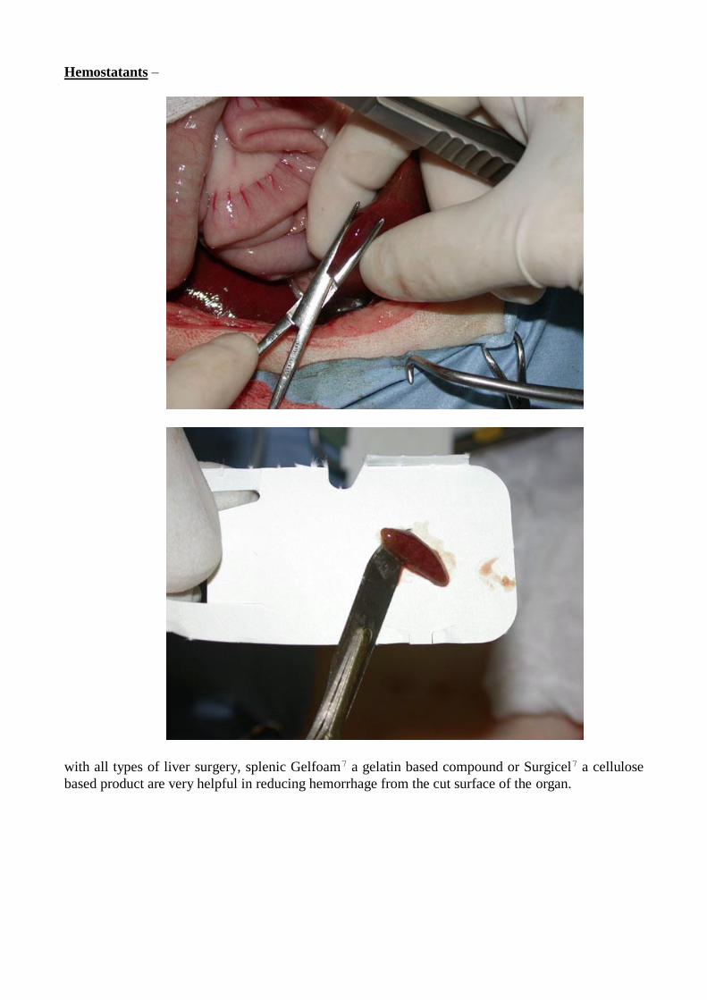
Hemostatants –
with all types of liver surgery, splenic Gelfoam7 a gelatin based compound or Surgicel7 a cellulose
based product are very helpful in reducing hemorrhage from the cut surface of the organ.
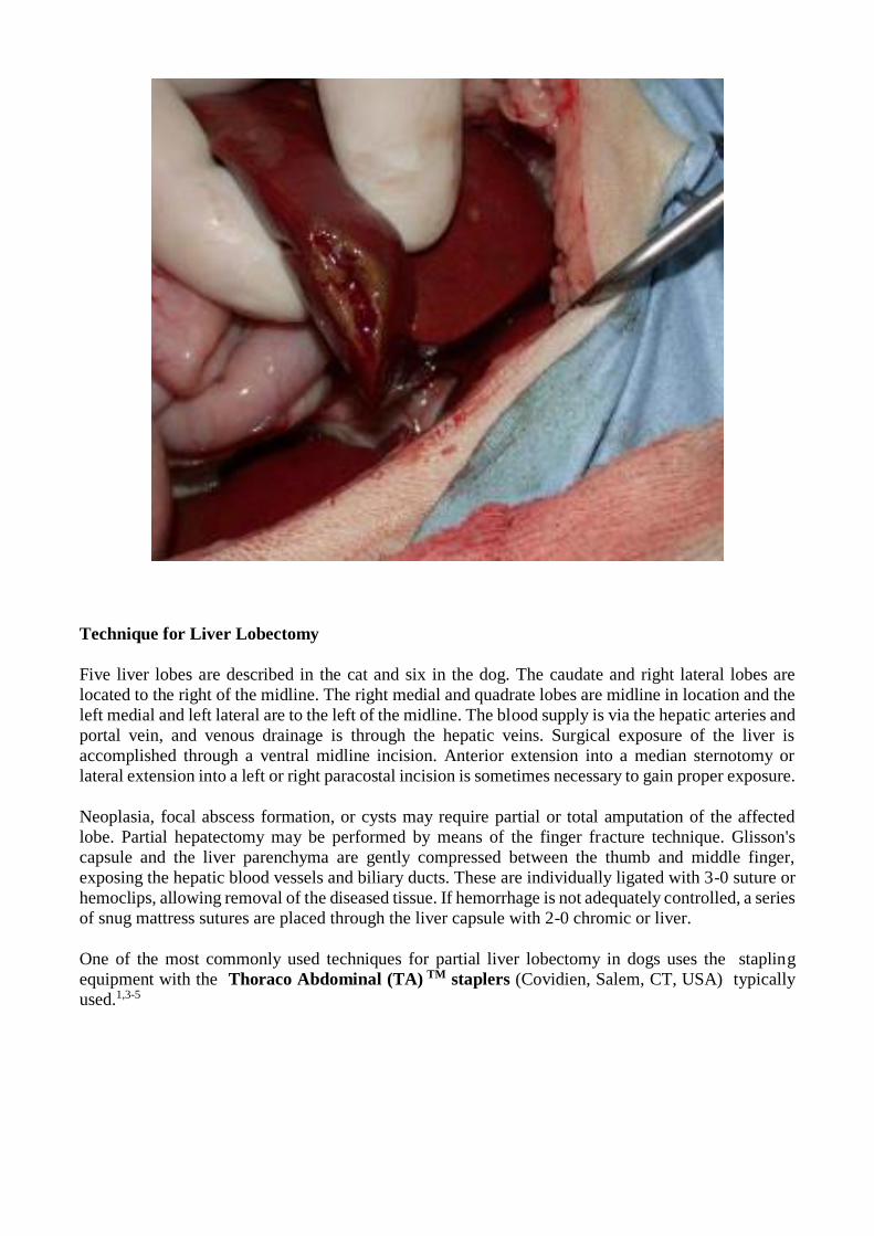
Technique for Liver Lobectomy
Five liver lobes are described in the cat and six in the dog. The caudate and right lateral lobes are
located to the right of the midline. The right medial and quadrate lobes are midline in location and the
left medial and left lateral are to the left of the midline. The blood supply is via the hepatic arteries and
portal vein, and venous drainage is through the hepatic veins. Surgical exposure of the liver is
accomplished through a ventral midline incision. Anterior extension into a median sternotomy or
lateral extension into a left or right paracostal incision is sometimes necessary to gain proper exposure.
Neoplasia, focal abscess formation, or cysts may require partial or total amputation of the affected
lobe. Partial hepatectomy may be performed by means of the finger fracture technique. Glisson's
capsule and the liver parenchyma are gently compressed between the thumb and middle finger,
exposing the hepatic blood vessels and biliary ducts. These are individually ligated with 3-0 suture or
hemoclips, allowing removal of the diseased tissue. If hemorrhage is not adequately controlled, a series
of snug mattress sutures are placed through the liver capsule with 2-0 chromic or liver.
One of the most commonly used techniques for partial liver lobectomy in dogs uses the stapling
equipment with the Thoraco Abdominal (TA) TM staplers (Covidien, Salem, CT, USA) typically
used.1,3-5

However, in some liver lobes, such as the left and right lateral, applying a TA stapler across the lobe
is nearly impossible due to the thickness of the lobe. If the lobe is too thick to apply the stapler,
skeletonization of the vessels can be performed using the inner tube of a Poole suction tip. The
lobectomy is then facilitated via application of hemostatic clips on the individual vessels or application
of a TA stapler across the entire skeletonized portion.
Several techniques developed for and used in laparoscopic surgery also have found acceptance in open
liver surgery. Among those are vessel sealant devices like the LigaSureTM (Covidien, Salem, CT,
USA) a high current low-voltage radiofrequency energy based device, and the Ultracision TM
Harmonic Scalpel (UAS, Ultracision Ethicon Endosurgery, Cincinnati, OH, USA), based on ultrasonic
principles. These work well but are expensive and not typically seen outside of specialty practices.

A practical and affordable alternative for practitioners for partial liver lobectomy is an encircling
ligature devices such as the Surgitie TM (Covidien, Salem, CT, USA) It has been used have been used
in dogs and cats for partial liver lobectomy. The SurgiTieTM consists of a pre-fabricated lasso like
loop of Polysorb TM on a single use applicator. After sliding it over the lobe to the point of resection
the applicator tip is broken off and the loop is pulled closed after which the remaining suture is
transected and the applicator is removed.
The suture loop is closed as tightly as possible without feeling a clearly defined closing of the vessels
or endpoint. Some resistance was felt when the suture cuts through the capsule, especially in the larger
lobes. Potentially, if used for mid lobar resections in the larger lobes (left lateral or right lateral)
attention should be paid to avoid doubling over of the lobe unto itself as this would increase the amount
of tissue and liver capsule the SurgiTieTM needs to close through. The SurgiTieTM also leaves a cuff of
tissue distally to the ligature, this aids in coagulation, but might also potentially decrease the margins
in surgery for neoplastic lesions.
Regardless of the technique used central venous bleeding is not uncommon and either a ligature or an
individual hemostatic clip must be used to stop the hemorrhage. The application of additional clips
and/or sutures to lobes that continue to bleed can be seen as reducing the possible total amount of
blood loss for that surgical procedure. Other methods of means of controlling hemorrhage include the
during a liver bleed include application of direct pressure on the wound, application of hemostatants
such as gelatin sponge, cellulose mesh or collagen sheeting or utilization of the Pringle maneuver.
In summary all five techniques have been used successfully in clinical practice at the University of
Florida. No significant difference has been found in surgical time between the five techniques. The
Suction+Clip technique has been associated with a higher amount of blood loss than the other
techniques.

How Manage Salivary Mucoceles
Gary W. Ellison, DVM, MS, Diplomate ACVS
University of Florida, Gainesville, FL [email protected]
Salivary mucoceles are accumulations of salivary secretions that have leaked from damaged salivary glands or
ducts into the soft tissues. Salivary mucoceles have also been referred to as sialoceles and salivary cysts. The
latter term is incorrect. The lining of a sialocele is not secretory nor is it epithelial, both of which are required
to qualify the mucocele as a true cyst. Instead, the lining of the mucocele is composed of fibrous granulation
tissue which results from the tissue's reaction to the presence of a foreign substance.
Incidence
Mucoceles are often seen in young mature dogs (2-4 years of age). Poodles and German Shepherds are at
increased risk and males are affected more often than females. Greater than 90% of the mucoceles are due to
sublingual gland or duct defects and 50% of the dogs also have a concurrent ranula.3 Parotid and zygomatic
mucoceles are rare.
Etiology
The exact etiology of salivary mucoceles remains obscure; however, trauma or inflammation of the sublingual
gland or duct inclusive of sialoliths and foreign bodies have been suggested. Ligation of the duct or incision
into the gland does not create a mucocele experimentally. The three basic types of mucoceles are: 1) cervical
mucoceles which are located subcutaneously in the ventral or ventrolateral intermandibular area of the neck, 2)
ranulas which are located along the base of the tongue,

and 3) pharyngeal mucoceles which are located in the wall of the pharynx or paratonsilar area.

Clinical Signs
A soft fluctuant intermandibular swelling is usually present with cervical mucoceles. Early in the course of the
disease, the area of swelling usually lateralizes to one side or the other, but with time, may gravitate to the
midline making identification of the affected side difficult. The swelling is usually non-painful and not infected
unless prior aspiration or drainage has been performed. With ranulas, ptyalism (excessive salivation) or
occasionally dysphagia (difficulty with eating) are noted due to sublingual swelling. Dogs with pharyngeal
mucoceles usually have a history of dysphagia but may also present in respiratory distress due to glottic
obstruction (a surgical emergency). Regardless of the initial type of mucocele identified, all areas of potential
occurrence should be evaluated.
Diagnosis
Aspiration of the mucocele usually reveals clear, translucent or blood tinged tenacious fluid which on cytology
is rich in mucin.
Sialography may identify the site of the ductal or glandular tear but cannulation of the duct is often difficult to
perform. Positive contrast material injected directly into the mucocele sometimes will reveal its communication
with the affected gland.

Determining the affected side is often difficult when the cervical mucocele is on the midline. A reliable history
is useful but the presence of a unilateral ranula or pharyngeal mucocele will sometimes create a bulge next to
the tongue on the affected side. If identification of the affected side is not possible, bilateral resection of the
sublingual and mandibular salivary gland is permissible. Adequate production of saliva will be maintained by
the remaining glands. Applying pressure to a cervical mucocele may increase the size of an otherwise inapparent
ranula assisting in lateralization of the disease.
Treatment
Definitive treatment of cervical mucoceles involves resection of the offending gland and drainage of the
mucocele. The sublingual gland closely intermingles with the rostral portion of the mandibular gland and its
duct necessitating removal of both glands simultaneously. Resection, cautery, aspirations or drainage of the
mucocele alone without gland removal will usually lead to recurrence.
Surgical Anatomy
The mandibular salivary gland is an oval, encapsulated, mixed (serous and mucous) gland located at the junction
of the maxillary and linguofacial veins as they empty into the jugular vein. The duct of the mandibular salivary
gland runs beside the sublingual gland and opens into the oral cavity at the base of the lingual frenulum. The
sublingual gland is a multilobulated mixed gland. It is divided into two portions. The monostomatic part which
begins at the rostral portion of the mandibular gland and runs along its duct and the polystomatic (multiple
microscopic openings into the oral cavity) portion which lies along the base of the tongue, medial to the body
of the mandible.

Surgical Technique - Unilateral Excision of Mandibular and Sublingual Salivary Glands
A 6 cm skin incision is made extending rostrally from the junction of the maxillary and linguofacial veins over
the mandibular and sublingual salivary glands. The incision is continued through the platysma muscle and
fibrous capsule of the mandibular salivary gland. The gland is separated from its capsule by blunt dissection
and grasped with an Allis tissue forceps. Glandular arteries are encountered dorsomedially and are ligated or
cauterized. Caudolateral traction is applied while sharp and blunt dissection around the sublingual glandular
chain is performed in a craniomedial direction.
Rostral dissection is facilitated by excising the bulk of the glandular tissue and passing the duct and remaining
sublingual gland medially and rostrally under the belly of the digastricus muscle with Carmalt forceps. The
external carotid artery and lingual nerve are carefully avoided. If a communicating isthmus between the duct
and mucocele is found, ligation of the duct distal to the communication is performed. Otherwise, the duct is
ligated at its most rostral point of dissection which is usually limited by the presence of the lingual nerve. The
gland capsule, platysma muscle, subcutaneous tissue and skin are closed in routine fashion. A stab incision is
placed in the cervical mucocele and a penrose drain is retained for 72 hours. Redundant skin is rarely a problem
after drain removal.
Surgical Treatment of Ranulas and Pharyngeal Mucoceles
The most dependable method of treating ranulas or pharyngeal mucoceles is to excise the offending sublingual
and mandibular salivary glands and establish drainage of the offending mucocele. Ranulas and pharyngeal
mucoceles associated with concurrent cervical mucoceles must be handled in this manner. Marsupialization
alone may be attempted if no concurrent cervical mucocele is apparent; however, this method usually results in
recurrence and is not recommended.

Marsupialization is accomplished by creating a 1 cm round or oval incision removing oral mucosa. The mucosa
is sutured to the sac lining with 3-0 chromic catgut in a continuous pattern providing an orifice for drainage. If
the ranula or pharyngeal mucocele returns, the mandibular and sublingual glands on the affected side need to
be excised. Remember marsupialization is not advised and is generally only a temporary solution.

Recurrence of Mucoceles
Recurrence of mucoceles is due to one of the following: 1) surgical removal of the unaffected mandibular and
sublingual salivary glands (wrong side), 2) failure of marsupialization openings to remain patent, or 3) the
glands or ducts were not dissected far enough rostrally and residual salivary tissue was not excised. Some
patients require oral resection of the rostral polystomatic portion of the sublingual gland. Identification of
residual tissue may be assisted by injecting methylene blue dye into the sublingual duct. An incision is made
between the tongue and vertical ramus of the mandible. The gland lies between the styloglossal and mylohyoid
muscles. The gland is freed by blunt dissection taking care to avoid the hypoglossal nerve and lingual nerve and
artery. When dissection is complete, the oral mucosa is closed with simple continuous 4-0 sutures. Infections
are rarely encountered.

GASTRIC DILATATION VOLVULUS (GDV) AN UPDATE
Gary W Ellison DVM MS Dipl ACVS
College of Veterinary Medicine
University of Florida, Gainesville, FL
Gastric dilatation volvulus complex also known as bloat is a medical and surgical emergency, which
is known to primarily affect large and giant breeds of dogs. The disease has also been reported in
smaller breeds such as the Pekingese and Dachshund. Mortality has been estimated as high as 30%.
There are no reliable estimates of how many dogs develop bloat in the United States each year, but in
certain breeds such as Irish Setters and Great Danes owners reported an incidence of 7 and 10%
respectively. It does appear that purebred dogs are more likely to develop bloat than are mixed breed
dogs.
Incidence
Several recent reviews by Dr. Larry Glickman at Purdue University utilized information from the
veterinary medical database (VMDB) have discovered some interesting findings. #1) amongst
veterinary institutions the frequency of bloat amongst all dogs ranged from 2.9-6.8 per 1,000 dogs. #2)
approximately 29% of the dogs with gastric dilatation and 33% of those with dilatation and volvulus
died. #3) aging of the dog increased risk. Dogs greater than 7 years of age are more than twice as likely
to have bloat as dogs 2-4 years of age. #4) purebreds were 3 times as likely to have bloat as mixed
breed dogs. #5) males are twice as likely to bloat as females yet spaying or neutering has no effect on
the risk of bloat.
Breed
GDV has been long reported to be more common in large and giant breeds of dogs yet until recently
the prevalence of bloat was not compared to the dog’s population at large. When this data was analyzed
statistically, it was found that the Great Dane, Saint Bernard, Weimaraner, Irish Setter, and Gordon
Setter were breeds at greatest risks. An accompanying chart outlines the remainder of the breeds.
Chest Conformation
Although it is established that large and giant breeds are the breeds at greatest risk it has been shown
there are profound differences in the risk of bloat within certain breeds. This possibility seems
related to the conformation of the animal=s chest. For instance, breeds such as Irish Setters, which
are at high risk, may weigh approximately the same as some of the Retriever breeds yet the
Retrievers are at much lower risk than Irish Setters for developing bloat.

It appears that the chest depth/width ratio is highly correlated with risk of bloat, ie. Those animals with
deep narrow chests within a certain breed are much more likely to develop bloat than those dogs with
deep wide chests. In using external measurements of chest conformation, it was found that within the
Great Dane breed the depth/width ratio may indeed be useful in identifying animals prone to bloat.
Also, Great Danes with moderate and high abdominal height to width ratios were approximately 52 to
8 times as likely to develop bloat as those with low abdominal height to width ratios. In Irish Setters,
the chest height to width ratio also correlated with those dogs having a higher depth to width ratio
being much more likely to develop bloat than those animals with a lower depth to width ratio. This
information is obviously very significant in terms of selective breeding for the reduction of bloat in
these breeds.
Diet
Exact determinations of types of diet on risk for developing bloat still cannot be made. Although
cereal-based have been incriminated, it is difficult to compare groups since almost all large and giant
breeds are fed cereal-based diets. Therefore, further controlled studies will be necessary to determine
if cereal-based diets are risk in fact a factor. However, several interesting findings have come to surface
with regard to the diet and nutritional management of breeds predisposed to bloat. For instance, it has
been shown that dogs that eat one meal a day are almost twice as likely to develop bloat as those fed
twice a day. The rate of eating is also very important. Those dogs characterized as slow eaters have
the lowest incidence of bloat whereas those dogs characterized as moderately fast eaters have about
22 times the chance of developing bloat and those characterized as fast eaters have almost 5 times the
chance of developing bloat as those being characterized as slow eaters.
Body weight may also be of some significance. Being overweight actually reduced the incidence of
bloat compared to dogs that were optimum weight. However, those animals characterized as
significantly underweight were about 3 times as likely to develop bloat as those animals characterized
as optimum weight.

Personality and Environment
There does seem to be a direct correlation of the animal’s temperament relating to its tendency to
develop bloat. Those animals being characterized as unhappy or fearful were about 22 times as likely
to develop bloat as those animals characterized as happy. In addition, the environment may play a role.
Stress appears to also significantly increase the chance of the animal developing bloat. Therefore,
animals that may undergo significant stress traveling to show, etc. are two to three times as likely to
bloat as those animals that are not significantly affected by the transport. Also activity level may be
important with those animals characterized as hyperactive and those animals being categorized as less
active were twice as likely to develop bloat as those animals characterized as having a normal activity
level.
Clinical Signs
Dogs usually demonstrate hyper salivation, retching or unproductive vomiting on presentation. Cranial
abdominal distention is apparent and gastric tympani are usually present on blunt percussion of the
right anterior quadrant. Hyperpnea or dyspnea accompanied by open mouth breathing indicates
hypoxia due to reduced diaphragmatic excursions. Shock is evidenced by pale or injected mucous
membranes, prolonged capillary perfusion, tachycardia and weak rapid femoral pulse.
Mechanisms of Rotation
A lack of coordinated gastric contractions due to gastric myoelectric dysrhythmias may slow gastric
emptying and contribute to the development of gastric dilatation volvulus (GDV). Food and fluid
distension from overeating or gaseous distension from aerophagia causes intra-abdominal angulation
of the gastroesophageal junction, which prevents belching or vomiting. Gastric dilatation results.
Volvulus occurs when the dilated gastric fundus becomes displaced from a left dorsal to a right ventral
position. The pylorus concurrently shifts from its right ventral position to a left, caudal and dorsal
position. When viewed from the rear a clockwise rotation occurs in the majority of the animals. The
spleen follows the greater curvature to the right. The gastro splenic ligament and short gastric arteries
are often torn during the volvulus.
Initial Medical Management of GDV
Initial lab work should include a CBC panel and coagulation panel. Dogs with gastric necrosis often
are in DIC and may have prolonged prothrombin time (PT); partial thromboplastic time (PTT) reduced
platelets, antithrombin III and an increase in fibrin degradation products (FDP). A more sensitive test
is for blood lactate. Those animals having values greater than 6 almost always have gastric necrosis.
Those animals with values less than 9 have a higher survival rate.
Initial patient management involves shock therapy, and gastric decompression followed by
management of cardiac arrhythmias. Shock therapy involves fluid loading with 90 ml/kg of lactated
Ringers solution over the first hour. The use of hypertonic saline may also be beneficial, as it has been
shown to be beneficial in increasing gastric arterial perfusion. Treatment for acid-base status is
controversial with one study indicating normal pH and another indicating the presence of metabolic
acidosis in cases of GDV. However, with mild metabolic acidosis Na bicarbonate infusion is not
necessary as long as adequate volume replacement with lactated Ringers solution is achieved.
Hypokalemia is a common finding associated with GDV and potassium replacement is sometimes
warranted. Cardiac dysrhythmias are commonly seen and require careful pre and postoperative
management. Paroxysmal ventricular tachycardia and premature ventricular contractions are most
commonly seen.

Gastric decompression is accomplished using a premeasured well-lubricated PVC plastic foal
nasogastric tube. Ability to pass the tube into the stomach does not mean that gastric volvulus is not
present. If intubation is not possible in the prone position, it is attempted in a sitting upright position.
Sometimes trocharization is necessary to reduce distension and facilitate tube passage. The character
of the fluid is sometimes important in predicting the status of the gastric lining. Black fetid smelling
fluid with flecks of devitalized mucosa indicates that mucosal ischemia is present and often predicts
the presence of gastric wall necrosis. After decompression, the stomach is lavaged with 4-5 liters of
H2O using gravity flow, dose syringe or stomach pump.
Radiography
Radiography is always postponed until after patient stabilization. With gastric dilatation, the stomach
appears as a grossly distended gas and fluid filled structure, which occupies the cranial abdomen
displacing all remaining viscera posteriorly. The spleen is usually not visible in its normal left ventral
location and is often located in a right dorsal position. Gastric volvulus is suspected when the pylorus
is located dorsal, cranial and to the left of the midline. After decompression it may take a classic
"upside down" appearance. Left and right lateral views are recommended. On the right lateral view
gas can be seen in the pylorus whereas on the left lateral view gas may be seen in the fundus. If stomach
position is questionable barium sulfate is administered to identify the pylorus.

Surgical Management
Definitive management of GDV involves 1) repositioning of the stomach with resection or involution
of any devitalized gastric wall
and 2) a prophylactic gastropexy technique to prevent recurrence. Up to 80% recurrence of GDV is
reported with gastric decompression or repositioning alone. We now advocate laparotomy as soon as
the patient is a reasonable anesthetic risk. This allows early derotation, which increases circulation and
allows assessment of gastric wall viability. Areas of necrosis are detected early and resected if possible.
With 270 to 360 clockwise gastric volvulus the dilated stomach is covered on its ventral aspect by
omentum. Reduction is accompanied by passing the hand down the left abdominal wall, grasping the
pylorus in its left dorsal position and rotating it in a caudal and counter-clockwise manner to its normal
right sided location.

Gastrectomy Techniques
Standard methods for gastrectomy involve ligation of branches of the left gastroepiploic arteries and
veins allowing areas along the greater curvature of the stomach to be resected.
The stomach is resected back to areas of healthy bleeding. Spillage is likely and prevented with
Babcock forceps or stay sutures. After resection is complete the stomach is closed in 2 layers. The
mucosa and submucosa are closed with a continuous inverted Cushing pattern of 2-0 or 3-0 PDS or
Maxon. The serosa and muscularis are then closed with a similar pattern. Recently we have relied
heavily on the autostapling equipment for rapid gastrectomy procedures with minimum risk of spillage.
The TA 90 autostapler is used with the green (4.8 mm) or blue (3.5 mm) cartridge. Often several end-
to-end staple lines have to be placed since each staple line is only 9 cm in length. The surgeon needs
to overlap the staple lines by a few mm to prevent leakage between the staples.

Gastric Involution
With this technique, the devitalized areas of stomach are invaginated and allowed to slough into the
lumen after necrosis. This procedure is performed by inventing the devitalized tissue with a Lembert
pattern using 2- or 3-0 PDS.
Postoperatively the dog must be fed small amounts of food multiple times daily for several weeks until
the stomach volume expands. We have found this technique very useful for us in dogs with smaller
areas of gastric necrosis.
Rational for Gastropexy
By definition, gastropexy describes the fixation of the stomach to nearby structures or body wall as a
means of preventing recurrence of GDV. Although gastropexy procedures reportedly diminish the
recurrence rate of GDV, their reliability in producing permanent adhesions between the stomach and
abdominal wall is not well documented.
Most North American surgeons use an antral gastropexy procedure to fix the gastric antrum to the right
abdominal wall. The 3 major categories of "permanent" antral gastropexy used in North America are
the tube gastrostomy described by Parks (1976); the incisional gastropexy described by McCoy (1982)
and the Circumcostal gastropexy described by Fallah (1982). In addition, two modifications of muscle
flap techniques one using a "muscular flap" from the abdominal wall (Shulman 1986) and another
using a "belt-loop" from the gastric muscularis (Whitney 1989) has recently been described.

Clinical Results
Potential advantages of the tube gastropexy are that 1) the surgery is rapid and easy, 2) that the tube
not only creates a permanent adhesion of the gastric antrum to the abdominal wall preventing
recurrence of volvulus but also 3) allows for continued gastric decompression in the early postoperative
period. In addition, 4) slurried food or medications can be offered through the tube. The main
disadvantages of the technique are the 1) nursing care and long hospital period required for tube
management and the potential for 2) fatal peritonitis secondary to leakage around the tube or early
removal by the dog.
Clinical studies of the tube gastrostomy have yielded encouraging results. Flanders (1984) reported
recurrence of volvulus in only 1 of 29 dogs treated with tube gastrostomy for a follow-up time ranging
from 14 to 40 months. However there was a mortality rate of 31% during the first week after surgery.
Johnson (1984) reported on 76 cases where this technique was used with only a 5% recurrence rate.
Older studies describe a recurrence rate as high as 29% (Walshaw 1976) as well as a 17% complication
rate (Fox 1985) including, premature dislodging of a tube, peritonitis, subcutaneous cellulitis or
persistent stoma drainage.
Advantages of the incisional gastropexy are that 1) the procedure is rapidly done, 2) the stomach lumen
is not entered and 3) fibrous connective tissue enters the abdominal rectus muscle
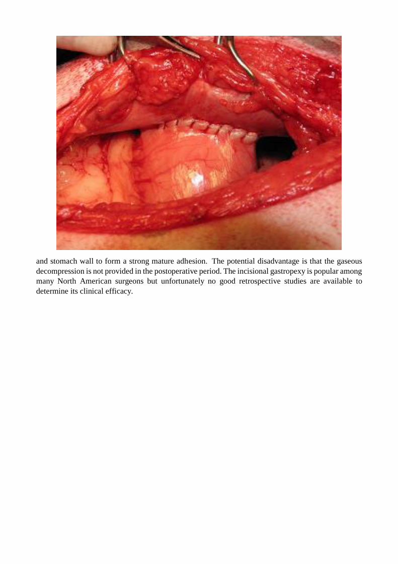
and stomach wall to form a strong mature adhesion. The potential disadvantage is that the gaseous
decompression is not provided in the postoperative period. The incisional gastropexy is popular among
many North American surgeons but unfortunately no good retrospective studies are available to
determine its clinical efficacy.

The Circumcostal technique has become popular for use in academic medicine because it probably
forms a stronger adhesion. It is reported to be more difficult to perform than the other techniques but
the author disagrees with this statement
.
Potential advantages include a 1) viable muscle flap adhesion as well as a 2) more proper anatomic
placement of the stomach. Potential disadvantages include a prolonged surgical time, potential for rib
fracture and potential for pneumothorax because of the close proximity to the diaphragm. Lieb (1984)
reported on 39 dogs with Circumcostal gastropexies to have a slightly lower recurrent rate (2.6% at
13.7 months) than dogs with tube gastrostomy.
Belt loop gastropexy offers similar advantages to the Circumcostal and incisional gastropexy in that
the gastric lumen is not entered and the risk of peritonitis is minimal. The technique is easily performed
by an unassisted surgeon. Although the belt loop gastropexy has not been evaluated biomechanically
one would suspect that breaking strengths would be superior to incisional or tube gastrostomy
techniques but not quite as secure as Circumcostal techniques since the base of the flap is narrower
than the latter technique.

Ventral midline gastropexy can be performed in dogs where anesthesia time is critical. The gastric
serosa is roughened with a surgical sponge and included with the linea alba during midline closure.
It is a popular technique in Europe. The obvious disadvantage is that a future midline laparotomy must
be done carefully or a gastrotomy incision might occur.
Laparoscopic assisted gastropexy – this is a “prophylactic procedure” commonly performed to prevent
the development of GDV in dogs predisposed to the condition. A video will be shown during lecture
to display the technique.
Postoperative Management Diligent postoperative care is mandatory for successful outcome of the
gastric dilatation volvulus patient. Most dogs that die in the postoperative period will do so within the
first 3-4 days after surgery. After major gastric resection the animal is kept NPO for a period of 24-48
hours. Maintenance fluid, electrolyte and acid base status is critical during this period. Maintenance
fluid should be given at a rate of 40-60 ml/kg per day. Although many dogs maintain normal serum
potassium levels following gastric dilatation volvulus a total body potassium deficit may exist because
of the NPO status, vomiting, oral gastric innervation and removal of gastric secretions. Therefore,
supplementation of 20 mEq of potassium chloride is usually added to each liter of fluids to help

maintain total body potassium. Hypokalemia can also contribute to the development of cardiac
arrhythmias, and gastrointestinal ileus.
Further Reading:
1. Small Animal Surgery, 4th ed. Fossum, ed. Mosby, St. Louis, 2014, pp 468-477.
2. Current Techniques in Small Animal Surgery, 5th ed. Bojrab, 2015, pp 263-275.
3. Glickman, L.T., Glickman, N.W., Shellenburg, D.B., et al. (1997), “Multiple Risk Factors for
the Gastric Dilatation Volvulus Syndrome in Dogs: A Practitioner/Owner Case Control
Study”, J. Am. Anim. Hosp. Assoc. 133:197-204.
4. Ellison, G.W. (1993), “Gastric Dilatation Volvulus: Surgical Prevention”, Vet. Clin. N. Am.
27:513-521.

STOPPING THE LEAKING INTESTINAL ANASTOMOSES; CAUSES AND TIPS FOR
PREVENTION
Gary W Ellison DVM MS Dipl ACVS
College of Veterinary Medicine
University of Florida, Gainesville, FL
Wound dehiscence of an intestinal anastomosis often leads to generalized bacterial peritonitis and
subsequent death. Therefore, factors which negatively affect visceral healing are potentially of great
clinical significance to the surgeon. Factors that cause intestinal anastomoses to leak include etiology
of obstruction, failure to adequately identify ischemic tissue, improper suturing or stapling technique
and factors that negatively effect wound healing such as sepsis, malnutrition and anti-neoplastic
therapy.
Importance of tissue apposition
Direct approximation of the wound edge allows for optimum rapid healing characterized by primary
intestinal wound healing. With good apposition rapid mucosal re-epithelialization and early formation
of young well-vascularized collagen between the submucosa, muscularis and serosa occurs.
Figure 1: example of an intestinal wound at 3 days postoperatively with using appositional sutures.
Note the good alignment of the mucosa, submucosa and muscularis with a fibrin seal on the serosal
surface.
Other advantages of approximating patterns for intestinal anastomosis are: 1) lumen diameter is not
compromised, 2) wound strength meets or exceeds everting or inverting wound strengths, and 3)

adhesions are minimal. The crushing suture has been shown to cause more tissue ischemia directly at
the suture line and its use is discouraged.
Figure 2: example of an intestinal wound at 3 days postoperative with crushing sutures applied to the
wound edges. Note the hemorrhage and necrosis within the muscularis and incomplete establishment
of mucosal integrity.
Mucosal eversion or tissue overlap retards healing and should be avoided. Delayed fibrin seal
formation, delayed mucosal re-epithelialization, increased mucocele formation, prolonged
inflammatory response, and marked adhesion formation all characterize everted healing. Eversion may

initially widen the lumen diameter, but the prolonged inflammatory response usually narrows the
lumen sometimes resulting in stenosis.
Figure 3: example of an intestinal wound at 7 days reveals mucosal eversion and poor tissue apposition
resulting in mucocele formation and reduced lumen diameter. Everting anastomoses also have an
increased tendency for leakage especially in the face of a septic abdomen and should never be used in
the colon.
Inversion of the wound edge creates an internal cuff of tissue that reduces lumen diameter.
Hemodynamic compromise of the inverted submucosa occurs resulting in mucosal edema and
necrosis. After five days the internal cuff usually sloughs.

Figure 4: example of an inverting anastomosis at 7 days revealing reduced lumen diameter at the
anastomotic site. Inverting anastomoses are characterized by a rapid serosa to serosa seal and minimal
adhesion formation. Because of their safety against leakage, inverting patterns may be the preferred
technique for the colon.
Suture material and pattern selection
Both absorbable and nonabsorbable suture materials have been used successfully for anastomosis. The
braided nonabsorbable suture materials such as silk or Dacron may harbor bacteria create
granulomatous inflammatory reaction or draining suture sinus. Monofilament non-absorbable sutures
such as Nylon and polypropylene are safe in contaminated environments. However polypropylene has
been associated with foreign body adherence in one case series. Absorbable suture materials are
usually used since the GI tract heals very rapidly and suture tensile strength is only needed for 2-
3weeks. Absorbable suture materials reported in the literature include chromic gut, polyglycolic acid
(Dexon), polygalactin 910 (Vicryl), polydioxanone (PDS) and polyglyconate (Maxon) and
poliglecaprone (Monocryl). Of these, surgical gut is not recommended for anastomosis because it is
rapidly broken down by collagenase. Polygalactin 910 and polyglycolic acid are multifilament
derivations of glycolic acid which retain good tensile strength for up to 28 days. Both sutures have
good knot tying and handling characteristics with the exception of significant tissue drag. Vicryl is
commonly used for intestinal anastomosis in Europe with good published success. Polydioxanone
(PDS) and polyglyconate (Maxon) are polyester monofilament suture materials which are also
absorbed by hydrolysis and therefore are unaffected by contaminated environment. They maintain up
to 40% of their original tensile strength after 3 weeks. Many surgeons are starting to use shorter acting
monofilaments such as Monocryl or Biosyn for intestinal anastomosis. They have similar handling
properties to PDS but its tensile strength are resorbed by within 10-21 days. The newer “Plus” sutures
are impregnated with the antibacterial agent Tryclosan. Their efficacy in reducing infection in
contaminated dermal wounds may foster an increased use in intestinal anastomosis.

Suture size, needle type and number of sutures are also important factors to consider. For cats, I use 4-
0 suture on an RB1 needle. Usually 16-20 sutures are needed to complete the anastomosis. For small
dogs I typically also use 4-0 suture on an RB1 needle whereas for larger dogs 3-0 suture on an SH
needle is used and 20-24 sutures are needed to complete the anastomosis. After transection, the wound
edges are trimmed to remove everted mucosa and suturing is begun at the mesenteric border. Sutures
are then placed on the anti-mesenteric border, then at the 3 and 9 o’clock position before filling in the
gaps.
I personally use a continuous suture pattern rather than interrupted pattern with the first suture being
placed at the mesenteric border and the second at the antimesenteric border.
Figure 5: with a continuous pattern the first suture is tied at the mesenteric border and the second at
the antimesenteric border. On one side the pattern is advanced from mesenteric to antimesenteric and
on the opposite side from antimesenteric to mesenteric. The suture line is then tied to the remaining
tag at the original knot to complete the anastomosis.
A rapid alterative to sutured anastomosis is the use of an Auto Suture 35 skin stapler with stainless
skin staples (United States Surgical Corp., Norwalk, CT). After triangulating the intestine with three
stay sutures, the skin stapler is used to place staples every 2-3 mm around the perimeter of the wound.
These closures are more rapidly done than hand sewn anastomosis and have similar bursting strengths
but mucosal eversion may occur between staples.

The GIA and TA auto staplers lay a double row of staples for security and when used in combination
create a functional “end to end anastomosis”. The GIA portion of the anastomosis is inverted whereas
the TA portion of the anastomosis is everted. Recent studies have shown that leakage rates are similar
to hand sewn techniques but auto stapler usage significantly reduces surgical time.
Small intestinal resection is limited to 70% of its length in adult dogs and 80% in puppies. Beyond that
short bowel syndrome with malabsorption, maldigestion and chronic diarrhea will result.
All anastomosis should be covered with a vascularized omental flap which is tacked in place. Omentum
is useful in 1) restoring blood supply to a devascularized area, 2) facilitating lymphatic drainage, and
3) minimizing mucosal leakage and secondary peritonitis. The role of omentum is significant when
one considers that in one study 90% mortality rates were seen with intestinal anastomoses after omental
resection was performed in dogs. Free omental flaps are not as effective as pedicle omental flaps and
may in fact lead to anastomosis failure.
After the anastomosis has been completed, the mesenteric defect is closed with a simple continuous
pattern taking care not to include the mesenteric vessels within the sutures. The anastomosis is then
covered with a pedicle of greater omentum. The omentum is critical to the successful healing of the
intestinal wounds especially in patients with peritonitis. In one study, 9 of 10 dogs with experimentally
induced peritonitis died after intestinal anastomosis whereas 10 out of 10 dogs survived when the
omentum remained.
Serosal patching utilizes the antimesenteric surface of the small bowel to cover or buttress an adjacent
area of questionable tissue viability or an area that cannot be reliably sutured. Jejunum is commonly
used because its freely movable mesentery allows it to be mobile. The serosal patch provides
mechanical stability and will help to induce and localize a fibrin seal over the questionable area.
Why do anastomoses leak?
Wound dehiscence of biopsy site, enterotomy or intestinal resection and anastomosis often leads to
generalized bacterial peritonitis and subsequent death. Therefore factors which negatively affect
visceral healing are potentially of great clinical significance to the surgeon. Factors that cause intestinal
anastomoses leakage include the etiology of obstruction, failure to adequately identify ischemic tissue,
improper suturing or stapling technique and factors that negatively affect wound healing such as sepsis,
malnutrition and anti-neoplastic therapy. In a retrospective study of 115 cases of intestinal anastomosis
in dogs and cats leakage occurred in 13 of 90 dogs but none of 25 cats. The incidence of postoperative
complications was related more to the etiology of the cause of resection. Mortality was also higher in
dogs needing intestinal surgery because of foreign body obstruction vs those secondary to neoplasia.
In this study discriminant analysis indicated that dogs with preoperative peritonitis, intestinal foreign
body and serum albumin concentration < =2.5 g/dl were most likely to have leakage of the intestinal
wound.
The etiology of the anastomosis Tissue trauma, sepsis, burns, and major surgery induce major
metabolic changes in small animal patients. With each of these stresses the animal's basic metabolic
rate is accelerated and protein metabolism occurs, leading to a potential state of negative nitrogen
balance. Protein-calorie malnutrition (PCM) occurs because of starvation, when a metabolic response
to injury becomes prolonged, or with hypermetabolism secondary to sepsis. It takes only five to 10
days of anorexia to compromise the immune system and deplete the body’s muscular and hepatic

glycogen stores. When PCM is present cell mediated immunity is impaired, there is a high risk of
infection, anemia and hypoproteinemia and impaired wound healing.
Caloric and protein depletion in animals has been shown to inhibit visceral healing, but only after
a loss of 15 to 20 percent of body weight. Decreases in wound breaking strength are directly
proportional to the carcass weight loss. It is estimated that 75 percent of animals with elective
surgical wounds attain functional wound union during the period of negative nitrogen balance;
however, extended PCM from muscle, visceral, or plasma tissue losses increases the risk for
visceral wound disruption. Impaired visceral wound healing is due to both a prolonged lag phase
of healing and diminished capacity for fibroplasia within the logarithmic phase. Malnutrition
induces intestinal mucosal atrophy, reduced motility, increased incidence of ileus and the potential for
bacterial translocation through the bowel wall, with resultant sepsis.
Glucocorticoids have a negative effect on wound healing when given in large doses prior to the
third day after wounding. NSAIDs appear to affect the early inflammatory phase of wound healing,
but do not appear to interfere with the proliferative phase of wound healing or have a significant
negative effect on visceral healing strength. Radiation therapy interferes with fibroblast
mobilization, replication, and collagen synthesis as well as causing sclerosis of microvasculature,
thereby reducing oxygenation at the wound site. Whenever possible, radiation therapy should be
initiated after visceral wound healing is complete. The negative effects of cancer on wound healing
appear to be secondary to nutritional deficiencies rather than direct tumor impairment on wound
healing. Visceral wound healing may actually be mildly augmented owing to release growth factors
by the neoplasm. Effects of chemotherapeutic agents on visceral wound healing are variable .
Drugs such as vincristine, vinblastine and azathioprine seem to be safe when used in therapeutic
doses. Drugs such as cyclophosphamide, methotrexate, 5-FU, and doxorubicin have been shown
to delay wound healing in both experimental and clinical studies. Cisplatin appears to significantly
impair intestinal wound healing in rats and should be used with caution after intestinal surgery.
Effect of early feeding on intestinal healing
Impaired wound healing due to nutritional causes may be ameliorated by feeding an enteral or
parenteral diet that supplies energy needs in the form of fatty acids and sugars and provides
essential amino acids. Feedings of high protein meals after injury can optimize conditions for
normal visceral wound healing. Amino acids provided through enteral nutrition are utilized for the
synthesis of structural proteins such as actin, myosin, collagen, and elastin.
Early if not immediate postoperative enteral feeding has been shown to have a positive influence on
the healing rate of intestinal anastomosis in dogs. Bursting pressures and collagen levels of ileal and
colorectal anastomosis were compared in Beagles fed elemental diets versus those fed only electrolyte
and water for four days. The dogs fed elemental diets had nearly twice the bursting strengths of the
control group and nearly double the amount of both immature and mature collagen at the wound site.
Total parenteral nutrition (TPN) does not appear to ameliorate the mucosal atrophy or increase collagen
deposition as does enteral nutrition. In human studies, the incidence of septic complications was
significantly lower in people fed between eight to 24 hours after surgery versus those maintained on
TPN. Additionally early fed patients had a reduced incidence of postoperative ileus and reduced
hospital stay.

REFERENCES
1. Braga M. Early postoperative enteral nutrition improves oxygenation and reduces costs
compares with total parenteral nutrition. Clin Nutr 2001;29:242-248.
2. Chatworthy HW, Saleby R, Lovingood C. Extensive small bowel resection in young dogs:
its effect on growth and development. Surg 1952;32:341.
3. Coolman BR, Ehrhart N, Pijanowski G, et al. Comparison of skin staples with sutures for
anastomosis of the small intestine of dogs. Vet Surg 2000; 29:392.
4. Ellison GW. Complications of gastrointestinal surgery in companion animals. Vet Clin North
Am Small Animal Practice 2011; 41: 915-34.
5. Erikoglu M, Kaynak A, Beyatli EA, et al. Intraoperative determination of intestinal viability:
a comparison with transserosal pulse oximetry and histopathological examination. J Surg Res
2005; 128(1):66.
6. McCaw DL. The effects of cancer and cancer therapies on wound healing. Sem Vet Med
Surg 1989; 4:281.
7. Ralphs SC, Jessen CR, Lipowitz AJ. Risk factors for leakage following intestinal anastomosis
in dogs and cats: 115 cases (1991-2000). J Am Vet Med Assoc 2003; 223(1):73.
8. Weisman DL, Smeak DD, Birchard SJ, et al. Comparison of a continuous suture pattern with
a simple interrupted pattern for enteric closure in dogs and cats: 83 cases (1991-1997). J Am
Vet Med Assoc 1999; 214(10):1507.
9. McLackin AD. Omental protection of intestinal anastomosis. Am J Surg 1973;125:134

Feline Intestinal Surgery- How does it differ?
Gary W Ellison DVM MS Diplomate ACVS
College of Veterinary Medicine
University of Florida, Gainesville, FL
OVERVIEW
Cats are monogastric carnivores and have similar anatomy to other monogastric animals but the physiology
of their digestive tract is not nearly as well studied as it is in the dog. Although the anatomic makeup and
relative length of the various portions feline intestinal tract is similar to that of dogs certain clinical conditions
such as cecal inversion and intestinal volvulus are not of clinical significance. It does appear that the cat’s
microvascular anatomy is similar to that of dogs with the vasa recta perforating the outer longitudinal and
inner circular muscularis layers and supplying a rich submucosal vascular plexus. In fact the collateral
circulation of the intestine in cats is superior that seen in the intestine of dogs. It also seems that the cat can
tolerate intestinal perforation more effectively and is less likely to develop peritonitis than is the dog.
INTESTINAL OBSTRUCTION
In the dog intestinal obstruction is commonly associated with vomiting whereas in the cat it’s far more
common to see anorexia and weight loss. It is essential to do a thorough physical examination on a cat
including abdominal palpation and oral examination. Cats are prone to ingestion of linear foreign bodies and
looking under the tongue will often lead to a quick diagnosis of yarn or fishing line entrapped at the frenulum.
Abdominal palpation has actually been shown to be more reliable than abdominal radiography with respect
to diagnosing intestinal obstruction with one study reporting a diagnosis rate of 70-75% on palpation and less
than 60% with plain radiographs. Linear foreign bodies will often cause pleating of the jejunum which can be
noted on palpation. Abdominal plain film radiographs may reveal a radiopaque foreign body, loss of visceral
detail or an obstructive gas pattern. Likewise bunching up of the small intestine can sometimes be seen on
plain films. Ultrasonography is a sensitive way of detecting foreign bodies and is especially useful in detecting
the “bull’s eye” sign typical of intussusception. Ultra-sonographic changes commonly seen with feline
intestinal neoplasia include symmetrical hypo echoic changes seen with lymphosarcoma and symmetric or
asymmetric echogenicity seen with intestinal adenocarcinoma.
Distinguishing between simple mechanical and strangulated (ischemic) bowel obstruction is critical because
the latter condition requires early and rapid surgical intervention. Mechanical obstructions can be luminal
(foreign bodies), intramural (neoplasia), or extramural (adhesions). With simple mechanical luminal
obstruction, blood flow to the distended bowel is not completely obliterated, but increased bowel wall tension
may cause both histological and physiologic changes. Venous and lymphatic hydrostatic pressures are
exceeded but arterial pressures are not which results in vascular congestion. Reduced capillary flow,
diminished tissue perfusion, and ultimate increase in vascular permeability result in extravasation of fluid into
the interstitial. Mural edema further compromises blood flow causing hypoxia, tissue ischemia, and mucosal
necrosis.
Strangulated obstruction may occur from intraluminal obstruction with local pressure necrosis and or
perforation of the bowel. More commonly, strangulation obstruction occurs secondarily to mesenteric
vascular disruption caused by intussusception or a strangulated hernia. Primary vascular disease leading to
thromboembolism and secondary ischemia as seen in human beings and horses is an uncommon finding in
small animals. With strangulation obstruction, the mucosa is sensitive to insufficient perfusion and anoxia and
undergoes necrosis. After the mucosal barrier is destroyed, bacteria and endotoxins pass transmurally into
the lymphatics and peritoneal cavity where they enter the systemic circulation. Vasoactive properties of
bacteria, endotoxins, and free peritoneal hemoglobin create systemic hypotension and septic shock.
Eventually full thickness infarction and perforation may occur.
ADENOMATOUS POLYPS OF THE DUODENUM IN CATS
Adenomatous polyps of the duodenum in cats have been reported in 18 cats. These are older cats with a
median age of about 12 years and tend to occur primarily in the Siamese and Himalayan breeds. These
polypoid mucosal masses are located in the upper duodenum and tend to cause vomiting and weight loss.
When the lesions ulcerate hematemesis may occur. About half the cats become anemic secondary to chronic
blood loss. Diagnosis is made on contrast radiography or by endoscopy. A skilled ultrasonographer can also
sometimes see the polypoid lesions within the lumen of the duodenum. This is a surgical disease and requires
excision of the lesions through an enterotomy incision or via resection and anastomosis. The lesions are benign
but regrowth is possible if inadequate resection occurs. In the one case study 13 of 18 cats were cured, four
had recurring signs and one cat died following surgery.
SMALL INTESTINAL ADENOCARCINOMA
These occur in older animals with a mean age of about 12 years. The so called napkin ring lesion is typically
located in the jejunum although duodenal or ileal lesions are also reported. Inappetence and weight loss is
typically seen and a radiographic obstructive pattern is rare. In one study of 32 cases 50% of the diagnosis was
made on palpation and only 35% made radiographically. Although intestinal AC is a terminal disease and there
are not really good chemotherapeutic agents to treat with it is worthwhile to true resection and anastomosis
if lymph node involvement is not present since in about 40% of the cats the mean survival time was greater
than 15 months.
FOREIGN BODIES
Surgical management of gastrointestinal foreign bodies varies depending on the type and location of the
foreign body. Sharp foreign bodies such as straight pins, safety pins, bones, nails, or glass will usually pass
through the gastrointestinal tract without creating intestinal perforation. Rubber balls, cellophane, or corn
cobs tend to pass slower or not at all and are more likely to cause complete mechanical obstruction requiring
emergency laparotomy.
Linear foreign bodies caused by such items as fishing line, meat wrappers, or sewing yarn present a difficult
surgical problem. The trailing end of string foreign bodies often catches over the base of the tongue or in the
stomach and act as an anchor. Intestinal peristalsis moves the foreign body aborally resulting in bowel
plication. The string often cuts through the wall on the mesenteric surface resulting in peritonitis. Linear
foreign bodies should be managed by initially identifying and releasing the anchor point. If wrapped around
the tongue, the foreign body should be released prior to laparotomy. More commonly, a gastrotomy is
necessary to free the wadding string or fish line from its gastropyloric anchor. Multiple enterotomies are then
usually required to facilitate complete removal of the foreign body. If too few enterotomies are made with
too much traction placed on the string, the mesenteric border may be perforated in an area which is difficult
to explore and suture. Occasionally the string has cut through at several locations and peritonitis is evident.
Sometimes, in long-standing cases, fibrosis has occurred around the foreign body so that even after its
removal, the bowel retains its pleated conformation. In these cases, intestinal resection and anastomosis may

be necessary. In some cases the number of enterotomies can be reduced by passing the end of the vinyl
feeding catheter to the string through the pleated bowel, attaching the linear foreign body to its end and
pulling the catheter distally to disengage the string.


ASSESSING TISSUE VIABILITY
With a complete obstruction, intestinal distention is often severe and the distended loops of bowel take on a
cyanotic appearance. Intestinal viability is best evaluated after: 1) decompression of dilated loops of intestine,
and 2) removal of the foreign body. Decompression of fluid and gas from the proximal segment of the
distended bowel is performed with a 20 gauge needle and suction apparatus or a 60 cc syringe with a three-
way stop cock. If intestinal wall ischemia and necrosis is present, then resection and anastomosis is performed
immediately. However, in most cases of simple non-strangulated obstruction, bowel viability is maintained
and the visual appearance of dark distended loops of bowel improves rapidly after removal of the obstruction.
Standard clinical criteria for establishing intestinal viability are color, arterial pulsations, and the presence of
peristalsis. Of these three parameters, experimental data has shown peristalsis to be the best and most
dependable determinant of viability. The "pinch test" should be performed on questionable bowel to
determine if smooth muscle contraction and peristalsis can be initiated. Intravenous fluorescein dye can also
be used to determine viability.
INTUSSUSCEPTION
Intussusceptions are most often seen in immature cats. The exact biomechanical cause of the condition is
unknown and has not been reproduced experimentally. Probably a local incongruency of the intestine caused
by indurations or spasticity (intestinal parasitism) or sudden diameter change (ileocecal area) occurs in which
a proximal bowel segment invaginates (intussusceptum) into a distal section of bowel (intussuscipiens). Three
layers of bowel wall are thus created if one were to make a cross section through the intussusception. Heavy

intestinal parasitism with ascarids or coccidia as well as severe enteritis is reported as predisposing causes.
Intussusceptions are also seen with increasing frequency after laparotomy on elective or non-elective intra-
abdominal procedures.
Clinical signs depend on the completeness and level of obstruction. The majority of intussusceptions occur at
the ileocecocolic junction in cats but jejunojejunal or even higher pylorogastric and gastro-esophageal
intussusceptions are reported in dogs. Patients with high intussusceptions usually undergo profuse vomiting,
rapid dehydration and early death. Ileocolic intussusceptions often present with a history of sporadic vomiting,
Inappetence, or bloody stools. Increased mortality of these cases may be due to 1) predisposing fluid and
electrolyte imbalances, 2) agranulocytosis and secondary bacterial septicemia, 3) increased tendency for the
intussusceptions to recur.
Although the invaginated bowel may become devitalized, perforation is rare because the outer ensheathing
layer retains its viability and fibrinous adhesions seal the proximal border of the intussusception. Occasionally
spontaneous recovery occurs when the nonviable intussusceptum is sloughed and patency of the intestinal
lumen is reestablished.
DIAGNOSIS
Diagnosis of intussusception can usually be accomplished by simple abdominal palpation and ultrasonography.
A cylindrical sausage shaped mass located in the mid to caudal sublumbar abdomen is pathognomonic for the
disease. An obstructive pattern is seen on plain radiographs. With an upper GI series or Barium enema, and
"coiled spring" appearance of the intussusceptum may be seen.
SURGICAL TREATMENT
Surgical management of intussusceptions involves: 1) reduction and or 2) resection and anastomosis coupled
with 3) prophylactic enteropexy. Upon identifying the intussusception, it is isolated and packed off from the
peritoneal cavity. Reduction is facilitated by gentle milking of the intussusceptum from the intussuscipiens.
The ensheathing layer is gently compressed over the apex of the intussusceptum while gentle traction is placed
on the ileum. In relatively acute cases, reduction is usually accomplished and bowel viability is closely
scrutinized. When mature adhesions have formed between the invaginated and ensheathing layers, reduction
is usually not possible, and resection and anastomosis are performed.
Following reduction and/or resection and anastomosis of the intussusception, a bowel plication or enteropexy
technique may be performed. Bowel plication involves laying the bowel side by side in a series of gentle loops.
At least three loops of plicated bowel are used proximally and three distally to the origin of the
intussusception. The loops are sutured together on their antimesenteric border using simple interrupted
sutures of 3-0 to 4-0 chromic surgical gut which penetrate the seromuscular layers of bowel but does not enter
the lumen. An alternative to this is an enteropexy technique where the intestinal serosa is sutured to the
adjacent areas of the bowel and to the peritoneal surface using surgical gut sutures which are placed every
eight to twelve cm. A recent report indicated that intestinal plication may not be necessary since the risk of
complications is higher and there was only a slight reduction of recurrence rate.

MANAGEMENT OF MEGACOLON IN CATS
CLINICAL SIGNS
Idiopathic megacolon is a disease of middle-aged cats. It has been reported in cats as young as one year and
as old as 15 years. The mean age however is about 5 years. Commonly, cats with megacolon won't pass stools
for days or weeks. The large dilated colonic segment can be easily palpated through the abdomen. Plain film
radiographs will disclose colonic dilatation and impaction. A barium enema may be performed after complete
evacuation of the feces to try to locate a stricture. Proctoscopy if available is also very useful in the diagnosis
of colorectal strictures.
MEDICAL MANAGEMENT
Complete evacuation of the colon usually requires general anesthesia, multiple soapy water or mineral oil
enemas and fecal evacuation with sponge or clamshell forceps. Bran or cereal-based diets, stool softeners
such as Metamucil or dioctyl sodium sulfosuccinate are then used in an attempt to manage the problem.
Hypertonic sodium phosphate (Fleet) enemas should be avoided in cats because they may cause dehydration,
hypernatremia, hyperphosphatemia, and tetany due to hypocalcemia.
The prokinetic agent cisapride (Propulsid - Jansen Pharmaceuticals) has been used in conjunction with oral
lactulose to successfully treat some cats with idiopathic megacolon. Cisapride works by causing the release of
acetylcholine from the enteric nervous system which stimulates colonic smooth muscle to contract. The
treatment is effective in some cats but not in all yet it is worth trying prior to the recommendation for surgery.
The dosage used at the University of Florida is 5 cc of lactulose and 5 mg of cisapride given BID to TID. Dosages
of up to 7.5 mg of cisapride can be given safely. Once constipation is relieved some animals can be maintained
on alternate drug treatment. Although the treatment is successful in some cats many owners have to have
subtotal colectomy performed because the cats develop an aversion to the lactulose therapy.
SURGICAL TECHNIQUE FOR SUBTOTAL COLECTOMY
When performing subtotal colectomy in cats the right colic and part of the left colic artery can be ligated but
the cranial rectal branch off the left colic artery should be preserved. Impacted feces are milked into the
enlarged colonic segment proximally and distally and intestinal forceps are applied. The proposed resection
sites are at the ileocecal junction and in 1-2 cm cranial to the brim of the pubis. The ileum is anastomosed to
the colon in end-to-end fashion using a simple interrupted pattern of 4-0 prolene. Luminal disparity is
corrected by cutting the mesenteric surface of the colonic stump back at a 60o angle and opposing the cut
edges with simple interrupted sutures until the stoma diameter approximates that of the ileal lumen. An
alternative approach is to do an end-to-side anastomosis. The final anastomosis is wrapped with omentum.
Preoperative bowel preparation is usually not possible and perioperative IV administration of Cefoxitin (40
mg/kg) 2 hours prior to and BID after surgery is recommended.

POSTOPERATIVE COURSE
Cats are often somewhat depressed and anorectic for 48 hrs following surgery. They will sometimes have a
moderate fever of 103-103.5F in the absence of leukocytosis. Dark tarry liquid feces are usually noted for
about 3-4 days. Feces remain liquid and poorly formed for 2-6 weeks. At which time they usually become soft
and poorly formed (cow pie consistency) for the remainder of the cats life. Some excellent studies have
recently been done looking at the physiologic after effects of this surgery. Most cats seem to maintain their
normal bad, weight or even gain a little after the surgery. The cats generally use the litter box 2-3 times a day
but the total amount of water loss in the feces equals that of normal cats. The ileum increases it absorptive
capacity by increasing villus height. Bacterial overgrowth is not a problem as evidenced by normal serum
coalbumin levels in operated cats. Folic acid deficiency or anemia does not seem to be a problem either. The
major complaint by some owners is chronic perineal soiling caused by the loose feces. If this becomes a
problem, it can often be managed by clipping hair in the perineal area. Antibiotic therapy is dependent on
presence or absence of peritoneal contamination. Onset of fever, abdominal tenderness, vomiting and
positive peritoneal tap warrants early reexploration of the abdomen.
Videos of Practical GI Surgical Procedures
Gary W Ellison DVM MS Diplomate ACVS
Professor and Service Chief Small Animal Surgery
College of Veterinary MedicineUniversity of Florida, Gainesville FL 32610
This lecture and accompanying videos will acquaint you the different suture patterns used for Gastric
intestinal and feeding tube Procedures.
Techniques you will learn:
Installation of an esophagostomy tube
Hepatic biopsy: Baker punch and guillotine techniques
Gastrotomy closure - Connell, the first layer; Lembert, the second layer
Incisional gastropexy procedure
Installation of a low profile Gastrostomy tube
Intestinal Biopsy - Incisional and Baker punch. Simple interrupted one layer closure
Enterotomy closure - Two methods: Cushing and Appositional
Jejunal serosal patch techniques for protection of enterotomy and anastomosis
All closures of the small intestine will should be tested by infusing water into the lumen using a syringe
and needle to check for leaks.

URINARY SURGICAL EMERGENCIES OF CAT AND PERINEAL URETHROSTOMY
Gary W. Ellison, DVM, MS, Diplomate ACVS
University of Florida,
Gainesville, FL USA
Urinary Tract Trauma
Diagnosis
Urinary tract trauma should be considered when there are associated injuries to the caudal thoracic area, lumbar
vertebrae or pelvis. Abdominal pain, shock, depression, hematuria, or anuria can result from the rupture of
kidneys, ureters, bladder or urethra. Abdominal distension and uremia slowly develops as urine leaks into the
peritoneal cavity or retroperitoneal space. With urinary tract trauma, plain abdominal films often reveal free
abdominal fluid and large kidney shadows or homogeneity of the retroperitoneal space. An intravenous
pyelogram (IVP) is necessary in most urinary trauma cases to properly evaluate the kidneys and ureters whereas
a positive contrast urethra cystogram is preferred to diagnose ruptured urethras or bladders. Abdominocentesis
or diagnostic peritoneal lavage is often beneficial in detecting free urine within the peritoneal cavity. Both serum
and abdominal fluid urea and creatinine values should be run if a diagnosis of uroperitoneum is suspected.
Creatinine is a much better indicator of uroperitoneum since the molecule is larger and does not equilibrate with
the serum as rapidly as the urea molecule.
Initial Management
The presence of free urine in the abdomen does not necessarily mandate emergency laparotomy. In addition to
having an elevated BUN and creatinine the patient also is usually hypovolemic, hypercalcemic, hyperphosphat-
emic, and suffers from severe metabolic acidosis. Most patients with uroperitoneum should be stabilized prior
to surgery. Urine is very irritating to the peritoneal surface and the abdomen should be emptied to not only
reduce uremic toxins but reduce the amount of protein loss from inflamed peritoneal membranes. This can be
done by needle abdominocentesis or by placing a Penrose tubes or dialysis catheter using a local anesthetic and
stab incision. The abdomen then can be lavaged with either isotonic crystalloid solution or preferably peritoneal
dialysis solution if available. At least 50 ml/kg of the solution should be used and allowed to remain in the
abdomen for a period of 30 minutes before being removed by gravity flow. The process can be repeated several
hours later. The purpose of drainage and lavage is to remove and dilute urea, creatinine, potassium and
phosphorus and thereby help correct the serum levels of these respective indices. The patient is concurrently
treated with intravenous crystalloids, preferably lactated Ringer's solution with bicarbonate added as needed to
help correct acid base irregularities. After several lavages and intravenous volume expansion the patient is often
a much better surgical candidate five to six hours after original presentation to the clinic.
Kidney
The kidney is susceptible to contusion, hemorrhage or avulsion from its vasculature. When this occurs renal
hemorrhage usually fills the retroperitoneal space with blood. Rupture of the renal pelvis may cause urine
pooling within the retroperitoneal space. If retroperitoneal fluid is seen on plain abdominal radiographs an IVP
should be performed to try to demonstrate urine leakage. With renal contusion and hemorrhage pressure within
the retroperitoneal space may arrest hemorrhage and laparotomy is not always required. Therefore, if urine
leakage is not seen on the IVP and the patient is stable, a conservative approach may be taken. However, if urine
leakage is seen on the IVP, surgery is ultimately necessary. If bleeding continues from renal contusion and the

circulatory status deteriorates even with shock treatment surgery is probably indicated. With severe
parenchymal damage to the kidney a partial and/or total nephrectomy is often necessary. When the IVP indicates
a nonfunctioning kidney due to vessel avulsion or severe parenchyma damage and the opposite kidney retains
function then a complete nephrectomy is also indicated. Total nephrectomy is performed by ligating the multiple
renal arteries, the renal vein and ureter individually. Renal function should be closely monitored postoperatively
with diuretics such as mannitol, required if urine production is not adequate.
Ureter
With ureteral trauma leakage of urine occurs into the retroperitoneal space. Ureters are often avulsed from the
renal pelvis or the neck of the bladder. Renal pelvis avulsions are often difficult to suture although sometimes
injuries can be stented by introducing a catheter through the abdominal wall through the renal parenchyma and
into the proximal ureter. The tear is then sutured as best possible and the in-dwelling catheter is kept in place
for ten days postoperatively. With ureteral tears at the neck of the bladder preimplantation is usually best the
traumatized section of ureter is excised and slightly tunneled through the bladder neck at the trigone. The
ureteral orifice is incised longitudinally and sutured to adjacent bladder mucosa using 7-0 absorbable suture
material. The most common ureter problem in cats involves obstruction due to calcium oxalate ureteroliths.
These can be managed by ureterotomy if the stone is located proximally or by preimplantation if the stone is
located in the distal third of the ureter Recently especially in cats ureteral calculi which can’t be easily removed
can be managed using in interventional radiology and using ureteral stents deployed through the use of guide
wires. These stents can be left in indefinitely if need be but also can be removed if they irritate the bladder or
create persistent infection. Also cats with a large number of ureteral stones can be managed with a subcutaneous
bypass system (SUB)
Bladder
Bladder tears are commonly seen and are often associated with pelvic fractures. Tears can occur in any area but
are most commonly seen on the dorsal surface near the trigonal area. Wound edges should be debrided prior to
closure. When repairing tears in the trigonal area it is best first catheterize the ureters to make sure they are not
encircled with sutures. Although a double layer inverting pattern has been suggested for bladder closure we find
that a single layer of 30-40 simple interrupted approximating sutures works well for repair of most bladder tears
and does not reduce lumen capacity as much as the inverting pattern.
Urethra
Urethral lacerations are usually traumatic and usually iatrogenic due to catheterization form Tom Cat Catheters.
Diagnosis of urethral tears is made by positive contrast study. Most small tears can be effectively repaired via
the use of urethral catheters which serve as a stent. Longitudinal tears may often heal within a one week period.
However, transverse tears with urethral defects treated by stenting may require three to four weeks to heal
properly and secondary strictures are common. When treating a urethral tear by stenting it is often wise to insert
a cystostomy catheter as well. This will allow the majority of the urine to be diverted directly from the lumen
of the bladder and thereby minimize the amount of urine flowing past the urethral defect. Two commercial
catheters can be used for prepubic cystostomy tubes, the Foley catheter and the Stamey catheter.

Urethral Obstruction
Incidence/Clinical Signs
In the cat, the feline Lower Urinary Tract Disorder (FLUTD) may be caused by urethral plugs seen
exclusively in males or uroliths seen in the males and females. Urethral plugs contain varying quantities of
proteinaceous material, cellular debris, and magnesium ammonium phosphate (struvite) crystals and are
usually not associated with bacterial cystitis. Conversely, uroliths are organized, crystalline aggregates that are
usually composed of struvite and are radio dense. Urease-producing bacteria are present in some of these
cases.
Diagnosis
Diagnosis of complete urethral obstruction is based on clinical signs and palpation of a greatly distended, turgid
bladder. Attempts at expressing urine are usually unrewarding, but occasionally a few drops of bloody urine
will dribble from the penile urethra. Plain film radiography may indicate the presence of radiopaque cystic or
urethral calculi. Contrast retrograde urethrography is occasionally helpful but ultrasound is preferred to identify
bladder sediment or stones. . The penile urethra is best viewed with the legs pulled caudally; the perineal urethra
is seen best with the patients legs pulled rostral. In the cat, the obstruction is commonly located in the penile
urethra near its tip. The tip of the penis is hyperemic, and often the calculi or urethral plug can be palpated by
gently rolling the penis between the thumb and index finger.
Treatment
Initial management of the patient depends on the acid-base, electrolyte, and hydration status as well as the
degree of uremia. Acidemia may be treated using alkalinizing electrolyte solutions such as Multisol (Abbott
Laboratories, North Chicago, IL) which contains 53 mEq/L NaHCO3. The volume of fluid administered is based
on the severity of dehydration. Dehydration is corrected as rapidly as possible to reestablish renal blood flow.
A five percent dehydration is mild, 8 percent is moderate, and 12 percent is severe. Fluid deficit replacement is
administered over 1-2 hours and should not exceed 50-60 ml/kg/hr in the cat, or pulmonary edema may develop.
Although hyperkalemia can be treated with intra-venous infusions of dextrose and/or insulin, increased
survivability with this technique has not been documented. A more reliable and safer method of treating the
hyperkalemia is to restore circulatory volume and increase renal perfusion, which, in turn, increases renal
excretion of potassium. In addition, correction of acidemia causes increased cellular uptake of potassium which
aids in the reduction of the serum hyperkalemia.

Relief of obstruction in the cat is usually accomplished by retrograde flushing of the penile urethra to
remove the urethral plug or calculus. Open-ended polyethylene catheters (Open End Tom Cat Catheter,
Sovereign Labs, St Louis, MO) or various stylet based systems such as the Mila international Tom Cat
Kit.
The obstruction can often be relieved without sedation, using lidocaine gel in the moribund animal, or
with a low dose of ketamine (2-4 mg/kg IV) in other cases. If a good stream of urine is noted with
adequate bladder detrusor function, the urethra is not left catheterized. Indwelling catheterization is
performed if the urethral stream is poor, if urethral trauma is severe, if bladder atony is present, or if
renal failure is suspected and urine output must be monitored. Indwelling catheters will increase gross
hematuria, due to bladder wall trauma, urethritis, and an increased tendency for postoperative cystitis.
If the urethral plug cannot be removed, cystocentesis is performed, followed by a repeated attempt at

catheterization. If this is unsuccessful, an emergency perineal urethrostomy may be required. Another
alternative may be the placement of a percutaneous, suprapubic, Stamey urinary drainage catheter
(Stamey Suprapubic Catheter, 10 French, Cook Urological, Spencer, IN). The animal may then be
stabilized and a perineal urethrostomy performed on an elective basis. The catheter is a 10 French,
polyethylene catheter with 4 wings at the tip and an 18-gauge stylet. It is introduced into the bladder
lumen percutaneous after aseptic preparation. When the stylet is removed, the wings of the catheter
extend outward, forming a circular configuration at its tip and preventing migration out of the bladder
lumen. The catheter is then secured to the skin with a piece of adhesive tape and 3-0 nylon. Removal
involves reinsertion of the stylet, which straightens the wings and facilitates catheter removal.
Perineal Urethrostomy in the Cat
Indications for perineal urethrostomy are: (1) Multiple recurrences of urethral obstruction 2) calculi or
sediment that cannot be retro flushed into the bladder, (3) strictures or tears caused by previous
catheterizations or chronic calculi lodgment. If cystic calculi are present at the time of obstruction I
often perform a cystotomy concurrently after the relief of obstruction, a cystotomy is performed
concurrently. If the animal is not uremic, the urethrostomy and cystotomy are performed at the same
time. If the animal is uremic, urethrostomy is performed to relieve obstruction, and cystotomy is
performed at a later date. A specimen for culture and sensitivity is taken from a piece of the bladder
mucosa at the time of the procedure. Some stones should be submitted for crystallography. Multiple
passages of the urethral catheter along with saline flushes are necessary to assure removal of all calculi.
Bladder closure is performed with a continuous pattern of 4-0 Monocryl.
Surgical Technique
Traditional PU in cats is done via perineal positioning in ventral recumbency. After incision around the
scrotum the penis is freed by sequentially dividing the ischio cavernosus muscles and finger dissection
up to the level but not pas the bulbourethral gland. After placing two dorsal sutures at a 45degree angle
I finish the urethrostomy drain board with a simple continuous pattern using 40 or 50 Monocryl. This
way no suture removal is required.


Dorsal poisoning technique
When concurrent cystotomy is needed it is often advantageous to perform the PU with the cat on its
back and legs pulled forward. The main advantage of this is that you can both flush the bladder of sludge
and stones while simultaneously performing the urethrostomy. This positioning is currently preferred
by many ACVS boarded surgeons. A video of this technique will be shown during the lecture.

Postoperative Care/Complications
Fluid therapy is continued postoperatively until azotemia, hyperkalemia, and postoperative diuresis have
resolved. Hyperkalemia usually abates within 24 hours, but postoperative obstructive hypokalemia may
result and may require supplementation of 20 mEq KCl to each liter of balanced electrolyte solution.
Azotemia often falls to within normal limits within 72 hours.
Bladder atony may result from bladder detrusor muscle dysfunction. An indwelling urethral catheter
may be required to keep the bladder emptied for several days and to allow tight junctions of the detrusor
muscle to reform. Oral bethanechol (2.5 mg TID) can also be used to facilitate bladder emptying.
Patency of the urethra must be insured when using this drug, because if residual obstruction is present,
rupture of the bladder may occur. Treatment of a concurrent bacterial cystitis is done with appropriate
antibiotics as determined by culture and sensitivity. Postoperative dietary management, urine
acidification or alkalization, antimetabolite therapy, and antimicrobial management are critical to
reducing the recurrence of specific types of feline uroliths.
Probably the most important aspect of the post PU patient is preventing the cat from gaining access to
the wound and disrupting the suture line. If this occurs wound dehiscence and secondary infection is
common. The more long term complications usually involve stricture of the urethrostomy site and
require revision of the urethrostomy.


PERINEAL HERNIA REPAIR-SOME NEW IDEAS
GARY W. ELLISON, DVM, MS, DIPLOMATE ACV UNIVERSITY OF FLORIDA GAINESVILLE, FLORIDA 32610 [email protected]
Incidence and Etiology
Perineal hernia (PH) is primarily a disease of intact male dogs with a mean age of about eight years. The disease is seen uncommonly in the female dog and is rarely reported in cats. Boston Terriers, Boxers, Collies, Welsh Corgies and Pekinese breeds are apparently at increased risk. Perineal hernia includes almost exclusively in males although it has been seen in post-partum bitches. Predisposed breeds of dogs include dogs with short tails including Old English Sheepdogs, Boxers and Corgi breeds. It is much higher in intact males and although most of the cases occur unilaterally (59%) many are bilateral (41%). Of the bilateral cases about two-thirds of these are on the right side. Most hernias have the external anal sphincter as their medial boundary, the levator ani and coccygeus muscle as their lateral boundary with the internal obturator muscle located ventrally. Occasionally the hernia will develop between the levator ani and coccygeus muscles.In 45 feline cases about three-quarters of these were male with bilateral herniation diagnosed in almost all cats.
The exact etiology and pathogenesis of perineal hernia remains obscure but several possible causes are suggested. A predisposition in the brachycephalic breeds suggests that inherent conformation deficiencies may be at fault in some animals. Hormonal imbalance is suggested as a cause and is supported by several interesting studies. It would appear that androgen secretions might be involved since a relatively high incidence of hernias were reported with interstitial cell tumors (15%), seminomas (19%), or mixed type tumors (11%) whereas Sertoli cell tumors had a low incidence of herniation (2%). Prostatic enlargement is commonly associated with perineal hernia and castration definitely has a profound sparing effect on the recurrence of the disease. However, the sparing effect may be due entirely to resolution of the prostatic disease and associated tenesmus rather than by hormonal influences. This is supported by the observation that perineal hernias may occur secondary to other medical conditions that cause tenesmus. With herniation of the supporting pelvic diaphragm becomes stretched. The levator and coccygeus muscles may either tear or undergo fascia weakening, retroperitoneal fat as well as paraprostatic fat may herniate between these muscles further compounding the hernia. Neurogenic atrophy of the affected muscles may then occur. The rectal wall undergoes stretching. Once the pelvic diaphragm is destroyed, fecal retention occurs and rectal deviation, diverticulum or sacculation may occur. Rectal sacculation is defined as a full thickness dilation of the rectal wall whereas diverticulum is a bulging of the submucosa and the mucosa through muscular layers of the rectal wall.
Clinical Signs and Diagnosis
Clinical signs include tenesmus and rectal dilation progressing to obstipation and gross distention of the perineal area. The hernia defect can sometimes be palpated externally immediately lateral to the external anal sphincter but definitive diagnosis comes from rectal palpation, identification of the rectal dilation and flaccid pelvic diaphragm. It is important to not only perform a rectal exam but also depress the perineal area on either side of the anus to detect the rent between the external anal sphincter coccygeus and internal obturator muscles. Occasionally the bladder or prostate retroflexes into the hernia creating dysuria and the potential for strangulation of the bladder. The urethra should be

catheterized in an attempt to empty the bladder, but commonly this is not possible and cystocentesis must be performed before the bladder can be reduced. Surgery is performed as rapidly as possible in these cases.

Complicated cases
The most severe preoperative complications occur with translocation of the urinary bladder, prostate into the hernial sac. This may be followed by severe lymphadenopathy of sublumbar lymph nodes, congestion and edema of the perineal area and ischemic entrapment of the bladder wall.
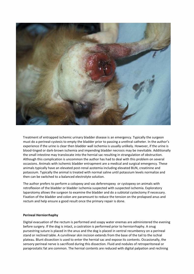
Treatment of entrapped ischemic urinary bladder disease is an emergency. Typically the surgeon must do a perineal-cystesis to empty the bladder prior to passing a urethral catheter. In the author’s experience if the urine is clear then bladder wall ischemia is usually unlikely. However, if the urine is blood-tinged or dark-brown ischemia and impending bladder necrosis may be inevitable. Additionally the small intestine may translocate into the hernial sac resulting in strangulation of obstruction. Although this complication is uncommon the author has had to deal with this problem on several occasions. Animals with ischemic bladder entrapment are a medical and surgical emergency. These animals typically have an elevated post-renal azotemia including elevated BUN, creatinine and potassium. Typically the animal is treated with normal saline until potassium levels normalize and then can be switched to a balanced electrolyte solution.
The author prefers to perform a colopexy and vas deferenspexy or cystopexy on animals with retroflexion of the bladder or bladder ischemia suspected with suspected ischemia. Exploratory laparotomy allows the surgeon to examine the bladder and do a subtotal cystectomy if necessary. Fixation of the bladder and colon are paramount to reduce the tension on the prolapsed anus and rectum and help ensure a good result once the primary repair is done.
Perineal Herniorrhaphy
Digital evacuation of the rectum is performed and soapy water enemas are administered the evening before surgery. If the dog is intact, a castration is performed prior to herniorrhaphy. A snug pursestring suture is placed in the anus and the dog is placed in ventral recumbency on a perineal stand or reclined table. A curvilinear skin incision extends from the base of the tail to the ischial plateau. Blunt dissection is used to enter the hernial sac and expose its contents. Occasionally, the sensory perineal nerve is sacrificed during this dissection. Fluid and nodules of retroperitoneal or paraprostatic fat are common. The hernial contents are reduced with digital palpation and reclining

of the table. The external anal sphincter is identified medially and the internal obturator muscle is located ventrally with the pudendal nerve and internal pudendal artery and vein coursing in a caudomedial direction. Inadvertent severance of the nerve on one side will not lead to incontinence. The levator ani muscle is often atrophied and not identifiable whereas the coccygeus can usually be identified on the dorsolateral boundary of the hernia immediately caudomedial to the sacrotuberous ligament. The rectal wall is examined closely prior to hernia repair. If a large diverticulum is present, it is reduced and the rent in the muscularis is apposed with simple interrupted sutures of 3-0 nylon. If a large sacculation is present, several Lembert type plication sutures are placed in the muscularis and submucosa to "gather" the redundant rectal wall. Resection of diverticula or sacculations has been described, but we have found this a largely unnecessary procedure. If the pelvic diaphragm is properly reestablished usually no rectal resection is necessary.
Suture placement is begun in the dorsal most aspect of the defect. I currently use non absorbable polypropylene suture for all repairs. The first suture engages the fibers of the coccygeus muscle dorsolaterally near the base of the tail and the external anal sphincter medially. Subsequent sutures are preplaced each 1.5 cm through the coccygeus and each 1 cm around the perimeter of the sphincter. Usually 5 or 6 sutures are needed. An alternative approach is to make two passes with the needle forming 3 or 4 cruciate mattress sutures. The levator ani muscle often has insignificant suture holding capacity, especially ventrolaterally and the sacrotuberous ligament is then engaged. In order to avoid encircling of the sciatic nerve which is immediately cranial to this structure we often pass the suture directly through the ligament. The preplaced sutures are crossed and pulled snug to test if obliteration of the cavity has occurred.
Often a defect remains in the ventral ischiorectal fossa and an internal obturator transposition is opted for. The caudal border of the origin of the internal obturator muscle is incised and elevated from the ischium for a distance of 5-10 mm not to proceed into the obturator foramen. The tendon of the muscle is incised taking care to avoid the sciatic nerve which passes over it laterally. The tendon and muscle are now rotated dorsomedially to fill the defect. The medial border of the muscle is sutured to the external anal sphincter and the lateral border is sutured to the sacrotuberous ligament using cruciate sutures of 2-0 Prolene. The remainder of the preplaced sutures are now tied and the reestablished pelvic diaphragm is checked for defects. A second layer closure of perineal fascia sutured to the external anal sphincter using simple interrupted sutures of 2-0 PDS is added and standard subcutaneous and skin closure is performed. If the hernia is bilateral, a second procedure either simultaneously or in 4 weeks, although some surgeons will operate both sides in the same sitting.
The author feels strongly that dogs and cats with diverticuli should have these out pouchings of the rectal wall repaired at the time of herniorrhaphy. Retention of feces in these out pouching is felt to increase the incidence of recurrence in these dogs. If primary approximation of the perineal diaphragm is not successful other methods have been utilized to support or repair the area. Occasionally the hernial defect is so large that mesh is used to repair the hernia. Risk of infection is very high with this technique. Lypholozed porcine submucosa or Biocyst has been used successfully along with other types of no absorbable mesh including polyproylene and latex sheeting material. Additionally, semi-tendinous membranous transpositions have been used and more recently free fascia lata grafts have been harvested from the opposite thigh to augment the herniorrhaphy.
In recent years the author has used more indirect repairs for perineal hernias in older dogs with multiple recurrences.. Typically, dogs that are in excess of ten years of age and have thin stretched perineal musculature are not good candidates for repair and in many times the outcomes are not optimal. Therefore, a planned colopexy and cystopexy is performed. The colopexy is performed by removing a 1 cm x 2 cm patch of peritoneum on the left body wall approximately 2 ½ cm off the midline.

A similar shaped area is aggressively divided using a number 15 blade and the areas are apposed using 3-0 non-absorbable suture material. The colopexy is done under tension to ensure adequate reduction of the redundant rectal wall and forward transposition of the anus. Additionally, the colopexy can be supported by multiple horizontal mattress sutures as described by Engen. Fixation of

the urinary bladder can be done with a vas deferens pexy or in which the vas deferens are amputated at the inguinal rings and placed through small fenestrations and the transverse abdominal muscles.

Alternatively the author likes to debride a similar shaped wound as described under colopexy on the right side of the dog’s abdomen and tacks the bladder over to the abdominal wall. A study of 40 some cases undergoing colopexy and cystopexy showed similar clinical results to animals undergoing primary herniorrhaphy repair. The morbidity and recurrence rate is often less with this technique in older animals.
Postoperative Care and Complications
Postoperative antibiotics are considered mandatory by some surgeons based on incision location, but we have used them only in cases of intraoperative contamination. Dietary modification and stool softeners are prescribed similar to that described under rectal prolapse. Postoperative tenesmus is often severe and we have noted eversion of redundant rectal mucosa or mild rectal prolapses in a good number of cases. Topical gel anesthetics or epidural analgesia may be effective in eliminating this tenesmus. We now routinely leave a loose pursestring suture in for the first 24 hours. Transient stretch incontinence is not uncommon for the first week postoperatively, but this is usually due to stretching of the external anal sphincter and not severance of the caudal rectal nerve. Extreme pain over the hip accompanied by carrying of the hind limb indicates sciatic nerve entrapment with a suture. If this sign is present the surgical repair should be immediately reopened and the offending ligature released. The most common complication following perineal herniorrhaphy is recurrence of the hernia, which occurs in 30 to 45% of the cases. Good shares of these cases recur after one year. Initial results with the internal obturator transposition have been very encouraging but longer term studies are necessary to evaluate its true efficacy.
References:
Fossum TW. Perineal hernia. In: Fossum TW (ed). Small Animal Surgery. 1st ed. Mosby: St. Louis, MO, 1997: 352-256.
Risselada M, Kramer M, van de Velde B, Pous I, Gortz K. Retroflexion of the urinary bladder associated ith a perineal hernia in a female cat. J Sm Anim Pract 2003;44:508-510.
Niles JD, Willaims JM. Perineal hernia with bladder retroflexion in a female Cocker spaniel. J Sm Anim Pract 1999;40:92-94.
Head LL, Francis DA. Mineralized paraprostatic cyst as a potential contributing factor in the development of perineal hernias in a dog. J Am Vet Med Assoc 2002;221:533-535.
Bongartz A, Carafiglo F, Balligard M, Heimann M, Hamaide A. Use of autogenous fascia lata graft for perineal herniorrhaphy in dogs. Vet Surg 2005;34:405-4113.
Szabo S, Wilkens B, Radasch R. Use of polypropylene mesh in addition to internal obturator transposition. 59 cases. J Am Anim Hosp Assoc 2007;43:136-142.
Huber DJ. Cystopexy and colopexy for the management of large or recurrent perineal hernias in dogs. Vet Surg 1992;26:218.