modeling of brain physics alexander kholmanskiy
TRANSCRIPT
1
Modeling of Brain Physics
Alexander Kholmanskiy
Scientific Center BEMCOM, Moscow
Laboratory of Medical and Biological Physics
[email protected], https://orcid.org/0000-0001-8738-0189
The physics of the human brain has two components – basic physics common to all
mammals and the physics of thinking inherent only in man. The development of the mental
component of the structural and functional organization of the brain in phylogeny was associated
with the chiral factor of the external environment, and in ontogenesis - with the social factor. The
sensitivity of the brain to these factors was based on the single-connected nature of its aqueous
basis, the mechanism of electromagnetic induction, and the features of the thermodynamics of the
brain in a state of night sleep. In order to unify the description of the mechanism of electromagnetic
processes in the brain, the concept of a quasiphoton has been introduced, combining all forms of
excitation of electronic and molecular-cellular structures of the brain. Equivalent schemes of
vibrational contours of neural network elements and macrostructures of the brain are proposed.
Estimates of the kinetic parameters (activation energy, velocity) of the physical processes
underlying the energy-information exchange of the brain with the external environment are made.
Mechanisms of operative (physical) and permanent (chemical) memory of the brain, including a
model of nonlocal quantum correlations, are discussed.
Keywords: electromagnetism; brain oscillator; quasiphoton; thermodynamic, kinetic;
quantum correlations
Contents Page
1. Conceptual introduction 2
2. Electromagnetic induction 5
2. 1. Oscillating contour 5
2. Electromagnetism of the neuron 6
2. 3. Neural networks 12
2. 4. Capsulated nerve endings 13
2.4.1. Fatera Pacini Body 13
2.4.2. Eye 14
Preprints (www.preprints.org) | NOT PEER-REVIEWED | Posted: 20 June 2019 doi:10.20944/preprints201906.0188.v1
© 2019 by the author(s). Distributed under a Creative Commons CC BY license.

2
3. Quasiphoton 17
3.1. Types of quasiphotons 17
3.2. Quasiphoton metric 19
3.3. Metabolic quasiphotons 21
4. Thermodynamics of the brain 23
5. The physics of brain organization 25
5.1. Functional hierarchy of the brain 25
5.1.1. Bark of the cerebral hemispheres 26
5.1.2. Thalamus, the ventricles of the brain 27
5.1.3. Epiphysis 28
5.2. Kinetic parameters of the physics of the brain 31
5.3. Nonlocal quantum correlations 35
5.3.1. Consciousness and memory 35
5.3.2. The mechanism of quantum correlations 37
6. Conclusion 39
References 40
1. Сonceptual introduction
The main structural and functional element of the brain is the nerve cell. It generates and
conducts electrical impulses - action potentials (PD). The motion of charge associated with the PD
induces local vortices of the electromagnetic (EM) field, which, in principle, can be defined as EM
quanta or quasiphotons (QPH). The metric, the principle and the speed of motion of a QPH will
be determined by the electrophysical properties and structural features of the neuron and its
surrounding medium. For energy-information support of the mechanism of pulse generation and
for the synthesis of metabolites, the cell body, its core and dendrites are responsible. Axons in
symbiosis with neuroglia (oligodendrocytes, astrocytes) translate metabolites and impulses,
realizing their energy and information through the synthesis and actions of neurotransmitters in
synapses. The ability of the nerve cell to provide energy for the reaction of oxidation of glucose,
which in the mitochondria is transformed into energy of macroergic bonds of ATP. In neural cells,
the energy of ATP is converted into the energy of QPH, into the energy of the chemical bonds of
the synthesized substances, into the kinetic energy of metabolites and molecules of the medium
Preprints (www.preprints.org) | NOT PEER-REVIEWED | Posted: 20 June 2019 doi:10.20944/preprints201906.0188.v1

3
(heat). At the expense of the same energy, axons grow, the development of neural networks and
neuroglial connections, which, in particular, are responsible for the mechanical integrity of the
cytoskeleton of the brain. The physicochemical properties of water, which form the basis of liquid
brain systems (cerebrospinal fluid, blood), are fully responsible for the electrophysics of the brain
and for its thermodynamic properties, both at the micro and macro levels of its organization.
Thus, the behavior of the brain as a single physical system is primarily subordinated to
the classical laws of electrophysics and the thermodynamics of continuous colloidal media. Within
the framework of these laws, the neuron metabolism is carried out, and the brain fulfills its basic
functions, controlling homeostasis and timely launching the mechanism of sexual reproduction.
The corresponding physics of the brain will be the same for all mammals, so it can be considered
the base. This is what allows us to extrapolate the results of animal studies on the human brain.
However, only the hominid organism (homo erectus) acquired sensitivity to the factor of
phylogenesis of geocosmic scale [1], under the influence of which the structures responsible for
speech and thinking began to form and develop in the brain in the conditions of the social
environment. Anatomical differences in the brain of modern man and monkey are clearly
expressed in the structure and volume of the frontal-temporal lobes of the neocortex. A key role in
the physics of thinking is played by the structural and functional asymmetry of the cerebral
hemispheres, which is absent in animals and has a racial-sexual differentiation in man. The genesis
of this asymmetry could be determined by the restructuring of the physics of genital organs, hands,
eyesight and hearing at the stage of erectness and in the process of developing skills to conscious
labor. Proceeding from these data, the cognitive functions of the brain will be based on the physics
of the frontal-temporal lobes of the neocortex and the chirality of the communication of the brain,
both interhemispheric and somatic, and with the external medium.
Given the presence in the brain of metastable and dynamic QPHs of various types and
energies, one can assume their active participation not only in metabolism, but also in the physics
of cognitive functions within the framework of the laws of classical quantum mechanics. The
nature of the external universal chiral factor, like the nature of the chiral energy quanta in the brain,
need not necessarily coincide with the nature of the QPH, the metric of which, nevertheless, may
be spiral. Mechanisms of absorption and actions in the brain of chiral energy quanta (eg, neutrino
nature [1]) are closely related to the physics of self-organization and phase transitions in
cooperative chiral systems [2, 3].
Preprints (www.preprints.org) | NOT PEER-REVIEWED | Posted: 20 June 2019 doi:10.20944/preprints201906.0188.v1

4
The main question of brain physics is the modeling of the mechanism of psychophysical
isomorphism [4], which, in essence, summarizes the following processes in itself:
- formation of the sense-word (thoughtform) at the level of the atomic-molecular system
of the EM-matrix;
- recognition and verbalization by another system of atoms of the content of the thought
form.
The space-time separation of two physical systems involved in the formation and
recognition of a thought-form presupposes the physical isolation of the thought-form in the form
of a bound system of discrete forms of matter isomorphic to the EM-matrix of the thoughtform.
The physical isolation of the thought form is a necessary condition for the adequacy of the
exchange of information on the mechanism of nonlocal quantum correlations. Ideal, in this sense,
the carrier of the thought form can be the simplest forms of matter, preceding the quanta of fields
and elementary particles. Then the problem of harmonizing and joining the physics of thinking
with the physics of the basic functions of the brain will be reduced to the problem of verbalization
of the fundamental dynamic form of matter, capable of becoming a carrier of energy and
information due to its movement [5]. The axiomatics of the simplest forms of matter (energy
forms) were constructed [6], relying on the laws of dialectics and extrapolating the authentic
positions of classical and quantum physics. The universalism of energy forms (EF) allows them to
be used for the modeling of thought forms, QPH and precursors of elementary particles.
Interactions of the EF with the brain material are mediated by QPH, combining the fractal-resonant
principle of the effect of EF [6] with the mechanism of nonlocal quantum correlations [7].
The energy forms and their condensates, in fact, include hypothetical "strings", "quarks",
"Abrikosov vortices", "density matrices" and other abstract models of subelementary discrete
forms of matter. In [8], in analyzing the thermodynamics of the brain's thinking activity, the gas
of hypothetical x-particles (fermions), distributed over the neural network of the cerebral cortex,
was proposed as the "working body" of the thinking apparatus. If the attempt to assign x particles
to a neutrino is groundless [4, 6], then certain features of the thermodynamics of x particles are
acceptable for bioactive EFs and QPH.
Thus, the physics of thinking can be isolated within the framework of the physics of the
basic functions of the brain, referring to it the unique ability of the brain substance under normal
conditions to resonantly absorb, generate, select, combine and preserve discrete forms of matter
Preprints (www.preprints.org) | NOT PEER-REVIEWED | Posted: 20 June 2019 doi:10.20944/preprints201906.0188.v1
5
(energy forms and QPH), recognizing in their actions the meaning- words, mental or other mental
information. To substantiate the use of energy forms and QPH for the modeling of the physics of
thinking, the present work analyzed the structural and functional features of the brain and made
estimates of the activation energies (EM quanta) of key physicochemical processes that provide
energy-information exchange inside the brain and between the brain and the external environment
to which the human body as well. The results of the analysis and estimates were used to carry out
extrapolations of known physical laws to the level of physics of energy forms.
2. Electromagnetic induction
2. 1. Oscillating contour
The kinematics of EF [6] illustrates the phenomenon of electromagnetic induction
(EMR), which formally obeys the first Maxwell equation [9]:
rotE = - дB/дt , (1)
where E and B are mutually orthogonal vectors of the intensity of the vortex electric and magnetic
fields.
With the help of (1) for a closed loop with current, an equation for the self-induction EDF
(U) is obtained:
U = – L (dJ/dt) = – dФ/dt, (2)
where L is the loop inductance; J is the current, and Φ = LJ is the flux linkage of the self-induction
of the circuit.
Effects of EMP in various structures and media of a living organism that have their local
magnetic (μ) and dielectric (ε) characteristics are subject to the second Maxwell equation:
rotH = j + дD/дt , (3)
where
D = εoε E , B = μoμH, (4)
j is the bias current, and the electrodynamic constant of vacuum (εoμo) and the medium are related
to the propagation velocities of EM quanta in vacuum (C) and medium (V) by the relationships
[9]:
C = (εoμo)–1/2 , V = С(εμ)–1/2 = С/n (5)
Extrapolation of the EMP phenomenon to the level of EF [6] can be illustrated by the
example of an oscillatory circuit (Fig. 1).
Preprints (www.preprints.org) | NOT PEER-REVIEWED | Posted: 20 June 2019 doi:10.20944/preprints201906.0188.v1

6
Figure 1. Vibrational contour - a) and its transformed forms corresponding to the
beginning of oscillations - b) and quarter of the period - c); d) - extrapolation of the state of the
contour (c) to the level of the energy form (ν/g-pair) having the momentum P and the equivalent
mass mg.
For an ideal circuit, the frequency of the harmonic electromagnetic oscillations is given by
the formula:
w = (LC)–1/2 (6)
Transformation of the oscillatory circuit by opening the capacitor and compressing the coil
is shown in Fig. 1. The state b) corresponds to the antenna circuit, which can, in principle, receive
and emit photons of the radio wave band. In this case, the vortex E and B fields fill the entire space.
The transformation c) corresponds to the state of the oscillatory circuit, when the energy of the E-
field is transferred to the energy of the vortex B field. The configuration of the EM field in state
c) can be identified with the EF (ν/g-pair [6]) by relating its momentum P or the energy of the E-
field, with the current pulse before it is twisted in the coil's spiral. Accordingly, the rotational
moment of the current or the associated B-field energy will correspond to the moment of the EF
pulse or its equivalent mass (mg). With the combination of different ν/g-pairs, field quanta
(photons, gravitons) are collected, and when they are condensed by a number equal to Avogadro's
number (6 1023), elementary particles are formed [6].
2.2. Electromagnetism of the neuron
It is obvious that EMP plays an essential role in the mechanisms of generation and action
of electromagnetic fields of electromagnetic nature in the human nervous system. At the heart of
Preprints (www.preprints.org) | NOT PEER-REVIEWED | Posted: 20 June 2019 doi:10.20944/preprints201906.0188.v1

7
its communicative and signaling functions lies the ability of nerve cells to generate and conduct
electrical impulses. Electrophysics and metabolism of the nervous system and neurons are
examined using ECG, EEG, MEG, NMR and positron emission tomography (PET) methods,
thermoencephaloscopy, psychopharmacology and direct probing of nerve cells by
microelectrodes. Quantum magnetometers (SQUID), in principle, allow one to register the
magnetic field of an individual neuron [10, 11]. The phenomena of EMR and resonance,
apparently, underlie the mechanism of sensitivity of the nervous system to direct effects of external
EM radiation of various ranges. The presence in the nervous system of LC-structures, in principle,
allows the "adjustment" of the sensory elements of the nervous system to the frequencies of both
internal and external biogenic radiations by the principle of heterodyne coupling.
To explain the electrical properties of the membrane, an equivalent circuit is used, in which
the conducting channels for various ions are modeled by the source of the EMF and the ohmic
resistance (R), and the insulating properties of the membrane are the capacitance (Fig. 2).
Figure 2. Equivalent electric model of
the nerve membrane: the batteries create a
total membrane potential U, ion conductivity
is denoted by resistances R, capacitance of the
membrane is capacitor C [12].
The parallel connection of several contours, shown in Fig. 2, simulates the neuron
membrane [12]. However, for the neuron model of the central nervous system, which has a myelin
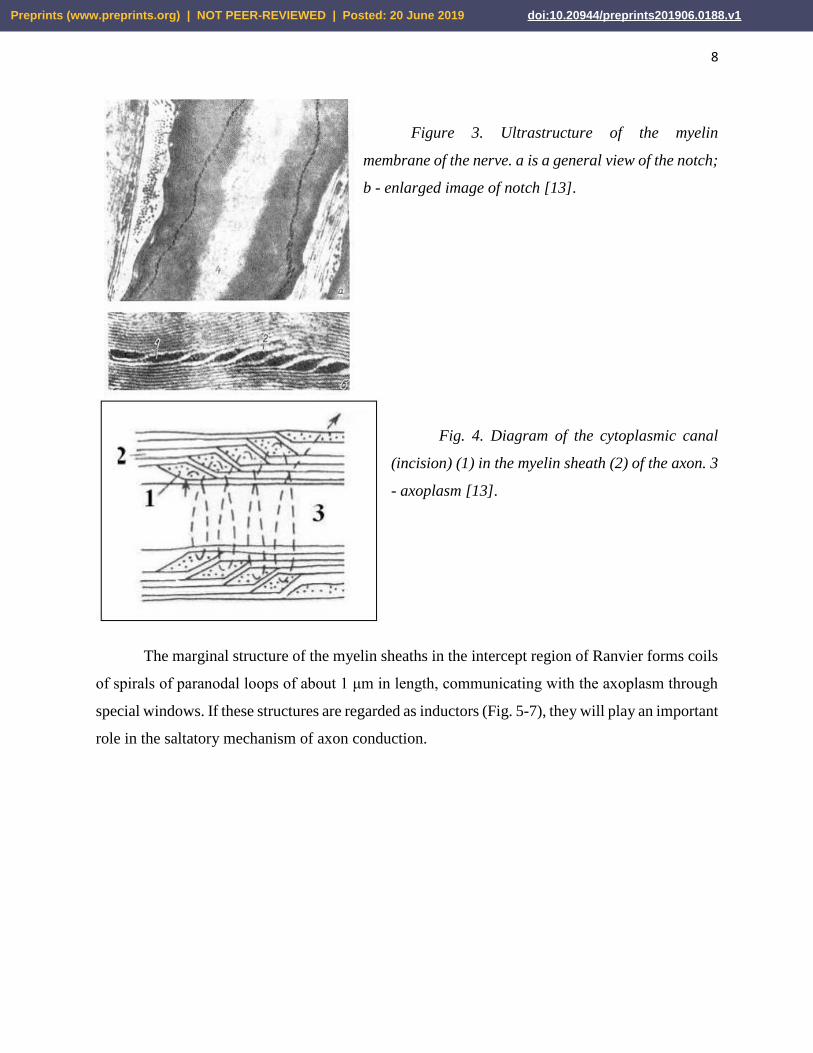
sheath, the capacitive characteristics of the membrane are not sufficient. Indeed, in the spiral
structure of myelin there are regular channels (notches) (Fig. 3), which, in the context of the
equivalent electric model of the membrane (Fig. 2), can play the role of local inductance coils. The
number of notches on one myelin fiber segment, the larger the thicker axon cylinder [13].
Preprints (www.preprints.org) | NOT PEER-REVIEWED | Posted: 20 June 2019 doi:10.20944/preprints201906.0188.v1
8
Figure 3. Ultrastructure of the myelin
membrane of the nerve. a is a general view of the notch;
b - enlarged image of notch [13].
Fig. 4. Diagram of the cytoplasmic canal
(incision) (1) in the myelin sheath (2) of the axon. 3
- axoplasm [13].
The marginal structure of the myelin sheaths in the intercept region of Ranvier forms coils
of spirals of paranodal loops of about 1 μm in length, communicating with the axoplasm through
special windows. If these structures are regarded as inductors (Fig. 5-7), they will play an important
role in the saltatory mechanism of axon conduction.
Preprints (www.preprints.org) | NOT PEER-REVIEWED | Posted: 20 June 2019 doi:10.20944/preprints201906.0188.v1

9
Figure 5. An electronic photograph of the interception of Ranvier, peripheral nerve [17].
Figure 6. The scheme of the structure of the interception of Ranvier. 1 - intercept slot; 2 -
the pulp cone; 5 - compact myelin; 6 - splitting of the main dense lines in the interception area; 7
- axial cylinder (axoplasma); 8 - cytoplasm of the Schwann cell [13].
Figure 7. Scheme of the edge helices of the myelin sheath
loop in the Ranvier intercept region. 1 - cytoplasm; 2 -
myelin; 3 - axoplasm [13].
Preprints (www.preprints.org) | NOT PEER-REVIEWED | Posted: 20 June 2019 doi:10.20944/preprints201906.0188.v1
10
Differences in the electrodynamic properties of the axoplasm, membrane, and intercellular
fluid, due to the difference in their ionic-molecular composition and structure, must leave their
imprint on the mechanism of PD generation. The stimulus that starts the charge exchange of the
membrane can be both physical and chemical, and the redistribution of charges can in one way or
another balance the transfer of ions through the membrane and their adsorption on its surfaces
[14]. With ionic currents of charge exchange of the surface of the axon membrane, impulse
displacement currents will be associated in the paranodal loops and in the scroll channels of the
notches, which allows them to be likened to magnetic dipoles [9]. The kinetics of ion currents and
displacement currents in the axon, membrane, and paranodal loops of the myelin sheath correlates
with the kinetics of growth and subsequent relaxation of the membrane potential. Since the phase
of the PD growth lasts about 0.1 - 0.2 ms, and the relaxation time of the membrane potential is of
the order of 1 ms [15], then the displacement impulse currents corresponding to the phase of
increase will be an order of magnitude greater than the relaxation currents. Changing the charge
on the inner side of the axon membrane in the Ranvier intercept region generates a polarization
wave or bias current in the paranodal region of the myelin segment [16]. The magnitude of this
perturbation will decay exponentially with distance [17], and the propagation velocity will not
exceed the velocity of motion of the PD in the unmyelized nerve (of the order of 1 m/s). The
presence of communication windows of the paranodal loops with axoplasm [16] ensures the
transformation of the polarization wave into a circular bias current in the helices of the loops.
Thus, the generation of PDs in the Ranvier interception is associated with the induction
and emission of vortex EM quanta, whose metrics are simulated by EM vortices c) and c) in Fig.
1. It is possible that this is the main function of the end coils of the myelin sheaths and incision
spirals. The direction of the vector of the flux density of the EM energy (the Poiting vector) will
be determined by the sign of the spiral. This chirality factor of the neuron will ensure one-sided
distribution of the EM-quantum, and therefore, PD on the myelinated nerve. Upon reaching the
EM quantum with the velocity V (5) of the end coil of the myelin segment, it can play the role of
a stimulus for the generation of PD in the next interception of Ranvier. In this model of the saltator
conductivity of the neuron, the speed of the spike motion will be limited by the process of current
excitation in the end coils, whose time is of the order of 10-6 s (1 μm: 1 m/s). At the same time, the
average transfer rate of PD from one end of the myelin segment to the other at a length of about
100 μm and will determine the rate of the saltative mechanism of conductivity ~ 100 m/s.
Preprints (www.preprints.org) | NOT PEER-REVIEWED | Posted: 20 June 2019 doi:10.20944/preprints201906.0188.v1

11
Using the value of the potential difference corresponding to the PD of a typical neuron (U
~ 70 mV [15]), we estimate the amount of electrical energy that is expended in excitation of the
PD in the Ranvier interception in the saltator mechanism of neuron conduction. To do this, imagine
the interception in the form of a cylindrical capacitor, the lining of which is formed from the neuron
membrane and the length is equal to the length of the interception (f). The change in the energy of
the capacitor (W) can be estimated from the formula:
W = (U2C)/2. (8)
The value of C for a cylindrical capacitor with the distance between the plates (d) and the
radius of the inner cylinder (R) under the condition d «R is equal to
C = (2πεoε f)/[ln (1 + d / R)] ≈ (2πεoε fR)/d
and the quantity
W = (U2 πεoε fR)/d (9)
We substitute in (9) such values for a nerve with R = 5 μm [17]: U ~ 0,07V; εo = 8.85 10-12 F/m;
ε ~ 5; f ~ 10-7 m; d ~ 10-8 m, we obtain
W ~ 510-17 J or 3107 J/mol. (10)
The same value of W is obtained if we substitute in C the value C = 10-2 F/m2 [17] for the
same parameters of Ranvier intercept and U. The value (10) is comparable with the energy released
during oxidation of ~ 10 glucose molecules and at the hydrolysis of ~103 ATP molecules.
It is known [12, 17] that during the hydrolysis of one ATP molecule, ~3 Na+ ions pass
through the membrane in exchange for two K+ ions, and when the PD is excited, the flux density
of Na+ ions through the intercept membrane is JNa ~ 4103 ions/μm2. Then the number of Na+ ions
entering into the axon will be equal to JNa (2πRf) ~ 104, it corresponds to ~ 3103 ATP molecules,
the total energy of which agrees in order of magnitude with (10). When ATP concentration in
axoplasm of axon of squid is ~1 mmol per kg of H2O [12], the total number of ATP molecules in
the Ranvier interception cylinder (radius 5 μm and length 1 μm) will be ~4 107 molecules.
Consequently, the value of W is only 0.01% of the total energy resource of the Ranvier
interception.
It is obvious that the energy of the EM quantum that plays the role of the stimulus of the
PD generation in the Ranvier interception will be one, two orders of magnitude less than the value
of W. For example, for the upper limit of the energy of the EM quantum one can take the energy
Preprints (www.preprints.org) | NOT PEER-REVIEWED | Posted: 20 June 2019 doi:10.20944/preprints201906.0188.v1

12
of a photon with a wavelength of 600 nm (4 10-19 J), which is sufficient to excite the signal in the
retinal cell of the retina [15].
2. 3. Neural networks
Submission of neurophysics to the law of EMP can be formalized by introducing an
inductor into the equivalent circuit of the nerve membrane with the myelin sheath together with
the capacitor (Fig. 8). Such a modification of the equivalent circuit, transforming it into an
oscillatory circuit, significantly expands the range of the electrophysical properties of the neuron.
Figure 8. Modified electrical circuit of the nerve
membrane. Rm, U - ion channel; C is the capacity of
the membrane; L - inductance of glial membrane
myelin helices; Rin - resistance of axoplasm
In addition, the introduction of inductors into the electrical circuit of the nerve membrane
allows us to model the chirality factor of a neuron and to associate it with the mechanism of
differentiation of nerve signals to stimulating and inhibitory ones. The combination of the chirality
factor with a biochemical factor (synaptic connections) empowers the logical element of neural
networks to encode the "yes" and "no" signals (Fig. 9).
Figure 9. Modified functional scheme of a formal neuron [18]. Xn - biochemical, Zn -
electrophysical factors of neuron activity; Y ("yes"), Y * ("no") are analogues of the exciting and
braking signals.
Preprints (www.preprints.org) | NOT PEER-REVIEWED | Posted: 20 June 2019 doi:10.20944/preprints201906.0188.v1

13
In addition, EM-quanta or QPH generated in a neural network can be combined into a
dynamic quantum system (Bose-gas) and represent the brain as a processor, the elemental base of
which is the entirety of the multilevel hierarchy of neuron-neuronal and neuroglial connections.
At the same time, the efficiency of the quantum level of the neural network organization will be
limited by the value V (5) and the transmission and processing time of the signal in the scale of
the neural network from 1 μm to 10 cm will vary in the range from 10-15 to 10-10 s. The first value
is comparable with the lifetime of the singlet electron-excited state of the molecule (optical QPH),
and the second with the characteristic lifetime of tetrahedral water clusters.
2. 4. Capsulated nervous terminations
2.4.1. Fatera Pacini Body
EMP can also be used to explain the mechanism of generation of electrical impulses in
encapsulated nerve endings. The most important for brain physicists the representative of this kind
of endings is the Fatera-Pacini body (FPB) (Fig. 10). FPB is abundantly present in the
subcutaneous layer of the palms and feet, in the female genitalia and in the connective tissues of
the internal organs [19].
Figure 10. Encapsulated ending nerve –
the body Fatera-Pacini [19]. Linear
dimensions reach 1-2 mm.
In addition to the sensory function, FPB can obviously accept gravitational and
geomagnetic energies and simultaneously play the role of EM-quanta generators. It is suggested
[19] that the basis for the mechanism of TP generation in FPB is the biochemical response of FPB
to its mechanical deformation. However, the isomorphism of FPBs and complex electromagnetic
devices, which have two inductors embedded in the rod, suggest the participation of EMP in the
mechanism of PD generation. Deformation of FPB, being associated with changes in its induction
or capacitive characteristics, can lead to the excitation of electromagnetic pulses that stimulate the
Preprints (www.preprints.org) | NOT PEER-REVIEWED | Posted: 20 June 2019 doi:10.20944/preprints201906.0188.v1

14
generation of PD. In this case, a decrease in the values of L and C in accordance with formula (6)
should lead to an increase in the frequency of generation of stimuli (w), and hence the repetition
rate of the PD, which is observed experimentally [19]. We note that in the absence of external
deformation of FPB, their background activity as "generators" of quasiphotons can provide a
rhythmic deformation of the fiber around the FPB, which corresponds to the pulsation of the
circulatory system. It is also possible that the LC-contour in the structure of FPB can move
resonantly in the motion of the arms and legs resonantly in the energy of the geomagnetic field.
2.4.2. Eye
The eye can be considered an integral capsular end of a large number of axons of the optic
nerve. Its main function is to convert the front of photons of the visible range into a complex spatio-
temporal mosaic of PD and QPH. The electrical energy (momentum) of the photon absorbed by
the retina stimulates the generation of PD in it and is partially transformed into a pulse of optic
nerve adhesion. In this role, about 10% of the photons enter the eye, the remaining 90% are
absorbed by the optical media of the eye [19]. When photons are absorbed, both in the retina and
in other elements of the eye, the probability of generation of states with intra- and intermolecular
charge transfer in the donor-acceptor fragments (D+δ-A-δ) is high. Such metastable states are called
excitons. The kinetics of the process of exciton relaxation in the retina correlates with the kinetics
of PD generation. A sufficiently long lifetime and a high photostationary concentration of these
states cause the dipole polarization of the retina surface [20]. The movement of retinal charges
during eye movement induces vortex magnetic fields (EM vortex), the maximum energy density
of which is achieved in the frontal and temporal lobes, as well as in the sinuses of the skull
(maxillary, wedge-shaped, frontal) (Fig. 11). It is known [11] that in these areas the functions of
attention and self-awareness are localized, that is why the EM-vortexes of the eyes can take a direct
part in their activation. With this in mind, suppose that the eyes and their nervous system, along
with their sensory function, play a dominant role in the physics of the cognitive functions of the
brain. That is why with increased mental work, not even related to reading, the eye muscles become
very tired, which provokes the development of a specific pattern of wrinkles around the eyes. Note
that with the congenital defeat of the CNS departments responsible for the formation of visual
representations of the external world ("central congenital blindness"), the child is doomed to
remain an idiot.
Preprints (www.preprints.org) | NOT PEER-REVIEWED | Posted: 20 June 2019 doi:10.20944/preprints201906.0188.v1

15
Figure 11. The distribution of magnetic induction for various eye movements (a, b, c) [20,
21] and the scheme of fluxes of magnetic induction in the frontal projection (e). Floor. - the
magnetic field is directed inward, ot. - outside the object. The value of the B-field is proportional
to the radius of the circle. A), b) - horizontal movement of the eyes from right to left within an
angle of 55°; c) - eye movement from the bottom up.
Fig. 11e shows the areas of the brain (front view) responsible for self-awareness: the medial
prefrontal cortex (connects the self-sensations and the memory of oneself) is highlighted in red;
yellow - predklinie (activation of retrospective memory of yourself) [11].
The geometry of the vortex B- and D-fields of the eyes is determined by the trajectories of
the charges of the retina and muscles during the movement of the eyes in the vertical and horizontal
directions. The localization of the maximum value of the B-field with the horizontal movement of
the eyes at the midpoint (Fig. 11a) indicates the summation of B-fields at this point from both eyes.
This is possible under the condition that the vortex metrics induced to the right and left eyes are
mirror symmetric. It is possible that the chirality of the electromagnetic stimulus, and hence the
signs of the myelin spirals of the nerves of the right and left eyes are opposite. It can be imagined
that EM vortices induce mirror-symmetrical pairs of EFs in the CSF cerebral liquor between the
Preprints (www.preprints.org) | NOT PEER-REVIEWED | Posted: 20 June 2019 doi:10.20944/preprints201906.0188.v1

16
hemispheres, their fusion, in principle, can produce a QPH and such a mechanism of EM-quantum
generation, obviously, underlies the exothermic process of recombination of two dissimilar electric
charges. It is obvious that the organizing effect of the eye EM-vortices in the process of formation
of the child's self-consciousness determines the formation in the medial prefrontal region of the
neuronal cortex of a unique form, called spindle cells [11, 15]. Similar reactions of EF fusion-
recombination induced in the right and left hemispheres can also take place in the cerebrospinal
fluid of the third and fourth ventricles, taking an active role in their bioenergetics. A simplified
diagram of the frontal projection of the D-, B-vortexes of the eyes is shown in Fig. 11e. The cross
in the center of the eye indicates the spike of the optic nerve that leaves in the plane of the picture.
This scheme also coincides with the distribution of the lines of force of the magnetic dipole,
oriented along the line of the nose. This is also consistent with the fact that the porous bones of the
walls of the nose, the sphenoid sinus and the latticed bone have a high residual magnetization value
[30].
In support of the important role of the eyes in the cognitive physics of the brain, there is a
complex relationship between eye energy and the baseline rhythm of brain electrophysics (α-
rhythm):
- only the higher mammals have an α-rhythm [22] and it is established synchronously with
puberty, after which it is not fixed in the frontal lobes [15];
- α-rhythm, like the beta-rhythm, can be localized separately in the right or left hemisphere
of the brain [23];
- The α-rhythm disappears with a loss of consciousness and opening of the eyes, but in the
blind it is either poorly expressed or absent [15];
- The frequency of the α-rhythm (~10 Hz) is correlated with the frequency of a standing
EM wave in a spherical resonator that forms the Earth's surface and its ionosphere [24].
The vitreous body of the eye in the background can condense the EM energy of the external
environment and, possibly, the energy of the solar neutrino [1, 6], directly feeding this energy to
the brain. The evidence supports this assumption:
- rapid eye movement in the phase of paradoxical sleep (phase-BDG) is associated with
the intensification of brain physics [15];
- the formation of a specific five-fingered relief on the surface of the eye socket, facing
the brain and curving the line of the nose [25];
Preprints (www.preprints.org) | NOT PEER-REVIEWED | Posted: 20 June 2019 doi:10.20944/preprints201906.0188.v1
17
- muscles of the eyeball spontaneously twitch at a frequency of 20 - 150 Hz
(microsaccharides, tremor) [15];
- the layer of ganglion cells of the retina bordering with the vitreous body in the dark and
with closed eyes show background activity with a frequency from 1 to ~20 pulses per second [26];
- exposure to closed eyes of mechanical pressure and a pulsed magnetic field initiates a
"vision" of white light (phosphenes) [15, 27]
- an increase in the intensity of light leads to an increase in the frequency of generation of
PD in the optic nerve [15];
- the crystalline lens and the vitreous of the eye are optically active [28, 29];
- glycolysis of glucose in the vitreous body gives, in addition to ATP, chiral lactic acid
[15];
- the characteristic time of hydrodynamics of the eye is ~ 900 s, during this time half of the
vitreous fluid is renewed [15].
The ontogeny of the asymmetry of sight, smell, hearing, facial (nose curvature) and
genitalia is synchronized with the process of stabilizing the frequency of the α-rhythm [31] and for
12-13 years repeats the stage of phylogenesis corresponding to the uprightness. Moreover, by the
age of two years, when the child begins to walk independently, the genesis of brain asymmetry
involves the energy of the TFP stop and the physics of the genital organs, the gender peculiarities
of which leave their imprint on the topology and functions of the brain of a man and a woman [32].
3. Quasifoton
3.1. Types of quasiphotons
To describe the properties of various condensed media, the concept of a quasiparticle is
widely used [33]. Since the water content in the brain reaches ~75% [15], it can be considered a
highly concentrated colloidal solution. To describe the mechanisms of energy-information
processes underlying the physics of the brain, it is convenient to use the concept of a QPH as a
generalization of the EM quantum. Thus, a QPH is a carrier of the excess energy of an
electromagnetic field localized on an electron or on the electron system of an ordered atomic-
molecular structure. The precursor of a QPH is a photon or an EM quantum, in the case of their
absorption by the system. Depending on the energy of the photon and the electronic structure of
the system, the metric, the lifetime and the fate of the QPH vary widely. The physics of QPH
Preprints (www.preprints.org) | NOT PEER-REVIEWED | Posted: 20 June 2019 doi:10.20944/preprints201906.0188.v1

18
genetically inherits the laws of atomic-molecular spectroscopy and the properties of excited states
of molecules of various types (electronic, vibrational, translational, rotational) [34]. Mixed
electron-nuclear configurations of excited states will correspond to rotational and vibrational QPH,
and optical QPH to purely electronic excited states. Examples of an optical QPH are an EM
stimulus generating a PD, an exciton, or an electronically excited state of a molecule. The
vibrational QPH in an elastic coupled structure will be similar to a phonon. A rotational QPH in a
system of coupled nuclear or electron spins can be identified with a magnon [33]. QPH can be free
and bound, depending on the properties of the medium and the mechanism of interaction of its
elements. Thus, the metric-dynamic characteristics of QPH will be determined by the type of
chemical bonds and the type of intermolecular interactions that determine the degree of orderliness
of the medium.
The energy of QPH of various types will vary over a wide range, the upper limit of which
can be regarded as the energy of the QPH stimulating the generation of PD in the interception of
Ranvier (~10-19 J). For the lowest energy limit of a QPH, one can take the energy of a vortex EM
field generated by the motion of the eyes. The density of a given energy at B ~ 4 nT is of the order:
E = В2 /(2μоμ) ~ 10-23 J/cm3 or ~ 0.01 kJ/mol in cm3. (11)
The degree of influence of this field on the magnetically susceptible micro and macro
structures and the environment of the brain will be determined by the energy density:
E = (M B)/2,
where M is the specific magnetization (the specific density of magnetic moments is m), equal to:
M = Σ m.
In the case of ring currents of any nature (J) m = J ΔS, where ΔS is the area of the surface
covered by the current. Moreover, the polarization effect of the magnetic field can be enhanced by
the thermal motion of the medium particles [36].
The energy of the thermal effect from the light stimulation of the rat eyes is comparable
with the value (11), which is manifested by an increase in the local temperature of the visual cortex
of the brain by ~ 0.06 оC [11]. The energy of the QPH of the thermal energy corresponding to a
given quantum is ~ 10-24 J or ~ 10-3 kJ/mol. In the range 10-3 - 102 kJ/mol, the energy of biogenic
MW radiation (λ = 100 - 1 mm, E = 10-3 - 0.1 kJ / mol) falls into the range [37]; including the
energy of the resonance frequencies of water (λ ~ 6 mm, E = 0.02 kJ/mol) [38]. It is known [3]
that the activation energy of the processes of associating sugars and quanta of biogenic MW
Preprints (www.preprints.org) | NOT PEER-REVIEWED | Posted: 20 June 2019 doi:10.20944/preprints201906.0188.v1

19
radiation is one or two orders of magnitude less than the thermal energy and is comparable in order
of magnitude with (11). It follows that quasi-photons of rotational type will play a key role in the
processes of self-organization of liquid brain media, and the physics of the fronto-temporal lobes,
responsible for the cognitive functions of the brain, will be directly related to the electrophysics of
the eyes.
3.2. Quasiphoton metric
The elementary structural cell of liquid water is a dynamic tetrahedron formed of four water
molecules connected by hydrogen bonds. The fifth molecule of water or a molecule or atom
commensurate with it can be in the center of a tetrahedron, then it is called a centered tetrahedron
(Fig. 13). Thanks to hydrogen bonds, water effectively interacts with dissolved molecules, thereby
expanding the range of their physico-chemical properties. This feature of aqueous colloids and
gels is especially important for brain physics, since its intercellular volumes are, as a rule,
comparable to the sizes of biomolecules, cells and organelles [35].
Because of this, it is necessary to assume a significant effect of the epitaxial effect on
processes regulating interneuronal and neuroglial interactions. It is known, for example, that in
chemical reactions occurring in an optically active medium or on the surface of quartz, the yield
of chiral products increases. The increase in the epitaxial effect of membranes and the walls of
various organs, in addition to the mediation of water, is apparently facilitated by bound or adsorbed
surface polypeptide and polysaccharide chains, as well as microvilli (Fig. 12) [15, 17]. The
epitaxial effect and the presence of chiral sugars affect the kinetics of reversible adsorption of ions
and neurotransmitters on the surfaces of neuronal membranes, both in Ranvier intercepts and in
synapses [14].
Figure 12. Diagram of the membrane and the
polysaccharide and polypeptide chains
emerging from it.
Metabolites having a charge, dipole, mechanical or magnetic moments, and also chirality,
affecting the electrodynamic constant (εμ) of the medium, the metric and the dynamics of
Preprints (www.preprints.org) | NOT PEER-REVIEWED | Posted: 20 June 2019 doi:10.20944/preprints201906.0188.v1

20
supramolecular structures, can vary widely the cooperative properties of solutions, the generation
efficiency, and the mechanism of motion of QPH. This applies primarily to ions (Na+, K+, Cl-, P3+)
(Table 1) and to molecules that play the role of carriers, acceptors and converters of QPH (oxygen,
carbon dioxide, water, sugars, ATP, neurotransmitters, hormones, enzymes).
Figure 13. Diagram of the fusion of two mirror-symmetric mobile EFs (v/g pairs) into a
quasiphoton at rest with a tetrahedral metric (a) and a scheme of electron orbitals of a water
molecule (b)
The main mechanism of motion of QPH will be their resonance absorption and reradiation
by medium molecules, metabolites and supramolecular structures. The main element of the three-
dimensional metric of the liquid medium and most of the organic metabolites is a tetrahedron
whose electron-nuclear matrix is the sp3-hybridized system of electron orbitals of carbon, nitrogen
and oxygen atoms. Consequently, a quasi-photon localized on a particular metabolite is most likely
to have a metric in the isomorphic geometry of sp3-hybridization.
Using the concept of v/g-pairs, a resting EF or a localized QPH with a tetrahedral metric
can be obtained by the scheme shown in Fig. 13. The rules for combining and condensing EF (v/g-
pairs) [6] allow one to model and calculate the metric of QPH of various types, including
isomorphic metrics of sp- and sp2-hybridized atomic orbitals. The energy of QPH associated with
π-electrons will be less than the energy of QPH corresponding to vibrational-rotational excitations
of atoms or deformations of the σ-skeleton. The lower vibrational levels of the carbon dioxide
molecule (O=C=O), having an energy of 10-21 to 10-20 J, can be populated by absorption of thermal
quanta (kT). The specificity of the location of the levels allows their inverted population, which
allows the use of carbon dioxide as the active medium of the laser (λ ~ 10 μm). In liquid media
predisposed to self-organization, the CO2 molecule can be a donor of vibrational QPHs for
molecules with a carboxyl group (-HCO). Similarly, molecules with aromatic rings will accept
Preprints (www.preprints.org) | NOT PEER-REVIEWED | Posted: 20 June 2019 doi:10.20944/preprints201906.0188.v1

21
QPHs corresponding to conformational vibrations of saturated carbon cycles isomorphic to them
and heterocycles. High activity, for example, of steroid hormones of cholesterol derivatives,
having condensed hexane cycles, can be caused by the saturation of their molecules with QPHs
with sp3-metric (Fig. 13). At the same time, aromatic molecules (benzene, anthracene, pyrene)
isomorphic to fragments of hormones, effectively deactivating hormones and distorting their
metabolic functions, can initiate carcinogenesis [39].
Table 1.
Nuclear-electronic characteristics of elements
Element
Characteristics of nucleus
Main
electronic.
state
of atom
Ionic
radius
(nm)
Spin
(I)
Magnetic
moment
(× μн)
Magnetic
susceptibility
(C13 = 1,00)
Quadruple
moment
(Q×1028, m2)
11Na23
3/2
2,22
525,0
0,12
[Ne]3s1
0,098
19K39
3/2
0,39
2,7
0,055
[Ar]4s1
0,133
17Cl35
17Cl37
3/2
0,82
0,68
20,2
3,8
– 0,08
– 0,06
[Ne]3s22р5
0,182
15Р31
15Р32 (14 дней)
1/2
1
1,13
–0,25
377
–
–
[Ne]3s23р2
0,044
3.3. Metabolic quasiphotons
For the unification of the language of bioenergetics, the energy of the QPH of the
corresponding metric is correlated with the values of the energies of chemical bonds, vibrational-
rotational and thermal motions of atoms and molecules. The ability of the brain to provide energy
enzymatic reactions of oxidation of glucose in the mitochondria and anaerobic glycolysis in the
eyeball. In these reactions, the energy of the chemical bonds of glucose and oxygen is transformed
into energy of the macroergic bonds of ATP, which in the subsequent ATP hydrolysis reactions
are converted into the kinetic and vibrational-rotational energy of metabolites and molecules of
the medium. The chemical activity of these molecules is then realized through the action of their
excitation energy, which is simulated by QPHs of the corresponding energy and metrics.
Preprints (www.preprints.org) | NOT PEER-REVIEWED | Posted: 20 June 2019 doi:10.20944/preprints201906.0188.v1

22
The total energy effect of all stages of the enzymatic reaction of glucose oxidation in
mitochondria has as its limit the thermal effect of the burning reaction of glucose in an oxygen
atmosphere:
C6H12O6 + 6 O2 → 6 CO2 + 6 H2O + 2800 (kJ/mol). (12)
The reaction of glucose oxidation in mitochondria is associated with the ATP synthesis
reaction, with one molecule of glucose accounting for 38 ATP molecules [40]. In the anaerobic
glycolysis of glucose, only two molecules of ATP and two molecules of chiral lactic acid are
formed, which obviously contribute to the chirality of the energy of the eyes and brain. The
maximum yield of metabolic energy will give hydrolysis of 38 ATP molecules according to the
following schemes:
ATP → ADP + P ~ P + 36 (kJ/mol)
P ~ P → P + P + 33.4 (kJ/mol).
The total energy of the macroergic bonds of 38 ATP molecules is 2640 kJ/mol, which is ~
95% of the limiting value of the combustion energy of one glucose molecule. This indicates the
high efficiency of enzymatic reactions of transformation of QPH corresponding to σ-bonds of С-
С, С-О-С, С-Н glucose into QPH localized on two macroergic bonds Р ~ О- in ATP.
Suppose quasi-photons are equally distributed over the bonds of the products of the
reactions of glucose oxidation and ATP hydrolysis, then the limiting values of the energies of the
QPH corresponding to these reactions will be 1/12 and 1/152 of the thermal effect of reaction (12)
equal to 4.5 10-18 J, that is, ~ 3 10-19 and ~ 3 10-20 J, respectively. If we apply to these QPH the
universal relation between energy and the characteristic size (r) of the discrete element of matter
(ν / g-pair, elementary particle) [6]:
E ~ ħc/r, (13)
then for a QPH acting as a quantum of metabolic energy, we obtain a radius of ~ 1 μm
comparable to the axon radius.
Metabolic QPH can take an active part in the enzymatic synthesis of proteins and nucleic
acids, as well as in the replication and transcription of DNA. One can imagine the participation of
QPHs in the interlacing of the double helix of DNA in the following way. In the region of the
replicative fork, the sugar-phosphate backbone of the DNA chain resonantly absorbs metabolic
quasi-photons of vibrational type. The elasticity of chains increases, which leads to the rupture of
hydrogen bonds between them. Taking into account that one hydrogen bond is necessary for two
Preprints (www.preprints.org) | NOT PEER-REVIEWED | Posted: 20 June 2019 doi:10.20944/preprints201906.0188.v1

23
sugar-phosphate links of the DNA helix and its energy is ~ 19 kJ/mol (3 10-20 J), it turns out that
for its rupture it is sufficient to take a single metabolic QPH by the DNA chain.
The presence of isomorphic amino acid fragments in peptide chains of the protein and in
the structure of neurotransmitters (glycine, acetylcholine, glutamic acid, dopamine, serotonin, etc.)
makes it possible to propose a resonant mechanism for the transfer of a quasi-photon of vibrational
type upon contact of the neurotransmitter with the receptor. Because of the presence in the
structures of the mediators of the electron and proton-donor and acceptor groups, their main
electronic state is characterized by intramolecular charge transfer D+δ-С-А-δ.. Here D - amino
groups, methoxy group, benzene ring and A - carbonyl and hydroxyl groups, and C - a chain of σ-
bonds. This factor and predisposition of mediators to the formation of hydrogen bonds are the basis
of their physical and chemical sorption at receptors of postsynaptic membranes. The receptor,
when taking or giving off a QPH upon contact with a neurotransmitter, changes its conformation
by locking or opening the membrane of the membrane.
4. Thermodynamics of the brain
The brain as a whole can be considered a reaction thermodynamic system, which is in a
stationary state. The influx of energy and the discharge of excess heat by the brain are balanced in
a narrow temperature range from ~ 37°С (the center of the brain) to ~ 36°C (brain cortex) [15].
This temperature gradient, due to the lower temperature of the external environment, can play a
significant role in the orientation of heat flows within the brain. A similar temperature gradient is
observed for the body, it has a maximum in the rectum, and a minimum in the surface layer of
cellulose and male testicles [15]. The range of the optimal temperature of metabolism is within the
limits of temperatures for which the isobaric heat capacity of pure water has a minimum [41]. The
peculiarities of the thermodynamics of phase transitions of aqueous solutions in the process of
phylogenesis formed the basis for the adaptation of living systems, which, in essence, are
isoenergetic transitions or transitions with an activation energy of the order of kΔT (at ΔT ~ 0.1-1
K) between states of different order of the protein molecules or homogeneous, molecular-cellular
ensembles. The decrease in the entropy component of the internal energy of a living system is
associated with the resonant absorption of a quantum of external EM or neutrino energy, which it
converts into an active metabolic QPH [3]. The liquid medium provides for the removal of the
quantum of thermal energy (entropy) beyond the boundaries of the system, and the function of a
Preprints (www.preprints.org) | NOT PEER-REVIEWED | Posted: 20 June 2019 doi:10.20944/preprints201906.0188.v1

24
QPH realizes the function of one or another brain structure that includes an ordered subsystem.
The energy released or absorbed during such transitions may be much smaller than kT. In
nonequilibrium conditions, the vibrations of individual macromolecules can be synchronized, in
particular, by means of an electromagnetic field [36].
Thus, the thermodynamics of the brain combines the equilibrium-stationary
thermodynamics of metabolism and the nonequilibrium thermodynamics of a neural network, the
"working body" of which is the Bose gas of QPHs. Accordingly, the internal energy U of the brain
as a function of its state will depend, in general, on the temperature (or entropy S), on the strain
tensor G, on the magnetic moment M of individual metabolites and macrostructures, on the total
angular momentum of nuclei and atoms L, and on the polarization of the medium P. Therefore,
the total differential of the internal energy U = U (S, G, M, L, P) will have the form:
dU ≡ TdS - PdG + BdM + DdP + FdJ, (14)
where T is the absolute temperature of the system; П is the pressure tensor; F is the vector
of the orientational polarization of the system of spins or momenta [42]. In expression (14), the
term FdL characterizes the work associated with the orientational polarization of the system of
nuclear spins or momenta of atoms and molecules (just as the terms DdP and BdM determine the
work associated with the polarization and magnetization of the system).
Orientational and polarization effects play an important role in initiating phase transitions
in homogeneous gas and liquid systems of the brain and organism. The high sensitivity of these
systems to the parameters included in (14) ensures the chirality of metabolites (mainly sugars [3,
29]) and the physicochemical features of molecular and liquid water. Such systems are formed in
the following structures and environments of the body and brain:
- stomach, uterus, trachea, craniofacial sinuses, cavity of the epiphysis;
- membranes and ventricles of the brain, venous sinuses, eyeball;
- blood and lymphatic systems;
- parenchyma of organs (lungs, liver, spleen, testicles, female breast);
- Subcutaneous tissue, connective and bone tissue.
All these systems function normally in two modes - stationary (quasi-equilibrium) and
nonequilibrium. The first is characteristic for the waking state of the body and brain not engaged
in mental work, and the second regime corresponds to the state of sleep or creative work. In the
first mode, energy exchange with the medium occurs continuously, and in the second, it is
Preprints (www.preprints.org) | NOT PEER-REVIEWED | Posted: 20 June 2019 doi:10.20944/preprints201906.0188.v1

25
quantized. The mechanism of accepting the quantum of external energy in the phase transition of
the cooperative system illustrates the processes of condensation of water vapor at the dew point
and quantum Bose condensation.
Acceptance of quanta of the energy of the MW-band or neutrino energy in these media is
facilitated by a decrease in body temperature in sleep by ~ 1K, as well as lower temperatures of
the vitreous of the eyes, peripheral (deposited) blood and testes. In the acceptance of chiral quanta
of neutrino energy, a large role is played by sugars, whose content in the blood increases in the
morning hours before sunrise. At this time, the neutrino component of solar radiation is filtered
from the EM radiation by the surface segment of the crust of the globe. An important role in the
acceptance of energy by the vitreous eye is played by the polysaccharide-hyaluronic acid [3, 29].
Almost half of all human hyaluronic acid is concentrated in its skin, where it is located in the
connective tissue of the dermis between collagen and elastin fibers, as well as in corneocyte corneal
cells. The dermis contains 70% water, which is ~ 20% of the body's water [15]. The energy
absorbed by the body, condensing on metabolites in the composition of liquid media (blood,
cerebrospinal fluid), is transmitted to the brain via neurohumoral and air channels (from the lungs).
The absorption of the external EM-quantum and the formation of a QPH from the energy
forms in the general case obeys the fractal-resonance mechanism and the principle of isoenergetic.
With allowance for (13), the principle of isoenergetic for resonant interactions and phase
transitions in cooperative systems can be expressed by the relation [6]:
ћC/r = N (ћC/R), (15)
here r characterizes the metric of the QPH, and R is the energy form and r = R/N; the
number N takes any values smaller than the Avogadro number when the EF is condensed into QPH
and reaches the Avogadro number with the participation of the EF in weak interactions [39].
5. Physics of the brain organization
5.1. Functional hierarchy of the brain
Functional hierarchy of the human brain is built on the physical properties of its following
structures: the neocortex, basal nuclei, limbic brain, thalamus, hypothalamus, pituitary gland,
epiphysis, reticular formation and cerebellum. The fluid system of the brain (blood and liquor), as
well as the whole complex of intracranial nervous communications, should be referred to separate
Preprints (www.preprints.org) | NOT PEER-REVIEWED | Posted: 20 June 2019 doi:10.20944/preprints201906.0188.v1
26
elements of the hierarchy, highlighting the corpus callosum, optic nerve and radiance of the
thalamus.
5.1.1. Bark of the cerebral hemispheres
In the structure of the cortex, surface special zones (up to 100 pieces) and six layers are
distinguished [15]. The topmost layer is formed by horizontally oriented apical dendrites of
pyramidal cells and axons of stellate cells, which provide intracortical connections between
neighboring neurons
The horizontal orientation of the neuron dipoles of this layer corresponds to a dynamic
electric field, which can play the role of a protective EM screen. The remaining five horizons of
the cortex are first structured into neural-glial modules (diameter ~ 100-150 μm), and then, in
columns up to 1 mm in diameter and with an average number of neurons of ~ 100 pc [15].
Synchronization of the electrical activity of neurons in modules and columns leads to the formation
in the beams of volleys pulsing out from them axons. The process of summing the PD into volleys
can be preceded by the condensation by (15) of external EM-quanta or QPH of PD stimuli on
individual neurons in modules, and then in columns. For example, with the condensation of ~ 100
QPH with a characteristic radius of 100 μm, a QPH with a radius of 1 μm (energy ~ 10-19 J) can
be formed in the salvo pulse, which will send the column to the conjugate structure of the subcortex
or inject it into the liquor.
The outer layer of the cortex, together with the layer of cerebrospinal fluid in the
subarachnoid space of the brain, obviously play the role of a spherical EM filter or an MWR
resonator (R ~ 7-10 cm), the absorption of which has several maxima, including those for the
wavelengths of external EM quanta λ) of the order of the radius of the sphere [37]. In the brain
tissues, the wavelength of the MKW quantum decreases by a factor of ~ 7, and the depth of its
penetration proves to be comparable to λ [37]. It can be assumed that the horizontal stratification
of the cortex (Fig. 14) is associated with the dispersion of the absorption by the brain material of
biogenic MKV quanta. And at a bark thickness of ~2 - 5 mm, its stratification into three myelin
layers (Fig. 14) can be phylogenetically caused by the action of three types of EM quanta with a
wavelength of 1 cm to 10 cm. Assuming that from EM quanta with λ ~ 1 cm, QPH-PD stimuli are
formed, according to Eq. (15), their number (N) can be estimated from the radius of a QPH of 100
μm. The estimate of N gives a value of ~ 1 cm/100 μm = 100, equal to the number of pyramidal
neurons in the column.
Preprints (www.preprints.org) | NOT PEER-REVIEWED | Posted: 20 June 2019 doi:10.20944/preprints201906.0188.v1

27
5.1.2. Thalamus, the ventricles of the brain
The geometric and energy information center of the brain is a dicotyledonous thalamus
having an axis orthogonal to the plane of the third ventricle (intertalamic fusion). Its fan nerve
connections with the bark (radiance, Fig. 15) can synchronize their electrical activity to generate
a macroscopic EM vortex with a magnetic vector directed along the axis of the medulla oblongata.
Figure 14. A micrograph of a section of a cortex dyed with a dye, an
absorbed myelin
Figure 15. Radiation of the thalamus
Similarly, volley pulses in the nerve fibers of the hippocampus will generate an EM vortex
with a magnetic vector parallel to the axis of intertalmic fusion. A thin layer of cerebrospinal fluid
in the brain's ventricles is saturated with metabolites under the influence of epitaxial effects and
under the conditions of sleep it evidently passes into the liquid crystal state acquiring the property
of a condenser-synthesizer of macroscopic QPH of the brain or external energy forms (including
neutrinos). Physicochemical connection of cerebrospinal fluid of the third ventricle with glandular
epiphysis and pituitary gland ensures their participation in mechanisms of adaptation, homeostasis
and reproduction. The bodies of both glands are immersed in the cerebrospinal fluid of the brain,
and their funnel necks are in contact with the cerebrospinal fluid of the third ventricle (Fig. 16).
Preprints (www.preprints.org) | NOT PEER-REVIEWED | Posted: 20 June 2019 doi:10.20944/preprints201906.0188.v1
28
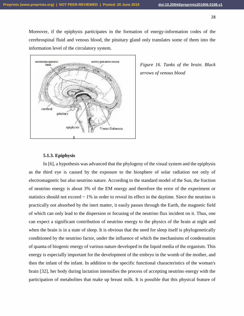
Moreover, if the epiphysis participates in the formation of energy-information codes of the
cerebrospinal fluid and venous blood, the pituitary gland only translates some of them into the
information level of the circulatory system.
Figure 16. Tanks of the brain. Black
arrows of venous blood
5.1.3. Epiphysis
In [6], a hypothesis was advanced that the phylogeny of the visual system and the epiphysis
as the third eye is caused by the exposure to the biosphere of solar radiation not only of
electromagnetic but also neutrino nature. According to the standard model of the Sun, the fraction
of neutrino energy is about 3% of the EM energy and therefore the error of the experiment or
statistics should not exceed ~ 1% in order to reveal its effect in the daytime. Since the neutrino is
practically not absorbed by the inert matter, it easily passes through the Earth, the magnetic field
of which can only lead to the dispersion or focusing of the neutrino flux incident on it. Thus, one
can expect a significant contribution of neutrino energy to the physics of the brain at night and
when the brain is in a state of sleep. It is obvious that the need for sleep itself is phylogenetically
conditioned by the neutrino factor, under the influence of which the mechanisms of condensation
of quanta of biogenic energy of various nature developed in the liquid media of the organism. This
energy is especially important for the development of the embryo in the womb of the mother, and
then the infant of the infant. In addition to the specific functional characteristics of the woman's
brain [32], her body during lactation intensifies the process of accepting neutrino energy with the
participation of metabolites that make up breast milk. It is possible that this physical feature of
Preprints (www.preprints.org) | NOT PEER-REVIEWED | Posted: 20 June 2019 doi:10.20944/preprints201906.0188.v1
29
mother's milk is due to the rapid rate of development and structuring of the neocortex in mammals,
both in phylogeny and in ontogeny.
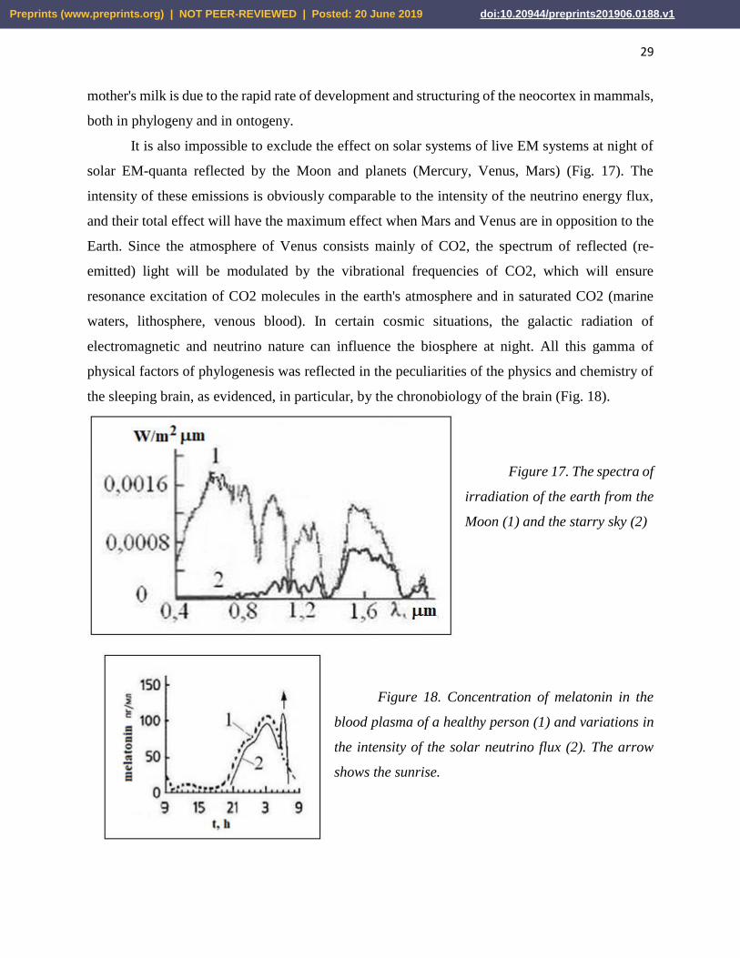
It is also impossible to exclude the effect on solar systems of live EM systems at night of
solar EM-quanta reflected by the Moon and planets (Mercury, Venus, Mars) (Fig. 17). The
intensity of these emissions is obviously comparable to the intensity of the neutrino energy flux,
and their total effect will have the maximum effect when Mars and Venus are in opposition to the
Earth. Since the atmosphere of Venus consists mainly of CO2, the spectrum of reflected (re-
emitted) light will be modulated by the vibrational frequencies of CO2, which will ensure
resonance excitation of CO2 molecules in the earth's atmosphere and in saturated CO2 (marine
waters, lithosphere, venous blood). In certain cosmic situations, the galactic radiation of
electromagnetic and neutrino nature can influence the biosphere at night. All this gamma of
physical factors of phylogenesis was reflected in the peculiarities of the physics and chemistry of
the sleeping brain, as evidenced, in particular, by the chronobiology of the brain (Fig. 18).
Figure 17. The spectra of
irradiation of the earth from the
Moon (1) and the starry sky (2)
Figure 18. Concentration of melatonin in the
blood plasma of a healthy person (1) and variations in
the intensity of the solar neutrino flux (2). The arrow
shows the sunrise.
Preprints (www.preprints.org) | NOT PEER-REVIEWED | Posted: 20 June 2019 doi:10.20944/preprints201906.0188.v1

30
One of the ways of metabolizing neutrino energy can be the synthesis reaction in the
epiphysis of neurohormone melatonin, which plays a key role in the process of puberty and in the
formation of the spectral and energy characteristics of the surface layer of the skin. The process of
melatonin synthesis is modulated by circadian rhythms and suppressed by bright light. Melatonin
is discharged into the cerebrospinal fluid of the third ventricle and into the venous blood, and its
maximum concentration in the cerebrospinal fluid is observed about three o'clock in the morning,
reaching an order of magnitude greater than its content in the blood [43]. It should be noted that at
three o'clock in the morning the blood pressure and body temperature are of minimal importance,
which contributes to the process of self-organization of liquid media and thereby increases the
efficiency of accepting quanta of biogenic energy [3, 29]. In the processes of acceptance and
utilization of energy in addition to melatonin involved chiral sugars and phosphorus. Their
concentrations reach a maximum also in the early morning hours before sunrise [15] (Fig. 18). In
the summer dew drops out at the same time. Saturation with neutrino energy of liquid media
intensifies the enzymatic oxidation of sugars, which together with an increase in the concentration
of phosphorus gives an increase in the rate of ATP synthesis. Thus, neutrino energy at night can
play the role of chiral kinetic factor of bioenergetics.
Figure 19. Schemes of structures of serotonin and melatonin
The phylogenetic and chemical precursor of melatonin is the neurohormone serotonin (Fig.
19). Substitution of substituents in the ring and under nitrogen during the transition from serotonin
to melatonin significantly changes its physicochemical properties. Due to -C = O group, melatonin
will accept quasi-photons from CO2 of venous blood. In addition, melatonin can serve as an
acceptor of energy quanta, which condense in the vitreous body of the eye and in the cerebrospinal
fluid in a state of sleep. In the metabolism of nutrient energy, other neurohormones can also
participate with melatonin. First of all, this refers to dopamine, as evidenced, for example, by such
experience. The intake of motilium (an antagonist of peripheral dopamine receptors) and
Preprints (www.preprints.org) | NOT PEER-REVIEWED | Posted: 20 June 2019 doi:10.20944/preprints201906.0188.v1
31
omeprazole (a hydrogen pump inhibitor) for two weeks along with a positive effect led to a
decrease in libido, sleep disturbances (regular awakenings at three o'clock in the morning) and an
increase in the allergic reaction of the skin (hives). Synchronization of these disorders with the
melatonin synthesis kinetics (Fig. 18) allows them to be associated with hypertrophy of the
hormonal function of melatonin, the cause of which may be a concentration imbalance between
melatonin and dopamine.
Since the epiphysis is completely immersed in incompressible cerebrospinal fluid and
close to it there is a large venous junction that includes a large cerebral vein of Galen (Fig. 16),
the volume of the epiphysis, and hence the volume and pressure in its internal cavity [44] will
pulsate. At the expansion stage, on the gas molecules in the volume and on the structures of the
inner surface of the cavity, quanta of biogenic energy will condense, which at the compression
stage can be translated together with metabolites into the parenchyma of the epiphysis, activating
its metabolism, and also into the cerebrospinal fluid of the third ventricle.
5.2. Kinetic parameters of the physics of the brain
The rhythm of some physical processes in the brain can be caused by periodic metabolic
reactions. Such processes obviously include the pulsation of the neuroglia - 12 seconds the phase
of tension and 240 with the relaxation phase, while their volume changes, their outgrowths swell
and swell [45]. The vibrations of the chemical activity of the synapses have a time constant of the
order of 100 ms, which corresponds to the total duration of the exciting and inhibitory postsynaptic
potentials ~30 and ~70 ms, respectively. Given the predominance of circadian rhythms in
chronobiology, it can be assumed that the basis of the mechanism of the pacemaker of the brain or
heart is the connection of the electrical activity of special nerve cells with this or that periodic
phenomenon of electromagnetic nature of geophysical or cosmic scale [46]. Spontaneous relic
radiation, pulsations of the geomagnetic field and its periodic disturbances by the Sun, the Moon
and other planets can serve as examples of such phenomena [47]. High sensitivity of pacemakers
to weak external signals is achieved due to cooperative effects in the ordered beams of neurons of
suprachiasmatic nuclei, the bundle of His and the nuclei of the reticular formation.
Preprints (www.preprints.org) | NOT PEER-REVIEWED | Posted: 20 June 2019 doi:10.20944/preprints201906.0188.v1

32
Figure 20. Amplitude-frequency
ratios of bioelectric signals [46]
The brain rhythm frequency spectrum corresponds to the electrical activity of various
structures in the brain hierarchy (Fig. 20). The stable rhythms of the EEG spectrum include: delta
rhythm (0.5-4 Hz); theta rhythm (5-7 Hz); alpha-rhythm (8-13 Hz); mu-rhythm - similar in
frequency-amplitude characteristics to alpha-rhythm but predominates in the anterior cortices of
the cerebral hemispheres; beta-rhythm (15-35 Hz) and gamma-rhythm (above 35 Hz). Mu-rhythm,
possibly associated with the electromagnetic activity of the eyes and the rhythm of scanning the
EM-vortex of the frontal lobes of the brain. The cause of other rhythms can be electromagnetic
oscillations that capture the cortex and various structures of the subcortex. In principle, for each
type of oscillations in the EEG spectrum, capacitive-induction LC structures can be isolated in the
brain and model their interconnection by an equivalent circuit of the oscillatory circuit, which has
its own frequency according to (6).
For example, the alpha rhythm, responding to the background electrical activity of the
cortex, maintains at the proper level the stability of the neocortical and thalamus connections.
When removing the thalamus or cutting off its bonds with the bark, the alpha rhythm disappears.
Right-left parts of the thalamus and the cortex of the hemispheres can be represented by dissimilar
plates of two spherical capacitors, and the nerve connections between them (the radiance of the
thalamus) will simulate ohmic connections and inductive coils in equivalent circuits of circuits
operating at the alpha-rhythm frequency (alpha-circuit) (Fig. 21). The asymmetry of the inductive
elements of the alpha contours of the right and left hemispheres can be the basis of their functional
specification. The frequency of oscillations in the alpha contour is presumably given by the
Preprints (www.preprints.org) | NOT PEER-REVIEWED | Posted: 20 June 2019 doi:10.20944/preprints201906.0188.v1

33
pacemaker nuclei of the reticular formation, which is closely related to the thalamus. The
characteristic time for tuning synchronized with the alpha rhythm is ~ 100 ms.
Figure 21. Equivalent oscillatory circuits modeling the alpha rhythms of the brain. L, L *,
R - inductive and ohmic models of the radiance of the thalamus (sign *) means a mirror inversion
of the chiral structures of the right hemisphere); r - intertalamic fusion; C and U are the
capacitance and potential difference between the thalamus and the cortex.
Currents in the nervous structures of the reticular formation and the medulla oblongata can
generate vortex magnetic fields in the structures of the variolium bridge and the cerebellum (Fig.
22). The appearance of this formation, in principle, is isomorphic to c) the model of the
transformed vibrational contour (Fig. 1). Consequently, pacemakers of the reticular formation can
resonantly tune to oscillations of a standing EM wave of the geomagnetic field (see Section 2.4.2)
and to regular perturbations of the geomagnetic field by solar activity or planets.
The time to assimilate one bit of visual information is 15 - 50 ms [28]. The time of the
elementary mental act lies in the range of 150-300 msec [48]. From the evaluation of the speed of
mastering the meaning of a readable text consisting of known words, it follows that on average,
about 200 ms are required to understand the meaning of a single word. This time constant can be
associated with the theta rhythm, which manifests cortico-limbic interactions, regulating emotions
and mental activity [15].
Preprints (www.preprints.org) | NOT PEER-REVIEWED | Posted: 20 June 2019 doi:10.20944/preprints201906.0188.v1
34
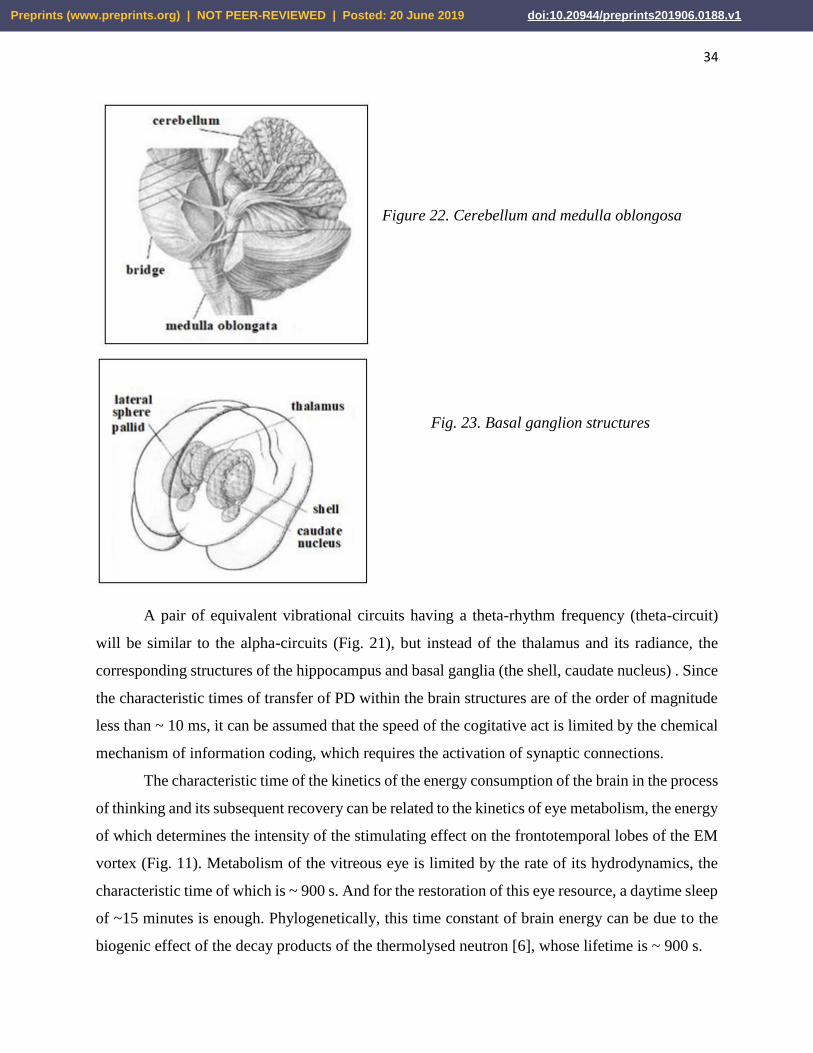
Figure 22. Cerebellum and medulla oblongosa
Fig. 23. Basal ganglion structures
A pair of equivalent vibrational circuits having a theta-rhythm frequency (theta-circuit)
will be similar to the alpha-circuits (Fig. 21), but instead of the thalamus and its radiance, the
corresponding structures of the hippocampus and basal ganglia (the shell, caudate nucleus) . Since
the characteristic times of transfer of PD within the brain structures are of the order of magnitude
less than ~ 10 ms, it can be assumed that the speed of the cogitative act is limited by the chemical
mechanism of information coding, which requires the activation of synaptic connections.
The characteristic time of the kinetics of the energy consumption of the brain in the process
of thinking and its subsequent recovery can be related to the kinetics of eye metabolism, the energy
of which determines the intensity of the stimulating effect on the frontotemporal lobes of the EM
vortex (Fig. 11). Metabolism of the vitreous eye is limited by the rate of its hydrodynamics, the
characteristic time of which is ~ 900 s. And for the restoration of this eye resource, a daytime sleep
of ~15 minutes is enough. Phylogenetically, this time constant of brain energy can be due to the
biogenic effect of the decay products of the thermolysed neutron [6], whose lifetime is ~ 900 s.
Preprints (www.preprints.org) | NOT PEER-REVIEWED | Posted: 20 June 2019 doi:10.20944/preprints201906.0188.v1

35
5.3. Nonlocal quantum correlations
5.3.1. Consciousness and memory.
The physics of consciousness or self-consciousness, being identical with the physics of
thinking and speech, is based on the ability of the brain to respond to external signals, transform
them into electromagnetic codes of the neural network, remember and recognize these codes,
transform and synthesize the template of them. All these operations are possible with the presence
in the brain of physicochemical mechanisms of reading-activation and memorization-conservation
codes. The process of reading and recognizing the thought-form itself implies the existence of a
stable dynamic reference system of reference codes, which constitutes the physical basis of the
subject's self-consciousness. This system is formed in the process of phylogenesis and ontogeny
according to the genetic program with continuous influence on the brain through the sense organs
(sight, hearing, touch, smell, taste) of the whole range of physical and social organizing factors. In
the absence of the latter, the ontogeny of the brain normal from the child's birth does not reach the
mental level at all. On the other hand, deaf-blind children from birth, having only tactile contact
with the teacher, are able to form a sufficiently adequate reference frame reference system that
allows them to learn the world and communicate with people.
The genetic program of the reference codes that determine the individuality of a person
is laid already when the metric of the genetic material is formed in the process of fertilization of
the female cell. The unfolding of this program at the stage of embryogenesis is subject to the law
of fractal-resonant action [6], which is realized through the physical mechanisms of energy-
information exchange and memory underlying the psycho- or spiritually-physical isomorphism.
Genetic codes, making up the basic level of memory, operate throughout the life of a
person, determining the racial, sexual and mental characteristics of his brain physics. The next
level of memory is made up of unconditioned reflexes, the principles of its organization are laid at
the stage of morphogenesis. At the same stage, the chemical basis of the memory resource of the
initial sensory impressions and sensations is created. Within the resource of unconscious memory,
under the influence of social factors, an array of a reference system of reference codes of
consciousness is formed. The mechanisms for memorizing and retrieving the necessary reference
code combine the electrophysics and biochemistry of the brain with the physics of dynamical
systems of QPH localized on the structural and functional elements of the brain.
Preprints (www.preprints.org) | NOT PEER-REVIEWED | Posted: 20 June 2019 doi:10.20944/preprints201906.0188.v1

36
The first, gene level of memory is realized spontaneously at the molecular-cellular level in
the synthesis of nuclear DNA, which, in turn, programs the growth and functions of the nerve cell.
In general, the thermodynamic features of the tertiary structure of protein molecules (DNA,
hemoglobin, enzymes, etc.) allow them to condense thermal or metabolic QPH, transforming them
into vibrational-rotational energy of atoms of characteristic amino acid groups. When the
excitation energy of the threshold level is reached, it is transmitted along the fractal-resonance
mechanism to the metabolite, thereby initiating its participation in the chemical synthesis of a
completely defined structure. In this way, gene information can be transmitted from the molecular
to the macroscopic level of the organization of living systems.
The following memory levels are formed by the development of stable neural
connections between the zones of the neocortex and the basal ganglia, limbic brain, thalamus and
cerebellum. The specialization of such memory blocks is determined by the functional dominance
of its second element. Given the synchronism of the morphogenesis of the fronto-parietal lobes of
the brain and the cerebellum, we assume that this block is the main repository of the base codes of
consciousness frames. The background activity of this memory unit ensures continuity of
orientation of the somatic and mental components of consciousness in space and time. The rhythm
of this mode of consciousness can be set by an alpha contour, providing a link between the visual
cortex, the cerebellum and the frontal lobes (Fig. 24).
Figure 24. Scheme of the structure (a) and connections with the cortex of the cerebral
hemispheres; (b) cerebellum – layered "crystal" of the basic block of brain memory. Dotted lines
layers and levels of the cerebellum organization are shown.
Preprints (www.preprints.org) | NOT PEER-REVIEWED | Posted: 20 June 2019 doi:10.20944/preprints201906.0188.v1

37
The switching of consciousness to the mode of thinking is associated with the replacement
of the alpha rhythm with the theta rhythm and, accordingly, with the activation of the memory
block of the mental reference codes, the key structures of which are obviously the basal ganglia.
It can be assumed that the functional asymmetry of the brain and the very logic of thinking are the
result of the synergy of the physics of chiral inductive-magnetic elements of the theta-contour
(caudate nucleus), olfactory organ (hippocampus) and hearing (Ammon horn) in each hemisphere.
Participation in the physics of thinking of the capacitive-electrical element of the organ of vision
(retina) is evidently the activation of the basic memory block by the action on the frontotemporal
lobes of the brain of the EM vortex of the eyes. A similar function of the activation of the parietal
and occipital (preklinya, Fig. 11e) regions of the cerebral cortex, apparently, is performed by the
second capacitively-electric element of the visual system - the external geniculate body. It may
well be the generation of quasiphotons-phosphenes in the fusion-recombination of mirror-
symmetric EFs from the right and left eyes.
The physics of thinking integrates in itself to some extent the functions of all brain
structures. The morphological basis for such an association is the fluid system of the brain. At the
initial stage of embryogenesis, the entire nervous system is localized in the neural tube [49], the
inner layer of which subsequently becomes a single-connected inner surface of the ventricles of
the brain and the central channel of the spinal cord. Subsequently, the cerebrospinal fluid, filling
these volumes and communicating with the cisterns and outer shells of the brain (Fig. 16), assumes
the role of an integrating "bus", on which, in one way or another, all the functional elements of the
nervous system are closed.
5.3.2. The mechanism of quantum correlations
Following the logic of [50], we model a hierarchy of physical states of structural and
functional brain blocks by a multilevel fractal from local dynamical systems of QPH. The basic
unit of this fractal, starting from the level of the neural network, to the hemispheres of the brain,
is the LC-structure, isomorphic to the vibrational contour (Fig. 9, 21). The geometric and
electrophysical parameters of LC structures will determine the type and energy of dynamic QPH,
and their action should formally satisfy the fractal-resonance principle [6]. From chains of LC-
circuits there are self-excited (reverberating by the principle of feedback) closed circuits in neural
networks, which can correspond to images of perception or images of operative memory. The
excitation data can be modeled by damped oscillations, linking their cyclicity and frequency with
Preprints (www.preprints.org) | NOT PEER-REVIEWED | Posted: 20 June 2019 doi:10.20944/preprints201906.0188.v1
38
Q-factor and resonance frequencies of LC-circuits, as well as with the length and degree of
branching of the loop of the neural network. In addition to electrodynamics, in principle, quantum
mechanics (Bose condensation, confusion of states), physics of wave and nonlinear processes
(interference, holography, coherence) can be used to model the cooperative properties of systems
of similar QPH.
The limiting time of a psychic reaction or mental act is of the order of ~ 100 ms. During
this time tens of thousands of synaptic connections are involved in the brain and tens of zones of
the cortex and structures of the subcortex are activated. Even if the operations in the neural network
are parallel and the associative and reflexive connections between the cortical zones and the
memory blocks are high in order to ensure the purposefulness of the functional activity of the
brain, a quantum mechanism of searching and selecting information must operate in it, the kinetics
of which should not be limited by the rates of transport of matter and energy. The morphological
basis of this mechanism is the single-connectedness of the aquatic environment of the brain and
the fractality of its EM structure. For example, each layer of the basic memory block (Figure 24),
consisting of a pair - a Purkinje tree cell and a lianon neuron [49], can be considered isomorphic
to the entire fractal of the neural cortical and spinal neural connections, together with the vagus
nerve (vagus). Isomorphism of the nerve cell - macrostructure provides high efficiency of the basic
memory block of the reference codes with the maximum value of its specific capacity.
The interconnection of quantum systems can be of two types. The first one presupposes
the overlapping of the wave functions of the electrons of two spatially separated structures of the
nervous system of the brain. In the energy-information exchange, QPH of various types (solitons,
phonons, EM quanta), and electron or proton (tunnel junctions) are involved. Such a relationship,
in fact, is analogous to the mechanism of saltatory conductivity and is effective for chemically
bound systems. The second type of connection - nonlocal quantum correlations [7] assumes an
information exchange between isomorphous structures of the brain by the mechanism of fractal-
resonant coupling [6]. This mechanism can be extended to the exchange of information between
the brain and the environment. It operates at the molecular level with DNA replication, and at the
socio-biological level, especially between mother and daughter.
In general, for the implementation of the mechanism of nonlocal correlations, systems must
satisfy two requirements. The first is that in the embryonic state they must form a simply-connected
system in a quantum state determined by one or another type of interaction (nuclear,
Preprints (www.preprints.org) | NOT PEER-REVIEWED | Posted: 20 June 2019 doi:10.20944/preprints201906.0188.v1
39
electromagnetic, weak, gravitational, biological, social). Examples: positronium, radioactive
nucleus, protostar, DNA molecule, neural tube, embryo and the mother's body. The second
requirement is called the inseparability or entanglement of states of chemically unrelated systems
and consists in the presence of a mechanism of quantum correlation of the physical characteristics
of states. For the above systems, their daughter correlating subsystems will be, respectively: two
photons, products of radioactive decay, binary stars or star-planetary systems, split DNA,
cerebellum and neocortex, mother and daughter.
The mechanism of quantum entanglement implies the preservation of the information
connection between the daughter subsystems at the level of interactions that determine the intrinsic
integrity of the subsystems. The universalism of quantum entanglement is genetically conditioned
by the subordination of the process of formation of discrete forms of matter, and then of elementary
particles to the bootstrap mechanism [6]. At the level of elementary particles and nuclei, on which
nonlocal correlations are experimentally observed [7], the nature of information interactions has
not yet been established, one can only assume that their kinetics is limited by the momentum
transfer rate in the physical vacuum (ether) ~ 1022 cm/s [6]. At the same speed, obviously,
information is exchanged at the level of social and space subsystems.
As a basis for the mechanism of entanglement of the structural and functional subsystems
of the brain, it is possible to take the single-connected nature of its aqueous basis, thanks to which
information exchange with a velocity V = C/n (5) is possible between systems of QPH. Submission
of this exchange to the fractal-resonance principle of the action reduces its probabilistic nature to
a purposeful selection between isomorphic structures of the pair, for which the level of
entanglement of the states of QPH is maximal.
6. Conclusion
A century and a half ago Maxwell Maxwell linked the development of fundamental physics
with the knowledge of the brain [5]. More than 30 years ago, F. Creek suggested that molecular
biology can very well establish the biochemical basis of the most complex mental and mental
functions of the brain, reaching a level of "biochemical theology" or "theochemistry" [51].
However, in 2000, F. Krik had already refrained from predicting the development of brain
biochemistry, emphasizing only the great importance of nonlinear dynamic systems in biology at
all levels. This clarification can easily be taken as an adaptation of the idea of Maxwell to
Preprints (www.preprints.org) | NOT PEER-REVIEWED | Posted: 20 June 2019 doi:10.20944/preprints201906.0188.v1

40
biological objects and conclude that the transition of brain biochemistry to "theology" can be
realized only simultaneously with the replacement of the materialistic paradigm of physics by the
spiritual. In other words, the physics of thinking can take place only as a natural theology or
theophysics. The evolution of natural science already has one dialectical leap in the form of a
transition from classical physics to quantum physics. Obviously, the next jump in the depths of
natural science has ripened - from quantum physics to physics of spirit or theophysics [52]. For all
its dramatic nature for the materialist physicists and idealist psychologists, this leap is inevitable,
and taking into account this, an analysis of the probable path of its realization is carried out in this
paper.
Reference
1. Kholmanskiy A.S., Strebkov D.S. Energy of the noosphere. Russian Agricultural Sciences.
2004 (1) 44-47
2. Eidi W.R. Cooperative mechanisms of susceptibility of brain tissue to external and internal
fields. M., Physiology of man. 1975
3. Kholmanskiy, A. Chirality anomalies of water solutions of saccharides. J. Mol. Liq 2016, 216,
pp. 683-687. http://dx.doi.org/10.1016/j.molliq.2016.02.006
4. Bekhtereva N.P., et al. Neurophysiological mechanisms of thinking. L., 1985. 272 p.
5. Maxwell J.K. Selected works on the theory of the electromagnetic field. M. 1954
6. Kholmanskiy, A.S. Chirality and quantum effects as factors of morphogenesis. Electron.
Math. Med.-Biol. J. 2010. 9(4) URL: http://www.sci.rostelecom67.ru/user/sgma/MMORPH/N-
28-html/kholmanskiy-2/kholmanskiy-2.htm ; . Fractal-resonance principle of action. 2003, MIS-
RT, 29(2). URL: http://www.ikar.udm.ru/sb/sb29-2.htm .
7. Menskii M B. Concept of consciousness in the context of quantum mechanics”, UFN, 175:4
(2005), 413–435; https://doi.org/10.3367/UFNr.0175.200504c.0413
8. Kobozev N.I. Selected works. T. 2. MSU. 1978. 396 p.
9. Tatur T.A., Fundamentals of the theory of the electromagnetic field. M. 1989
10. Kholodov Yu. A., et al. Magnetic fields of biological objects. M .: Nauka, 1987.
11. Naatanen R., Attention and function of the brain. Moscow State University. 1998
12. Huho F. Neurochemistry, Fundamentals and Principles. M. 1990
13. Sotnikov O.S. Dynamics of the structure of a living neuron. M. Nauka, 1982.
Preprints (www.preprints.org) | NOT PEER-REVIEWED | Posted: 20 June 2019 doi:10.20944/preprints201906.0188.v1

41
14. Matveev V.V., Revolution and counter revolution in cell physiology. Cell Biol. Int. 2002, 26;
http://bioparadigma.narod.ru/reviewonpollack.htm
15. The Physiology of Man, ed. R. Schmidt, G. Tevs. 1- 3 tons, M., 1996
16. Poliak S., Peles E. Nature Reviews Neuroscience V. 4. No. 12. P. 968-980 (2003)
17. Alberts B. et al. Molecular biology of the cell. T 3. M., Mir, 1994
18. Terekhov S.A, Lectures on the theory and applications of artificial neural networks.
Snezhinsk, 1994-1998.
19. Hotels A.A. Taurus Fatter Pacini. L. 1976
20. Katila T. et al. Magnetic fields produced by human eye. J. Appl. Phys. 1981, V. 52 (3) 2565
21. Williamson, S.J., Kaufman, L., Brenner, D., Evoked neuromolecular fields of the human
brain, J. Appl. Phys. 1979, V. 50 (3), 2418 - 2421
22. Vvedensky V.L, Ozhogin V.I, Supersensitive magnetometry and biomagnetism. M. 1986
23. Raevskaya OS, Geomagnetic field and human organism, Uspekhi Fiziolog Sci.. 1988 (9)4
24. Kholodov Yu. A., Lebedeva NN Reactions of the human nervous system to
electromagnetic fields. M. 1992
25. Fenish H. Pocket Atlas of Human Anatomy. Minsk 1997
26. Hubel D. Eye, brain, vision, M., 1990
27. Soloviev AN To the question of the mechanism of the biological action of a pulsed
magnetic field. DAN USSR, 1963, 149(2);
28.Alexander H. S. The biological effects of magnetic fields. Am. J. Med. Electronics, 1962, v. 1,
No. 4
29. Khatsevich TN, Medical Optical Devices: Physiological Optics: Tutorial. Novosibirsk:
SSGA. 1998. Part 1. 98 sec.
30. Baker, R., Magnetic bone in sinus of man, Nature, 1983, v. 301, p. 78-80
31. Bragina N. N., Dobrokhotova T.A. Functional asymmetries in humans, M., 1981
32. Cahill L. His brain, her brain: http://www.nanonewsnet.com
33. Lifshits I.M, Quasiparticles in modern physics, in: In the depths of the atom, Moscow, 1964
34. Kizel V.A. Practical molecular spectroscopy. M. MIPT. 1998. 254 p.
35. Kassil G. Brain barrier. Science and life. 1986. № 11
36. Temur'yants N.A. et al. Ultrahigh-frequency electromagnetic signals in biological world.
Kiev, 1992.
Preprints (www.preprints.org) | NOT PEER-REVIEWED | Posted: 20 June 2019 doi:10.20944/preprints201906.0188.v1

42
37. Kudryashov Yu.B. Biophysical basis of the action of microwaves. Moscow: MSU. 1980
38. Sinitsyn N.I. et al. Biomedical radioelectronics. 1998. 1.
39. Kholmanskiy , A. Alzheimer's in Post-Industrial Epoch. Preprints 2019, 2019050345 (doi:
10.20944/preprints201905.0345.v1 .
40. Musil Ya., Novakova O., Kuntz K. Modern biochemistry in the schemes. M. 1984, 214 p.
41. Kholmanskiy, A.; Zaytseva, N. Physically adequate approximations for abnormal
temperature dependences of water characteristics. J. Mol. Liq. 2019, 275, pp. 741–748.
https://doi.org/10.1016/j.molliq.2018.11.059
42. Etkin V.A. On the specifics of spin-spin interactions; On the orientational interaction of spin
systems. NiT, 2002.
43. Kovalzon, V.M. Foundations of somnology: physiology and neurochemistry of the cycle
«wakefulness-sleep». M, 2014, 239 p.
44. Khelimskiy A.M Epiphysis, M, 1969
45. Katalymov L.L. Physiology of excitation and inhibition, Ulyanovsk, 1997
46. Gnezditsky V.V. Evoked brain potentials in clinical practice. Taganrog: TSTU, 1997.
47. Biological rhythms, ed. J.Ashoff. 1, 2 t. M. 1984
48. Ivanitsky A.M, Strelets V.B, Korsakov I.A. Information processes of the brain and mental
activity. M. 1984.
49. Cowen W. Development of the brain. In the collection. Brain. M. 1984
50. Peled A., Geva A.B., Brain Organization and Psychodynamics. J. Psychotherapy Practice
Res. 1999; 8: 24-39.
51. Krik F. Molecular biology in 2000. Nature. 1970. 228. 613-615.
52. Kholmansky, A. Beginning of theophysics. M. 124 р. ISBN 5-86020-375-6. URL:
https://dlib.rsl.ru/viewer/01000633036#?page=1
Preprints (www.preprints.org) | NOT PEER-REVIEWED | Posted: 20 June 2019 doi:10.20944/preprints201906.0188.v1