mlab 1227: coagulation keri brophy-martinez coagulation disorders: secondary hemostasis part two
TRANSCRIPT
MLAB 1227: CoagulationMLAB 1227: CoagulationKeri Brophy-MartinezKeri Brophy-Martinez
Coagulation Disorders: Secondary HemostasisPart Two
Acquired Coagulation Acquired Coagulation DisordersDisordersTwo or more factors generally
affected, more complicatedBleeding from multiple sitesMore common than hereditary
disordersClassification
1. DIC2. Primary Fibrinogenolysis3. Liver Disease4. Vitamin K Deficiency5. Acquired Pathologic Inhibitors
1. DIC: Disseminated 1. DIC: Disseminated Intravascular CoagulationIntravascular CoagulationConsumption Coagulopathy
◦ As fibrin is formed, clotting proteins and naturally occurring inhibitors and platelets are consumed faster than they are made
◦ Thrombo-hemorrhagic disorder Clotting and lysing occurring in blood vessel, at
the same time◦ Life threatening◦ Bleeding is the most apparent
characteristic◦ Initiating events are thrombotic, where
material enters circulation ◦ Occurs due to lack of the negative
feedback mechanism◦ Affects young and elderly
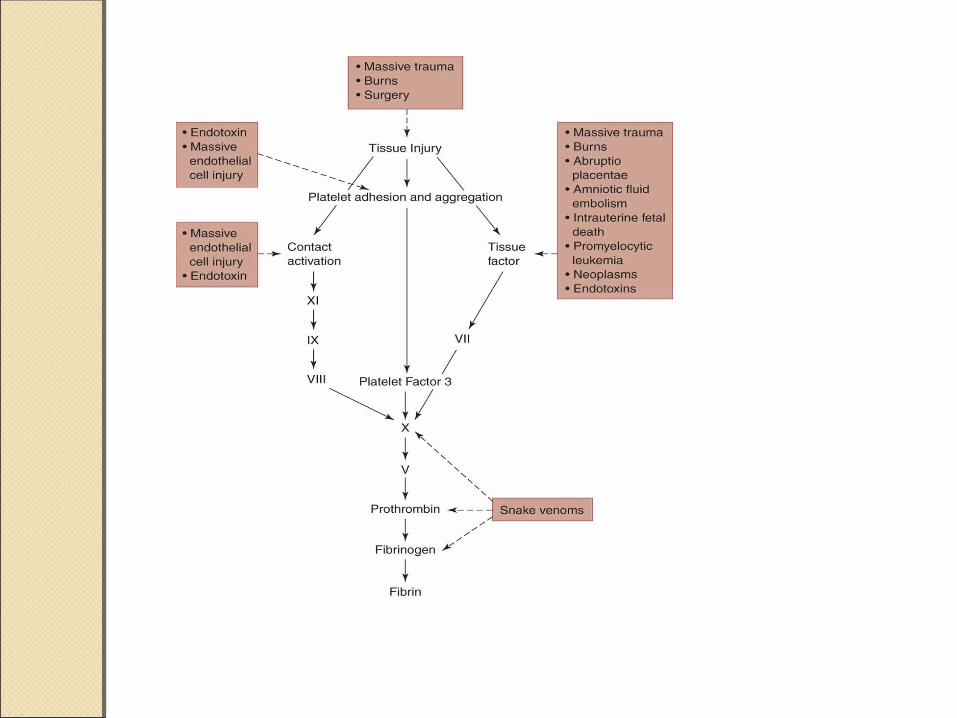
DIC: TriggersDIC: Triggers Obstetric – usually due to major tissue damage such as
retained dead fetus, abruptio placentae, or placenta previa
Acute leukemias – Promyelocytic – increase number of granules released into circulation as cells break down
Intravascular hemolysis – ex: transfusion reaction Massive trauma (especially crushing injuries), burns,
surgical procedures Heat stroke Snake venoms Septicemias and infections – viral, bacterial, rickettsial,
fungal, protozoan (especially gram negative that release endotoxins)
Tumors – foreign tissues and cells Prosthetic devices – heart valves, aortic balloon,
peritoneal shunting Vascular disease – damaged endothelial lining

Course of DICCourse of DIC
DIC: How Does It Occur?DIC: How Does It Occur?
Step 1: Out of control clotting◦Causes widespread fibrin
deposits in vessels of tissues and organs
◦Subsequent event: Hemorrhage Clotting proteins consumed at a high
rate Causes multiple factor deficiencies,
especially fibrinogen group
Platelets caught in thrombi and removed
DIC: How Does It Occur?DIC: How Does It Occur?
Step 2: Triggers Fibrinolytic system to remove fibrin◦Results in:
Circulating degradation products (FDPs) that interfere with platelet function & normal clot formation
Degradation of Factor V & VIII
DIC: How Does It Occur?DIC: How Does It Occur?
Step 3: Uncontrolled plasmin and thrombin enter circulation◦Why?
Inhibitors such as AT have been depleted
DIC: How Does It Occur?DIC: How Does It Occur?
Step 4: Appearance of Symptoms◦Bleeding from multiple sites◦Petechiae◦Purpura◦Occlusions in organs◦Oozing from needle puncture
sites◦Shock
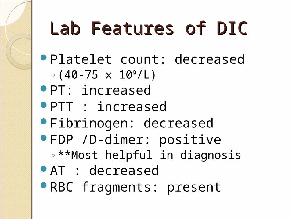
Lab Features of DICLab Features of DIC
Platelet count: decreased ◦(40-75 x 109/L)
PT: increasedPTT : increasedFibrinogen: decreased FDP /D-dimer: positive
◦**Most helpful in diagnosisAT : decreasedRBC fragments: present
DICDIC
Treatment◦Goal is to treat the underlying
condition Remove the triggering process –
treat with antibiotics, antineoplasms, remove dead tissue, treat the diseases or conditions
◦Heparin – to prevent or limit further coagulation
◦Replace factors, platelets = give FFP
2. Primary 2. Primary FibrinogenolysisFibrinogenolysisSimilar to DICPlasminogen is inappropriately
activated to plasminPlasmin circulates overwhelming
the antiplasmin inhibitors and degrading fibrinogen and factors V,VIII, XIII
No thrombin is generatedLiver disease is a common trigger
3. Liver Disease3. Liver DiseaseAffects all proteins made in the
liver that function in fibrin formation, fibrinolysis and inhibition.
Patients show minimal bleeding, except in severe cases
Lab features◦Increased
PT,PTT◦Decreased
Platelets
4. Vitamin K Deficiency4. Vitamin K DeficiencyLiver cells able to make precursor
protein but the calcium binding site is nonfunctional
Causes◦Malabsorptive syndromes
Sprue Obstruction in biliary tract Ingestion of vitamin K inhibitors- like warfarin
◦Antibiotic therapy Kills off normal flora in gut which made
vitamin K
5. Acquired Pathologic 5. Acquired Pathologic InhibitorsInhibitorsDevelop in patients with certain
disease states and others with no underlying conditions
Circulating anticoagulants which may develop against any clotting factor
Classed as immunoglobulins◦Either IgG or IgM◦Can be alloantibodies or autoantibodies
Types of InhibitorsTypes of Inhibitors1. Directed towards a single
coagulation factor◦ Seen in patients with inherited factor
deficiencies that have had replacement therapy for bleeding complications
◦ Less commonly seen in healthy people and those taking certain drugs
◦ Rare, except Factor VIII & IX◦ How do we find them?
Interfere with clotting factor activity PTT prolonged, other tests normal Mixing study: test will still be prolonged

Types of InhibitorsTypes of Inhibitors2. Lupus Inhibitor/Anticoagulant
◦ Seen in patients with autoimmune diseases, drug reactions, but also in normal patients
◦ Autoantibodies interfere with phospholipid-dependent reagents used in PTT tests
◦ Patients have no bleeding problems (though some have an increase risk of thrombosis)
◦ In vitro, any coag test using a phospholipid reagent will be falsely prolonged (PT, PTT)
◦ Coag studies must be performed using reagents that do not contain phospholipids
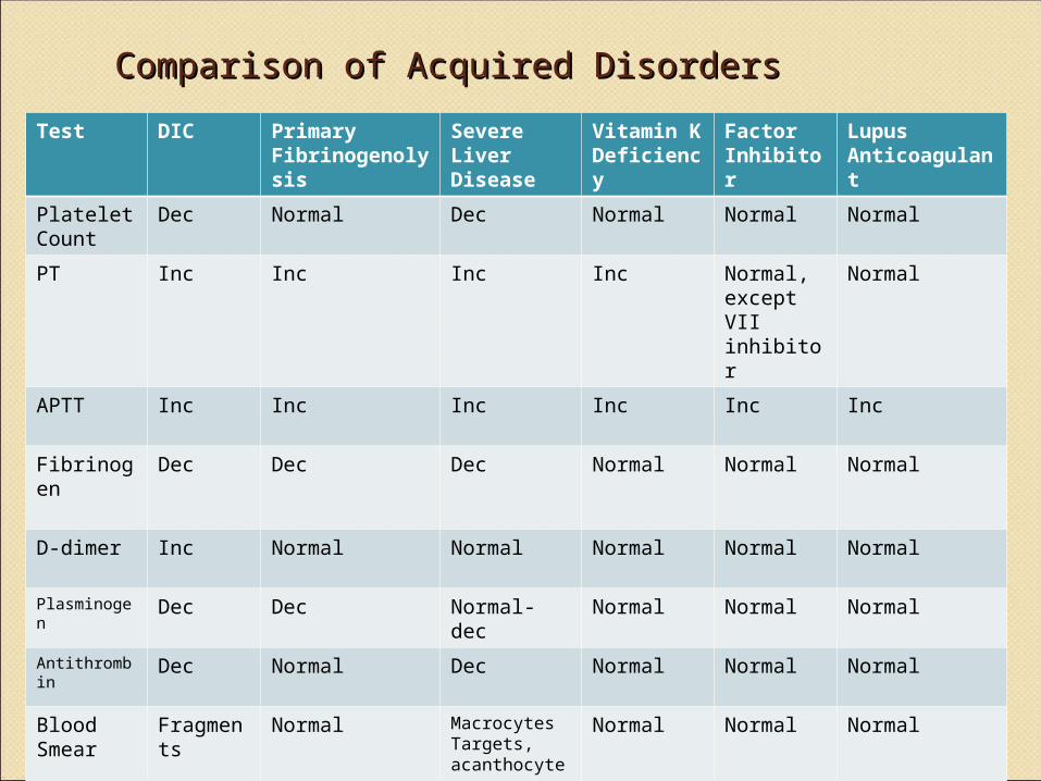
Comparison of Acquired DisordersComparison of Acquired Disorders
Test DIC Primary Fibrinogenolysis
Severe Liver Disease
Vitamin K Deficiency
Factor Inhibitor
Lupus Anticoagulant
Platelet Count
Dec Normal Dec Normal Normal Normal
PT Inc Inc Inc Inc Normal, except VII inhibitor
Normal
APTT Inc Inc Inc Inc Inc Inc
Fibrinogen
Dec Dec Dec Normal Normal Normal
D-dimer Inc Normal Normal Normal Normal Normal
Plasminogen
Dec Dec Normal-dec Normal Normal Normal
Antithrombin
Dec Normal Dec Normal Normal Normal
Blood Smear
Fragments
Normal MacrocytesTargets, acanthocytes
Normal Normal Normal
ReferencesReferences
McKenzie, Shirlyn B., and J. Lynne. Williams. "Chapter 32." Clinical Laboratory Hematology. Boston: Pearson, 2010. Print.