mitochondrial point mutations do not limit the natural lifespan of mice
TRANSCRIPT
Mitochondrial point mutations do not limit the naturallifespan of miceMarc Vermulst1, Jason H Bielas1, Gregory C Kujoth2, Warren C Ladiges3, Peter S Rabinovitch1,Tomas A Prolla2 & Lawrence A Loeb1
Whether mitochondrial mutations cause mammalian aging,or are merely correlated with it, is an area of intense debate1.Here, we use a new, highly sensitive assay2 to redefine therelationship between mitochondrial mutations and age. Wemeasured the in vivo rate of change of the mitochondrialgenome at a single–base pair level in mice, and wedemonstrate that the mutation frequency in mousemitochondria is more than ten times lower than previouslyreported. Although we observed an 11-fold increase inmitochondrial point mutations with age, we report that amitochondrial mutator mouse3 was able to sustain a 500-foldhigher mutation burden than normal mice, without anyobvious features of rapidly accelerated aging. Thus, our resultsstrongly indicate that mitochondrial mutations do not limit thelifespan of wild-type mice.
Mitochondria are functionally diverse organelles with a central role inboth oxidative phosphorylation4 and apoptosis5. Accordingly, post-mitotic tissues with high rates of oxygen consumption, such as brainand heart, are exceptionally sensitive to mitochondrial dysfunction6.The mitochondrial theory of aging postulates that a lifelong accumu-lation of mitochondrial DNA (mtDNA) mutations in multiple tissueseventually results in mitochondrial failure, which, in synergy withdownstream processes such as apoptosis, results in loss of cellularityand the progressive decline of tissue functioning known as aging3,7.These mutations may compromise the integrity of the electrontransport chain and increase the formation of reactive oxygen species(ROS), creating a vicious cycle of mutagenesis that continuouslyamplifies the production of cytotoxic oxygen radicals8,9.
Although several studies have quantified either tissue-specific muta-tion frequencies10 or mutation accumulation as a function of age11,most methods rely heavily on PCR and cloning-based strategies12,13,techniques that are limited in throughput and that may be con-founded by polymerase infidelity on damaged templates and bycloning artifacts. Therefore, we explored the relationship betweenage and mutation accumulation in mtDNA with an adaptationof the random mutation capture (RMC) assay2,14, a quantitative
PCR-based approach that relies on PCR amplification of singlemolecules for mutation detection (Supplementary Fig. 1 online)but is not limited by polymerase fidelity2,14. This methodology allowsfor exact determination of mutation frequencies in high-throughputscreens that interrogate millions of base pairs simultaneously2,14.Approximately 150 million bp were screened for mutation detectionin this study.
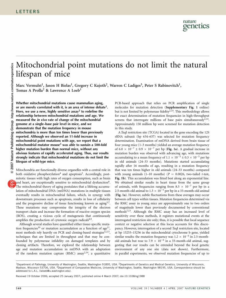
ATaqI restriction site (TCGA) located in the gene encoding the 12SrRNA subunit (bp 634–637) was selected for mutation frequencydetermination. Examination of mtDNA derived from brain tissue offour young mice (1–3 months) yielded an average mutation frequencyof 6.0 � 10�7 ± 0.9 � 10�7 per bp (Fig. 1a). A gradual increase inmutation burden was observed with advancing age, with mutationsaccumulating to a mean frequency of 1.1 � 10�5 ± 0.3 � 10�5 per bpin old animals (24–33 months). Mutations started accumulatingrapidly after 16 months of age, resulting in a mutation frequencythat was ten times higher in old animals (24–33 months) comparedwith young animals (1–10 months) (P ¼ 0.0026, two-tailed t-test,Fig. 1b). This accumulation was fitted best along an exponential line.We obtained similar results in heart tissue from the same groupof animals, with frequencies ranging from 8.5 � 10�7 per bp in a2.5-month-old animal to 1.3 � 10�5 per bp in a 33-month-old animal(Fig. 1a). However, subtle fluctuations from these averages may occurbetween cell types within tissues. Mutation frequencies determined viathe RMC assay in young mice are approximately one to two ordersof magnitude lower than previously documented by conventionalmethods7,13. Although the RMC assay has an increased level ofsensitivity over these methods, it registers mutational events at theinterrogated restriction site only; thus, it is possible that local sequencecontext or negative selection at this locus accounts for this discre-pancy. However, interrogation of a second TaqI restriction site, locatedat bp 15253–15256 in the mitochondrial cytochrome b gene, yieldedsimilar results: the mutation frequency was 1.2 � 10�6 in 2.5-month-old animals but rose to 7.9 � 10�6 in a 33-month-old animal, sug-gesting that our results can be extended beyond the local geneticenvironment of any one site (data not shown). Furthermore,in parallel experiments, we observed mutation frequencies of up to
Received 19 October 2006; accepted 25 January 2007; published online 4 March 2007; doi:10.1038/ng1988
1Department of Pathology, University of Washington, Seattle, Washington 91895, USA. 2Departments of Genetics and Medical Genetics, University of Wisconsin,Madison, Wisconsin 53706, USA. 3Department of Comparative Medicine, University of Washington, Seattle, Washington 98195, USA. Correspondence should beaddressed to L.A.L. ([email protected]).
540 VOLUME 39 [ NUMBER 4 [ APRIL 2007 NATURE GENETICS
LET TERS©
2007
Nat
ure
Pub
lishi
ng G
roup
ht
tp://
ww
w.n
atur
e.co
m/n
atur
egen
etic
s
1.5 � 10�3 at both restrictionsites in brain tissue and mouseembryonic fibroblasts (MEFs) derived from 2.5-month-old animalscompletely deficient in the proofreading activity of DNA polymerase g(Polg), the mitochondrial replicative enzyme. Because these animalsare healthy at this age and cell lines are viable despite a B2,500-foldincrease in mutation burden, these data strongly suggest that negativeselection is not an influential factor in determining mutationload at either site. Nevertheless, we cannot exclude the possibilitythat mutational hotspots that lie outside of our mutational targetcould raise the overall mutation frequency per bp reportedhere. However, the contribution of hotspots would be mitigatedby their rareness and diluted by the presence of large regions ofuniform mtDNA. Thus, we expect their influence to the mutationfrequency per bp to be modest, and we conclude that our resultsindicate that the mutation burden in mitochondria of wild-type miceis more than ten times lower than previously reported. This discre-pancy is most likely to be the result of mutations introduced ex vivoon (damaged) DNA templates during PCR before cloning steps inconventional assays, as PCR amplification, prior to applicationof the RMC assay, increased the mutation frequency at least 32-fold(Supplementary Fig. 2 online). In contrast, the RMC assay does notseem to be substantially influenced by DNA damage (SupplementaryFig. 3 online).
mtDNA is located in the vicinity of the electron transport chain, theprimary site of ROS production. To investigate whether oxidativedamage is responsible for mutation acquisition, we measured themutation load in MEFs and heart tissue of transgenic animals carryingan extra gene that targets human catalase (mCAT), an ROS scavenger,to the mitochondrion15. In the hearts of three 26- to 28-month oldmice that showed stable expression of the mCAT gene, we measuredan average mutation frequency of 1.4 � 10�6 ± 0.1 � 10�6, whereasfive age-matched wild-type animals showed an average mutationfrequency of 4.0 � 10�6 ± 1.0 � 10�6 (P ¼ 0.036, Mann-Whitneytest, Supplementary Fig. 4 online). In addition, we measured amutation frequency of 1.3 � 10�6 in primary MEFs of a wild-typeanimal but did not find any mutations in a screen of 6.3 million bpfrom a similar primary cell line derived from an mCAT littermate. Weobserved a marked difference in the mutation spectrum of wild-typeand mCAT-expressing animals (Supplementary Fig. 5 online). Col-lectively, these findings are in agreement with extensive literature that
proposes a substantial role for oxidativedamage in mitochondrial mutagenesis16.
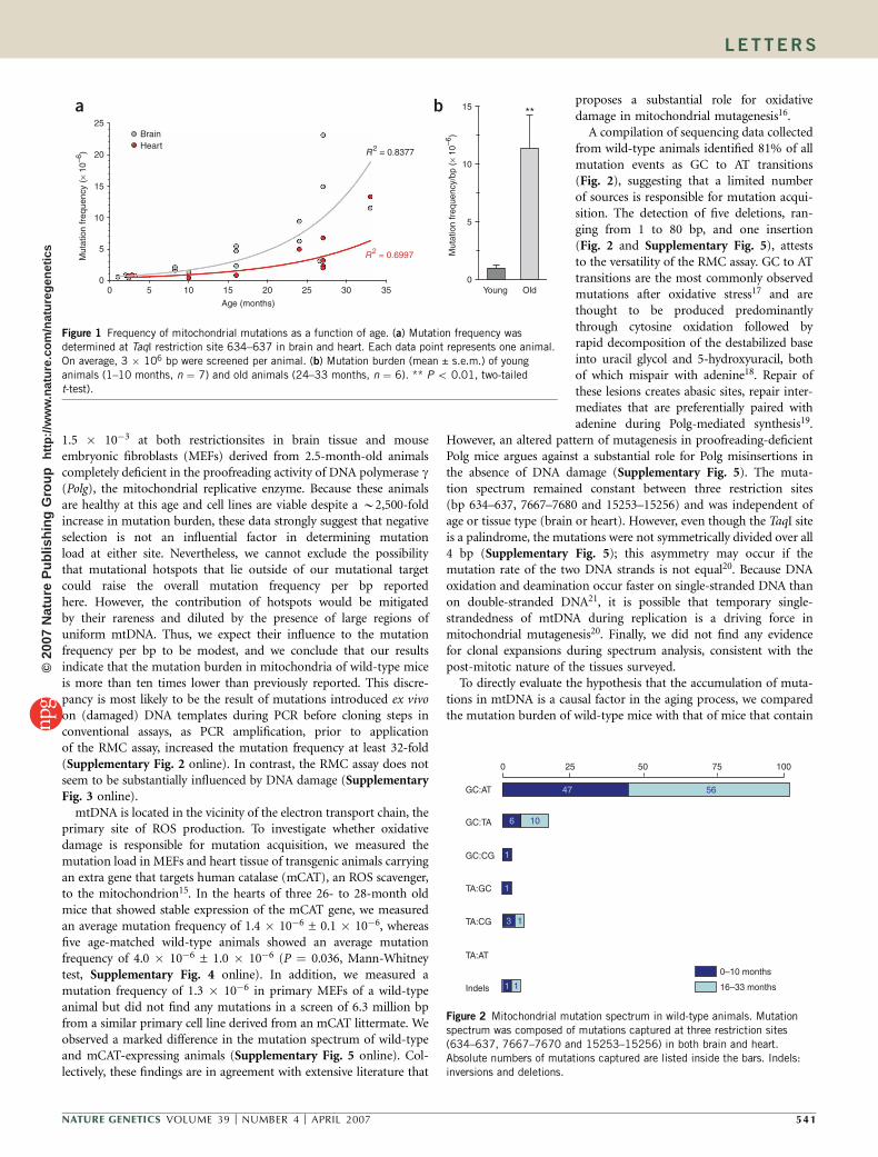
A compilation of sequencing data collectedfrom wild-type animals identified 81% of allmutation events as GC to AT transitions(Fig. 2), suggesting that a limited numberof sources is responsible for mutation acqui-sition. The detection of five deletions, ran-ging from 1 to 80 bp, and one insertion(Fig. 2 and Supplementary Fig. 5), atteststo the versatility of the RMC assay. GC to ATtransitions are the most commonly observedmutations after oxidative stress17 and arethought to be produced predominantlythrough cytosine oxidation followed byrapid decomposition of the destabilized baseinto uracil glycol and 5-hydroxyuracil, bothof which mispair with adenine18. Repair ofthese lesions creates abasic sites, repair inter-mediates that are preferentially paired withadenine during Polg-mediated synthesis19.
However, an altered pattern of mutagenesis in proofreading-deficientPolg mice argues against a substantial role for Polg misinsertions inthe absence of DNA damage (Supplementary Fig. 5). The muta-tion spectrum remained constant between three restriction sites(bp 634–637, 7667–7680 and 15253–15256) and was independent ofage or tissue type (brain or heart). However, even though the TaqI siteis a palindrome, the mutations were not symmetrically divided over all4 bp (Supplementary Fig. 5); this asymmetry may occur if themutation rate of the two DNA strands is not equal20. Because DNAoxidation and deamination occur faster on single-stranded DNA thanon double-stranded DNA21, it is possible that temporary single-strandedness of mtDNA during replication is a driving force inmitochondrial mutagenesis20. Finally, we did not find any evidencefor clonal expansions during spectrum analysis, consistent with thepost-mitotic nature of the tissues surveyed.
To directly evaluate the hypothesis that the accumulation of muta-tions in mtDNA is a causal factor in the aging process, we comparedthe mutation burden of wild-type mice with that of mice that contain
OldYoung0
5
10
15
Mut
atio
n fr
eque
ncy/
bp (
× 10
–6)
Mut
atio
n fr
eque
ncy
(× 1
0–6)
35302520151050
Age (months)
R2 = 0.6997
R2 = 0.8377
BrainHeart
25
20
15
10
5
0
a b **
Figure 1 Frequency of mitochondrial mutations as a function of age. (a) Mutation frequency wasdetermined at TaqI restriction site 634–637 in brain and heart. Each data point represents one animal.
On average, 3 � 106 bp were screened per animal. (b) Mutation burden (mean ± s.e.m.) of young
animals (1–10 months, n ¼ 7) and old animals (24–33 months, n ¼ 6). ** P o 0.01, two-tailed
t-test).
11
13
1
6 10
1
47 56
100 7550250
GC:AT
GC:TA
GC:CG
TA:GC
TA:CG
TA:AT
Indels
0–10 months
16–33 months
Figure 2 Mitochondrial mutation spectrum in wild-type animals. Mutation
spectrum was composed of mutations captured at three restriction sites
(634–637, 7667–7670 and 15253–15256) in both brain and heart.
Absolute numbers of mutations captured are listed inside the bars. Indels:inversions and deletions.
NATURE GENETICS VOLUME 39 [ NUMBER 4 [ APRIL 2007 541
LET TERS©
2007
Nat
ure
Pub
lishi
ng G
roup
ht
tp://
ww
w.n
atur
e.co
m/n
atur
egen
etic
s

an allelic substitution in the exonuclease domain of Polg. Thissubstitution abolishes the proofreading activity of Polg, causing it tobe error prone. We3 and others22 have recently demonstrated thatmice homozygous for this substitution (Polgmut/mut) show a markedreduction in lifespan and several features of premature aging. Thisphenotype could be attributed to an increased rate of mtDNAmutation, which we initially reported to be three- to eightfold higherin homozygous mutant mice compared with wild-type mice,as measured by a standard DNA sequencing approach. Here, wecorrect this to be B2,500-fold. Because our measurements inyoung Polgmut/mut mice (1.5 � 10�3 ± 0.2 � 10�3) are equivalent towhat was previously reported3,22 with conventional assays, this differ-ence can be attributed solely to the increased sensitivity of the RMCassay, which allowed us to correctly determine the very low mutationfrequency in wild-type mice. Thus, a key finding of this study isthat the exonuclease domain of Polg is a principal caretaker ofmtDNA integrity.
Heterozygous animals (Polg+/mut) did not show a statisticallysignificant reduction in mean lifespan (P ¼ 0.875, Fig. 3), consistentwith previous reports3,22. Additionally, no significant increase in age-related pathology has thus far been detected in these animals22 (T.A.P.,unpublished data). We re-examined the mutation load of the Polg+/mut
mice with the RMC assay. Notably, we recorded an average muta-tion frequency of 3.3 � 10�4 ± 0.9 � 10�4 per bp (Fig. 4a) in braintissue of young (2- to 3-month-old) Polg+/mut animals at the 12SrRNA locus—approximately 500 times higher than in age-matchedwild-type animals (P¼ 0.008). Notably, this mutation burden was also29 times higher than the burden in old wild-type animals (24–33months, Fig. 4a, P ¼ 0.001). The lack of a classic mismatch repairmechanism in mouse mitochondria23 probably allows for this vastincrease in mitochondrial mutagenesis in an exonuclease-deficientbackground. Because the frequency of intracellular expansions ofmtDNA mutations depends directly on the mutation rate24, thesedata suggest that both heteroplasmic and homoplasmic mutations aremarkedly higher in Polg+/mut cells than in wild-type cells. We con-firmed these results in heart tissue of three young Polg+/mut mice,which showed an average mutation frequency of 1.6 � 10�4 ± 0.5 �10�4, approximately 220 times higher than in four young wild-typeanimals, which carried a mutation burden of 7.1 � 10�7 ± 0.1 � 10�7
(P ¼ 0.0175) and 30 times higher than in six old wild-type animalswith a frequency of 5.4 � 10�6 ± 1.7 � 10�6 (Fig. 4b, P¼ 0.004). Thisobservation was strengthened by similar results from a probe of threeadditional TaqI restriction sites, randomly dispersed throughout themitochondrial genome in both brain (Supplementary Fig. 6 online)and heart (data not shown).
As the mutation frequency is determined at the same loci inall genotypes, these measurements unambiguously describe therelative differences in mutation rate between them. Because hetero-zygous mice are born with a 30-fold higher mutation burden than theoldest wild-type animals without suffering a phenotype that resemblespremature aging, we conclude that the threshold at which mitochon-drial mutations become limiting for lifespan is unlikely to be reachedin wild-type mice. Notably, heterozygous carriers of human variantsof polymerase g (POLG) are asymptomatic as well, suggestingthat there may be many healthy human carriers of POLG alleles thatharbor large numbers of undetected mtDNA mutations. Althoughour data strongly argue against a causal role for mitochon-drial mutations in natural aging, it should be noted that despite ourability to detect small deletions, large mtDNA deletions are notdetected by our methods. Large mtDNA deletions have beencorrelated with the demise of certain specialized tissues suchas the substantia nigra25,26. However, twinkle transgenic mice,which have an organism-wide increase in the accumulation of largedeletions, do not show a decrease in lifespan or a prematureaging phenotype27.
The mutation burden rapidly grew after 16 months of age, aroundthe time when reproduction has ceased and the average life expectancyof these animals in the wild has expired. As ROS seem to be theprimary source of mutagenesis in mitochondria, this study is con-sistent with the hypothesis that the production of ROS increases withage28 and that mtDNA is a primary target of ROS, but that the mostprevalent result of oxidative lesions, point mutations, are extremelyrare and do not determine the rate of aging of wild-type mice.
METHODSTissue homogenization and organelle separation. All tissues were harvested
within 5 min of death. All animals were cared for according to approved
guidelines at the University of Washington. Tissues were sliced in pieces with a
scalpel and rinsed in 1� PBS before homogenization in a Dounce-type glass
3630241812600
Age (months)
25
50
75
100
Per
cent
age
surv
ival
Polgmut/mut
Polg+/mut
Wild-type
Figure 3 Kaplan-Meier survival curves. Median survival is 423 d forPolgmut/mut mice, 758 d for Polg+/mut mice and 864 d for wild-type mice.
The curves for wild-type and Polg+/mut mice do not differ statistically
significantly (log rank test, P ¼ 0.875).
10–2
10–3
10–4
10–5
10–6
10–7 10–7
10–6
10–5
10–4
10–3
Mut
atio
n fr
eque
ncy/
bp
Mut
atio
n fr
eque
ncy/
bp
Youn
g WT
Old W
T
Youn
g WT
Old W
T
Youn
g Polg
+/mut
Youn
g Polg
+/mut
Youn
g Polg
mut/mut
a b**
**
***
Figure 4 Mutation burden in wild-type and Polg exonuclease–deficient
mice. (a,b) Mutation frequency (mean ± s.e.m.) was determined at TaqI
restriction site 634–637 in brain (gray) and heart (red) and is plotted on
a logarithmic scale. For brain tissue, young wild-type (WT) animals are1–3 months of age (n ¼ 4); old wild-type animals, 24–33 months of age
(n ¼ 6); young Polg+/mut animals, 2.5 months of age (n ¼ 3) and young
Polgmut/mut animals, 2.5 months of age (n ¼ 2). * P o 0.05. ** P o 0.01.
For heart tissue, young wild-type animals are 1–16 months of age (n ¼ 4),
old wild-type animals are 24–33 months of age (n ¼ 6) and young Polg+/mut
animals are 2.5 months of age (n ¼ 3).
542 VOLUME 39 [ NUMBER 4 [ APRIL 2007 NATURE GENETICS
LET TERS©
2007
Nat
ure
Pub
lishi
ng G
roup
ht
tp://
ww
w.n
atur
e.co
m/n
atur
egen
etic
s
homogenizer with 25 firm strokes of a hand-driven glass pestle. Homogeniza-
tion buffer contained 0.075 M sucrose, 0.225 M sorbitol, 1 mM EGTA, 0.1%
fatty acid–free BSA and 10 mM Tris HCl (pH 7.8). Differential centrifugation
was performed as described previously12 to obtain a crudely purified mito-
chondrial pellet.
DNA extraction and restriction digest. Mitochondrial pellets were digested for
1 h in a buffer containing 0.2 mg/ml proteinase K (Sigma), 0.75% SDS, 0.01 M
Tris HCl, 0.15 M NaCl and 0.005 M EDTA at pH 7.8. mtDNA was subsequently
isolated using phenol-chloroform extraction (1:1, vol/vol) followed by ethanol
precipitation. mtDNA was then diluted and digested in a specialized TaqI buffer
(New England Biolabs) in the presence of 100 units TaqaI (New England
Biolabs) and 1� BSA (New England Biolabs) for 10 h, with the addition of
100 units of TaqaI per h.
Mutation detection. PCR was performed using a DNA Engine Opticon
Monitor 2, a continuous fluorescence detection system (BioRad), and
amplicons were visualized with Stratagene’s Brilliant SYBR Green qPCR
Master Mix. Primers used for amplification are listed in Supplementary
Table 1 online.
All real-time qPCR reactions were performed in 25-ml reactions containing
1� Brilliant SYBR Green qPCR Master Mix from Stratagene, 20 pmol forward
and reverse primers and 2 units uracil DNA glycosylase (UDG). The samples
were amplified as follows: UDG incubation at 37 1C for 10 min and 95 1C for
10 min followed by 45 cycles of 95 1C for 30 s, 60 1C for 1 min and 72 1C for
1.5 min. Samples were held at 72 1C for 5 min and then immediately stored
at –20 1C. All PCR products were either (i) incubated with TaqaI and then
verified by agarose gel electrophoresis to be insensitive to digestion or (ii)
isolated with the QiaQuick PCR Purification Kit (Qiagen) and sequenced to
identify the mutation at the TaqI recognition site.
Cell culture. Mouse embryonic fibroblasts were cultured in DMEM
(Gibco-BRL) containing 10% (vol/vol) FBS (HyClone), 1% L-glutamine and
1% penicillin-streptomycin (Gibco). An atmosphere of 5% CO2 was main-
tained in a humidified incubator at 37 1C. mCAT and corresponding wild-type
cell lines were grown with 2% oxygen.
Note: Supplementary information is available on the Nature Genetics website.
ACKNOWLEDGMENTSThis work was supported by US National Institutes of Health grants AG001751(L.A.L., P.S.R.), CA102029 (L.A.L.), ES11045 (L.A.L., W.C.L.) and AG021905(T.A.P., G.C.K.). J.H.B. was supported by a research fellowship from the CanadianInstitutes of Health. The authors thank G.M. Martin, R.S. Mangalindan,R.N. Venkatesan and C.-Y. Chen for editing this manuscript, technical assistanceand discussions.
AUTHOR CONTRIBUTIONSM.V. carried out all the experiments described and wrote the paper. M.V., J.H.B.and L.A.L. conceived the project. G.C.K. and T.A.P. provided Kaplan-Meier curvesand statistical analysis of mouse cohorts. J.H.B., W.C.L., G.C.K., T.A.P. andP.S.R. provided technical assistance, animal care and tissues. L.A.L. supervised theexperimental work and interpretation of data. All authors commented on anddiscussed the paper.
COMPETING INTERESTS STATEMENTThe authors declare no competing financial interests.
Published online at http://www.nature.com/naturegenetics
Reprints and permissions information is available online at http://npg.nature.com/
reprintsandpermissions
1. Khrapko, K., Kraytsberg, Y., de Grey, A.D., Vijg, J. & Schon, E.A. Does premature agingof the mtDNA mutator mouse prove that mtDNA mutations are involved in naturalaging? Aging Cell 5, 279–282 (2006).
2. Bielas, J.H. & Loeb, L.A. Quantification of random genomic mutations. Nat. Methods2, 285–290 (2005).
3. Kujoth, G.C. et al. Mitochondrial DNA mutations, oxidative stress, and apoptosis inmammalian aging. Science 309, 481–484 (2005).
4. Saraste, M. Oxidative phosphorylation at the fin de siecle. Science 283, 1488–1493(1999).
5. Newmeyer, D.D. & Ferguson-Miller, S. Mitochondria: releasing power for life andunleashing the machineries of death. Cell 112, 481–490 (2003).
6. Wallace, D.C. A mitochondrial paradigm of metabolic and degenerative diseases, aging,and cancer: a dawn for evolutionary medicine. Annu. Rev. Genet. 39, 359–407 (2005).
7. Trifunovic, A. Mitochondrial DNA and ageing. Biochim. Biophys. Acta 1757, 611–617(2006).
8. Balaban, R.S., Nemoto, S. & Finkel, T. Mitochondria, oxidants, and aging. Cell 120,483–495 (2005).
9. Miquel, J., Economos, A.C., Fleming, J. & Johnson, J.E., Jr. Mitochondrial role in cellaging. Exp. Gerontol. 15, 575–591 (1980).
10. Khrapko, K. et al. Mitochondrial mutational spectra in human cells and tissues. Proc.Natl. Acad. Sci. USA 94, 13798–13803 (1997).
11. Michikawa, Y., Mazzucchelli, F., Bresolin, N., Scarlato, G. & Attardi, G. Aging-dependent large accumulation of point mutations in the human mtDNA control regionfor replication. Science 286, 774–779 (1999).
12. Copeland, W.C. Mitochondrial DNA: methods and protocols. Methods Mol. Biol. 197,v–vi (2002).
13. Zhang, D. et al. Construction of transgenic mice with tissue-specific acceleration ofmitochondrial DNA mutagenesis. Genomics 69, 151–161 (2000).
14. Bielas, J.H., Loeb, K.R., Rubin, B.P., True, L.D. & Loeb, L.A. Human cancers express amutator phenotype. Proc. Natl. Acad. Sci. USA 103, 18238–18242 (2006).
15. Schriner, S.E. et al. Extension of murine life span by overexpression of catalasetargeted to mitochondria. Science 308, 1909–1911 (2005).
16. Mandavilli, B.S., Santos, J.H. & Van Houten, B. Mitochondrial DNA repair and aging.Mutat. Res. 509, 127–151 (2002).
17. Wang, D., Kreutzer, D.A. & Essigmann, J.M. Mutagenicity and repair of oxidative DNAdamage: insights from studies using defined lesions. Mutat. Res. 400, 99–115(1998).
18. Kreutzer, D.A. & Essigmann, J.M. Oxidized, deaminated cytosines are a source of C -T transitions in vivo. Proc. Natl. Acad. Sci. USA 95, 3578–3582 (1998).
19. Pinz, K.G., Shibutani, S. & Bogenhagen, D.F. Action of mitochondrial DNA polymerasegamma at sites of base loss or oxidative damage. J. Biol. Chem. 270, 9202–9206 (1995).
20. Tanaka, M. & Ozawa, T. Strand asymmetry in human mitochondrial DNA mutations.Genomics 22, 327–335 (1994).
21. Frederico, L.A., Kunkel, T.A. & Shaw, B.R. A sensitive genetic assay for the detection ofcytosine deamination: determination of rate constants and the activation energy.Biochemistry 29, 2532–2537 (1990).
22. Trifunovic, A. et al. Premature ageing in mice expressing defective mitochondrial DNApolymerase. Nature 429, 417–423 (2004).
23. Larsen, N.B., Rasmussen, M. & Rasmussen, L.J. Nuclear and mitochondrial DNArepair: similar pathways? Mitochondrion 5, 89–108 (2005).
24. Elson, J.L., Samuels, D.C., Turnbull, D.M. & Chinnery, P.F. Random intracellular driftexplains the clonal expansion of mitochondrial DNA mutations with age. Am. J. Hum.Genet. 68, 802–806 (2001).
25. Bender, A. et al. High levels of mitochondrial DNA deletions in substantia nigraneurons in aging and Parkinson disease. Nat. Genet. 38, 515–517 (2006).
26. Kraytsberg, Y. et al. Mitochondrial DNA deletions are abundant and cause functionalimpairment in aged human substantia nigra neurons. Nat. Genet. 38, 518–520 (2006).
27. Tyynismaa, H. et al. Mutant mitochondrial helicase Twinkle causes multiple mtDNAdeletions and a late-onset mitochondrial disease in mice. Proc. Natl. Acad. Sci. USA102, 17687–17692 (2005).
28. Hamilton, M.L. et al. Does oxidative damage to DNA increase with age? Proc. Natl.Acad. Sci. USA 98, 10469–10474 (2001).
NATURE GENETICS VOLUME 39 [ NUMBER 4 [ APRIL 2007 543
LET TERS©
2007
Nat
ure
Pub
lishi
ng G
roup
ht
tp://
ww
w.n
atur
e.co
m/n
atur
egen
etic
s