minireview - jcm.asm.org · or intestinal biopsy specimens containing less debris and arti-fact...
TRANSCRIPT

JOURNAL OF CLINICAL MICROBIOLOGY, June 2002, p. 1892–1901 Vol. 40, No. 60095-1137/02/$04.00�0 DOI: 10.1128/JCM.40.6.1892–1901.2002Copyright © 2002, American Society for Microbiology. All Rights Reserved.
MINIREVIEW
Laboratory Identification of the MicrosporidiaLynne S. Garcia*
LSG & Associates, Santa Monica, California 90402-2908
The microsporidia belong to the phylum Microspora withinthe taxonomic group Protozoa; there are more than 140 generaand 1,200 species that are parasitic in all major animal groups.Already, seven genera (Enterocytozoon, Encephalitozoon,Nosema, Pleistophora, Vittaforma, Trachipleistophora, and Bra-chiola) and some that are unclassified have been confirmed tocause human infection. Septata intestinalis, an organism thatwas identified in 1994 as belonging to a new genus, i.e., Septata(Encephalitozoon-like), has been reclassified as Encephalito-zoon intestinalis, although there is not universal agreement onthis generic change. The genus Microsporidium is essentially acatch-all genus for organisms that have not yet been classified(or may never be classified due to a lack of specimen). De-pending on acceptance of these generic designations, theremay be seven or nine microsporidial genera.
The microsporidia are obligate intracellular, spore-formingprotists with no active metabolic stages outside of the host cell.The life cycle involves a proliferative merogonic stage followedby sporogony, which results in spores containing a tubularextrusion apparatus (polar tubule) for injecting infective sporecontents into the host cell (Fig. 1).
Because microsporidia have a membrane-bound nucleus, anintracytoplasmic membrane system, and chromosome separa-tion on mitotic spindles, they are considered to be true eu-karyotes. However, they are somewhat unusual in that theyhave 70S ribosomes, have no mitrochondria or peroxisomes,and have simple Golgi membranes. Also, the microsporidialgenome is smaller and less complex than those of other eu-karyotes. Several characteristics, including the presence ofchitin in the spore wall, suggest a potential link to the fungi.
Microsporidiosis, a disease resulting from infection withthese important emerging opportunistic organisms, has beennoted in human immunodeficiency virus (HIV)-infected pa-tients, as well as in other immunocompromised patients, in-cluding transplant recipients. The first human case was re-ported in 1959; since then, cases of microsporidiosis in boththose who are immunocompromised and those who have nounderlying immune problems and are considered to be immu-nocompetent have been reported (Table 1) (2, 7, 21).
SPECIMEN COLLECTION
It is important to remember that microsporidial spores arequite resistant to environmental conditions and can remain
infectious for several years, particularly if they are protectedfrom desiccation. Stool or duodenal drainage specimens can besubmitted fresh or preserved in 5 or 10% formalin, or sodiumacetate-acetic acid-formalin. Biopsy specimens are also accept-able. In cases of disseminated infection, it is recommendedthat urine (fresh or preserved) be submitted for analysis; otherbody fluids (sputum, bronchoalveolar lavage [BAL] fluid, nasalsecretion, or cerebrospinal fluid [CSF]), conjunctival smears,corneal scrapings, or tissue can also be submitted (Table 2).Formalin fixation is recommended for routine histology, whilegluteraldehyde is preferred for electron microscopy. Freshspecimens should be collected for cell culture and for molec-ular studies.
LABORATORY METHODS
There are a number of methods available for the recovery ofmicrosporidial spores and confirmation of microsporidiosis(Table 3). Most of these methods were developed in order todiagnose infections in the immunocompromised population.Microsporidial infections in the immunocompetent patientmay result in self-cure after mild symptoms appear, a situationsimilar to that seen with cryptosporidiosis and isosporiasis. Asawareness of microsporidiosis increases and more-sensitivetechniques become available, we may find that these infectionsare not uncommon in the immunocompetent host. The recom-mendations for the use of current, less-sensitive methodologysuggest that multiple diagnostic methods may be required todiagnose a microsporidial infection, particularly when fecalspecimens are examined. Since microsporidial infections ofteninvolve multiple body sites, detection of organisms from anyclinical specimen should be followed by examination of otherbody tissues and fluids. In patients for whom disseminatedmicrosporidiosis is suspected, urine specimens should alwaysbe examined.
LIGHT MICROSCOPY
Modified trichrome stains. Smears are prepared using 10 to20 �l of concentrated specimen (stool specimen or urine orother fluid) that is thinly spread onto the slides. Some labora-tories use concave well slides; these are very helpful whenexamining the stained preparations. It is important to remem-ber to centrifuge the specimen for 10 min at 500 � g prior tosmear preparation. There is also some evidence to indicatethat pretreatment of fecal specimens (1:1) with 10% KOH mayprovide a better-quality smear when modified trichrome stainsare used. Specimens can be fresh or preserved (with 5 or 10%
* Mailing address: LSG & Associates, 512-12th St., Santa Monica,CA 90402-2908. Phone: (310) 393-5059. Fax: (310) 899-9722. E-mail:[email protected].
1892
on April 9, 2019 by guest
http://jcm.asm
.org/D
ownloaded from

formalin or with sodium acetate-acetic acid-formalin or byusing one of the newer single-vial systems). The modifiedtrichrome stain methods are based on the fact that stain pen-etration of the microsporidial spore is very difficult; thus, thedye content in the chromotrope 2R component of the formulais greater than that used to prepare Wheatley’s modification ofthe Gomori’s trichrome stain, and the staining time is muchlonger (90 min) (11, 12, 19, 20). Several of these stains (e.g.,Weber-Green modified trichrome stain [Fig. 2] and Ryan-Bluemodified trichrome stain [Fig. 3]) are available commerciallyfrom a number of suppliers. The use of positive control mate-rial is highly recommended. Spore detection requires adequateillumination and magnification (oil immersion; total magnifi-cation, �1,000).
The spore wall should stain pinkish to red, with the interiorof the spore being clear or perhaps showing a horizontal ordiagonal stripe, which represents the polar tube. The back-ground will appear green or blue, depending on the method.Other bacteria, some yeast cells, and some debris will also beevident (stained shades of pink and red); the shapes and sizesof the artifacts may be helpful in differentiating the sporesfrom other structures. Results should be reported only if thepositive control smears are acceptable.
The choice of counterstain, with either fast green or anilineblue dyes in the stain formulation, depends on laboratory pref-erence and does not change the color results of the actualmicrosporidial spores, which stain pink. In addition to thecounterstain (green or blue), several modifications of the orig-inal chromotrope staining solution have been proposed, in-cluding changes in temperature of the staining solution andstaining time. Results indicate that staining at 50°C for 10 minor staining at 37°C for 30 min may improve the detection ofspores; the background appears to contain less debris, and thespores may stain more intensely (5, 7, 13, 22).
An acid-fast trichrome method stains both acid-fast crypto-sporidial oocysts and microsporidial spores in the same smear
(Fig. 4) (10). Also, a rapid-hot Gram-chromotrope stainingmethod with a staining time of 5 min is available; the micro-sporidial spores stain dark violet against a pale-green back-ground (16).
Giemsa stain. Although Giemsa staining of stool can beperformed and results in a light-blue staining of microsporidialspores, the spores can be very difficult to differentiate fromdebris in the smear. However, body fluid cytology preparationsor intestinal biopsy specimens containing less debris and arti-fact material can be stained with Giemsa stain; identification ofspores will be much easier with these types of specimens thanwith preparations from stool (Fig. 5).
Chemofluorescent (optical brightening) agents. Chemofluo-rescent (optical brightening) agents are chitin stains that re-quire the use of a fluorescent microscope (8). Depending onthe agent used as well as the wavelength, the walls of themicrosporidial spores fluorescence. Using Calcofluor White2MR (American Cyanamid Corp., Princeton, N.J.) and a wave-length of 395 to 415 nm (observation light, 455 nm), the sporesappear as bluish-white or turquoise oval halos. It is importantto remember that this type of stain is nonspecific; small fungiand some artifact material present in stool may also fluoresce.However, when this approach is used with specimens otherthan stool, the microsporidial spores tend to be much easier todetect and identify (Fig. 6). Many laboratories tend to use oneof the modified trichrome stains as well as the Calcofluormethod for clinical specimens. The sensitivity of both methodsis relatively good, and small numbers of spores that might bemissed by the modified trichrome methods might be seen withthe optical brightening agents. Again, clinical specimens otherthan stool tend to contain less debris and artifact material andare much easier to examine. Other optical brightening agentsthat can be used include Fungi-Fluor and Cellufluor (bothfrom Polysciences Inc., Warrington, Pa.), Fungiqual A (Med-ical Diagnostics, Kandern, Germany), and Uvitex 2B (not com-mercially available; Ciba Geigy, Basel, Switzerland).
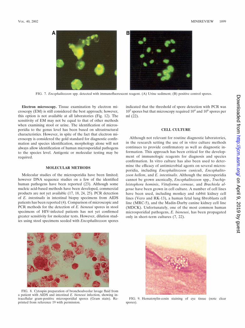
Immunofluorescent reagents. Although immunofluorescentreagents for the detection of microsporidial spores have beenused, they are not yet commercially available and most havebeen limited to Encephalitozoon species spores (Fig. 7) (3, 15).Unfortunately, background staining, cross-reactions with yeastspecies and bacteria, and overall lower sensitivity than is ob-served with chromotrope or chemofluorescent staining preventthe polyclonal antibodies from being applicable to routine di-agnostic use. Fluorescing microsporidial spores have a darkercell wall, and the polar tubule can be seen as cross or diagonallines. With the use of an antiserum, it was observed that 9 of 27patients (30%) who were positive for cryptosporidiosis alsohad microsporidial spores in the stool (8). Although there issome cross-reactivity with bacteria, immunofluorescent stain-ing is more-sensitive than the routine staining methods cur-rently available.
More specific reagents are in various stages of developmentand testing; it can be hoped that these will become commer-cially available in the near future. With the development ofmonoclonal antibodies to Encephalitozoon intestinalis and En-terocytozoon bieneusi, it can also be hoped that diagnostic re-agents will become available soon (1, 6). Since E. intestinalisand E. bieneusi are the two most important microsporidianpathogens in humans, the availability of commercial reagents
FIG. 1. Life cycle of microsporidia. Reprinted from reference 9.
VOL. 40, 2002 MINIREVIEW 1893
on April 9, 2019 by guest
http://jcm.asm
.org/D
ownloaded from

TA
BL
E1.
Mic
rosp
orid
iaca
usin
ghu
man
infe
ctio
n
Org
anis
m(s
)C
linic
alpr
esen
tatio
nC
omm
ents
a
Imm
unoc
ompr
omis
edho
stIm
mun
ocom
pete
ntho
st
Com
mon
Ent
eroc
ytoz
oon
bien
eusi
Chr
onic
diar
rhea
,was
ting,
chol
angi
tis,a
calc
ulou
sch
olec
ystit
is,c
hron
icsi
nusi
tis,c
hron
icco
ugh,
and
pneu
mon
ia;i
sola
ted
from
5to
30%
ofH
IV-
posi
tive
patie
nts
with
very
low
CD
4ly
mph
ocyt
eco
unts
(one
ofth
em
ost
impo
rtan
tH
IV-a
ssoc
iate
din
test
inal
path
ogen
s)an
dor
gan
(liv
er,h
eart
-lung
,an
dbo
nem
arro
w)
tran
spla
ntre
cipi
ents
Self-
limiti
ngdi
arrh
eaan
dtr
avel
er’s
diar
rhea
;as
ympt
omat
icca
rrie
rs
Shor
tte
rmcu
lture
only
;thr
eest
rain
sid
entifi
edbu
tno
tna
med
;clin
ical
dise
ase
base
don
cont
inuo
usex
cess
loss
ofep
ithel
ialc
ells
,cry
pthy
perp
lasi
a,pa
rtia
lvill
usat
roph
y,de
crea
sed
brus
hbo
rder
sucr
ase,
lact
ase,
and
mal
tase
activ
ities
,or
mal
abso
rptio
n;no
nhum
anho
sts
incl
ude
pigs
and
prim
ates
Enc
epha
litoz
oon
helle
mD
isse
min
ated
infe
ctio
n;ke
rato
conj
unct
iviti
s,si
nusi
tis;b
ronc
hitis
,pne
umon
ia,n
ephr
itis,
uret
eriti
s,cy
stiti
s,pr
osta
titis
,and
uret
hriti
s;na
sal
epith
eliu
mco
nfirm
edas
infe
cted
inon
eA
IDS
patie
nt;i
sola
ted
inca
ses
ofas
ympt
omat
icre
spir
ator
ysy
stem
infe
ctio
n
Not
iden
tified
orde
scri
bed
Gro
wn
incu
lture
;may
beac
quir
edby
inha
latio
n;ce
rebr
alle
sion
ssi
mila
rto
thos
ese
enin
case
sof
toxo
plas
mos
is;
nonh
uman
host
sin
clud
eps
ittac
ine
bird
s
Enc
epha
litoz
oon
inte
stin
alis
(Sep
tata
inte
stin
alis
)C
hron
icdi
arrh
ea;c
hola
ngio
path
y;si
nusi
tis,
bron
chiti
s,pn
eum
oniti
s,ne
phri
tis;b
one
infe
ctio
n;no
dula
rcu
tane
ous
lesi
ons;
ofte
nas
soci
ated
with
AID
Spa
tient
s;te
nds
todi
ssem
inat
e;ki
dney
ispr
omin
ent
site
Self-
limiti
ngdi
arrh
ea;
asym
ptom
atic
carr
iers
Gro
wn
incu
lture
;can
prod
uce
mul
tiorg
anfa
ilure
rese
mbl
ing
hype
rinf
ectio
nsy
ndro
me
(Str
ongy
loid
esst
erco
ralis
);no
nhum
anho
sts
incl
ude
dogs
,don
keys
,pig
s,co
ws,
and
goat
s
Enc
epha
litoz
oon
cuni
culi
Dis
sem
inat
edin
fect
ion;
kera
toco
njun
ctiv
itis;
sinu
sitis
,bro
nchi
tis,p
neum
onia
;nep
hriti
s;he
patit
is;p
erito
nitis
;sym
ptom
atic
and
asym
ptom
atic
inte
stin
alin
fect
ion;
ence
phal
itis;
asym
ptom
atic
resp
irat
ory
syst
emin
fect
ions
Not
iden
tified
orde
scri
bed;
two
HIV
-se
rone
gativ
ech
ildre
nw
ithse
izur
edi
sord
erpr
obab
lyw
ere
imm
unoc
ompr
omis
ed
Gro
wn
incu
lture
;non
hum
anho
sts
incl
ude
rabb
its(s
trai
nI)
(ide
ntifi
edin
hum
ans
inSw
itzer
land
),m
ice,
blue
foxe
s(s
trai
nII
)(n
otid
entifi
edin
hum
ans)
;dom
estic
dogs
(str
ain
III)
(ide
ntifi
edin
hum
ans
inth
eU
nite
dSt
ates
and
Spai
n)
Unc
omm
onB
rach
iola
alge
rae
Not
iden
tified
orde
scri
bed
Ker
atiti
sF
orm
erly
know
nas
Nos
ema
alge
rae
Bra
chio
laco
nnor
iD
isse
min
ated
infe
ctio
nN
otid
entifi
edor
desc
ribe
dF
orm
erly
know
nas
Nos
ema
conn
ori;
Nos
ema
spp.
ofte
nin
fect
inse
cts;
diss
emin
ated
inin
fant
with
SCID
Bra
chio
lave
sicu
laru
mM
yosi
tis;a
lso
foun
din
corn
eals
trom
aN
otid
entifi
edor
desc
ribe
dPr
evio
usly
calle
da
Nos
ema-
like
spec
ies;
nonh
uman
host
sno
tid
entifi
edM
icro
spor
idiu
maf
rican
umN
otid
entifi
edor
desc
ribe
dC
orne
alul
cer,
kera
titis
(cor
neal
stro
ma)
Cat
ch-a
llge
nus
for
orga
nism
sth
atco
uld
not
beid
entifi
ed;
HIV
-ser
oneg
ativ
ein
divi
dual
atau
tops
y;no
nhum
anho
sts
not
iden
tified
Mic
rosp
orid
ium
ceyl
onen
sis
Not
iden
tified
orde
scri
bed
Cor
neal
ulce
r,ke
ratit
is(c
orne
alst
rom
a)C
atch
-all
genu
sfo
ror
gani
sms
that
coul
dno
tbe
iden
tified
;H
IV-s
eron
egat
ive
indi
vidu
alat
auto
psy;
nonh
uman
host
sno
tid
entifi
edN
osem
aoc
ular
umD
isse
min
ated
inat
hym
icch
ildC
orne
alul
cer,
kera
titis
(cor
neal
stro
ma)
HIV
-ser
oneg
ativ
ein
divi
dual
(chi
ld);
nonh
uman
host
sno
tid
entifi
edP
leis
toph
ora
sp.
Myo
sitis
(ske
leta
lmus
cle)
Not
iden
tified
orde
scri
bed
HIV
-ser
oneg
ativ
ean
dH
IV-s
erop
ositi
vein
divi
dual
,P
leis
toph
ora
spp.
tend
toin
fect
fish,
inse
cts,
amph
ibia
ns,
and
rept
iles
Tra
chip
leis
toph
ora
anth
ropo
phth
era
Dis
sem
inat
edin
fect
ion
inA
IDS
patie
nt(b
rain
,he
art,
kidn
ey)
Not
iden
tified
orde
scri
bed
App
ears
tobe
dim
orph
ic:t
wo
diffe
rent
spor
efo
rms
have
been
seen
;rin
g-en
hanc
ing
lesi
ons
sugg
estiv
eof
CN
Sto
xopl
asm
osis
;non
hum
anho
sts
not
iden
tified
Tra
chip
leis
toph
ora
hom
inis
Myo
sitis
(ske
leta
lmus
cle)
,ker
atoc
onju
nctiv
itis;
sinu
sitis
Not
iden
tified
orde
scri
bed
AID
Spa
tient
;non
hum
anho
sts
not
iden
tified
Vitt
afor
ma
corn
eae
Dis
sem
inat
edin
fect
ion
Ker
atiti
s(c
orne
alst
rom
a);i
ritis
For
mer
lykn
own
asN
osem
aco
rneu
m
aC
NS,
cent
raln
ervo
ussy
stem
.SC
ID,s
ever
eco
mbi
ned
imm
unod
efici
ency
dise
ase.
1894 MINIREVIEW J. CLIN. MICROBIOL.
on April 9, 2019 by guest
http://jcm.asm
.org/D
ownloaded from

TABLE 2. Body sites where microsporidia infecting humans have been detecteda
Body site or specimen type
Report of detection of:
Enterocytozoonbieneusi
Encephalitozoon (Septata)intestinalls
Encephalitozoonhellem
Encephalitozooncuniculi Other speciesb
Body fluidsStool �� �� � � �Duodenal aspirate �� � � � �Bile � � � � �Urine � � �� �� V. corneaeSputum, BAL fluid � � �� �� �Nasal secretion or washings � � � � T. hominisCSF � � � � �
Mucosal smearsConjunctival � � � � �Corneal � � � � V. corneae
N. ocularum
TissueIntestinal tract
Duodenum �� �� � � B. connoriJejunum �� �� � � B. connoriTerminal ileum �� �� � � B. connoriColon � � � � B. connori
Gallbladder � � � � �
Biliary tract � � � � �
Liver � � � � T. anthropophtheraB. connori
Pancreas � � � � T. anthropophthera
Pancreatic duct � � � � T. anthropophthera
Peritoneum � � � � �
Respiratory systemBronchial epithelium � � � � B. connoriSinus � � � � B. connoriNasal epithelium � � � � B. connori
EyeCornea � � � � V. corneae
N. ocularumM. ceylonensisM. africanumT. hominis
Conjunctiva � � � � V. corneaeN. ocularumM. ceylonensisM. africanumT. hominis
Kidney � � � � T. anthropophthera
Urinary system B. connoriUreter � � � � B. connoriBladder � � � � B. connoriProstate � � � � B. connoriUrethra � � � � B. connori
Brain � �c � � T. anthropophtheraMuscle � � � � Pleistophora sp.
T. hominisB. vesicularumd
Circulatory systemHeart � � � � T. anthropophthera
B. connoriBone marrow � � � � T. anthropophthera
Immune systemLymph nodes � � � � T. anthropophtheraSpleen � � � � T. anthropophthera
Bone � � � � T. anthropophtheraEndocrine system
Thyroid � � � � T. anthropophtheraParathyroid � � � � T. anthropophtheraAdrenal glands � � � � B. connori
a Data are from references 7 and 21 to 23. Symbols: �, not reported; �, case report(s) of microsporidia detection; ��, reliable specimens for detection of microsporidia.b If detection of another species has been reported, the species name is given. The species listed may include one or more of the following: Vittaforma corneae,
Trachipleistophora hominis, Nosema ocularum, Brachiola connori, Trachipleistophora anthropophthera, Microsporidium ceylonensis, Microsporidium africanum, and Brachiolavesicularum.
c Detected in pituitary gland.d Formerly known as Nosema-like microsporidian.
1895
on April 9, 2019 by guest
http://jcm.asm
.org/D
ownloaded from

for these organisms would enhance diagnostic capabilities ofthe routine clinical laboratory.
As clinicians begin to suspect these infections and microbi-ologists become more familiar with the diagnostic methods, thenumber of patients who test positive for microsporidial infec-tion may increase dramatically. It is strongly suspected thatwith the use of more sensitive and specific methods many of
these cases will be found in the immunocompetent population,as well as in immunocompromised patients.
CYTOLOGY
Techniques that do not require tissue embedding are be-coming more widely used. Touch preparations of fresh biopsy
TABLE 3. Microsporidia: Recommended diagnostic techniquesa
Technique Use Comments
Light microscopyStool specimens
Modified trichromeb �� Reliable, available; light infections difficult to identifyGiemsa � Not recommended for routine use, hard to readChemofluorescence �� Calcofluor, Fungifluor, Unitex 2B; sensitive but nonspecificImmunofluorescence (��) Commercial availability limits use; products in development
Other bodily fluidsModified trichrome �� Reliable, available; light infections difficult to identifyGiemsa � Urine, conjunctival swab, BAL, CSF, duodenal aspirateChemofluorescence �� Calcofluor, Fungifluor, Unitex 2B; sensitive but nonspecificImmunofluorescence (��) Commercial availability limits use; products in development
Cytology testingc
Modified trichrome �� Reliable, available; light infections difficult to identifyGiemsa � Urine, conjunctival swab, BAL, CSF, duodenal aspirateGram � Recommended, especially for specimens with little debrisChemofluorescence �� Calcofluor, Fungifluor, Unitex 2B; sensitive but nonspecificImmunofluorescence (��) Commercial availability limits use; products in development
Routine histologyHematoxylin & eosin � Sensitivity uncertain with low parasite numbersPAS � Controversy over effectiveness; PAS-positive granuleModified Gram stains (Brown Brenn,
Brown-Hopps)�� Sensitive, generally recommended
Giemsa � Sensitivity uncertain with low parasite numbersWarthin-Starry � Not standardized, may not be necessary, but often usedModified trichrome �� Reliable, sensitiveImmunofluorescence (��) Commercial availability limits use; products in development; have been used in
research setting for conformation of microsporidia to species level
Plastic-embedded sectionsToluidine blue �� Recommended; sensitive methodMethylene blue-azure II-basic fuchsin �� Recommended as alternative to toluidine blue
Electron microscopyBody fluids � Specific, sensitivity unknown; used for identification to species level (some exceptions)Tissue sections �� Gold standard for confirmation, but sensitivity lower than for detection of spores in
stool or urine; used for identification to species level (some exceptions)
Molecular testing � Availability limited to research laboratories; studies ongoing and appear promising;molecular identification to the species level possible
Serologic testing (serum) � Reagents not commercially available; preliminary results controversial; have beenreported for Encephalitozoon; not available for Enterocytozoon; not relevant forimmunocompromised patients
Culture � Generally used in the research setting; continued advances in culture and organismsurvival and growth; Encephalitozoon, Nosema, Trachipleistophora, Vittaforma can beisolated; delivery to specialty lab within 2 to 3 days recommended; use UniversalPrecautions when handling specimens
a Abbreviations: BAL, bronchoalveolar lavage; CSF, cerebrospinal fluid. Symbols: �, not available or not recommended for routine use; �, reported; ��, techniquesin general use (most widely used); (��), excellent results, but reagents not yet commercially available.
b Various stain modifications have been developed: Chromotrope (original modified trichrome stain [Weber green]) trichrome blue (modification of originalcounterstain from fast green to aniline blue, different concentration of photphotungstic acid [Ryan blue]); modified chromotrope stain (staining temperature increasedto 50°C, staining time reduced to 10 min [Kokoskin]); modified chromotrope stain (staining temperature increased to 37°C, staining time 30 min [Didier]); quick-hotGram-chromotrope stain [Moura]); acid-fast trichrome stain (can detect coccidia and microsporidia [Ignatius]) (5, 7, 10, 14, 16, 19, 20, 22).
c Requires high-speed centrifugation.
1896 MINIREVIEW J. CLIN. MICROBIOL.
on April 9, 2019 by guest
http://jcm.asm
.org/D
ownloaded from

material that are air dried, methanol fixed, and Giemsa stainedhave been used; however, microscopic examination must beperformed using the oil immersion objective (100� objective;total magnification, �1,000). This approach can also be usedfor smears prepared from conjunctival swabs and for sputumand nasal discharge fluids.
Cytocentrifugation followed by Giemsa staining has alsobeen recommended. Spores have been detected in centrifugedsediment of duodenal aspirate, bile, biliary aspirates, urine,BAL fluid, and CSF. Oil immersion examination of stainedsmears of duodenal aspirates is recommended for the diagno-sis of intestinal microsporidiosis. Because disseminated diseaseoften involves multiple organs, detection of microsporidia inany tissue or fluid should be followed by examination of spec-imens from other body sites. The examination of urine shouldalways be included in cases of disseminated infection. High-speed centrifugation (at least 1,500 � g for 10 min) may berequired to concentrate the spores, particularly if the specimencontains debris, mucus, or other particulate material.
Appropriate stains used for light microscopy can be appliedto these clinical specimens. For body fluids that do not containdebris, other fungi or bacteria, the routine Gram stain can beused. The spores are often gram variable and may stain par-tially gram positive (Fig. 8). Giemsa stain and chemofluores-cent stains can also be used for these types of specimens.
Urine. Microsporidial spores are often shed sporadically;multiple single or 24-h urine specimens should be submittedfor examination. A single negative specimen would not rule outthe possibility of infection with the microsporidia.
Duodenal aspirate. Microscopic examination of centrifugedaspirate sediment obtained by endoscopy can be very helpful indiagnosing intestinal infection. Duodenal aspirate specimenseliminate some of the problems inherent in stool examination,including less debris, ease of high-speed centrifugation, andeasier recognition of spores.
Other body fluids. Specimens of sinonasal washings, sputum,BAL fluid, ascites fluid, CSF, or other body fluids are preparedusing high-speed centrifugation and standard smear prepara-tion techniques. Gram, modified trichrome, or Giemsa stain-ing, use of chemofluorescent agents, or immunofluorescenttesting (if available) is recommended.
Conjunctival smears and corneal scrapings. Confirmationof microsporidial conjunctivitis or keratitis can be made fromthe examination of conjunctival or corneal swab, scraping, orbiopsy specimens. The staining method is optional and mayinclude the use of Gram, Giemsa, or modified trichrome stains.In cases of confirmed ocular microsporidiosis, urine and respi-ratory specimens should also be submitted for examination.For immunocompetent patients who may have localized cor-neal infection with no disseminated disease, diagnosis can be
FIG. 2. Weber-Green modified trichrome staining of microsporidial spores. (A) Spores in stool specimen; (B) higher magnification of the imageshown in panel A.
FIG. 3. Ryan-Blue modified trichrome staining of microsporidial spores. (A) Spores in stool specimen; (B) spores in intestinal tract tissue.
VOL. 40, 2002 MINIREVIEW 1897
on April 9, 2019 by guest
http://jcm.asm
.org/D
ownloaded from

confirmed using routine histologic methods or electron micros-copy.
ROUTINE HISTOLOGY AND ELECTRON MICROSCOPY
Routine histology. There are a number of techniques avail-able for recovery and identification of microsporidia in tissues.Although the organisms have been identified in routine histo-logic tissue preparations, they do not tend to stain with pre-dictable results. Occasionally, the spores appear as refractilegold bodies in formalin-fixed, paraffin-embedded routine he-matoxylin-eosin-stained sections. Using routine hematoxylinand eosin staining on tissue sections, only experienced pathol-ogists have consistently been able to identify microsporidia(Fig. 9).
However, with a variety of special staining methods, sporescan be easily demonstrated in formalin-fixed and paraffin-em-bedded tissues. Birefringence is seen in paraffin-embeddedtissue sections and results from the presence of chitin in theendospore layer. This property can be observed in sectionsstained with Gram and modified Warthin-Starry stains and isused to differentiate the spores from lysosomes, intracytoplas-mic neuroendocrine granules, karyorrhexic debris, and mucindroplets.
Tissue Gram stains (Brown-Brenn or Brown-Hopps) havebeen used on routine paraffin-embedded tissue sections andappear to provide reliable results for microsporidial spores ofall species; these stains are often the preferred stains for tissuespecimens. Mature spores stain gram-positive (violet to pur-ple), with some intensity variations; red spores tend to beimmature. The spores may be darkly stained, but with carefulexamination, the spore wall and horizontal stripe representingthe polar tubule can be seen in some spores.
Also, spores can occasionally be seen very well using peri-odic acid-Schiff (PAS) stain, silver stains (e.g., Warthin-Starry),acid-fast stains, a chromotrope-based stain, or chemofluores-cent agents. The spore has a small, PAS-positive posteriorbody, the spore coat will stain with silver, and the spores areacid-fast variable (Fig. 10). The Warthin-Starry method stainsboth developing and mature stages in intestinal tissues (Fig.11). Warrin-Starry staining also makes the spores appear largerthan their actual size. One disadvantage is that the internalstructure of the spore, the polar tubule, is much more difficultto see with preparations stained by the Warrin-Starry method(silver staining) than it is with Gram-stained preparations.
Plastic sections. Regardless of the fixative used, there isevidence to indicate that ultrathin plastic sections stained withmethylene blue-azure II-basic fuchsin or toluidine blue mayfacilitate spore detection.
FIG. 4. Single smear stained by an acid-fast trichrome stain methodshowing both an Isospora belli oocyst (modified acid-fast positive stain)and microsporidial spores (modified trichrome stain).
FIG. 5. Giemsa staining of microsporidial spores in intestinal tract cells. The images show the development of the spores.
FIG. 6. Calcofluor white staining of microsporidial spores in urinesediment.
1898 MINIREVIEW J. CLIN. MICROBIOL.
on April 9, 2019 by guest
http://jcm.asm
.org/D
ownloaded from
Electron microscopy. Tissue examination by electron mi-croscopy (EM) is still considered the best approach; however,this option is not available at all laboratories (Fig. 12). Thesensitivity of EM may not be equal to that of other methodswhen examining stool or urine. The identification of micros-poridia to the genus level has been based on ultrastructuralcharacteristics. However, in spite of the fact that electron mi-croscopy is considered the gold standard for diagnostic confir-mation and species identification, morphology alone will notalways allow identification of human microsporidial pathogensto the species level. Antigenic or molecular testing may berequired.
MOLECULAR METHODS
Molecular studies of the microsporidia have been limited;however DNA sequence studies on a few of the identifiedhuman pathogens have been reported (23). Although somenucleic acid-based methods have been developed, commercialproducts are not yet available (17, 18, 24, 25). PCR detectionof E. intestinalis in intestinal biopsy specimens from AIDSpatients has been reported (4). Comparison of microscopic andPCR methods for the detection of E. bieneusi spores in stoolspecimens of HIV-infected patients has not yet confirmedgreater sensitivity for molecular tests. However, dilution stud-ies using stool specimens seeded with Encephalitozoon spores
indicated that the threshold of spore detection with PCR was102 spores but that microscopy required 104 and 106 spores perml (22).
CELL CULTURE
Although not relevant for routine diagnostic laboratories,in the research setting the use of in vitro culture methodscontinues to provide confirmatory as well as diagnostic in-formation. This approach has been critical for the develop-ment of immunologic reagents for diagnosis and speciesconfirmation. In vitro culture has also been used to deter-mine the efficacy of antimicrobial agents on several micros-poridia, including Encephalitozoon cuniculi, Encephalito-zoon hellem, and E. intestinalis. Although the microsporidiacannot be grown axenically, Encephalitozoon spp., Trachip-leistophora hominis, Vittaforma corneae, and Brachiola al-gerae have been grown in cell culture. A number of cell lineshave been used, including monkey and rabbit kidney celllines (Vero and RK-13), a human fetal lung fibroblasts cellline (MRC-5), and the Madin-Darby canine kidney cell line(MDCK). Unfortunately, one of the most common humanmicrosporidial pathogens, E. bieneusi, has been propagatedonly in short-term cultures (7, 22).
FIG. 7. Encephalitozoon spp. detected with immunofluorescent reagent. (A) Urine sediment; (B) positive control spores.
FIG. 8. Cytospin preparation of bronchoalveolar lavage fluid froma patient with AIDS and intestinal E. bieneusi infection, showing in-tracellular gram-positive microsporidial spores (Gram stain). Re-printed from reference 19 with permission.
FIG. 9. Hematoxylin-eosin staining of eye tissue (note clearspores).
VOL. 40, 2002 MINIREVIEW 1899
on April 9, 2019 by guest
http://jcm.asm
.org/D
ownloaded from

SEROLOGIC TESTING
A variety of serologic testing methods (carbon immunoas-say, indirect immunofluorescent-antibody testing, enzyme-linked immunosorbent assay, counterimmunoelectrophoresis,and Western blotting) have been used to detect immunoglob-ulin G and immunoglobulin M antibodies to microsporidia(primarily E. cuniculi) in animals. Antibodies to E. cuniculi and
E. intestinalis have been found in HIV- and non-HIV-infectedhumans; however, whether this represents true infection, cross-reactivity with other species, or nonspecific reactions is un-clear. Unfortunately, due to the lack of success in long-termculture and development of available antigens, there are noserologic assays available for E. bieneusi. Also, whether anti-bodies to this organism will cross-react with those of E. cuniculi
FIG. 10. PAS staining of eye tissue (note PAS-positive granule atthe end of each spore). FIG. 11. Warthin-Starry silver staining of eye tissue (note dark
spores).
FIG. 12. Transmission electron micrograph of a jejunal biopsy demonstrating numerous septated parasitophorous vacuoles of Encephalitozoonintestinalis, which are located in the Golgi-rich supranuclear cytoplasm. Reprinted from reference 14 with permission.
1900 MINIREVIEW J. CLIN. MICROBIOL.
on April 9, 2019 by guest
http://jcm.asm
.org/D
ownloaded from

is unknown; serologic evidence of human infection is basedprimarily on results for E. cuniculi as the parasite antigen.Currently, the available serologic data are interesting, but re-liable tests for the diagnosis of human microsporidiosis are notyet available.
REFERENCES
1. Accoceberry, I., M. Thellier, I. Desportes-Livage, A. Achbarou, S. Biligui, M.Danis, and A. Datry. 1999. Production of monoclonal antibodies directedagainst the microsporidium Enterocytozoon bieneusi. J. Clin. Microbiol. 37:4107–4112.
2. Canning, E. U. 1993. Microsporidia, p. 299–370. In J. P. Kreier (ed.), Para-sitic Protozoa, vol. 6. Academic Press, San Diego, Calif.
3. Croppo, G. P., G. S. Visvesvara, G. J. Leitch, S. Wallace, and D. A. Schwartz.1998. Identification of the microsporidian Encephalitozoon hellem using im-munoglobulin G monoclonal antibodies. Arch. Pathol. Lab. Med. 122:182–186.
4. Del Aguila, C., G. P. Croppo, H. Moura, A. J. Da Silva, G. J. Leitch, D. M.Moss, S. Wallace, S. B. Slemenda, N. J. Peiniazek, and G. S. Visvesvara.1998. Ultrastructure, immunofluorescence, Western blot, and PCR analysisof eight isolates of Encephalitozoon (Septata) intestinalis established in cul-ture from sputum and urine samples and duodenal aspirates of five patientswith AIDS. J. Clin. Microbiol. 36:1201–1208.
5. Didier, E. S., J. M. Orenstein, A. Aldra, D. Bertucci, L. B. Rogers, and F. A.Janney. 1995. Comparison of three staining methods for detecting micros-poridia in fluids. J. Clin. Microbiol. 33:3138–3145.
6. Enriquez, F. J., O. Ditrich, J. D. Palting, and K. Smith. 1997. Simplediagnosis of Encephalitozoon sp. microsporidial infections by using a pan-specific antiexospore monoclonal antibody. J. Clin. Microbiol. 35:724–729.
7. Garcia, L. S. 2001. Diagnostic medical parasitology, 4th ed., p. 87–97. ASMPress, Washington, D.C.
8. Garcia, L. S., R. Y. Shimizu, and D. A. Bruckner. 1994. Detection of mi-crosporidial spores in fecal specimens from patients diagnosed with crypto-sporidiosis. J. Clin. Microbiol. 32:1739–1741.
9. Gardiner, C. H., R. Fayer, and J. P. Dubey. 1988. An atlas of protozoanparasites in animal tissues, U.S. Department of Agriculture handbook no.651. U.S. Department of Agriculture, Washington, D.C.
10. Ignatius, R., S. Henschel, O. Liesenfeld, U. Mansmann, W. Schmidt, S.Koppe, T. Schneider, W. Heise, U. Futh, E. O. Riecken, H. Hahn, and R.Ulrich. 1997. Comparative evaluation of modified trichrome and Uvitex 2Bstains for detection of low numbers of microsporidial spores in stool speci-mens. J. Clin. Microbiol. 35:2266–2269.
11. Isenberg, H. D. (ed.). 1992. Clinical microbiology procedures handbook, vol.1 and 2. American Society for Microbiology, Washington, D.C.
12. Isenberg, H. D. (ed.). 1995. Essential procedures for clinical microbiology.ASM Press, Washington, D.C.
13. Kokoskin, E., T. W. Gyorkos, A. Camus, L. Cedilotte, T. Purtill, and B.Ward. 1994. Modified technique for efficient detection of microsporidia.J. Clin. Microbiol. 32:1074–1075.
14. Kotler, D. P., and J. M. Orenstein. 1999. Clinical syndromes associated withmicrosporidiosis, p. 258–292. In M. Wittner (ed.), The microsporidia andmicrosporidiosis. ASM Press, Washington, D.C.
15. Moura, H., F. C. Sodre, F. J. Bornay-Llinares, G. J. Leitch, T. Navin, S.Wahlquist, R. Bryan, I. Meseguer, and G. S. Visvesvara. 1999. Detection byan immunofluorescence test of Encephalitozoon intestinalis spores in rou-tinely formalin-fixed stool samples stored at room temperature. J. Clin.Microbiol. 37:2317–2322.
16. Moura, H., D. A. Swartz, F. Bornay-Linnares, F. C. Sodré, S. Wallace, andG. S. Visvesvara. 1997. A new and improved “Quick-Hot Gram-Chromo-trope” technique that differentially stains microsporidian spores in clinicalsamples, including paraffin-embedded tissue sections. Arch. Pathol. Lab.Med. 121:888–893.
17. Muller, A., K. Stellermann, P. Hartmann, M. Schrappe, G. Fatkenheuer, B.Salzberger, V. Diehl, and C. Franzen. 1999. A powerful DNA extractionmethod and PCR for detection of microsporidia in clinical stool specimens.Clinical and Diagnostic Laboratory Immunology 6:243–246.
18. Raynaud, L., F. Delbac, V. Brousolle, M. Rabodonirina, V. Girault, M.Wallon, G. Cozon, C. P. Vivares, and F. Peyron. 1998. Identification ofEncephalitozoon intestinalis in travelers with chronic diarrhea by specificPCR amplification. J. Clin. Microbiol. 36:37–40.
19. Ryan, N. J., G. Sutherland, K. Coughlan, M. Globan, J. Doultree, J. Mar-shall, R. W. Baird, J. Pedersen, and B. Dwyer. 1993. A new trichrome-bluestain for detection of microsporidial species in urine, stool, and nasopharyn-geal specimens. J. Clin. Microbiol. 31:3264–3269.
20. Weber, R., R. T. Bryan, R. L. Owen, C. M. Wilcox, L. Gorelkin, G. S.Visvesvara, and The Enteric Opportunistic Infections Working Group. 1992.Improved light-microscopical detection of microsporidia spores in stool andduodenal aspirates. N. Engl. J. Med. 326:161–166.
21. Weber, R., R. T. Bryan, D. A. Swartz, and R. L. Owen. 1994. Humanmicrosporidial infections. Clin. Microbiol. Rev. 7:426–461.
22. Weber, R., D. A. Swartz, and P. Deplazes. 1999. Laboratory diagnosis ofmicrosporidiosis, p. 315–361. In M. Wittner (ed.), The microsporidia andmicrosporidiosis. ASM Press, Washington, D.C.
23. Weiss, L. M., and C. Vossbrinck. 1999. Molecular biology, molecular phy-logeny, and molecular diagnostic approaches to the microsporidia, p. 129–171. In M. Wittner (ed.), The microsporidia and microsporidiosis. ASMPress, Washington, D.C.
24. Xiao, L., L. Li, H. Moura, I. Sulaiman, A. A. Lal, S. Gatti, M. Scaglia, E. S.Didier, and G. S. Visvesvara. 2001. Genotyping Encephalitozoon hellem iso-lates by analysis of the polar tube protein gene. J. Clin. Microbiol. 39:2191–2196.
25. Xiao, L., L. Li, G. S. Visvesvara, H. Moura, E. S. Didier, and, A. A. Lal. 2001.Genotyping Encephalitozoon cuniculi by multilocus analyses of genes withrepetitive sequences. J. Clin. Microbiol. 39:2248–2253.
VOL. 40, 2002 MINIREVIEW 1901
on April 9, 2019 by guest
http://jcm.asm
.org/D
ownloaded from