mineralslibvolume7.xyz/nursing/bsc/1styear/nutrition/minerals/mineralspresentation2.pdf · minerals...
TRANSCRIPT

Minerals
Dr Reed Berger
Nutrition Course Director
Visiting Clinical Professor
GI/Nutrition

General Lecture Format
� -test questions will come from clinical correlations--these will be relevant in clinical training and practice
� -items with *** and those with photos are important!!

Minerals
� A naturally occurring , homogeneous,
inorganic substance required by humans in
amts of 100 mg/day or more
� -functions
� -high and low serum levels
� -absorption
� -excretion
� -deficiency
� -toxicity

calcium

Calcium
-most abundant mineral in the body
-99% of calcium is in the bones and teeth
-the remaining 1% is in the blood and ECF in
cells and soft tissues

Skeletal Calcium
-if there is no reserve, calcium is drawn from bone—leading to deficiency
�Serum levels: 8.8 to 10.8 mg/dl
� **when albumin is low (malnutrition,
liver dz), calcium is decreased
�Ratio: for each gram albumin is
decreased below 4, add 0.8 to calcium
� -ionized calcium is increased in acidosis and decreased in alkalosis (increased bicarb binds calcium)
� ***-example: in resp alkalosis, total serum calcium is normal, but ionized is low—always check ionized level with acid/base disorders

Functions
� -building and maintaining bones and teeth
� -transport fxn of cell membranes and membrane stabilizer
� ***-nerve transmission and regulation of heartbeat—use calcium gluconate IV to treat hyperkalemia (EKG—peaked T waves)
� -ionized form initiates formation of the blood clot
� -cofactor in conversion of prothrombin to thrombin
Absorption
� -***absorbed mainly in the acidic part of the duodenum
� -absorption is decreased in the lower GI tract which is more alkaline
� 20-30% of digested calcium is absorbed
� Absorption is thru 1,25 (OH)2D3 (vit D derivative)--stimulates production of calcium binding protein and alk phos
� -unabsorbed form is excreted in feces

Factors that increase calcium
absorption
� -***more efficiently absorbed when the body is deficient
� -best absorbed in acidic environment (upper duodenum)
� -HCL in stomach allows better absorption in the proximal duodenum
� -taking calcium with food increases abs
� -fat increases intestinal transit time and increases absorption

Factors that decrease
absorption
� -***lack of vitamin D
� -oxalic acid forms insoluble complex which decreases absorption (rhubarb, spinach, chard, beet greens)
� -phytic acid found in outer husks of cereal grains also form insoluble complex
� -alkaline medium decreases abs.(lower GI tract)
� Aging decreases absorption

Maintenance of serum level
� -parathormone (PTH) by the parathyroid gland and thyrocalcitonin secreted by the thyroid gland maintain serum levels
� -***with decreased serum calcium levels, PTH increases and causes transfer of calcium from bone to blood to increase serum levels
� -decreased levels also cause kidney to reabsorb calcium more efficiently (might normally be excreted in the urine) and to increase intestinal absorption
� -when blood levels are increased, calcitonin acts by the opposite mechanisms as PTH to decrease serum levels
Maintenance of serum level
cont’d
� ***-always need to correct low Mg level
before treating a low calcium level
� -hypomagnesemia decreases tissue
responsiveness to PTH
Causes of hypocalcemia
-***malabsorption
-small bowel bypass, short bowel
-vit D deficiency
-alcoholism
-***chronic renal insufficiency
-***diuretic therapy
Causes of hypocalcemia
cont’d
-hypoparathyroidism
-***hypomagnesemia
-sepsis
-pseudohypoparathyroidism
-calcitonin secretion with medullary
carcinoma of the thyroid

Causes of hypocalcemia
cont’d
-***associated with low serum albumin
(ionized calcium will be wnl)
-decreased end organ response to vit D
-hyperphosphatemia
-***aminoglycosides, plicamycin, loop
diuretics, foscarnet

Causes of hypercalcemia
-milk-alkali syndrome
-vit D or vit A excess
-primary hyperparathyroidism
-secondary hyperparathyroidism (renal insuff, malabsorption)
-acromegaly
-adrenal insufficiency
Causes of hypercalcemia
cont’d
***Neoplastic Disease
-tumors producing PTH-related proteins (ovary, kidney, lung)
-***mets to bone
-lymphoproliferative disease including multiple myeloma
-secretion of prostaglandins and osteolytic factors

Causes of hypercalcemia
cont’d
-***thiazide diuretic
-sarcoidosis
-paget’s disease of bone
-***immobilization
-familial hypocalciuric hypercalcemia
-complications of renal transplant
-iatrogenic

Excretion
� -normal is 65-70% of ingested calcium
to be excreted in the feces and urine
� -strenuous exercise increases loss (in
sweat)
� -***immobility with bed rest and space
travel increase calcium loss because of
lack of bone tension
RDA
� -see handout

sources

Deficiency
� 1)***bone—to be discussed in osteoporosis lecture
� 2) tetany—decreased serum levels increase the irritability of nerve fibers resulting in muscle spasms, fatal laryngospasm� ***-Chvostek’s sign: contraction of the facial m. after tapping the facial n.
� ***-Trousseau’s sign: carpal spasm after occlusion of the brachial a. with blood pressure cuff for 3 min
� 3) HTN—controversial
� 4) prolonged QT--arrythmias

Toxicity
� -***polyuria, constipation, bone pain,
azotemia, coma
� -”stones, bones(bone pain), groans,
psychiatric overtones”

Phosphorus
�Levels maintained by parathyroid gland
Functions
� -structure of teeth and bones
� -essential component in cell
membranes, nucleic acids,
phospholipids
� -phosphorylation of glucose
� -buffer system in ICF and kidney
absorption
-best occurs when calcium and phos are
ingested in equal amts (milk)
-vit D also increases absorption

RDA
� -see table (and for all RDA’s)
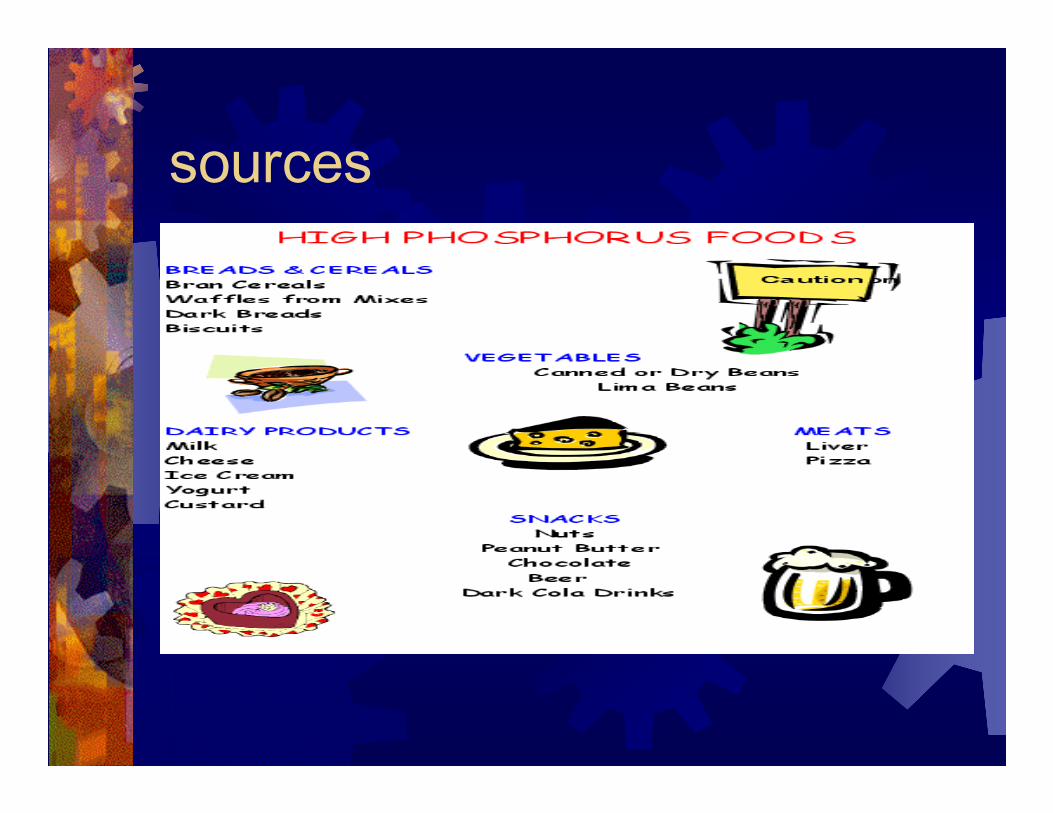
sources

Sources
***dietary sources should be restricted in renal disease (usually see increased phos, decreased Ca)
� -protein sources
� -meat, poultry, fish, eggs, legumes, nuts, milk, cereals, grains
Renal Disease
Causes of hypophosphatemia
-starvation
-TPN with inadequate phos content
-malabsorption, small bowel bypass
-vit D deficient and vit D resistant
osteomalacia
Causes of hypophosphatemia
cont’d
-phosphaturic drugs: theophylline, diuretics, bronchodilators, corticosteroids
-hyperparathyoidism (primary or secondary)
-hyperthyroidism
-renal tubular defects
-hypokalemic nephropathy
-inadequately controlled DM
-***alcoholism

Causes of hypophosphatemia
cont’d
Intracellular shift of phosphorus
-administration of glucose
-anabolic steroids, estrogen, OCP
-respiratory alkalosis
-salicylate poisoning
Electrolyte abnormalities
-hypercalcemia
-hypomagnesemia
-metabolic alkalosis

Causes of hypophosphatemia
cont’d
Abnormal losses followed by inadequate
repletion
-***DM with acidosis—with aggressive therapy
-***recovery from starvation or prolonged catabolic
state—refeeding syndrome
-***chronic alcoholism, especially with nutritional
repletion, assoc with hypomagnesemia—”
-recovery from severe burns

Causes of hyperphosphatemia
-excessive growth hormone
(acromegaly)
-hypoparathyroidism assoc with low Ca
-pseudohypoparathyroidism assoc with
low Ca
-***chronic renal insufficiency
-acute renal failure
Causes of hyperphosphatemia
cont’d
Catabolic states, tissue destruction
-stress or injury, rhabdomyolysis (esp with
renal insufficiency)
-chemotherapy of malignant disease,
particularly lymphoproliferative disease
Excessive intake or absorption
-laxatives or enemas containing phosphate
-hypervitaminosis D

Deficiency
� -fatal
� -usually rare with food intake
� -***respiratory muscle collapse
� -heart failure
� -muscle aches, bone pain, and fracture
Toxicity
� -symptoms of the primary disorder

Magnesium

Function
-bone, muscle contractility, nerve
excitability
-antagonistic to calcium
--in a muscle contraction, Mg relaxes, and
calcium contracts
--low Mg can cause pregnancy induced
HTN

Absorption / Excretion
� -absorption varies
� -similar to calcium (low pH, upper GI), however, no Vit D required-kidney conserves Mg when intake of Mg is low
� -large losses with vomiting because of high levels of gastic juice

sources

Sources
� -seeds, nuts, legumes, unmilled cereal
grains, dark greens
� -fish, meat, milk, fruits
� -lost during refining of flour, rice, vinegar


Causes of hypomagnesemia
-malabsorption, chronic diarrhea, laxative abuse
-prolonged GI suction
-small bowel bypass
-malnutrition
-***alcoholism
-refeeding
-TPN with inadequate Mg

Causes of hypomagnesemia
cont’d-DKA
-diuretics
-hyperaldosteronism, Barrter’s syndrome
-hypercalcuria
-renal Mg wasting
-hyperparathyroidism
-postparathyroidectomy
-vit D therapy
-aminoglycosides, ***cisplatin, ampho B

Causes of hypermagnesemia
Decreased renal fxn
***Increased intake—abuse of Mg
containing antacids (MOM) and
laxatives in renal insufficiency

Deficiency
� -anorexia, growth failure, cardiac and
neuromuscular changes—weakness,
irritability, mental derangement
� -tetany, muscle cramps

Toxicity
� -respiratory—depression, apnea
� -CV—hypotension, cardiac arrest, EKG (prolonged QRS and QT, heart block, peaked T waves)
� -GI—N/V
� -neuromuscular—paresthesias, somnolence, confusion, coma, hyporeflexia, paralysis, apnea

Iron

Function
� -respiratory transport of O2 and CO2
� -immune system
� -cognitive performance
� -found in Hgb (in RBC’s) and myoglobin
(in muscles)
� -cytochrome p450 system

Absorption and transport
� -dietary iron exists in heme (Hgb and myoglobin) and non-heme
� -***heme Fe is absorbed better
� -non-heme Fe has to be present in the duodenum or upper jejunum in soluble form if it is to be absorbed
� -in Fe deficiency, 50% can be absorbed
� -***2-10% of Fe from veggies is absorbed and 10-30% is absorbed from animal protein

Factors affecting absorption
� -***ascorbic acid is the most potent enhancer
� -animal proteins (beef, pork, veal, lamb, liver, fish, chicken) enhance
� -but, proteins from cow’s milk, cheese, eggs, don’t
� -gastric acidity enhances absorption (antacids interfere)
� -pregnancy, increased growth, Fe defic all increase deficiency
� -phytate and tannins decrease abs
� -Fe used for enrichment are less
absorbed than elemental Fe
� -increased intestinal motility decreases
absorption because it decreases
contact time for absorption
Storage
� -stored as ferritin and hemosiderin
� -long term high Fe ingestion or frequent blood transfusions can lead to accumulation of Fe in the liver
� -***hemosiderosis develops in individuals who consume a lot of Fe or have a genetic defect resulting in increased Fe absorption
� -in associated with tissue damage, it is called hemochromatosis

Excretion
� -lost thru bleeding, feces, sweat,
exfoliation of hair and skin
� -none in urine
Sources and Intakes
� -best source is liver
� -oysters, shellfish, kidney, lean meat, poultry, fish
� -dried beans, veggies, dark molasses
� -egg yolks, dried fruit, enriched breads,
� -requirements are highest in infancy and adolescence
� -females stay high because of menstruation
� -decrease with menopause and increased with pregnancy

Deficiency
� -most common deficiency
� -most at risk: <2 yrs old, teens, pregnancy, elderly
� -***anemia (hypochromic, microcytic)
� -tx: diets high in absorbable Fe and/or Fe supplements (ferrous sulfate, ferrous gluconate)
� -can be caused by injury, hemorrhage, illness, poor diet
Zinc
� -involved in synthesis or degradation of CHO, proteins, lipids, nucleic acids
� -stabilizes RNA and DNA
� involved in transcription and replication
� -needed for bone enzymes and osteoblastic activity

absorption
� Impaired absorption in Crohn’s or
pancreatic insufficiency
� -plasma zinc levels act as acute phase
reactants and fall by 50% with injury
(like platelets)
Inhibiting Factors
� -fiber, phytate
� -high doses of copper
� -Fe competes with zinc for absorption

Enhancing Factors
� -glucose, lactose, and soy protein
� -red wine
� -human milk

Excretion
� -feces—almost entirely
� -***in urine with starvation, nephrosis,
DM, alcoholism, hepatic cirrhosis (zinc
supplementation in encephalopathy),
porphyria

Sources and Intakes
� -meat, fish, poultry, milk
� -oysters, shellfish, meat, liver, cheese,
whole grains, dry beans, nuts

Deficiency
� -short stature, hypogonadism, anemia
� -with diets high in unrefined cereal and
unleavened bread
� -delayed wound healing, alopecia
� ***-acrodermatitis enteropathica=AR dz with
zinc malabsorption
� -eczematoid skin lesions, alopecia, diarrhea,
bacterial and yeast infections, death
� -immunologic deficits—lymphopenia,
thymic atrophy

***Causes of
deficiency�Anorexia Nervosa
� TPN without zinc (diarrhea, small bowel fistulas)
�High intake of phytate, tannins, binding drugs (EDTA), oxalate
�High iron intake
�Malabsorption syndromes
�Acrodermatitis enteropathica
�Diarrhea
�Pancreatico-cutaneous fistula
�Proximal entero-cutaneous fistulas
�Hemolytic anemias (sickle cell anemia)
�Renal failure patients on dialysis
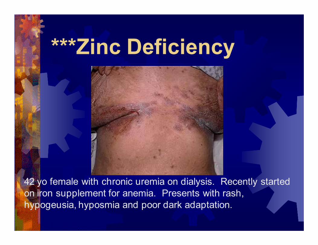
***Zinc Deficiency
42 yo female with chronic uremia on dialysis. Recently started
on iron supplement for anemia. Presents with rash,
hypogeusia, hyposmia and poor dark adaptation.
Acrodermatitis
Enteropathica
�Autosomal recessive disease
associated with a defect causing a
reduction in zinc absorption
�Can be treated by pharmacologic
doses of oral zinc

Acrodermatitis
Enteropathica
Toxicity
� ->100-300 mg/d
� -rare
� -interferes with copper absorption
� -decrease in HDL
� -GI irritation, vomiting
Fluoride
� -tooth enamel
� -resistance to dental caries
� -fluoridation of h20 has decreased
caries by half
� -found in drinking h20, teflon pots and
pans (cooked in these)
� -toxicity at doses >0.1 mg/kg/d

Prevention of dental caries
� ***Incidence of dental fluorosis (mottled
teeth) occurs with increased intake
above 1-2 ppm.
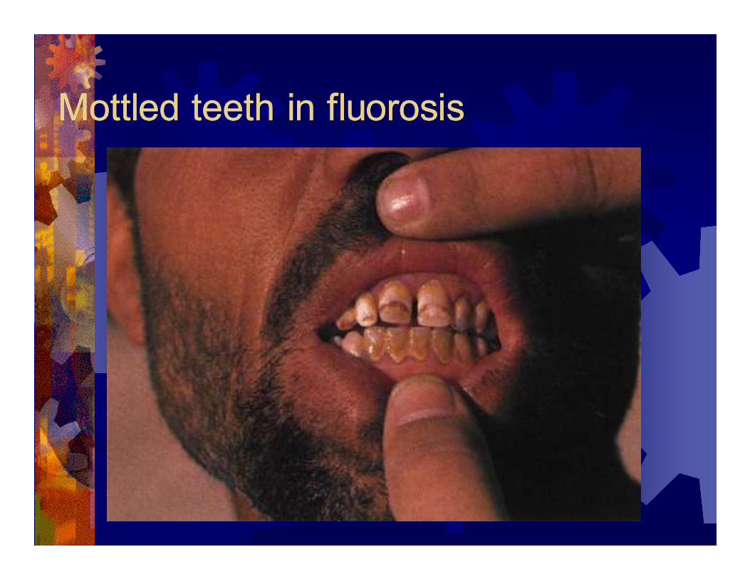
Mottled teeth in fluorosisMottled teeth in fluorosis

Maganese
� -found in many enzymes
� -connective and bony tissue formation
� -growth and reproduction
� -CHO and lipid metabolism

Absorption and Excretion
� -after absorption, it appears rapidly in
the bile and is excreted in the feces
� -concentrated in liver and increases with
liver disease

Sources and Intakes
� -whole grains, legumes, nuts, teas, fruit,
veggies, instant coffee, and tea
Deficiency
� -wt loss, ataxia, dermatitis, N/V,
decreased hair growth, impaired
reproductive activity, decreased
pancreatic function and CHO
metabolism

Toxicity
� -accumulates in liver and CNS—
parkinsonian sx