midline radial glia translocation and corpus callosum formation … complete... · midline radial...
TRANSCRIPT

Midline radial glia translocation and corpus callosumformation require FGF signaling
Karen Muller Smith1,4, Yasushi Ohkubo1,4, Maria Elisabetta Maragnoli1, Mladen-Roko Rasin2,3,Michael L Schwartz2, Nenad Sestan2,3 & Flora M Vaccarino1,2
Midline astroglia in the cerebral cortex develop earlier than other astrocytes through mechanisms that are still unknown.
We show that radial glia in dorsomedial cortex retract their apical endfeet at midneurogenesis and translocate to the overlaying
pia, forming the indusium griseum. These cells require the fibroblast growth factor receptor 1 (Fgfr1) gene for their precocious
somal translocation to the dorsal midline, as demonstrated by inactivating the Fgfr1 gene in radial glial cells and by RNAi
knockdown of Fgfr1 in vivo. Dysfunctional astroglial migration underlies the callosal dysgenesis in conditional Fgfr1 knockout
mice, suggesting that precise targeting of astroglia to the cortex has unexpected roles in axon guidance. FGF signaling is sufficient
to induce somal translocation of radial glial cells throughout the cortex; furthermore, the targeting of astroglia to dorsolateral
cortex requires FGFr2 signaling after neurogenesis. Hence, FGFs have an important role in the transition from radial glia to
astrocytes by stimulating somal translocation of radial glial cells.
Radial glial progenitor cells of the cortical ventricular zone extend anapical foot to the ventricular surface and a basal process to the pialmembrane. After their neurogenic phase (around embryonic day (E)18.5 in mice), these cells begin translocating their cell bodies toward thepia and differentiate into astrocytes1–3. Cortical astrocytes upregulatethe intermediate filament glial fibrillary acidic protein (GFAP) severaldays after birth. However, astroglial cells at the telencephalic midlinehave been observed to develop GFAP immunostaining by E17.5, muchearlier than other astrocytes4,5. These cells populate the indusiumgriseum, the glial wedge and the midline zipper. The indusium griseumis composed of neurons and astrocytes located underneath the pialmembrane of the dorsomedial pallium, above the path of the corpuscallosum. The glial wedge is formed by radial glial cell bodies arrangedin the medial pallial ventricular zone, underneath the indusiumgriseum6. The correct morphogenesis of these pioneer glial populationsalong the midline is thought to be crucial for guiding the axons of thedeveloping corpus callosum6–8.
The factors that drive the transformation of radial glia into astrocytesat the appropriate stage in development have not been elucidated. Cellsof the glial wedge and indusium griseum are born as early as E13.5 andE14.5, respectively4. Before expressing GFAP, cells of the glial wedgeexpress glutamate astrocyte transporter (Glast/Slc1a3), brain lipidbinding protein (Blbp/Fabp7) and nestin (Nes) gene products typicalof radial glial cells9, in keeping with their radial glial identity4. Hence, itis possible that cells of the indusium griseum derive from these radialglial progenitors that migrate to the cortical midline. If that were true,indusium griseum astroglia, unlike other cortical astrocytes, must begenerated synchronously with upper layer cortical plate neurons and
must start their migratory process concurrently with neurons andmuch earlier than the astrocytes that populate other regions of thetelencephalon. How the precise timing of migration and corticaltargeting of these different populations of astroglial cells is regulatedis unknown.
Previous in vitro studies have implicated the neuropeptide FGF2, amember of the fibroblast growth factor (FGF) family, in renderingprogenitor cells competent to switch from neuronal to glial cellfates10–12. Here, we show that the FGF receptor 1 gene (Fgfr1) isrequired for the early translocation of radial glial cells to the medialpallium, the prospective indusium griseum region, and that Fgfr2 isrequired for the targeting of astroglia to the cerebral cortex at the end ofgestation. In vertebrates, FGFs encompass 22 ligands13 that bind to 4tyrosine kinase FGF receptors (FGFr1–4)14. FGFs are required forprogenitor proliferation, specification and survival at early stages ofCNS development12,15–20. FGF receptors are widely expressed bytelencephalic progenitors at these early stages16,21. We and othersfound that at later stages of development, Fgfr1 continues to beexpressed in radial glial cells of the dorsomedial ventricular zone, inthe hippocampal primordium and, as shown here, in its anteriorrudiment, the indusium griseum. On the other hand, Fgfr2 is expressedby radial glial cells of the dorsolateral ventricular zone and ganglioniceminences21,22. The targeted inactivation of the Fgfr1 gene within radialglial cells produces alterations in hippocampal development23. Wereport here that these mice also show a prominent dysgenesis of thedorsal telencephalic commissures, including the corpus callosum andhippocampal commissure, without other obvious connectivity defects.In humans, homozygous inactivating mutations in the FGFR1 gene
Received 3 April; accepted 24 April; published online 21 May 2006; doi:10.1038/nn1705
1Child Study Center, 2Department of Neurobiology and 3Kavli Institute for Neuroscience, Yale University School of Medicine, New Haven, Connecticut 06520, USA. 4Theseauthors contributed equally to this work. Correspondence should be addressed to F.M.V. ([email protected]).
NATURE NEUROSCIENCE VOLUME 9 [ NUMBER 6 [ JUNE 2006 787
ART ICLES©
2006
Nat
ure
Pub
lishi
ng G
roup
ht
tp://
ww
w.n
atur
e.co
m/n
atur
eneu
rosc
ienc
e

result in Kallman syndrome with agenesis of the corpus callosum24.Hence, the role for Fgfr1 in midline commissure development isevolutionarily conserved. Using mouse mutants that lack Fgfr1 and/or Fgfr2 in radial glia along with shRNA-mediated knockdown of Fgfr1and overexpression of FGF2 and FGF8 proteins in cortical explants, wedemonstrate that commissural dysgenesis is secondary to a defectivetranslocation of radial glial cells from the ventricular zone to theanterior pallial midline, an area that is normally enriched inFGFr1 signaling. Misexpression of FGF proteins in other areas ofthe cortical plate was sufficient to prematurely induce the upwardtranslocation of radial glial cell bodies, underscoring a wider require-ment for FGFr2 signaling in somal translocation throughout thecerebral cortex.
RESULTS
Loss of Fgfr1 in radial glia causes callosal dysgenesis
Targeted inactivation of the Fgfr1 gene in radial glia was accomplishedvia Cre-dependent recombination of the loxP-flanked Fgfr1f allele25.Cre was driven by a human GFAP-Cre transgene (hGFAP-Cre) active intelencephalic radial glial cells beginning at E13.5–E14.5 (refs. 23,26).Mice homozygous for the recombined Fgfr1 null allele (referred to asFgfr1f/f;hGFAPCre) lacked the corpus callosum and hippocampal com-missure (Fig. 1 and Supplementary Fig. 1 online). These mice hadProbst bundles, collections of tangled axons adjacent to the corticalmidline (Fig. 1b, arrowheads). Whereas the defects in the corpuscallosum and hippocampal commissure were quite severe in most ofthe Cre transgene–bearing mice, they did not lead to a completeagenesis of these structures in all the Fgfr1f/f;hGFAPCre mice examined.We therefore assessed a series of control and mutant littermate mice inorder to examine both the penetrance and expressivity of this pheno-
type. The average midline width of commissures was markedly smallerin the Fgfr1f/f;hGFAPCre mice compared to controls (Fig. 1d). An analysisof variance (ANOVA) showed a significant effect of genotype oncommissure width (F1,90 ¼ 123; P o 0.0001), and a significantinteraction between genotype and region (P o 0.01), in that anteriorand posterior regions were significantly worse than the middle.Categorically, we found that 47% of the Fgfr1f/f;hGFAPCre mice(n ¼ 15) had complete agenesis of the corpus callosum, andan additional 33% percent of these mutants had a severely dysgeniccorpus callosum in the middle section, adjacent to the fimbria, thatwas less than 50% of the average width in the control mice(n ¼ 17). Another 20% of the Fgfr1f/f;hGFAPCre mice were similar tocontrol mice (Fig. 1e). In marked contrast to the dorsal commissures,the ventral commissures, such as the anterior and the habenularcommissures, were not affected in Fgfr1 mutant mice (arrows inFig. 1a,b; Supplementary Fig. 1).
This partial phenotype could potentially be attributed to a late orincomplete recombination of the Fgfr1 allele. To inactivate theFgfr1 gene at an earlier time point and in a broader distribution, wemated mice bearing the Fgfr1f/f alleles with a Cre strain driven by theCNS-specific nestin enhancer, which drives Cre recombination in alltelencephalic neural precursors by E11.5 (ref. 27). The Fgfr1f/f;NesCre
mice also showed defects in the dorsal telencephalic commissures butnot in the ventral commissures (Fig. 1a,c and Supplementary Fig. 1).Scoring adult Fgfr1f/f;NesCre mice (n ¼ 5) for midline defectsdemonstrated that 100% of the mutant mice had a complete absenceof midline crossing at the most anterior and posterior regions exam-ined; however, 20% of the Fgfr1f/f;NesCre mice had a small number ofcallosal fibers in the region above the hippocampal commissure(level ‘M’ in Fig. 1d). Hence, the defects were more penetrant in the
Control
Width of commissure
(µm)300
200
100
0A M P Type A (Normal)
IG
GW
MZG
P
Type B
100%
20%
20% 80%
33.3% 46.7%
Type C
Phenotype frequency
Cre
syl v
iole
t
Fgfr1f/f;hGFAPCre Fgfr1f/f;NesCre
Control
P0
GFA
P
Fgfr1
Fgf8
Spry1
Fgfr1f/f;hGFAPCre
Control Control Fgfr1f/f;hGFAPCre
Fgfr1f/f;NesCre
Control
Control
Fgfr1f/f;hGFAPCre
Fgfr1f/f;hGFAPCre
Fgfr1f/f;NesCre
Fgfr1f/f;NesCre
a
d
f g h
i j k
l m n
o p q
e
b c
Figure 1 Disruption of dorsal telencephalic commissures in glial-specific Fgfr1 mutant mice and enriched expression of FGF-related genes in anterior midline.
(a–c) Cresyl violet–stained sections illustrating the most severe and common phenotype of Fgfr1f/f;hGFAPCre (b) and Fgfr1f/f;NesCre (c) mice as compared to
Cre– controls harboring Fgfr1f/f alleles (a). Arrowheads, Probst bundles; red line, corpus callosum; arrows, anterior commissure. Scale bar, 1 mm.
(d) Commissure widths for anterior (A), medial (M) and posterior (P) cortical areas in control (n ¼ 17), Fgfr1f/f;hGFAPCre (n ¼ 15) and Fgfr1f/f;NesCre (n ¼ 5)
mice. (e) Frequency histogram categorizing mice into type A (normal), type B (commissures measuring o50% of normal in one or more area) or type C
(commissures completely absent). (f–h) GFAP immunostaining in coronal sections of control (f), Fgfr1f/f;hGFAPCre (g) and Fgfr1f/f;NesCre (h) mice at P0 to
vizualize the glial wedge (GW), indusium griseum (IG) and midline zipper glia (MZG). Arrowheads, GFAP+ cells; P, Probst bundles; black arrow, radial glial
fibers. (i–q) Expression of FGF-related genes in the anterior telencephalic midline at E14.5. In situ hybridization for Fgfr1 in control (i,j) and Fgfr1f/f;hGFAPCre
mice (k). Arrow, indusium griseum; arrowhead, glial wedge. In situ hybridization for Spry1 in control mice (l,m) and Fgfr1f/f;hGFAPCre mice (n). In situ
hybridization for Fgf8 in control mice (o,p) and Fgfr1f/f;hGFAPCre mice (q). Fgf8 expression was maintained in Fgfr1f/f;hGFAPCre mice (q). Scale bars: 200 mm in
f–h, 200 mm in i, 400 mm in l and o, 100 mm in j,k,m,n,p and q.
788 VOLUME 9 [ NUMBER 6 [ JUNE 2006 NATURE NEUROSCIENCE
ART ICLES©
2006
Nat
ure
Pub
lishi
ng G
roup
ht
tp://
ww
w.n
atur
e.co
m/n
atur
eneu
rosc
ienc
e
Fgfr1f/f;NesCre mice, although fewer mice with this mutation wereexamined (Fig. 1d,e).
Anterograde axonal tracing studies in adult mice using biotinylateddextran amines (BDA) stereotaxically injected into the frontal cortexdemonstrated that both Fgfr1f/f;hGFAPCre and Fgfr1f/f;NesCre mice did nothave any detectable labeling of axons within the contralateral cortex orwhite matter; instead, labeled cortical fibers were trapped within Probstbundles (Supplementary Fig. 2 online). However, corticostriatal andcorticothalamic axon tracts in Fgfr1f/f;hGFAPCre and Fgfr1f/f;NesCre micewere similar to those in controls (Supplementary Fig. 2).
We next examined commissure development using lipophilic dyetracers. In 83% of Fgfr1f/f;hGFAPCre mice, a complete loss of callosalaxons from the motor and somatosensory cortex was already present atbirth (controls, n ¼ 12; Fgfr1f/f;hGFAPCre, n ¼ 12; SupplementaryFig. 2). There was also a loss of hippocampal commissural axons inneonatal Fgfr1f/f;hGFAPCre mice (controls, n¼ 6; Fgfr1f/f;hGFAPCre, n¼ 3;Supplementary Fig. 2). Furthermore, corticothalamic and corticos-triatal projections were preserved in these mutant and control neonates(Supplementary Fig. 2). Together, these findings underscored arequirement for Fgfr1 in the development of dorsal telencephaliccommissures. Also, the presence of Probst bundles and the preservedcorticostriatal and corticothalamic projections indicated that thedefects were not due to a problem with axon extension or a generallack of neurite outgrowth.
Absent indusium griseum in glial-specific Fgfr1 mutants
Commissural axon guidance is thought to depend upon the integrity ofmidline cells in the glial wedge, indusium griseum, midline zipper andglial sling. To examine the formation of these glial structures in Fgfr1mutant neonates, we performed immunohistochemistry for GFAP inknockout (n¼ 6) and control (n¼ 6) littermates at postnatal day (P) 0.The glial wedge, indusium griseum and midline zipper astroglia wereclearly visible at this age in control mice throughout the anteroposter-ior extent of the telencephalon (Fig. 1f and Supplementary Fig. 1), aspreviously described4. In contrast, indusium griseum astroglia wereabsent in the anterior regions of the Fgfr1f/f;hGFAPCre mice (Fig. 1g and
Supplementary Fig. 1). Furthermore, in mutants, the GFAP-positive(GFAP+) processes emerging from the glial wedge and adjacentventricular zone remained attached at both the ventricular zone andthe pia, and very few astrocytes were observed in between the glialwedge and the indusium griseum (Fig. 1g and Supplementary Fig. 1,black arrows). In comparison, in control littermates, GFAP+ astrocyteshad already reached the indusium griseum or were noticeable in theregion between the glial wedge and indusium griseum (Fig. 1f andSupplementary Fig. 1, arrowheads). The Fgfr1f/f;NesCre mice had amore extensive defect in the morphogenesis of the indusium griseum,with posterior regions also lacking glial cells within this structure. Therewere no GFAP+ cell bodies migrating toward the indusium griseum;however, GFAP+ cells were seen in the glial wedge or appeared to betrapped within the Probst bundles (Fig. 1h and Supplementary Fig. 1,arrowheads). The uniform lack in accumulation of astroglial cells at themidline correlates with the more severe defect in callosal morphogen-esis and may be a reflection of the more widespread Fgfr1 recombina-tion in radial glial cells driven by the nestin promoter.
FGF signaling is enriched at the anterior cortical midline
At E14.5, when a majority of indusium griseum cells undergo their lastmitosis8, the highest expression of Fgfr1 was in the dorsomedialportion of the ventricular zone (arrowhead in Fig. 1i), correspondingto the glial wedge, and in the primordium of the indusium griseum(arrow in Fig. 1j). Fgfr1 was also expressed at lower levels in the corticalplate. This expression pattern remained essentially unchanged at E16.5,when a large proportion of pioneer neocortical axons project acrossthe anterior midline (ref. 28 and data not shown). The expressionof Fgfr1 was undetectable in corresponding areas of FGFr1f/f;hGFAPCre
mice (Fig. 1k).The restricted expression of Fgfr1 may be attributable to the positive
autoregulation of Fgfr1 transcription via ligand-induced activation ofFGFr1 signaling23. Indeed, in control mice, the expression of Spry1, agene product that is immediately downstream to the activation of FGFreceptors15,29, partially overlaps that of Fgfr1 (Fig. 1l,m). The Spry1signal was highest in the glial wedge (Fig. 1m, arrowhead) with someexpression in the indusium griseum (Fig. 1m, arrow). In both areas, thesignal for Spry1 was greatly reduced in Fgfr1f/f;hGFAPCre mice (compareFig. 1m with Fig. 1n).
FGF8, together with other FGF ligands, is enriched at the dorso-anterior midline at early stages of development20,30,31. At E14.5(Fig. 1o–q), Fgf8 mRNA was expressed within the presumptive glialwedge (arrowheads) and indusium griseum (arrows). Therefore, thissecreted ligand is likely to form a concentration gradient emergingfrom this region. Notably, the expression pattern of Fgf8 wasunchanged in the Fgfr1f/f;hGFAPCre mice (Fig. 1q).
The presence of FGFr signaling molecules within the glial wedge, aswell as in the presumptive indusium griseum, is consistent with the
Fgfr1f/f;Syn1Cre;Rosa26R
Fgfr1f/f;Syn1CreControl
LacZ
Cre
syl v
iole
tTA
G1
GFA
P
IG IG
GW GW
MZG MZG
ba b
c d
g h
e f
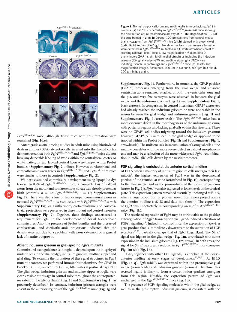
Figure 2 Normal corpus callosum and midline glia in mice lacking Fgfr1 in
neurons. (a) LacZ histochemistry in Fgfr1f/f;Syn1Cre;Rosa26R mice showing
the distribution of Cre recombinase activity at P0. (b) Magnification (2�) of
the area framed in a. (c–h) Coronal 100-mm sections from control mouse
brains (c,e,g) or from Fgfr1f/f;Syn1Cre mice (d,f,h) stained with cresyl violet
(c,d), TAG-1 (e,f) or GFAP (g,h). No abnormalities in commissure formation
were detected in Fgfr1f/f;Syn1Cre mutants (in e,f, white arrowheads point to
crossing callosal fibers). Insets, low magnification 4,6-diamidino-2-phenylindole (DAPI) stain. Midline glial structures including the indusium
griseum (IG), glial wedge (GW) and midline zipper glia (MZG) were
indistinguishable in control (g) and Fgfr1f/f;Syn1Cre mice (h). Insets, low
magnification images. Scale bars: 400 mm in a,e and f, 800 mm in c and d,
200 mm in b, g and h.
NATURE NEUROSCIENCE VOLUME 9 [ NUMBER 6 [ JUNE 2006 789
ART ICLES©
2006
Nat
ure
Pub
lishi
ng G
roup
ht
tp://
ww
w.n
atur
e.co
m/n
atur
eneu
rosc
ienc
e

hypothesis that Fgfr1 may be important for the proper formation ofmidline glial structures and/or may secondarily affect the distributionof the chemotactic signals that they emanate. Furthermore, thepresence of Fgfr1 mRNA in the developing neocortex also suggeststhat FGFr1 may function within neurons fated to cross the midline.For example, FGFr1 might act as a receptor/coreceptor for midlineguidance cues or may regulate the expression of receptors for knownchemotactic signals.
To clarify the role of FGF signaling in commissure development, weinvestigated whether the targeted inactivation of Fgfr1 in radial glialprogenitors might result in neuronal or glial differentiation defects thatcould have an impact on axon guidance. The Fgfr1f/f;hGFAPCre andFgfr1f/f;NesCre mice appeared normal with respect to the differentiationof neurons and astroglial cells as shown by bIII tubulin, calretinin andGFAP immunostaining (Fig. 1f–h and Supplementary Fig. 3 online).At E16.5, control and Fgfr1f/f;hGFAPCre mice showed similar amounts ofDCC, TAG-1/Cntn2, L1 and N-cadherin (Supplementary Fig. 3),proteins that are involved in commissural axon fasciculation andguidance. In the Fgfr1 mutants, some of the DCC-positive axonsreached the midline, but were less compactly organized and did notappear to cross it. Furthermore, the expression of Slit2, Slit3, Robo1 andgrowth-associated protein 43 (Gap43) genes, whose mutations alsoresult in commissural defects32, did not appear to be altered in Fgfr1mutant embryos (Supplementary Fig. 4 online). Therefore, we exclu-ded perturbations in the expression levels of several axonal and glialfactors necessary for midline guidance as a potential explanation for theobserved phenotype in Fgfr1 mutant mice. However, the expression ofNtn1 appeared to be decreased in the indusium griseum ofFgfr1f/f;hGFAPCre mice, while being maintained in the glial wedgeand other regions (Supplementary Fig. 4). This could potentiallybe a consequence of the altered morphogenesis of astroglia in theindusium griseum.
Neuronal specific Fgfr1 mutants lack commissure dysgenesis
To directly assess the potential contribution of FGFr1 signaling withinneurons during commissure formation, we generated mice carrying aneuron-specific inactivation of the Fgfr1 gene. We did so by crossingthe Fgfr1f/f strain with mice bearing the Syn1-Cre transgene, to obtainFgfr1f/f;Syn1Cre mutants. The Syn1-Cre transgene is specific to neuronsand begins to be expressed at approximately E12.5 (ref. 33). Using theRosa 26R lacZ reporter gene for Cre-mediated recombination34, we
observed robust b-galactosidase (b-gal) staining in all cortical layers ofFgfr1f/f;Syn1Cre at P0 (Fig. 2a,b). Most of this cortical staining ispresumed to be neuronal in nature as most of the cortical cells at P0are neurons. At higher magnification, labeled fibers could be seendescending from the cortex into the white matter of Fgfr1f/f;Syn1Cre
mice. This staining was identical to that obtained with heterozygousFgfr1f/+;Syn1Cre littermates (data not shown). Hence, Cre recombinationwas widespread throughout the cerebral cortex.Fgfr1f/f;Syn1Cre mice did not show any detectable differences in brain
morphology or histological organization when compared to controllittermates (Fig. 2c,d). Furthermore, Fgfr1f/f;Syn1Cre mice appearedto have normal commissures. This was confirmed with staining forTAG1 (Fig. 2e,f). Finally, midline glial structures were preserved in theFgfr1f/f;Syn1Cre mice. These mice did not have any apparent defects inglial wedge, midline zipper or indusium griseum astroglia (Fig. 2g,h).Therefore, defects in the formation of the indusium griseum and thetelencephalic commissures are specific to mutants with targeted inac-tivation of Fgfr1 in radial glial progenitors, as they are not present inneuron-specific mutants of Fgfr1.
Fgfr1 is required for midline radial glia translocation
Fgfr1 loss of function was associated with a decrease in the number ofGFAP+ cells in the apparent process of migrating toward the prospec-tive indusium griseum area, as well as with the maintenance ofradial glial cell processes in this region (Fig. 1f–h). To assess whetherFgfr1f/f;hGFAPCre mice had defective migration of cells from the glialwedge to the indusium griseum, coronal cortical slices were preparedfrom E14.5 control and Fgfr1f/f;hGFAPCre embryos and maintainedin vitro. The fluorescent dye tracer PKH26 was applied to the dor-somedial ventricular zone, in order to label presumptive radial glialcells in the glial wedge (Fig. 3a,b). The movement of the cells in theseexplants was monitored by fluorescence microscopy over a 48-h period.
a b
f g
h i
j k
l m
n o
c
e
d
Dorsal
Ventral
1 h
24 h
Control
M
IG
M
h
lm
i
LVLV
M
IG GWGW
n = 7
n = 6
100
Dis
tanc
e fr
om v
entr
icle
75
50
25
0
M
n o
M
LV
LVLV
LV
Fgfr1f/f;hGFAPCre
Control
E14.5 GLAST / BrdU (1 h)
E16.5 GLAST / BrdU (←E14.5 BrdU, 48 h)
Fgfr1f/f;hGFAPCre
Control Fgfr1f/f;hGFAPCre
PKH26-labeled E14.5 slice culture
LV
M
Figure 3 Migration of astroglial cells to the indusium griseum is disrupted
in glial-specific Fgfr1 mutant. (a–e) Movement of PKH26-labeled cells
from ventricular zone to midline pia in coronal slices of E14.5 mouse
telencephalon. Labeled cells in the ventricular zone 1 h after labeling
(a,b). White dashed lines, slice shape as determined by bright-field images.
(c) Control slices at 24 h showing labeled cells (arrowheads) translocated
toward the indusium griseum while little translocation occurred in
Fgfr1f/f;hGFAPCre slices (d). (e) Average distance from the ventricular wall tothe leading edge of PKH-labeled cells after 24 h in culture, normalized by
the cortical thickness (cells close to the pia would have a value of 100).
Mean values are 97.8 in control (n ¼ 7) and 41.2 in Fgfr1f/f;hGFAPCre embryos
(n ¼ 6). P o 10–5, one-tailed Student’s t-test. (f–o) Coronal sections from
control (f,h,j,l,n) and Fgfr1f/f;hGFAPCre embryos (g,i,k,m,o) harvested 1 h
(f–i) or 48 h (j–o) after a single BrdU administration at E14.5 and immuno-
stained for GLAST (green) and BrdU (red). Dashed white lines, midline pia.
Accumulation of GLAST+ and BrdU+ cells in the indusium griseum of control
embryos 48 h after BrdU injection (j,l; confocal stack analyse in n) is not
observed in Fgfr1f/f;hGFAPCre embryos (k,m; confocal stack analyses in o). LV,
lateral ventricle; M, midline; GW, glial wedge; IG, indusium griseum. Scale
bars: 100 mm in a–d, 150 mm in f,g,j and k, 75 mm in h,i,l and m, 30 mm in
n and o. Error bars represent s.e.m.
790 VOLUME 9 [ NUMBER 6 [ JUNE 2006 NATURE NEUROSCIENCE
ART ICLES©
2006
Nat
ure
Pub
lishi
ng G
roup
ht
tp://
ww
w.n
atur
e.co
m/n
atur
eneu
rosc
ienc
e
By 24 h, labeled cells had reached the midline pial membrane in controlembryos (arrowheads in Fig. 3c). In cortical slices prepared fromFgfr1f/f;hGFAPCre embryos, however, we observed little or no cell migra-tion away from the ventricular zone, and no label accumulated in theindusium griseum area (Fig. 3d). The average distance of migration ofmidline cells in Fgfr1f/f;hGFAPCre embryos was less than half of thatachieved in controls (n¼ 7 control and n¼ 6 Fgfr1f/f;hGFAPCre embryos;Fig. 3e). Labeled cells reached the pia in 4 of 7 explants in control mice,but in none of the explants prepared from Fgfr1f/f;hGFAPCre mice. Thesedata indicate that Fgfr1 is required for the migration of cells from theventricular zone to the subpial region of the dorsomedial pallium.
These experiments, however, did not distinguish whether thesemigrating cells corresponded to the astroglial cells of the indusiumgriseum. Most indusium griseum cells are born around E14.5 (ref. 4).Hence, we labeled dividing cells at E14.5 by in vivo 5-bromodeoxyur-idine (BrdU) incorporation and traced their location and fate in themedial pallium of both control and Fgfr1 mutant embryos (Fig. 3f–o).
Both control and Fgfr1f/f;hGFAPCre littermates had abundant BrdUlabeling in the ventricular zone, including the glial wedge region, 1 hafter a single BrdU injection at E14.5 (Fig. 3f–i). Very little BrdUlabeling was visible in the cortical plate, except in the meninges. ByE16.5, in control embryos many BrdU-positive (BrdU+) cells double-labeled with the astroglial marker GLAST were visible in the presump-tive indusium griseum at the midline, as well as throughout the regionbetween the glial wedge and the indusium griseum (Fig. 3j,l; magnifiedconfocal images in Fig. 3n). A few BrdU+ cells that were GLAST-negative (GLAST–), presumably neurons, were also present in thisregion in E16.5 control embryos. In Fgfr1f/f;hGFAPCre littermates, theBrdU/GLAST double-labeled cells were absent in both the presumptiveindusium griseum and the area between the glial wedge and theindusium griseum, whereas BrdU single-labeled cells were detectablein the cortical plate (Fig. 3k,m; magnified confocal image inFig. 3o). Furthermore, an accumulation of GLAST-positive (GLAST+)cells was visible in the presumptive indusium griseum in controlembryos (Fig. 3j,l,n) but not in Fgfr1f/f;hGFAPCre littermates(Fig. 3k,m,o). We detected no changes in radial glial cell proliferationin the glial wedge and adjacent dorsomedial ventricular zone inFgfr1f/f;hGFAPCre mice. The density of BrdU+ was 6.5 � 10�4 (± 0.6)cells mm–3 in controls versus 7.8 � 10�4 (± 0.4) cells mm–3 inFgfr1f/f;hGFAPCre mice (mean (± s.e.m.); P ¼ 0.16, Student’s two-tailedt-test). Combined, these data suggest that a population of radial glialcells born at E14.5 migrated from the nearby ventricular zone to thepresumptive indusium griseum in control mice, but not in Fgfr1mutant littermates.
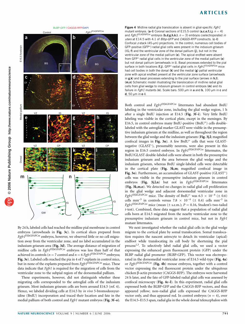
We next investigated whether the radial glial cells in the glial wedgemigrate to the cortical plate by somal translocation. Somal transloca-tion requires the nascent astrocyte to detach its ventricular (apical)endfoot while translocating its cell body by shortening the pialprocess1,3. To selectively label radial glial cells, we used a vectorexpressing the enhanced green fluorescent protein (GFP) under theBLBP radial glial promoter (BLBP-GFP). This vector was electropo-rated in the dorsomedial ventricular zone of E14.5 wild-type (Fig. 4a)or Fgfr1f/f;hGFAPCre (Fig. 4b) mouse embryos, together with a controlvector expressing the red fluorescent protein under the ubiquitouschicken b-actin promoter (CAGGS-RFP). The embryos were harvested24 h later, and the fate of GFP-labeled radial glial cells was assessed byconfocal microscopy (Fig. 4c–l). In this experiment, radial glial cellsexpressed both the BLBP-GFP and the CAGGS-RFP vectors, and thusappeared yellow; non–radial glial cells expressed the CAGGS-RFPvector only, and thus appeared red. In control embryos (n ¼ 4), overthe E14.5–E15.5 span, radial glia in the whole dorsal telencephalon with
IG
GW
IG
GW
CPVZCPVZ
Dor
sal
IGVZIGVZ
Med
ial p
alliu
m
LV
LV
BGBGc
d
E15
.5 fo
rebr
ain
Control Fgfr1f/f;hGFAPCre
BLBP-GFP / CAGGS-RFP/DAPI
a b
c d
e
i j k l
f g h
m n
Figure 4 Midline radial glia translocation is absent in glial-specific Fgfr1
mutant embryos. (a–l) Coronal sections of E15.5 control (a,c,e,f,i,j; n ¼ 4)
and Fgfr1f/f;hGFAPCre embryos (b,d,g,h,k,l; n ¼ 3) embryos coelectroporated in
utero at E14.5 with 4:1 of Blbp-GFP and CAGGS-RFP constructs. (c–l)
Confocal z-stack (45-mm) projections. In the control, numerous cell bodies of
GFP-positive (GFP+) radial glial cells were present in the indusium griseum
(IG; f) and the ventricular zone of the dorsal pallium (j), but not in the
ventricular zone of the medial pallium (e). The apical endfeet were absentfrom GFP+ radial glial cells in the ventricular zone of the medial pallium (e)
but not dorsal pallium (arrowheads in i). Basal processes extended to the pial
surface in both locations (f,j). GFP+ radial glial cells in Fgfr1f/f;hGFAPCre mice
had cell bodies in both the dorsal (k) and the medial (g) pallial ventricular
zone with apical endfeet present at the ventricular zone surface (arrowheads
in g,k) and basal processes extending to the pial surface (arrows in h,l).
(m,n) Schematic model illustrating the translocation of midline radial glial
cells from glial wedge to indusium griseum in control embryos (m) and its
failure in Fgfr1 mutants (n). Scale bars: 500 mm in a and b, 100 mm in c and
d, 50 mm in e–l.
NATURE NEUROSCIENCE VOLUME 9 [ NUMBER 6 [ JUNE 2006 791
ART ICLES©
2006
Nat
ure
Pub
lishi
ng G
roup
ht
tp://
ww
w.n
atur
e.co
m/n
atur
eneu
rosc
ienc
e

the exception of the medial region were bipolar, with their apicalprocess firmly attached to the ventricular basement membrane(Fig. 4a,i,j). In the medial region of the pallium, however, the GFP-labeled radial glial cells had already detached their apical endfeet fromthe ventricular zone (Fig. 4a,c,e), and most had already settled in theprospective indusium griseum (Fig. 4c,f) while a few were still visibleen route to this area (Fig. 4c,e). Notably, astroglial cells were seenpulling themselves up by their intact basal process attached to theprospective indusium griseum area (Fig. 4c,e).
To understand whether this cell movement was disrupted in theFgfr1 mutants, we electroporated these constructs in Fgfr1f/f;hGFAPCre
embryos (n ¼ 3). Notably, radial glial cell bodies throughout thetelencephalon remained in the ventricular zone with their apicalendfeet attached to the ventricular basement membrane in Fgfr1mutants (Fig. 4b,d). In the medial pallial region, GFP-labeled radialglial cells were attached by their apical endfeet to the lateral ventricle(Fig. 4g), and no labeled cell populated the indusium griseumprimordium (Fig. 4h), in marked contrast to what was observed incontrols. Radial glial morphology in the dorsal pallium of Fgfr1mutants (Fig. 4k,l) resembled that of wild-type embryos (Fig. 4i,j).These experiments illustrate the early detachment of radial glia apicalprocesses from the dorsomedial ventricular zone and their somaltranslocation from the ventricular zone to the indusium griseumprimordium at E14.5–E15.5, a time when upper layer cortical neuronsare generated in the ventricular zone of the cerebral cortex (schematicdrawing in Fig. 4m). Furthermore, this process did not occur whenFGFr1 signaling was disrupted in these cells (Fig. 4n). Although thedetachment and translocation process could be simply delayed in thesemutants, the absence of indusium griseum astroglia at P0 (Fig. 1f–h)argues against this notion.
Cell-autonomous regulation of glial translocation by FGFr1
To further understand whether FGFr1 signaling within midline radialglial cells is required for their translocation, we performed an in vivoknockdown of Fgfr1 mRNA by electroporating radial glial cells in uterowith pCGLH vectors expressing either small-hairpin RNA (shRNA)targeting the Fgfr1 (n ¼ 7), control shRNA with four point mutationsin the Fgfr1 target sequence (n ¼ 7) or empty shRNA vectors (n ¼ 5).The shRNA targeting Fgfr1 resulted in decreased Fgfr1 expression(Supplementary Fig. 5 online). All pCGLH vectors coexpressedshRNA and GFP, which allowed us to directly visualize the behaviorof the transfected cells in the wild-type background. Electroporation ofcontrol shRNA harboring four mutations with respect to the targetRNAi sequence or electroporation of the empty shRNA vector pro-duced the same phenotype previously observed after electroporatingthe BLBP-GFP vector in control embryos (Fig. 4a,c): 3 d aftertransfection, cells had already left the medial ventricular zone anddensely populated the subpial region in the prospective indusiumgriseum area (Fig. 5a,b). No radial glial endfeet or cell bodies couldbe visualized in the dorsomedial ventricular zone (Fig. 5b), suggestingthat these radial glial cells had left behind no progeny. Transfected cellsin the dorsal telencephalon remained in the ventricular zone (Fig. 5a),further demonstrating that these different populations of radial glialcells translocate to the upper layers according to completely differentschedules. Notably, radial glial cells transfected with Fgfr1 shRNA didnot leave the dorsomedial ventricular zone (Fig. 5c,d). At highermagnification (Fig. 5e–j), it was clear that whereas midline radialglial cells electroporated with control vector left the ventricular zoneand accumulated in the indusium griseum (Fig. 5e–i), midline radialglial cells harboring a knockdown of Fgfr1 mRNA could not undergotheir early translocation toward the indusium griseum prospectiveregion and behaved similarly to all the other radial glial cells (Fig. 5f,h).Further, midline radial glia left no endfeet in the ventricular zone(Fig. 5g), whereas many radial glial cell endfeet were attached to theventricular surface in radial glial cells transfected with Fgfr1 shRNA(arrowheads in Fig. 5h). Because all the surrounding cells had normalFgfr1 expression, the data demonstrate that Fgfr1 is directly requiredwithin the translocating cells and is not secondarily affecting themthrough cell-to-cell interactions. Notably, cells with an Fgfr1 mRNAknockdown could reach the dorsomedial cortical plate 3 d afterelectroporation, but appeared to have neuronal morphology identicalto that of other cortical plate areas (Fig. 5j), suggesting that thedisruption of Fgfr1 does not alter neuronal migration.
FGFs are sufficient to induce radial glial translocation
Our in situ hybridization data show that endogenous Fgf8 and Fgfr1messages are restricted to the midline at the same time and place at
IGV
ZM
edia
l pal
lium
LV
j IG
h
LVi
gLV
IG
IG
LV
IG
LV
b
Fgfr1 siRNA E14.5 → E17.5Control siRNA E14.5 → E17.5
d
a
e f
g h
i j
b c dFigure 5 Cell-autonomous regulation of midline radial glia translocation by
FGF signaling. (a–j) Coronal sections of E17.5 embryos electroporated in
utero at E14.5 with pCGLH vectors expressing control shRNAs (a,b,e,g,i;
n ¼ 7 point-mutated and n ¼ 5 empty shRNA vector) or Fgfr1 shRNAs
(c,d,f,h,j; n ¼ 7). Anti-GFP diaminobenzidine immunohistochemistry (a–d)
and fluorescence (e–j) in electroporated brains revealed that dorsomedial
ventricular zone cells expressing control shRNAs (a,b,e,g,i) but not Fgfr1
shRNAs (c,d,f,h,j) migrated to the indusium griseum (IG). Numerous GFP+
cells with bipolar morphology and apical endfeet (arrowheads) remained in
the dorsomedial ventricular zone when expressing Fgfr1 shRNAs (h compared
to control in g). In contrast, many GFP+ cells in the dorsal pallium remained
in the ventricular zone irrespective of electroporation with control or Fgfr1
shRNAs. Images in g,i and h,j are high magnifications of the respective boxed
areas shown in e and f. Panels b and d are high magnification of red boxes
in panels a and c. Scale bars: 600 mm in a and c, 100 mm in e and f,
30 mm in g–j.
792 VOLUME 9 [ NUMBER 6 [ JUNE 2006 NATURE NEUROSCIENCE
ART ICLES©
2006
Nat
ure
Pub
lishi
ng G
roup
ht
tp://
ww
w.n
atur
e.co
m/n
atur
eneu
rosc
ienc
e

which the indusium griseum astroglia are migrating. We thereforetested whether the exogenous application of FGF8 protein to an area ofthe cortical plate that normally has little or no expression of FGF8and Fgfr1 is capable of inducing the migration of cells from theventricular zone to the cortical plate. Cell migration was detectedusing the vital dye PKH26 applied to the dorsolateral ventricularzone of cortical slices, after a bead soaked in FGF8 or controlbovine serum albumin (BSA) was implanted into the dorsal corticalplate (Fig. 6a,b). Numerous cells were visible in the intermediatezone and cortical plate in the area adjacent to the FGF8 bead 24 hafter dye application (Fig. 6b). These cells appeared to be movingdirectly up to the cortical plate in alignment with radial glial fibers. Incomparison, the implantation of BSA-soaked beads did not have anyeffect (Fig. 6a).
We further investigated whether FGF protein gradients stimulatedneuronal or glial cell translocation in explants of whole E13.5 dorsalcortex. After FGF or BSA bead implantation and a 48-h culture period,explants were sectioned and immunostained with various cell markers.Dorsal cortices embedded with control beads showed the expectedorganization into ventricular zone, intermediate zone and corticalplate. The ventricular zone contained tightly associated cell bodies ofthe radial glial cells. In contrast, in dorsal cortices embedded with FGF2or FGF8 beads, radial glial cells were displaced toward the cortical plate.This displacement was demonstrated by double immunolabeling of theproliferating radial glial cell somata with the cell cycle markers PCNAand BLBP (Fig. 6c,d) or BLBP and RC2 (Fig. 6e,f). There was a dearthof immunostained radial glial apical projections in the ventricular zonelayer (compare Fig. 6c with Fig. 6d), suggesting that these cells haddetached their apical process from the ventricular zone. Notably,GFAP-immunoreactive cells were not present in control explants(Fig. 6g) and were only present in FGF-treated explants in the upper-most region of the explants (white arrows in Fig. 6h).
To confirm that FGFs induced an upward movement of radial glialcells, we pulse-labeled the explants with BrdU at the beginning ofculture. After 48 h, most BrdU-labeled nuclei in control corticesremained within the ventricular zone (Fig. 6i). In contrast, dorsalcortices embedded with either FGF2 or FGF8 beads showed a disper-sion of BrdU-labeled nuclei within the explant and a heavy accumula-tion at the pia (Fig. 6j). Most of the BrdU+ cells displaced in the upperlayers extended RC2-positive (RC2+) fibers consistent with a radial glia
phenotype (arrowheads in Fig. 6j inset). These data suggest that theBrdU-labeled cells in these areas comprise both the cells coming fromthe ventricular zone, which were initially labeled by BrdU, and theiroffspring after cell division.
Proliferating radial glial cells were quantified in different areas ofthe explants 48 h after bead implantation. In comparison to BSA,FGF2 and FGF8 induced a 5.9-fold and a 2.9-fold increase, respectively,in the density of PCNA-positive (PCNA+) cells in the cortical plateand intermediate zone, whereas neither factor caused a change in
Medial Lateral Medial Lateral
Ven
tral
VZ
CP
VZ
CP
VZ
CP
VZ
CP
Dor
sal
24 h
PC
NA
/BLB
PB
LBP
/RC
2P
CN
A/G
FAP
/DA
PI
48 h
Brd
U/R
C2
PCNA+ cells
VZ
**
**
CP/IZ VZ CP/IZ
10.0
7.5
5.0
2.5
0
2.0
4.0
6.0
0
BSAFGF8
FGF2BSA
BSAFGF8
FGF2FGF2
BSAFGF2C
ells
per
1,0
00 µ
m2
48 h BrdU+ cellsk l
B
B
VV
B
B
B
B
B
B
B
B
BSA FGF8
BSA FGF2
a b
c d
e f
g h
i j
Figure 6 FGF signaling is sufficient to induce premature radial glial cell
translocation in developing cortex. (a,b) Coronal slices of wild-type E14.5
mouse telencephalon 24 h after implantation in the dorsolateral cortical plate
with beads (‘B’) soaked in either BSA (a; n ¼ 5) or FGF8 (b; n ¼ 4) and
labeled with PKH26 in the adjacent ventricular zone. In FGF8-treated
explants, cells migrated radially to the area adjacent to the bead
(arrowheads). Scale bar, 100 mm. (c–j) Sections from E14.5 dorsal cortical
explants implanted with BSA-soaked beads (c,e,g,i) or beads soaked in
FGF2 protein (d,f,h,j) and cultured for 48 h. (c,d) PCNA/BLBP double
immunostaining. (e,f) BLBP/RC2 double immunostaining. (g,h) GFAP/PCNA
immunostaining with DAPI counterstaining, showing GFAP-immunoreactive
cells in the cortical plate of FGF2-treated explants (arrows in h).
(i,j) Proliferating cells pulse-labeled with BrdU at the beginning of FGF beadapplication were analyzed after 48h by anti-BrdU immunohistochemistry.
BrdU+ cells in both control and FGF2-treated explants had RC2+ fibers
(arrowheads in i,j; insets show 3� magnification). CP, cortical plate; V,
ventricle; VZ, ventricular zone. Scale bar, 50 mm. (k,l) Quantification of
PCNA+ and BrdU pulse-labeled cells (48 h BrdU+) in two bins of tissue
adjacent to the bead (n ¼ 4 independent experiments). Both BrdU+ and
PCNA+ cell densities were increased by treatment with FGF2 and FGF8 as
compared to BSA in the cortical plate and intermediate zone. *P ¼ 0.05
and **P o 0.001, one-tailed Student’s t-test. Error bars represent s.e.m.
NATURE NEUROSCIENCE VOLUME 9 [ NUMBER 6 [ JUNE 2006 793
ART ICLES©
2006
Nat
ure
Pub
lishi
ng G
roup
ht
tp://
ww
w.n
atur
e.co
m/n
atur
eneu
rosc
ienc
e

PCNA+ cells in the ventricular zone area (Fig. 6k). Furthermore,the density of BrdU+ cells 48 h after pulse labeling increased by2.5-fold in the cortical plate and intermediate zone after FGF2 treat-ment (Fig. 6l). These data illustrate that exogenous FGFs emanatingfrom the bead elicited an increase in number of radial glial cells,probably due to their well-known mitogenic effect. In addition, FGFsare sufficient to induce a premature upward movement of radialglial cells. Cells did not show a chemotropic movement toward thebead, but rather translocated their cell bodies in a radial fashion towardthe cortical plate. This is consistent with the idea that they were not freeto move within the tissue but were ‘pulled up’ by their pial processes.
FGFr2 regulates to astrocyte targeting to dorsolateral cortex
Whereas Fgfr1 mRNA is expressed in a gradient peaking at thedorsomedial ventricular zone and indusium griseum, the Fgfr2 messageis enriched in the ventricular zone and cortical plate of the dorsolateralcortex during mid- and late embryogenesis21,22,35. To investigatewhether Fgfr2 has a role in astroglial somal translocation that normallyoccurs in the cerebral cortex at the end of gestation, we generated micelacking functional Fgfr2 in radial glia by hGFAP-Cre–mediated recom-bination of Fgfr2f/f alleles36; we intercrossed these mice withFgfr1f/f;hGFAPCre to generate Fgfr1;Fgfr2f/f;hGFAPCre double mutantmice. Both the Fgfr2f/f;hGFAPCre and Fgfr1;Fgfr2f/f;hGFAPCre doublemutants were viable and fertile and demonstrated a decrease in theFgfr2 message by semiquantitative real-time reverse transcriptase–
polymerase chain reaction (RT-PCR;Supplementary Fig. 5). Similarly to theFgfr1f/f;hGFAPCre mice, Fgfr2f/f;hGFAPCre miceshowed a subtle reduction in cortical sizecompared to control mice (Fig. 7a,b),which was accentuated in the double mutantmice (Fig. 7a,c). Furthermore, the thicknessof the ventricular zone was not affected inFgfr2f/f;hGFAPCre mice, although it appeared tobe reduced in Fgfr1/Fgfr2 double mutant mice(Fig. 7d–f), consistent with the previously
documented role of Fgfr1 and Fgfr2 in radial glial cell proliferation22,23.In both Fgfr2f/f;hGFAPCre single and Fgfr1;Fgfr2f/f;hGFAPCre doublemutant mice, we did not observe any abnormalities in the morphologyor distribution of radial glial cells at embryonic stages of development.In particular, radial glial cells had densely packed cell bodies in theventricular zone and extended regularly arrayed processes to the piallayer, as assessed by BLBP and RC2 immunostaining at E18.5 (Fig. 7a–cand data not shown). Despite the lack of apparent abnormalities in theradial glia of late gestation embryos, there was a decreased density inGFAP+ astrocytes within the dorsal cortex in both single and doublemutant mice at P7 (Fig. 7g–i). Unbiased counting confirmed that thenumber of astroglia that reached the cortex in Fgfr2f/f;hGFAPCre (n ¼ 3)and Fgfr1;Fgfr2f/f;hGFAPCre (n ¼ 3) mice was significantly reduced(F1,23 ¼ 25; P o 0.0001) compared to that in Cre-negative (Cre–)control mice at P7 (n¼ 4). Moreover, the severity of this reduction wassimilar in Fgfr1/Fgfr2 double mutant and Fgfr2 single mutant mice,suggesting that Fgfr2 had a major role (Fig. 7j). In Fgfr2f/f;hGFAPCre
mice, the greatest loss of astrocytes was observed in the upper corticallayers (60% decrease), and the smallest loss was in the subcorticalwhite matter (22% decrease); an intermediate reduction in astrocytedensity was seen in the inferior cortical layers (39% decrease). The factthat the loss of GFAP+ astrocytes became increasingly severe withgreater distance from the subventricular zone is consistent with a rolefor Fgfr2 in astrocyte displacement from the ventricular zone to thecortex. Double staining for GFAP and the Cre reporter gene b-gal in
Control
E18
.5 B
LBP
E18
.5 V
ZP
7 G
FAP
Fgfr2 f/f;hGFAPCre Fgfr1;Fgfr2 f/f;hGFAPCre
Control
Ast
rocy
te d
ensi
ty (
1,00
0 ce
lls m
m–3
)
10.0
8.0
6.0
4.0
2.0
0.0Supragranular Infragranular White matter
Fgfr2 f/f;hGFAPCre
Fgfr1;Fgfr2 f/f;hGFAPCre
Fgfr1;Fgfr2 f/f;hGFAPCre, P7 cortex, GFAP / β-Gal
a b c
d e f
g
j
h i
k
Figure 7 Fgfr2 is required for astroglial targeting
to the dorsolateral cortex (a–c) BLBP staining
in control (a), Fgfr2f/f;hGFAPCre (b) and Fgfr1;
Fgfr2f/f;hGFAPCre (c) E18.5 embryos, showing
normal radial glial morphology in Fgfr2 single and
Fgfr1/Fgfr2 double mutant mice. (d–f) DAPI
staining in E18.5 control (d), Fgfr2f/f;hGFAPCre (e)
and Fgfr1;Fgfr2f/f;hGFAPCre (f) embryos, showingnormally arranged and densely packed nuclei in
the ventricular zone of Fgfr2 and Fgfr1/Fgfr2
mutant embryos. (g–j) GFAP staining in the
cortical plate of control (g), Fgfr2f/f;hGFAPCre (h)
and Fgfr1;Fgfr2f/f;hGFAPCre (i) mice at P7.
(j) Density of GFAP+ cells in the supragranular and
infragranular dorsolateral cortex and subcortical
white matter. The Fgfr2 and Fgfr1/Fgfr2 mutant
mice differed significantly from controls in the
density of GFAP+ cells in the supragranular layer
(P ¼ 0.005), infragranular layer (P ¼ 0.05) and
white matter (P ¼ 0.009) (ANOVA using Scheffe
post-hoc tests). (k) Confocal images of GFAP and
b-gal (reporter) double immunostaining in
Fgfr1;Fgfr2f/f;hGFAPCre mice. Arrowhead, double-
labeled cell; arrow, GFAP+ astrocyte that failed to
show b-gal reporter expression. Scale bars: 100
mm in a–c, 50 mm in d–f, 25 mm in g–i, 12.5 mm
in k. Error bars represent s.e.m.
794 VOLUME 9 [ NUMBER 6 [ JUNE 2006 NATURE NEUROSCIENCE
ART ICLES©
2006
Nat
ure
Pub
lishi
ng G
roup
ht
tp://
ww
w.n
atur
e.co
m/n
atur
eneu
rosc
ienc
e

Fgfr2 knockout mice revealed that only a portion of the GFAP+
astrocytes found in the cerebral cortex were reporter-positive(Fig. 7k). This heterogeneity implies that some of the astrocytes thatwere able to translocate to the cortex in Fgfr2 mutant mice might haveescaped Cre recombination.
DISCUSSION
We showed that radial glial cells in the dorsomedial ventricular zonedetach their apical processes at midneurogenesis to translocate to thepia, where they contribute to form the indusium griseum at the corticalanterior midline. These midline glial cells are born concurrently withneurons, contradicting the common belief that neurons must be bornfirst, followed by glial cells. Loss-of-function mouse models, shRNAknockdown and gain-of-function experiments demonstrated that theFgfr1 gene is essential for the translocation of these radial glial cells tothe medial pallium (schematic in Fig. 4m,n). FGFr1 is likely to act bybinding FGF8 and other ligands that are enriched in this region. Wesuggest that a failure of this mechanism is a causative factor in thecallosal dysgenesis present in Fgfr1 mutant mice. By contrast to themidline regions, radial glia in lateral regions of the cerebral cortexundergo somal translocation only at the end of neurogenesis. Thisprocess is disrupted when the Fgfr2 gene is excised in radial glial cells.
The differentiation of astroglial cells in the dorsomedial palliumbegins much earlier than that of other cortical astrocytes, as midlineglial cells have largely completed their migration to the indusiumgriseum by E17.5, a time when other cortical astroglia are still anchoredat the ventricular zone. It has been shown that mechanical or geneticdisruptions of midline glial cell morphogenesis result in acallosalphenotypes6–8, strongly suggesting that these cells must be properlypositioned when commissural axons begin crossing the midline. Weshowed that inactivating Fgfr1 in radial glial cells produces a specificdisruption of the corpus callosum and hippocampal commissure thatdoes not reflect a general defect in neurite outgrowth. The miceharboring Fgfr1 mutations lacked the indusium griseum, but had noimpairments in the formation of the glial wedge or midline zipper. Wesuggest that the specific lack of the indusium griseum may be theprimary cause of disrupted commissure development.
Our results suggest that the failure in the formation of the indusiumgriseum is attributable to a role for Fgfr1 in the translocation of radialglial cells from the dorsomedial ventricular zone (glial wedge region) tothe subpial region of the dorsomedial pallium. This interpretation issupported by multiple lines of evidence. First, targeting an Fgfr1mutation to radial glial cells produced a loss of GFAP+ cells in theindusium griseum or in transit from the glial wedge region to theindusium griseum, but not in the glial wedge. This suggests that theFGFr1-deficient radial glial progenitors of the glial wedge can differ-entiate into GFAP+ cells but cannot migrate out from this region. Thisdefect was not seen in mice with a disruption of Fgfr1 in neurons,suggesting that Fgfr1 is required in glia to mediate these effects. Second,in Fgfr1 mutant mice, astroglial cells born in the ventricular zone atE14.5 did not migrate in the prospective indusium griseum. Hence, theindusium griseum does not form in situ but arises, as suggested here, byan FGF-dependent migration of astroglia into the midline pallium.Third, there is active movement of cells from the dorsomedial ven-tricular zone toward the presumptive indusium griseum at E14.5, andtargeting the Fgfr1 mutation to radial glial cells impaired this move-ment, suggesting that FGF signaling is critical for cell migration.Marking ventricular zone radial glial cells in vivo by a GFP constructdriven by a radial glial promoter showed that a population of radialglial cells at the dorsal midline detached their apical processes from theventricular zone and pulled themselves up to the subpial layer in the
prospective indusium griseum. These midline radial glial cells wereunlike all the other cortical radial glial cells in that they appeared tohave left no daughters in the ventricular zone (schematic outline inFig. 4m). This early detachment of dorsomedial radial glial cells wasnot observed in glial-specific Fgfr1 mutant mice.
The dorsomedial cortical wall is an FGF signaling center. We andothers30 have shown that Fgf8, Fgf17 and Fgf18 mRNAs are expressed atE14.5 in the glial wedge and nascent indusium griseum. Fgf8 hypo-morphic mice have defects in corpus callosum formation37,38, and theinactivation of the Fgf8 gene in zebrafish results in commissuraldefects39. Therefore, these FGF sources near the midline may signalto nearby radial glial cells via Fgfr1 expressed in the latter. Consistentwith this, at stages of development when callosal connections begin toform, active FGFr signaling as detected by Spry1 is enriched in the glialwedge and is drastically reduced in glial-specific Fgfr1 mutants.
The role of Fgfr1 in directing somal translocation is both cell type–specific (that is, restricted to astroglia) and cell-autonomous (that is,independent from cell-to-cell communication). Neuronal migrationdid not seem to be affected by the knockdown of Fgfr1 mRNA and theloss of FGFr1 in neurons did not produce cortical laminar defects.Furthermore, cells harboring shRNA-mediated knockdown of Fgfr1mRNA were unable to translocate to the prospective indusium griseumin brains where other surrounding cells expressed the wild-type Fgfr1gene product. Hence, Fgfr1 is required in radial glia and, according tothe hypothesis presented here, regulates their detachment from theventricular zone to migrate subpially in response to gradients of FGFs.
We suggest that the failure of this translocation process is responsiblefor the dysgenesis of the dorsal telencephalic commissures. This issupported by the close correlation noted in our series of mutantsbetween midline glial defects and the extent to which the formation ofthe callosum is disrupted. Incidentally, this is consistent with thepersistence of normal ventral commissures in Fgfr1 mutants, as theseareas are not enriched in Fgfr1 expression and do not show abnormalglial positioning in the Fgfr1 mutant mice. Notably, the DrosophilaFGFr homolog breathless is also involved in midline glial cell migrationand commissure formation in the CNS (ref. 40). In Fgfr1 mutant flies,posterior midline glial cells (MGP cells) are produced properly buttheir migration is defective and they remain in their original segments.The dislocation of MGP leads to abnormal arrangement of commis-sural axons. Hence, the role of FGF receptors in glial cell positioningand commissure development is evolutionarily conserved.
Although FGF signaling has been implicated in neurite outgrowthand axon guidance41,42; the selective inactivation of Fgfr1 in neuronsdid not produce any detectable phenotype. It is possible that some ofthe neurons escaped Fgfr1 inactivation; however, no intermediatephenotype was observed in the Fgfr1f/f;Syn1Cre mice, although theother models clearly demonstrate that they are possible. The normalformation of midline glial structures in the neuron-specific Fgfr1mutants demonstrates that the glial defects are not secondary to aprimary requirement for Fgfr1 in neurons.
The question remains as to the mechanism by which glia of theindusium griseum promote axon guidance at the dorsal telencephalicmidline. In the mouse, the first cortical axons to cross are those of theanterior cingulate cortex at E15.5 (ref. 28,43), directly underneath thecells of the indusium griseum. The malpositioning of these cells mayprevent them from expressing a callosal chemoattractant such asnetrin-1. It is also possible that the persistence of radial fibers in themutants represents a mechanical obstacle or a source of chemorepul-sant factors in front of the early callosal axon path.
Notably, both FGF2 and FGF8 are sufficient for inducing thepremature translocation of actively proliferating radial glial cells in
NATURE NEUROSCIENCE VOLUME 9 [ NUMBER 6 [ JUNE 2006 795
ART ICLES©
2006
Nat
ure
Pub
lishi
ng G
roup
ht
tp://
ww
w.n
atur
e.co
m/n
atur
eneu
rosc
ienc
e

embryonic explants of dorsal cortex. These cells did not appear to bemoving toward the FGF gradient, but rather were directed radiallytoward the pial surface, suggesting a process of soma translocationrather than chemotropic attraction. Because all radial glia seem to becapable of responding to FGF stimulation, the circumscribed expres-sion of FGFs to the midline presumably restricts radial glial detachmentto a restricted area of the ventricular zone at midneurogenesis.Eventually, some other mechanism(s) must trigger the widespreadradial glial cell translocation to the cerebral cortex after birth. Althougha complete examination of the factors involved in this phenomenon isbeyond the scope of this paper, our evidence implicates Fgfr2, but notFgfr1, in radial glial cell translocation targeting the dorsolateral cerebralcortex. In the cerebral cortex of Fgfr2 mutant mice GFAP+ cells weredecreased despite the absence of any disruption in the morphology ofradial glial cells during late embryogenesis. The deletion of Fgfr2 causeda 60% decrease in cortical astrocytes at P7, in the absence of asubstantial decrease in the thickness of the ventricular zone. Astrocytedensity was decreased by only 20% in white matter, arguing against aprimary defect in the differentiation of astroglial cells in these Fgfr2mutant mice. Considering the close homology in signal transductionamong FGFr1 and FGFr2, these data are consistent with the idea thatFgfr2 is involved in the translocation of astroglia from the ventricularzone to the cerebral cortex. More studies are required to understand thespecific ligands that trigger this process at the appropriate time, andtheir provenance.
FGF2 and FGF receptors have a role in the proliferation of corticaland hippocampal progenitors16,44,45. FGF2 and FGFR2 are also involvedin the maintenance of radial glial cell fates, presumably by repressingneuronal differentiation22,46. However, in agreement with previousdata47, Fgfr1 mutants did not reveal any defects in proliferation ordifferentiation in the cerebral cortex, as assessed by BrdU incorporationand phenotypic analyses using astroglial (GLAST, GFAP and Slit2) orneuronal (TAG-1, neuropilin-1, Gap43, bIII tubulin and calretinin)markers. Furthermore, the disruption of the Fgfr1 gene in Fgfr1f/f;NesCre
and Fgfr1f/f;hGFAPCre mice did not lead to a substantial abnormality ineither brain morphology or patterning, as assessed by Pax6, Titf1, Foxg1,Lhx2/5 or Pou3f1 gene expression (ref. 23 and data not shown). Weconclude that Fgfr1 has a role in radial glial cell proliferation in thehippocampal primordium23, but this receptor does not seem to mediatethe actions of FGF2 in cortical development. Although the FGFr2 geneproduct could, in principle, compensate for the lack of FGFr1, ourinitial results with Fgfr2f/f;hGFAPCre mice and FGFr1;FGFr2f/f;hGFAPCre
double mutant mice show normal radial glial morphology and relativelymild defects in total brain size at the stages of development examined.
Later in development, FGF2 enhances the competence of corticalprogenitors to respond to astroglial differentiation signals, allowingtheir transition into astrocytes11,12,22,46. Consistent with these notions,the earliest differentiation of cortical astroglia occurs in the midlineregion, which strongly expresses FGFs and downstream signalingmolecules. The inactivation of Fgfr1 or Fgfr2, however, did not precludeprogenitor cells from acquiring astrocyte fates, but prevented their earlytranslocation to the pia. Hence, it can be hypothesized that not FGFr1/FGFr2 directly, but possibly other FGF-regulated molecules may beinvolved in the fate switch from neuronal to astrocyte progenitors.
METHODSMice. The genetically modified mouse lines Tg(GFAP-Cre)25Mes, Fgfr1f/f,
Fgfr2f/f, Tg(Nes-Cre) and Tg(SynI-Cre) and mating strategies have been
previously described23,25,27,33,48. All experimental procedures involving mice
were performed in accordance with the policies of the Yale Institutional Animal
Care and Use Committee.
Commissure measurements. Three areas were chosen using landmarks within
cresyl violet–stained coronal sections from Fgfr1f/f;hGFAPCre (n ¼ 15),
Fgfr1f/f;NesCre (n ¼ 5) and littermate controls (n ¼ 17). Scale bars were used
to measure the commissural white matter. The areas (Fig. 1d) corresponded to
the corpus callosum, nucleus accumbens, and beginning of the anterior
commissure (A), the dorsal fornix and fimbria (M), and the rostral hippo-
campus, hippocampal commissure and the medial habenular nucleus (P).
Axonal tracing. BDA (10,000 Mw, Molecular Probes) was injected with a
stereotaxic surgery restraint system equipped with a nanoinjector (Stoelting;
details in Supplementary Methods online). BDA was detected with avidin-
conjugated horseradish peroxidase (avidin-HRP) and diaminobenzidine (DAB,
Vector Labs) or fluorescein isothiocyanate–conjugated avidin DCS (FITC-
avidin DCS, Vector Labs). For tracing with 1,1¢-dioctadecyl-3,3,3¢,3¢-tetra-
methylindocarbocyanine perchlorate (DiI) and 4-(4-(dihexadecylamino)styryl)
-N-methylpyridinium iodide (DiA) (Molecular Probes), neonatal mice (P0–P1)
were killed by intracardial perfusion and postfixed in 4% paraformaldehyde
(PFA). DiI- or DiA-coated hand-pulled glass pipette tips were implanted into
the anterior cingulate, somatosensory cortex or hippocampus.
In utero electroporation and RNAi. For in utero gene transfer by electropora-
tion, 1–2 ml of DNA solution (4 mg ml–1) was injected into the lateral ventricle
and electroporated as previously described49 (Supplementary Methods). For
RNA interference experiments, a template for short-hairpin RNA (shRNA)
synthesis was made by annealing a pair of oligonucleotides representing the
sense and antisense strands of the Fgfr1 target sequences into pCGLH shRNA
vector coexpressing GFP. Mutated (four mutations per sequence) Fgfr1 target-
ing sequences were used as a control (oligonucleotides sequences in Supple-
mentary Methods). Equal amounts of three shRNA vectors targeting the Fgfr1
gene or of three control vectors with mutated shRNA sequences were injected.
An empty pCGLH shRNA vector coexpressing GFP was injected as an
additional control in separate experiments. Fluorescent images were obtained
using a Zeiss LSM500 confocal microscope. In selected cases, GFP was
visualized using DAB immunohistochemistry with an antibody to GFP (anti-
GFP, 1:3,000; A11122, Molecular Probes). The plasmid containing GFP driven
by the mouse Fabp7 promoter (pBLBP-GFP) and the CAGGS-RFP plasmid
have been previously described50.
In situ hybridization and immunohistochemistry. Digoxigenin-labeled RNA
probes were synthesized from cDNAs by in vitro transcription (Digoxigenin
RNA labeling kit, SP6/T7, Roche) using previously described techniques16.
Primary antibodies for immunohistochemistry were detected with Alexa-
conjugated fluorescent secondary antibodies (Molecular Probes). For nonfluor-
escent detection, we used biotinylated goat anti-rabbit or goat anti-mouse
antibodies (Vector Labs) followed by avidin-HRP and DAB detection (Vector
Labs). For the analyses of cell proliferation and lineage tracing, BrdU was
injected (50 mg g–1, i.p.) and detected as previously described17. Probe and
antibody details are given in the Supplementary Methods.
RT-PCR RNA was prepared from microdissected cortical and hippocampal
tissue from E16.5 embryos or from P0 cortical tissue using Trizol reagent
(Invitrogen), and cDNA was prepared from this RNA using the Superscript III
first strand synthesis kit (Invitrogen). PCR primers for Gap43, Robo1, Slit2,
Fgfr2 and Gapdh were designed with the PRIMER 3 program and published
database sequences (available upon request).
Slice culture, cortical explant culture. Telencephalic slice cultures were
prepared as described in Supplementary Methods. PKH26 (Sigma) was
applied over the ventricular zone in the coronal slice with a picoinjector fitted
with a fine glass tip and, in some cases, FGF- or BSA-soaked beads were
inserted into the dorsal side of the explant culture with a tungsten needle. Slice
cultures were removed from the incubator and visualized by fluorescent and
bright-field microscopy at 1-h, 24-h and 48-h time points.
Dorsal cortical explants were dissected from the E13.5 mouse telencephalon
(FVB strain, Supplementary Methods). After 16 h in culture, heparin acrylic
beads (Sigma) presoaked with either 20 mM FGF2 (18 kDa, Invitrogen),
20 mM FGF8b (R&D Systems) or 20 mM BSA (Sigma) were embedded into
these explants. BrdU labeling was performed by adding 0.1 mM BrdU (Sigma)
to the culture media. Fixed explants were frozen, sectioned at 10 mm and
796 VOLUME 9 [ NUMBER 6 [ JUNE 2006 NATURE NEUROSCIENCE
ART ICLES©
2006
Nat
ure
Pub
lishi
ng G
roup
ht
tp://
ww
w.n
atur
e.co
m/n
atur
eneu
rosc
ienc
e

analyzed by immunohistochemistry. For details regarding cell counting, see
Supplementary Methods.
Morphometry and statistical analysis. Unbiased estimates for the density of
GFAP+ or BrdU+ cells in tissue sections were obtained using a computer
coupled to a Zeiss Axioskope 2 Mot Plus equipped with a motorized stage,
running the StereoInvestigator software (Microbrightfield; Supplementary
Methods). Data were analyzed by ANOVA or Student’s t-test using
the DataDesk statistical program. The number of samples is specified in
each case in the figure legends. Post-hoc analyses were performed via the
Scheffe post-hoc test.
Note: Supplementary information is available on the Nature Neuroscience website.
ACKNOWLEDGMENTSWe thank A. Uchida for explant preparation and valuable discussions; S.L. Ellisand J. Silbereis for technical assistance; R. Slack (University of Ottawa, Ottawa,Ontario) for the Nestin-Cre mice; J. Marth (University of California San Diego)for the SynI-Cre mice; J. Partanen (Institute of Biotechnology, Universityof Helsinki, Helsinki, Finland) for the FGFr1f/f mice; D. Ornitz (WashingtonUniversity Medical School, Saint Louis) for the FGFr2f/f mice; K. Kwan (YaleUniversity, New Haven) for generating pCGLH; E. Anton (University of NorthCarolina, Chapel Hill, North Carolina) for providing pBLBP-EGFP; andJ. Miyazaki (Osaka University, Osaka, Japan) for providing pCAGGS. We aregrateful to M. Tessier-Lavigne (Genentech, South San Francisco) for probesand J. Rubenstein (University of California San Francisco) for sharing data. Thiswork was supported by the US National Institutes of Health (grants MH067715,NS35476, 5T32-MH18268, NS054273 and HD045481) and the National Alliancefor Research on Schizophrenia and Depression Foundation.
COMPETING INTERESTS STATEMENTThe authors declare that they have no competing financial interests.
Published online at http://www.nature.com/natureneuroscience
Reprints and permissions information is available online at http://npg.nature.com/
reprintsandpermissions/
1. Schmechel, D.E. & Rakic, P. A Golgi study of radial glial cells in developing monkeytelencephalon: morphogenesis and transformation into astrocytes.Anat. Embryol. (Berl.)156, 115–152 (1979).
2. Pixley, S.K.R. & de Vellis, J. Transition between immature glia and mature astrocytesstudied with a monoclonal antibody to vimentin. Dev. Brain Res. 15, 201–209(1984).
3. Mission, J.P., Takahashi, T. & Caviness, V.S., Jr. Ontogeny of radial and other astroglialcells in murine cerebral cortex. Glia 4, 138–148 (1991).
4. Shu, T., Puche, A.C. & Richards, L.J. Development of midline glial populations at thecorticoseptal boundary. J. Neurobiol. 57, 81–94 (2003).
5. Silver, J., Edwards, M.A. & Levitt, P. Immunocytochemical demonstration of earlyappearing astroglial structures that form boundaries and pathways along axon tracts inthe fetal brain. J. Comp. Neurol. 328, 415–436 (1993).
6. Shu, T. & Richards, L.J. Cortical axon guidance by the glial wedge during the develop-ment of the corpus callosum. J. Neurosci. 21, 2749–2758 (2001).
7. Silver, J. & Ogawa, M.Y. Postnatally induced formation of the corpus callosum inacallosal mice on glia-coated cellulose bridges. Science 220, 1067–1069 (1983).
8. Shu, T., Butz, K.G., Plachez, C., Gronostajski, R.M. & Richards, L.J. Abnormal devel-opment of forebrain midline glia and commissural projections in Nfia knock-out mice.J. Neurosci. 23, 203–212 (2003).
9. Kriegstein, A.R. & Gotz, M. Radial glia diversity: a matter of cell fate. Glia 43, 37–43(2003).
10. Qian, X., Davis, A.A., Goderie, S.K. & Temple, S. FGF2 concentration regulates thegeneration of neurons and glia from multipotent cortical stem cells. Neuron 18, 81–93(1997).
11. Song, M.R. & Ghosh, A. FGF2-induced chromatin remodeling regulates CNTF-mediatedgene expression and astrocyte differentiation. Nat. Neurosci. 7, 229–235 (2004).
12. Morrow, T., Song, M.R. & Ghosh, A. Sequential specification of neurons and glia bydevelopmentally regulated extracellular factors. Development 128, 3585–3594(2001).
13. Ornitz, D. & Itoh, N. Fibroblast growth factors. Genome Biol. 2, 3005–3012 (2001).14. Itoh, N. & Ornitz, D.M. Evolution of the Fgf and Fgfr gene families. Trends Genet. 20,
563–569 (2004).15. Storm, E.E., Rubenstein, J.L. & Martin, G.R. Dosage of FGF8 determines whether cell
survival is positively or negatively regulated in the developing forebrain. Proc. Natl.Acad. Sci. USA 100, 1757–1762 (2003).
16. Vaccarino, F.M. et al. Changes in cerebral cortex size are governed by fibroblast growthfactor during embryogenesis. Nat. Neurosci. 2, 246–253 (1999).
17. Raballo, R. et al. Basic fibroblast growth factor (Fgf2) is necessary for cell proliferationand neurogenesis in the developing cerebral cortex. J. Neurosci.20, 5012–5023 (2000).
18. Garel, S., Huffman, K.J. & Rubenstein, J.L.R. Molecular regionalization of the neocortexis disrupted in Fgf8 hypomorphic mutants. Development 130, 1903–1914 (2003).
19. Fukuchi-Shimogori, T. & Grove, E.A. Neocortex patterning by the secreted signalingmolecule FGF8. Science 294, 1071–1074 (2001).
20. Ohkubo, Y., Chiang, C. & Rubenstein, J.L. Coordinate regulation and synergistic actionsof BMP4, SHH and FGF8 in the rostral prosencephalon regulate morphogenesis of thetelencephalic and optic vesicles. Neuroscience 111, 1–17 (2002).
21. Gregg, C. & Weiss, S. Generation of functional radial glial cells by embryonic and adultforebrain neural stem cells. J. Neurosci. 23, 11587–11601 (2003).
22. Yoon, K. et al. Fibroblast growth factor receptor signaling promotes radial glial identityand interacts with Notch1 signaling in telencephalic progenitors. J. Neurosci. 24,9497–9506 (2004).
23. Ohkubo, Y., Uchida, A.O., Shin, D., Partanen, J. & Vaccarino, F.M. Fibroblast growthfactor receptor 1 is required for the proliferation of hippocampal progenitor cells and forhippocampal growth in mouse. J. Neurosci. 24, 6057–6069 (2004).
24. Dode, C. et al. Loss-of-function mutations in FGFR1 cause autosomal dominantKallmann syndrome. Nat. Genet. 33, 463–465 (2003).
25. Pirvola, U. et al. FGFR1 is required for the development of the auditory sensoryepithelium. Neuron 35, 671–680 (2002).
26. Malatesta, P. et al. Neuronal or glial progeny: regional differences in radial glia fate.Neuron 37, 751–764 (2003).
27. Graus-Porta, D. et al. Beta1-class integrins regulate the development of laminae andfolia in the cerebral and cerebellar cortex. Neuron 31, 367–379 (2001).
28. Ozaki, H.S. & Wahlsten, D. Prenatal formation of the normal mouse corpus callosum: aquantitative study with carbocyanine dyes. J. Comp. Neurol. 323, 81–90 (1992).
29. Minowada, G. et al. Vertebrate Sprouty genes are induced by FGF signaling and cancause chondrodysplasia when overexpressed. Development 126, 4465–4475 (1999).
30. Maruoka, Y. et al. Comparison of the expression of three highly related genes, FgF8,Fgf17 and Fgf18, in the mouse embryo. Mech. Dev. 74, 175–177 (1998).
31. Crossley, P.H. & Martin, G.R. The mouse FGF8 gene encodes a family of polypeptidesand is expressed in regions that direct outgrowth and patterning in the developingembyo. Development 121, 439–451 (1995).
32. Shu, T., Sundaresan, V., McCarthy, M.M. & Richards, L.J. Slit2 guides both precrossingand postcrossing callosal axons at the midline in vivo. J. Neurosci. 23, 8176–8184(2003).
33. Zhu, Y. et al. Ablation of NF1 function in neurons induces abnormal development ofcerebral cortex and reactive gliosis in the brain. Genes Dev. 15, 859–876 (2001).
34. Soriano, P. Generalized lacZ expression with the ROSA26 Cre reporter strain. Nat.Genet. 21, 70–71 (1999).
35. Hasegawa, H. et al. Laminar patterning in the developing neocortex by temporallycoordinated fibroblast growth factor signaling. J. Neurosci. 24, 8711–8719 (2004).
36. Yu, K., Herr, A.B., Waksman, G. & Ornitz, D.M. Loss of fibroblast growth factor receptor 2ligand-binding specificity in Apert syndrome. Proc. Natl. Acad. Sci. USA 97, 14536–14541 (2000).
37. Meyers, E.N., Lewandoski, M. & Martin, G.R. An FGF8 mutant allelic series generated byCre-and Flp-mediated recombination. Nat. Genet. 18, 136–141 (1998).
38. Huffman, K.J., Garel, S. & Rubenstein, J.L. FGF8 regulates the development of intra-neocortical projections. J. Neurosci. 24, 8917–8923 (2004).
39. Shanmugalingam, S. et al.Ace/FGF8 is required for forebrain commissure formation andpatterning of the telencephalon. Development 127, 2549–2561 (2000).
40. Klambt, C., Glazer, L. & Shilo, B.Z. breathless, a Drosophila FGF receptor homolog, isessential for migration of tracheal and specific midline glial cells. Genes Dev. 6, 1668–1678 (1992).
41. Williams, E.J., Furness, J., Walsh, F.S. & Doherty, P. Activation of the FGF receptorunderlies neurite outgrowth stimulated by L1, N-CAM, and N-Cadherin. Neuron 13,583–594 (1994).
42. Webber, C.A., Hyakutake, M.T. & McFarlane, S. Fibroblast growth factors redirect retinalaxons in vitro and in vivo. Dev. Biol. 263, 24–34 (2003).
43. Rash, B.G. & Richards, L.J. A role for cingulate pioneering axons in the development ofthe corpus callosum. J. Comp. Neurol. 434, 147–157 (2001).
44. Shin, D.M. et al. Loss of glutamatergic pyramidal neurons in frontal and temporal cortexresulting from attenuation of FGFR1 signaling is associated with spontaneous hyper-activity in mice. J. Neurosci. 24, 2247–2258 (2004).
45. Cheng, Y., Black, I.B. & DiCicco-Bloom, E. Hippocampal granule neuron production andpopulation size are regulated by levels of bFGF. Eur. J. Neurosci. 15, 3–12 (2002).
46. Faux, C.H., Turnley, A.M., Epa, R., Cappai, R. & Bartlett, P.F. Interactions betweenfibroblast growth factors and Notch regulate neuronal differentiation. J. Neurosci. 21,5587–5596 (2001).
47. Hebert, J.M., Lin, M., Partanen, J., Rossant, J. & McConnell, S.K. FGF signaling throughFGFR1 is required for olfactory bulb morphogenesis. Development 15, 1101–1111(2003).
48. Zhuo, L. et al. hGFAP-cre transgenic mice for manipulation of glial and neuronal functionin vivo. Genesis 31, 85–94 (2001).
49. Chen, J.G., Rasin, M.R., Kwan, K.Y. & Sestan, N. Zfp312 is required for subcorticalaxonal projections and dendritic morphology of deep-layer pyramidal neurons of thecerebral cortex. Proc. Natl. Acad. Sci. USA 102, 17792–17797 (2005).
50. Schmid, R.S., Yokota, Y. & Anton, E.S. Generation and characterization of brain lipid-binding protein promoter-based transgenic mouse models for the study of radial glia.Glia 53, 345–351 (2006).
NATURE NEUROSCIENCE VOLUME 9 [ NUMBER 6 [ JUNE 2006 797
ART ICLES©
2006
Nat
ure
Pub
lishi
ng G
roup
ht
tp://
ww
w.n
atur
e.co
m/n
atur
eneu
rosc
ienc
e
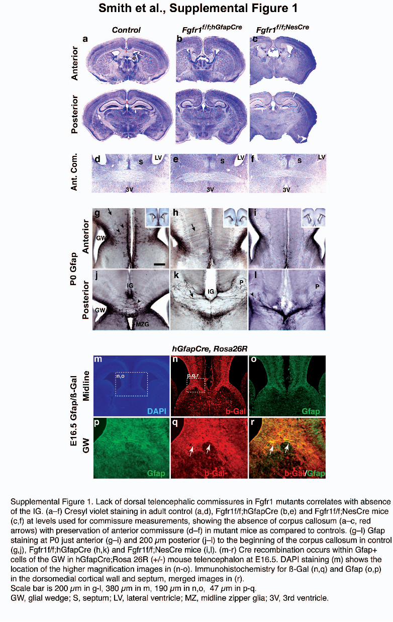

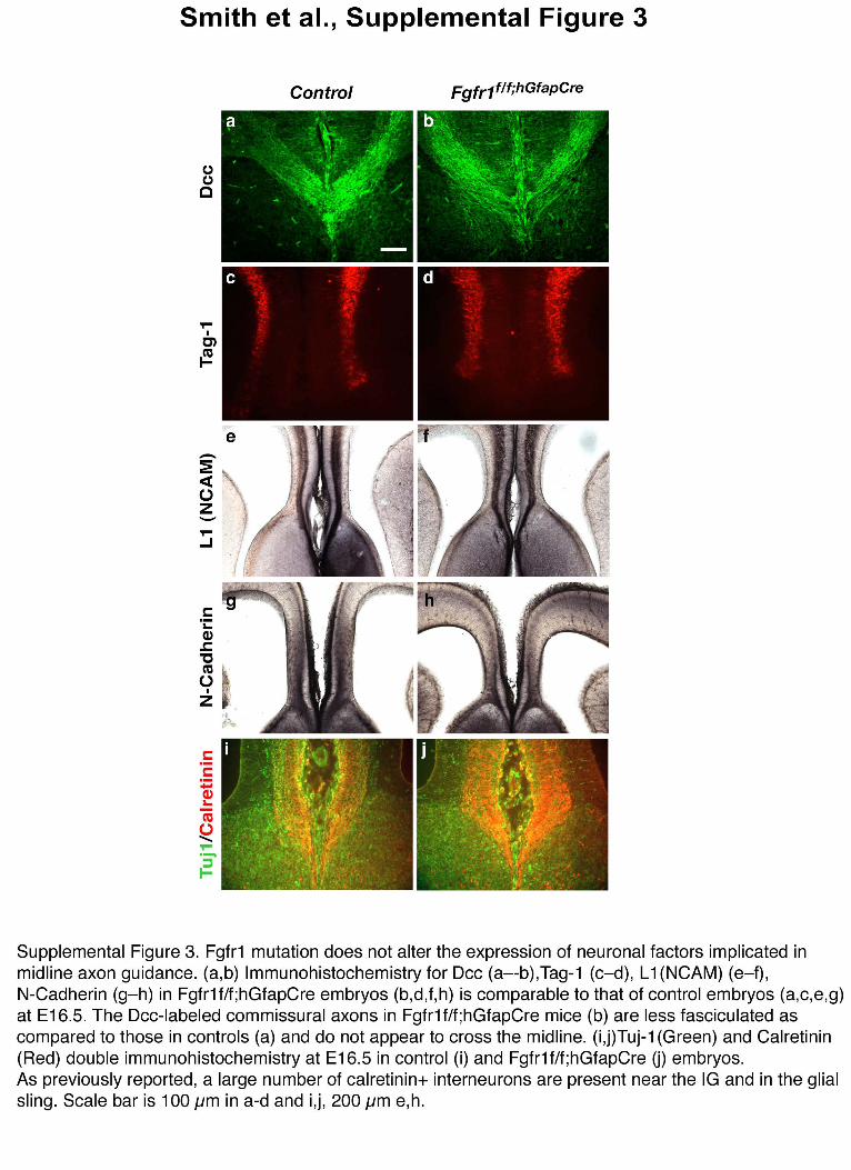
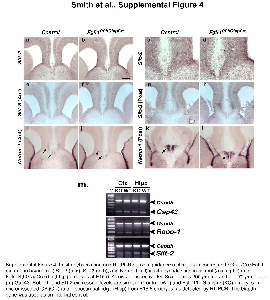
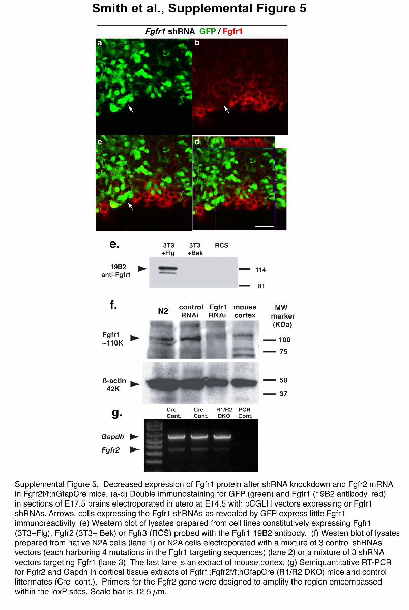
Supplemental Methods
Axonal Tracing
Two hundred nanoliters of a 10% Biotinylated Dextran Amine (10,000 MW,
Molecular Probes) solution in PB buffer was injected into the brains of adult
animals, deeply anesthetized with ketamine and xylazine (100 mg/kg and 10
mg/kg). Injections were performed in a stereotaxic surgery restraint system
equipped with a nanoinjector (Stoelting) using the following coordinates: Anterior-
Posterior +1.94 mm, Medial-Lateral +.5 mm, Dorsal-Ventral –1.40 mm (all
coordinates taken from bregma). Seven days after injection, animals were
sacrificed by intracardial perfusion with 4% paraformaldehyde (PFA) in PBS.
Brains were cryoprotected and sectioned at 50 µM on a cryostat. BDA was
detected with avidin-HRP and DAB (Vector) or FITC-avidin DCS (Vector) and
analyzed by microscopy.
For tracing with DiI and DiA (Molecular Probes), neonatal mice (P0-P1) were
sacrificed by intracardial perfusion, and brains were postfixed in 4%PFA at 4°C
for three days. Pulled glass pipette tips previously coated with DiI or DiA were
implanted into anterior cingulate, somatosensory cortex, or the hippocampus.
Brains were embedded in agar and incubated at 37°C for 6-10 weeks, after
which implants were removed, and pictures were taken to document implant site.
Brains were sectioned in agar at 100 µM on a VT1000 Vibratome (Leica),
mounted on slides and counter stained with DAPI.

In Utero Electroporation and RNAi. For in utero gene transfer by
electroporation, 1-2 µl of DNA solution (4 mg/ml) was injected into the lateral
ventricle and electroporated (five 50 ms square pulses of 40 V with 950 ms
intervals). For RNA interference experiment, a template for short hairpin RNA
(shRNA) synthesis was made by annealing a pair of oligonucleotides
representing the sense and antisense strands of the target sequence. The
oligonucleotides sequences chosen for Fgfr1 targeting were: 5’-
AGGTTTCTGTTTAGGCCT-3’; 5’-GGCCAGACAACTTGCCGTA-3’; 5’-
CCAGTACTCACCCAGCTT-3’. These oligos target, respectively, the 3'-UTR (bp
2624 to 2641), the second the extracellular IG-like region (bp 657 to 675) and the
third the intracellular tyrosine kinase domain (bp 2077 to 2093) (all bases are
given with reference to Genbank accession no. BCO3347). BLAST searches
indicated that none of the oligonucleotides sequences chosen had any significant
matches to any other known mammalian genes. The annealed template was
cloned into pCGLH shRNA vector coexpressing GFP. pCGLH was created by
replacing RFP from pCRLH with GFP (Chen et al. 2005. PNAS 102:17792-
17797). Mutated (4 mutations per sequence) Fgfr1 targeting sequences were
used as a control. The sequences of point-mutated control oligos were: 5’-
AGATTACTCTTTACGCCT-3’; 5’-GGCATACCACTCGCCATA-3’; 5’-
CCATTAATCACTCCGCTT-3’. Equal amounts of the three shRNA vectors
targeting the Fgfr1 gene or of the three control vectors with mutated shRNA
sequences were injected. Empty pCGLH shRNA vector coexpressing GFP was
also injected as additional controls in separate experiments. Cells expressing

shRNA were reliably identified by the co-expression of GFP. To assess the
degree of Fgfr1 knockdown, FACS sorted transfected Neuro2a cells or
untransfected Neuro2a cells were homogenized in 6x SDS sample buffer (12%
SDS, 600mM DTT, 300mM Tris-HCl, pH 6.8), and used for Western blotting with
anti-FGFR1 antibodies (Yu302, gift of J. Schlessinger or 19B2, UBI) or an anti-ß
actin antibody as previously described (Ohkubo et al. 2005. J Neurosci 24:6057-
6059) (Supplementary Fig. 5f). To assess Fgfr1 knockdown in vivo, sections
obtained from shRNA electroporated brains were reacted with the 19B2
monoclonal antibody (Supplementary Fig. 5a-e). Robust inhibition of Fgfr1
protein expression was obtained with either method by transfecting shRNA
targeted to Fgfr1, while control shRNA vectors produced no change in Fgfr1
expression as compared to untransfected cells (Supplementary Fig. 5).
Fluorescent images were obtained using a Zeiss LSM500 confocal microscope.
In selected cases, GFP was visualized using diaminobenzidine
immunohistochemistry with anti-GFP antibody (1:3,000; A11122, Molecular
Probes).
In situ Hybridization and Immunohistochemistry
The following cDNA were used as probes: Fgfr1 (exon 9-15; comprising the
intracellular portion of Fgfr1, which is deleted in hGfap/Cre KO animals);
mSprouty1 (complete protein coding region and 3’UTR; Minowada et al., 1999.
Development 126:4465-4475); FGF-8 (3’UTR and C-terminal coding sequences;
Crossley and Martin. 1995. Development 121:439-451); Slit-2 and Slit-3 (Brose

et al. 1999. Cell 96:796-806), and Netrin-1 (Serafini et al. 1996. Cell:86:1001-
1014) .
The following primary antibodies were used for immunohistochemistry:
βgalactosidase (Cappel or Promega), Blbp (gift from Dr. Nathaniel Heintz or
Chemicon), BrdU (Accurate Chemical), Calretinin (Swant), Dcc (BD
Pharmingen), Fgfr1 (19B2, Upstate), Gfap (DAKO), Glast (gift from Dr. M.
Watanabe), Nestin (Chemicon), PCNA (Upstate), L1 (Chemicon), MNCD2 (N-
cadherin, gift from Dr. M. Takeichi), Tuj1 (Promega), RC2 and Tag1
(Developmental Studies Hybridoma Bank).
Slice Culture, Cortical Explant Culture
Telencephalic slices culture was performed as previously described (Anderson et
al. 1997. Science 278:474-476). Embryonic explants from the dorsal
telencephalon were prepared as described (Furuta et al. 1997. Development
124: 2203-2212). Dorsal cortical explants were dissected from the E13.5 mouse
telencephalon (FVB strain). Cortical tissue was carefully trimmed to remove the
ganglionic eminences and developing hippocampi. After 16 hours in culture,
heparin acrylic beads (Sigma) presoaked for 30 minutes with either 20 µM FGF2
(18 kDa, Invitrogen), 20µM Fgf8b (R&D Systems, MN) or 20 µM BSA (Sigma)
were embedded into these explants. BrdU labeling was performed by adding
0.1mM BrdU (Sigma) to the culture media prior to returning explants to the
incubator. Fixed explant tissue was sectioned at 10 µm and analyzed by
immunohistochemistry. PCNA and BrdU positive cells were counted in 2 bins of

tissue per experiment encompassing the VZ and the IZ/CP. In the VZ, an
average of 97 DAPI-stained cells per experiment over a total area of 125 µm2
were evaluated, among which (depending upon the treatment) an average of 54
to 73 were stained for Pcna, and 35 to 40 were stained for BrdU. In the IZ/CP an
average of 260 DAPI-stained cells per experiment over a total area of 375 µm2
were evaluated, among which (depending upon the treatment) an average of 28
to 165 were stained for Pcna, and 5 to 78 were stained for BrdU. Counts were
normalized by the area to obtain cell density. Number of independent
experiments: n = 4 control, n = 5 FGF2, n = 4 FGF8.
Cell counting
Unbiased estimates for density of Gfap or BrdU-positive cells were obtained
using a computer coupled to a Zeiss Axioskope 2 Mot Plus equipped with a
motorized stage, running the StereoInvestigator software (Microbrightfield,
Colchester, VT). Nuclear profiles were counted in 3-dimensional counting boxes,
using a randomly placed sampling grid of 500 x 350 µm on the cerebral cortex
and 100 x 100 µm on the SVZ. Counting boxes (100 x 100 x 8 µm) were placed
in 1 µm below the surface at each of the grid intersection points. Cells
immunoreactive for Gfap were only counted if they overlapped a DAPI-stained
cell nucleus.