middleear muscles of the frog - proceedings of the ... · pdf filetheanatomyof the frog's...
TRANSCRIPT

Proc. Nati. Acad. Sci. USAVol. 76, No. 6, pp. 3031-3033, June 1979Physiological Sciences
Middle ear muscles of the frog(amphibian ear/hearing)
ERNEST GLEN WEVERAuditory Research Laboratories, Princeton University, Princeton, New Jersey 08544
Contributed by E. C. Wever, March 28, 1979
ABSTRACT The anuran middle ear in its complete formincludes two skeletal elements, the columella and operculum,each occupying a portion of the oval window of the otic capsuleand each provided with a middle ear muscle. The two elementshave an interlocking arrangement of a form that makes it pos-sible for these muscles to exercise a high degree of control ofsound transmission from tympanic membrane to inner ear re-
ceptors. From the anatomical relations it is inferred that the twomuscles operate as antagonists so that contraction of the oper-
cular muscle and relaxation of the columellar muscle leave thecolumella free to move in and out of the oval window in re-
sponse to sound vibrations, whereas a contraction of the colu-mellar muscle and relaxation of the opercular muscle tend toimmobilize the columella and reduce the transmission inward.The frog thus achieves a degree of control of sound receptionthat probably is unmatched among vertebrate ears. The purposeof the middle ear mechanism is no doubt the protection of theinner ear receptors (the amphibian and basilar papillae) fromoverstimulation by sounds, including the animal's own cries andthe intense clamor produced by a group of frogs calling inchorus.
Gaupp (1) in his monumental treatment of the Anatomie desFrosches in 1896 first figured and named the opercular musclein the anuran middle ear. He described this muscle as a deriv-ative of the levator scapulae superior, which is a strong bundlerunning from the prootic region to the ventral surface of thesuprascapula and serving to retract this cartilage. Gaupp was
not the first to see the opercular muscle; Eiselt (2) in a thoroughhistorical search found mention of it by Blainville as early as
1822 as a part of the shoulder musculature, and Huschke shortlythereafter spoke of this muscle as a homolog of the stapediusmuscle of mammals. However, most subsequent writers on theanuran middle ear have referred to Gaupp's account and haveagreed with him concerning the derivation of the muscle fromthe levator scapulae superior. A muscle that usually is regardedas corresponding to this one, and likewise called the opercularmuscle, is present in the urodeles. However, the third Order ofamphibians, the Gymnophiona or caecilians, lack this musclealong with the operculum itself.Numerous treatments of the middle ear mechanism in frogs
have repeated Gaupp's descriptions of both the operculum andits muscle with little variation, but sometimes consideration hasbeen given to the possible function of this mechanism in thehearing process. Earlier theorizing dealt extensively with thisproblem in urodeles, and a curious hypothesis was advancedinvolving the transmission of substrate vibrations through theforelegs, with the opercular muscle providing the path fromshoulder region to ear. Much less attention has been given tothe function of the middle ear in frogs, though it usually is as-
sumed that the opercular muscle produces a damping of vi-brations transmitted inward along the columella. De Burlet (3),in a general review of the vertebrate middle ear, offered a more
The publication costs of this article were defrayed in part by page
charge payment. This article must therefore be hereby marked "ad-vertisement" in accordance with 18 U. S. C. §1734 solely to indicatethis fact.
3031
specific suggestion for the anurans in which he compared theopercular muscle with the tensor tympani of mammals andsupposed that this muscle on contraction conveys its restraintsthrough the linkage of operculum and columella to the tym-panic membrane and thus produces a flattening (and hence astiffening) of its surface.
Present observationsThe anatomy of the frog's middle ear has been studied in avariety of species as a part of a general examination of the earand hearing in the amphibia. This study was stimulated by theobservation of large variations in the ear's sensitivity, as mea-sured in terms of its electrical potential in response to sounds.These variations were especially prominent in lightly anes-thetized animals and at times amounted to as much as 40 db-a100-fold variation in terms of the sound pressure required fora given electrical output. The variations continued in spite ofa strict maintenance of experimental conditions, such as bodytemperature, the moist condition of the skin, and respiration,and pointed to the existence of some kind of internal controlmechanism.
Observations were made both by dissection under the bin-ocular microscope and by the use of serial sections. The sec-tioning used the three standard planes (frontal, transverse, andsagittal) and sometimes also an oblique anterolateral plane thatreveals the columellar connections with special clarity. Thestructure to be described was investigated in greatest detail inthe ranids, but has been noted also in other anurans (all thoseexamined thus far) in which the middle ear is fully devel-oped.
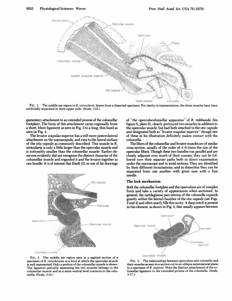
These observations show that the classical description of theanuran middle ear is seriously inadequate. The levator scapulaesuperior does not simply send off a branch that serves as theopercular muscle, as the usual accounts suggest. In the ear re-gion there are three muscles that are essentially independent.These are the opercular muscle, the columellar muscle, and thelevator scapulae superior, represented for Rana utricularia inFig. 1.The opercular muscle is the one previously described under
this name, but, as shown, it bears no special relation to the le-vator scapulae superior. It has a distinct place of origin on theanteromedial undersurface of the suprascapula, and runs di-rectly to an insertion on the operculum.The fibers of this muscle have a certain distinctive character;
they are noticeably smaller in cross section than those of theother muscles in the vicinity, they have a more translucentappearance on visual examination, and with certain histologicaltreatments they stain differently from the others. These featuresmake the muscle readily identifiable in serial sections. Thedifference in fiber size between opercular and columellarmuscles is evident in Fig. 2.The columellar muscle has its place of origin on the supra-
scapula posterolateral to that of the opercular muscle. It runsparallel to the opercular muscle, but independent of it, to a li-
3032 Physiological Sciences: Wever
Otic capsule
7/Hyoid process ( iluell'
FIG. 1. The middle ear region in R. utricularia, drawn from a dissected specimen. For clarity in representation, the three muscles have beenartificially separated at their upper ends. (Scale, X12.)
gamentary attachment to an extended process of the columellarfootplate. The form of this attachment varies regionally froma short, blunt ligament as seen in Fig. 3 to a long, thin band asseen in Fig. 4.The levator scapulae superior has a still more posterolateral
attachment on the suprascapula, and runs to the lateral surfaceof the otic capsule as commonly described. This muscle in R.utricularia is only a little larger than the opercular muscle andis noticeably smaller than the columellar muscle. Earlier ob-servers evidently did not recognize the distinct character of thecolumellar muscle and regarded it and the levator together asone bundle. It is of interest that Eiselt (2), in one of his drawings
Otic capsule
- Coluniella
(}peersuIt-viOpercUlar II SC If.
of "the operculocolumellar apparatus" of R. ridibunda (hisfigure 6, plate 2), clearly portrayed two muscles in addition tothe opercular muscle, but had both attached to the otic capsuleand designated both as "levator scapulae superior" though oneof these in his illustration definitely makes contact with thecolumella.The fibers of the columellar and levator muscles are of similar
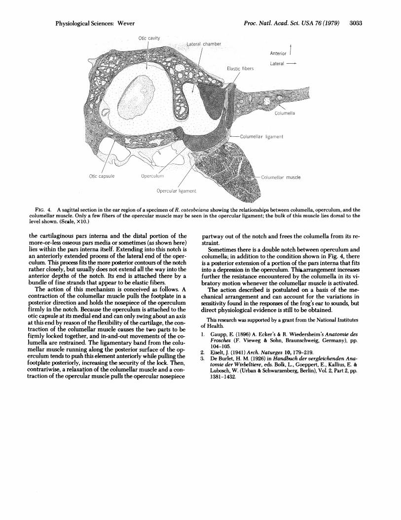
cross section, usually of the order of 4-6 times the size of theopercular fibers. Though these two bundles run parallel and areclosely adjacent over much of their courses, they can be fol-lowed over their separate paths both in direct examinationunder the microscope and in serial sections. They are identifiedby their different terminations, and in dissection they can beseparated from one another with great ease with a fineneedle.The lock mechanismBoth the columellar footplate and the operculum are of complexform and take a variety of appearances when sectioned. Ingeneral, the cartilaginous pars interna of the columella expandsgreatly within the lateral chamber of the otic capsule (see Figs.2 and 4) and often nearly fills this cavity. A deep notch is presentin this element, as shown in Fig. 4, that usually appears between
C pe rculC'IIra>
OpercLOEar UIIWSC^f\LrC :L e d
C : Li m1elIa rlliC '.'i II" t !
ColUrmellar rL'.LJSCle
FIG. 2. The middle ear region seen in a sagittal section of aspecimen of R. catesbeiana at a level at which the opercular muscleis well represented. Only a portion of the columellar muscle is shown.The ligament partially separating the two muscles belongs to thecolumellar muscle and at a more ventral level connects to the colu-mella. (Scale, X10.)
Columellar muscle
FIG. 3. The relationships between operculum and columella andtheir muscles as seen in a section cut in an oblique anterolateral planein a specimen of R. pipiens. Note the distinct attachment of the co-lumellar ligament to the extended process of the columella. (Scale,X17.)
Proc. Natl. Acad. Sci. USA 76 (1979)
Proc. Natl. Acad. Sci. USA 76 (1979) 3033
Otic cavity
FIG. 4. A sagittal section in the ear region of a specimen of R. catesbeiana showing the relationships between columella, operculum, and thecolumellar muscle. Only a few fibers of the opercular muscle may be seen in the opercular ligament; the bulk of this muscle lies dorsal to thelevel shown. (Scale, X10.)
the cartilaginous pars interna and the distal portion of themore-or-less osseous pars media or sometimes (as shown here)lies within the pars interna itself. Extending into this notch isan anteriorly extended process of the lateral end of the oper-culum. This process fits the more posterior contours of the notchrather closely, but usually does not extend all the way into theanterior depths of the notch. Its end is attached there by abundle of fine strands that appear to be elastic fibers.The action of this mechanism is conceived as follows. A
contraction of the columellar muscle pulls the footplate in aposterior direction and holds the nosepiece of the operculumfirmly in the notch. Because the operculum is attached to theotic capsule at its medial end and can only swing about an axisat this end by reason of the flexibility of the cartilage, the con-traction of the columellar muscle causes the two parts to befirmly locked together, and in-and-out movements of the co-lumella are restrained. The ligamentary band from the colu-mellar muscle running along the posterior surface of the op-erculum tends to push this element anteriorly while pulling thefootplate posteriorly, increasing the security of the lock. Then,contrariwise, a relaxation of the columellar muscle and a con-traction of the opercular muscle pulls the opercular nosepiece
partway out of the notch and frees the columella from its re-straint.
Sometimes there is a double notch between operculum andcolumella; in addition to the condition shown in Fig. 4, thereis a posterior extension of a portion of the pars interna that fitsinto a depression in the operculum. Thirarrangement increasesfurther the resistance encountered by the columella in its vi-bratory motion whenever the columellar muscle is activated.The action described is postulated on a basis of the me-
chanical arrangement and can account for the variations insensitivity found in the responses of the frog's ear to sounds, butdirect physiological evidence is still to be obtained.
This research was supported by a grant from the National Institutesof Health.1. Gaupp, E. (1896) A. Ecker's & R. Wiedersheim's Anatomie des
Frosches (F. Vieweg & Sohn, Braunschweig, Germany), pp.104-105.
2. Eiselt, J. (1941) Arch. Naturges 10, 179-219.3. De Burlet, H. M. (1926) in Handbuch der vergleichenden Ana-
tomie der Wirbeltiere, eds. Bolk, L., Goeppert, E., Kallius, E. &Lubosch, W. (Urban & Schwarzenberg, Berlin), Vol. 2, Part 2, pp.1381-1432.
Physiological Sciences: Wever