middle-aged woman with delayed diagnosis of severe mitro...
TRANSCRIPT

Middle-aged woman with delayed diagnosis of severe mitro-aorticValvular rheumatic heart disease following tachyarrhythmic event
Mulher de média idade com diagnóstico tardio de valvulopatia mitro-aórtica remumática
Andrés Ricardo Pérez-Riera M.D. Ph.D.Physician of Hospital do Coração (HCor) - Sao Paulo-Brazil
In charge of Electro-vectorcardiogram sector – Cardiology Discipline –ABC Faculty of Medicine –ABC Foundation - Santo André –São Paulo – Brazil.

Case report
A 41-year-old Caucasian woman, housewife, natural of the countryside of São Paulo Brazil, from a low socioeconomic background presented in our clinical consultation after internment 2 Month ago consequence of tachyarrithtymic event At this time, her only symptom was intermittent palpitations. She had no recollection of previous rheumatic fever. History of Present Illness (HPI) She refer the last Month presented fatigue, especially during times of increased physical activity, progressive shortness of breath especially with exertion or when lie down a dry cough presented at rest and unique fast regular palpitation episode that its motives emergency internment and intra-hospitalar treatment with electric cardioversion and use of intravenous drugsThe patient denied fever, chest pain, or other systemic symptoms.
She has no recollection of any previous symptoms or antecedent of repetitive streptococcal throat infections, murmurs or joints pain in the pass.There was no personal or familial history of cardiovascular disease or rheumatic fever picture.Physical She was breathing well, acianotic, without fever and cored. She was in sinus rhythm, BP: normal systolic blood pressure and low diastolic=140/30-0 mmHg with Watson's water hammer pulse.On precordial auscultation She had an accentuated first heart sound, an opening snap that occurs earlier during diastole and with a 3/6 mid-diastolic rumble murmur was heard best at the apex. There is accentuation of P2 on pulmonary focus. Additionally, an early diastolic and decrescendo murmur is heard at aortic area. The murmur is soft wit radiation to the right parasternal region. An ejection systolic 'flow' murmur is present when auscultating the same aortic area. This systolic murmur not start with an ejection click.Lungs: inspiratory pulmonary rales at the lung bases.Abdomen Absence of hepatomegalyExt: No clubbing, cyanosis, edemaNeurological examination was normal.Preliminary diagnosis: mitral stenosis (MS) associated with aortic insufficiency.

Presentación del casoMujer de 41 años de edad, blanca, ama de casa, natural del Estado de São Paulo, Brasil, del estracto socio-económico bajo se presenta en nuestra consulta clínica después de haver tenido una internación aproximadamente dos meses atrás consecuencia de episodio taquiarrítmico. En este momento, su único síntoma eran palpitaciones intermitentes. Ella no tenía ningún recuerdo de fiebre reumática anterior.Historia de la enfermedad actual (IPH) refieren que el último 2 meses presenta fatiga, especialmente durante las actividades físicas, dificultad progresiva para respirar, especialmente con el ejercicio o cuando se acuesta aparece una tos y un único episodio de palpitaciones rápidas regular que motivó internamiento de emergencia y tratamiento con cardioversión eléctrica y fármacos por vía intravenosaNiega fiebre, dolor de pecho o otros síntomas. No recuerda síntomas anteriores o antecedentes de infecciones de garganta por estreptococos repetitivas, soplos o dolor de las articulaciones en el pasado.No tiene antecedentes personales ni familiares de enfermedad cardiovascular o un cuadro compatible con fiebre reumática.EF eupneica, acianótica, sin fiebre.En ritmo regular con presión arterial sistólica normal y diastólica baja = 140/30-0 mm Hg Pulsos de Watson en martillo de agua.Auscultación precordial Primer ruido de intensidad aumentada,. chasquido de apertura, durante la protodiástole y soplo mesodiastólico 3.6 que se oye mejor en el ápice. P2 acentuada en foco pulmonar. Soplo diastólico suave precoz y decrescendo que se escucha en el foco aórtico irradiado para el borde paraesternal derecho. Soplo sistólico de “hiperflujo" eyecctivo en foco aórtico. Este soplo sistólico no va precedido de click.Pulmones: estertores pulmonares inspiratorios en las bases pulmonares.Abdomen ausencia de hepatomegaliaExt: Ausencia de baqueteamiento digital, cianosis o edema.El examen neurológico fue normal.Diagnóstico Preliminar: estenosis mitral (EM) asociado a la insuficiencia aórtica.

First ECGName: JSS.; Age: 41 yo.; Sex: Feminine.; Weight: 70 Kg.; Heigh: 1,63m.
ECG realized: May/09/2012. Time: 15:55h

Second
Name: JSS Age: 41 y.o. Sex: Feminine Weight: 70 Kg Height: 1,63mECG realized in March, 09/2012 Time:17:50

Third
Nome: JSS Idade: 41 anos Sexo: Feminino Peso: 70 Kg Altura: 1,63ECG realized: May/10/2012 Time: 15:55h

Fourth
Name: JSS Age: 41 y.o. Sex: Fem Weight: 70 Kg Heigth: 1,63ECG realized: May/ 16/ 2012 Time: 15:00

Name: JSS Age: 41 yo Sex: Fem Weight: 70 Kg Heigh: 1,63mMay 28/ 2012
Fifth ECG

Collages opinions

Dear Andrés and colleagues,The clinical picture is that of rheumatic heart disease with mitral stenosis, aortic regurgitation and
secondary pulmonary hypertension. Workup would include an echocardiogram and cardiac catheterization which would likely indicate need for valve surgery.ECG 1 (March 9): atrial flutter with 2:1 AV block (pseudo RBBB) with right axis deviation (~+90 degrees); atrial rate ~ 260 bpmECG 2 (March 9): atrial flutter with variable AV block probably the result of AV nodal slowing drugs (digoxin, beta-blocker, or non-dihydropyridine Ca++ blocker); QRS axis now ~80 degrees)ECG 3 (May 10): atrial flutter with 2:1 AV block and QRS axis ~+90 degrees; atrial rate now ~ 240 bpm perhaps suggesting a larger right atriumECG 4 (May 16): sinus rhythm (~90 bpm) with one PAC, QRS axis ~80 degrees, left atrialenlargement, LVH voltage (? diastolic LV overload from aortic regurgitation)ECG 5 (May 28): sinus rhythm (~60 bpm), QRS axis ~70 degrees, left atrial enlargement, LVH of diastolic overload.I await more erudite comments from you and our colleagues.
Warmest regards,Frank Yanowitz MDProfessor of MedicineUniversity of Utah School of MedicineMedical Director, ECG DepartmentLDS Hospital Salt Lake City, Utah
8th Ave. and C StreetSalt Lake City, Utah 84143 USA
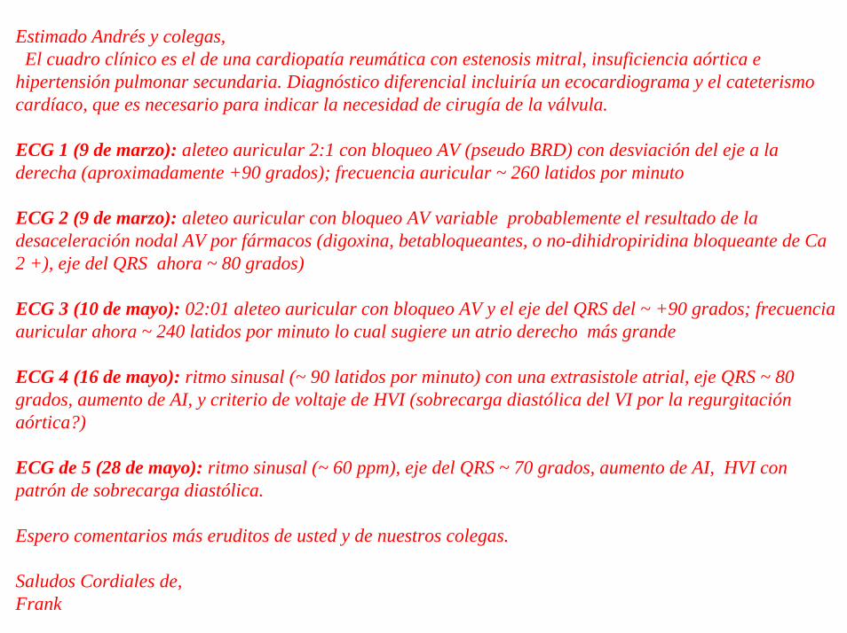
Estimado Andrés y colegas,El cuadro clínico es el de una cardiopatía reumática con estenosis mitral, insuficiencia aórtica e
hipertensión pulmonar secundaria. Diagnóstico diferencial incluiría un ecocardiograma y el cateterismo cardíaco, que es necesario para indicar la necesidad de cirugía de la válvula.
ECG 1 (9 de marzo): aleteo auricular 2:1 con bloqueo AV (pseudo BRD) con desviación del eje a la derecha (aproximadamente +90 grados); frecuencia auricular ~ 260 latidos por minuto
ECG 2 (9 de marzo): aleteo auricular con bloqueo AV variable probablemente el resultado de la desaceleración nodal AV por fármacos (digoxina, betabloqueantes, o no-dihidropiridina bloqueante de Ca 2 +), eje del QRS ahora ~ 80 grados)
ECG 3 (10 de mayo): 02:01 aleteo auricular con bloqueo AV y el eje del QRS del ~ +90 grados; frecuencia auricular ahora ~ 240 latidos por minuto lo cual sugiere un atrio derecho más grande
ECG 4 (16 de mayo): ritmo sinusal (~ 90 latidos por minuto) con una extrasistole atrial, eje QRS ~ 80 grados, aumento de AI, y criterio de voltaje de HVI (sobrecarga diastólica del VI por la regurgitación aórtica?)
ECG de 5 (28 de mayo): ritmo sinusal (~ 60 ppm), eje del QRS ~ 70 grados, aumento de AI, HVI con patrón de sobrecarga diastólica.
Espero comentarios más eruditos de usted y de nuestros colegas.
Saludos Cordiales de,Frank

Queridos amigos del forum nuestro maestro Profesor Andrés Ricardo Pérez Riera Ph.D. hizo un análisisextraordinaria de esta paciente sufriente de un caso crónico de estenosios mitral e insuficiencia aórtica.Estas discusiones eran, el pan de todos los dias, en los años 60-70 del siglo pasado.Después de la descripción del maestro se podria dibujar el ECG y la Radiografia. Evidentemente el primer ECG corresponde al axioma que decia: “fluter o fibrilacion auricular con eje a la derecha es estenosis mitral hasta que no se demuestre lo contrario”Este ECG a mi parecer muestra una dilalatación del ventriculo derecho con HVILa morfologia en V1 no sugiere HVD , y las R,s altas con S,s pequeñas en V6 ,V5 sugieren HVILas ondas P expresasan una aurícula izquierda con de gran dimensión, talvez el ECO muestre unadilatación de alrededor de los 50 mm. Supongo que la auricula izquierda estaba protegida por la vasontricción de las arteriolas precapilarespulmonares lo que explica porque esta paciente ,con estenosis mitral severa nunca desarrolló edema agudode pulmón Los capilares venosos no se ingurgitan porque estaban protegidos por el efecto torniquete. La aparicion casi brusca de la insuficiencia cardiaca es mas derecha que izquieda
Seria importante aqui describir los latidos venosos del cuello, para revelar si hay insuficiencia tricuspidea, que determinara la conducta quirúrgicaFantástico caso querido amigo y para los residentes o estudiantes que siguen estas discusiones , El profe Edgardo Schapashnik, se referia a la importancia se la distancia entre el click diastólico, con respecto al segundo ruido. Cuando mas cerca esta distancia mas severa la estenosis y cuando mas alejadomas leveCon respecto a los soplos aórticos, el maestro M B Rosenbaum decia "este tiene hiperacusia diastólica"porque estaba de moda decir: “escucho un soplo diastólico aórtico” . y quien le demostraba que no?Un fraternal abrazo
Samuel Sclarovsky
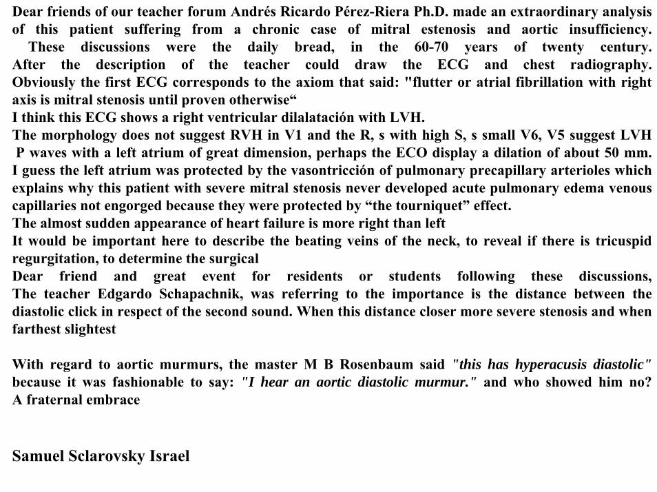
Dear friends of our teacher forum Andrés Ricardo Pérez-Riera Ph.D. made an extraordinary analysis of this patient suffering from a chronic case of mitral estenosis and aortic insufficiency.
These discussions were the daily bread, in the 60-70 years of twenty century.After the description of the teacher could draw the ECG and chest radiography.Obviously the first ECG corresponds to the axiom that said: "flutter or atrial fibrillation with right axis is mitral stenosis until proven otherwise“I think this ECG shows a right ventricular dilalatación with LVH.The morphology does not suggest RVH in V1 and the R, s with high S, s small V6, V5 suggest LVHP waves with a left atrium of great dimension, perhaps the ECO display a dilation of about 50 mm.
I guess the left atrium was protected by the vasontricción of pulmonary precapillary arterioles which explains why this patient with severe mitral stenosis never developed acute pulmonary edema venous capillaries not engorged because they were protected by “the tourniquet” effect.The almost sudden appearance of heart failure is more right than leftIt would be important here to describe the beating veins of the neck, to reveal if there is tricuspid regurgitation, to determine the surgicalDear friend and great event for residents or students following these discussions,The teacher Edgardo Schapachnik, was referring to the importance is the distance between the diastolic click in respect of the second sound. When this distance closer more severe stenosis and when farthest slightest
With regard to aortic murmurs, the master M B Rosenbaum said "this has hyperacusis diastolic"because it was fashionable to say: "I hear an aortic diastolic murmur." and who showed him no?A fraternal embrace
Samuel Sclarovsky Israel

Final comments
By Andrés Ricardo Pérez-Riera M.D.Ph.D.

First ECGName: JSS.; Age: 41 yo.; Sex: Feminine.; Weight: 70 Kg.; Heigh: 1,63m.
ECG realized: May/09/2012. Time: 15:55h
Regular sustained narrow QRS tachycardia, heart rate 125bpm, typical atrial flutter 2:1 F atrial rate= 260bpm. “F” waves of negative polarity in II, III and aVF without isoelectrical line “plateau”.. F waves have sawtooth or picket fence appearance, observed better in the inferior leads with slowly descending and rapidly ascending ramp. These waves resemble an inverted P wave, followed by an ascending ramp: “Tp”waves.

Second ECG
Name: JSS Age: 41 y.o. Sex: Feminine Weight: 70 Kg Height: 1,63mECG realized in March, 09/2012 Time:17:50
Typical atrial flutter with variable atrioventricular (AV) block (with CCW rotation atrial macro-reentrant circuit involving most of the right atrium.) Atrial rate 250 to 350/min (typically 300/min).with variable atrioventricular (AV) block probably the result of drugs (digoxin, β-blocker, or Ca2+
antagonist such as verapamil or diltiazen); QRS axis in 80º.
Carotid sinus massage can increase AV block and better expose the typical flutter waves.
Triphasic rSr´pattern in V1 “pseudo RBBB”
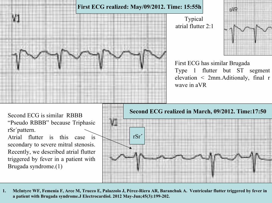
First ECG realized: May/09/2012. Time: 15:55h
Typical atrial flutter 2:1
Second ECG realized in March, 09/2012. Time:17:50Second ECG is similar RBBB“Pseudo RBBB” because TriphasicrSr´pattern.Atrial flutter is this case is secondary to severe mitral stenosis. Recently, we described atrial flutter triggered by fever in a patient with Brugada syndrome.(1)
First ECG has similar Brugada Type 1 flutter but ST segment elevation < 2mm.Aditionaly, final r wave in aVR
rSr´
1. McIntyre WF, Femenía F, Arce M, Trucco E, Palazzolo J, Pérez-Riera AR, Baranchuk A. Ventricular flutter triggered by fever in a patient with Brugada syndrome.J Electrocardiol. 2012 May-Jun;45(3):199-202.

Third
Nome: JSS Idade: 41 anos Sexo: Feminino Peso: 70 Kg Altura: 1,63ECG realized: May/10/2012 Time: 15:55h
Again typical atrial flutter with 2:1 AV block and QRS axis ~+90º; atrial rate ≈ 240 bpmQRS complex in FP with Counter Clock Wise Rotation (CCWR): I and aVL RS/Rs and qR pattern inInferior leads QIII>QII. Similar Brugada ECG pattern in V1

Fourth
Name: JSS Age: 41 y.o. Sex: Fem Weight: 70 Kg Heigth: 1,63ECG realized: May/ 16/ 2012 Time: 15:00
Sinus rhythm, heart rate: 71bpm, P axis +50º, bimodal broad P-wave with slow final component in V1 with one PAC, QRS axis ~80 degrees, left atrial enlargement, negative Sokolow-Lyons index.
Bimodal broad P wave
Premature Atrial Contraction (PAC)

Name: JSS Age: 41 yo Sex: Fem Weight: 70 Kg Heigh: 1,63mMay 28/ 2012
Fifth ECG
Sinus Rhythm, left atrial enlargement with P axis in +60º: biatrial enlargement?

aVR aVL
I
IIIII
aVF
X
Y
TP QRS axis +80º
P axis +60º
P wave isodiphasic(“plus-minus”) in aVI. = P axis +60º

P duration = 150ms
The normal shape of P wave is rounded and monophasic, and there may be small notches (more frequent in V3 and V4 ) and the distance between these notches should not exceed 30ms (0.03s). Notches in P wave with distance between the apexes of ≥ 40 ms (0.04 s) constitutes a sign of left atrial enlargement (LAE) or interatrialblock by Bachman’s bundle (BB), in charge of activating the left atrium (LA).P waves of duration ≥120 ms and bimodal with SÂP on frontal plane (+60º) is a possible indication of biatrial enlargement
LARA
RA: right atriumLA: left atrium
III

V6
V1
V4
V5
V2
V3
X
Z
T
PP-loop on posterior quadrants

Z Z
XX
V1
V2
V6
V1
V2
V6
0E 0
E
Z Z
XX
V1
V2
V6
V1
V2
V6
Z Z
XX
V1
V2
V6
V1
V2
V6
0E 0
E
Z Z
XX
V1
V2
V6
V1
V2
V6
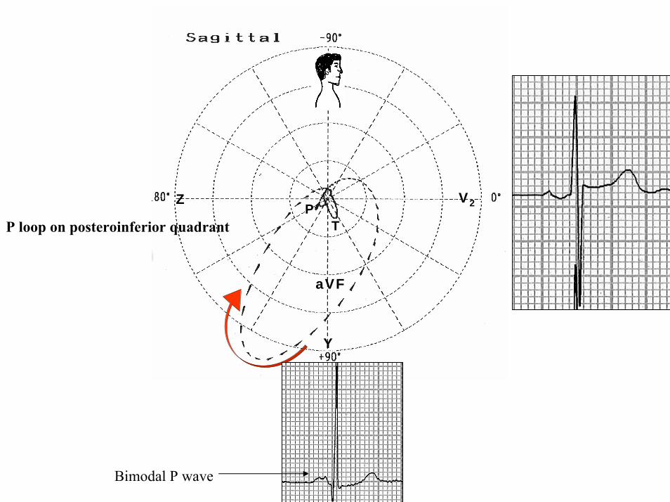
Normal P loop LAE P loop
P
The present case P loop on posterior location
LAE: Left Atrial EnlargementVCG criteria
1. The maximal P vector is locatedto the left posterior quadrant: ≥0.10 mV in adults and ≥0.14 mV in < 15 years old
2. P- loop with CCW or in eight rotation
3. Maximal P vector >0.05 mV4. “Bow Tie” morphology.

V1
V1
RA
Normal P wave
LA
RA RA
Left Atrial EnlargementLA
LA
LA2/: Final deep and slow component: LAE ≥ the area of one small square the final minus portion indicates left atrial enlargement, abnormality or advanced interatrial block
Final slow and deep negative component > 1 mm in depth and 40 ms in duration
LAE
Y
aVF
Z V2
TP
P loop on posteroinferior quadrant
Bimodal P wave

Valvular heart disease resulting from rheumatic fever is referred to as "rheumatic heart disease". While developed countries once had a significant burden of rheumatic fever and rheumatic heart disease, medical advances and improved social conditions have dramatically reduced their incidence. Many developing countries, as well as indigenous populations within developed countries, still carry a significant burden of rheumatic fever and rheumatic heart disease and there has been a resurgence in efforts to eradicate the diseases in these populations. Inflammation of the heart valves due to any cause is called endocarditis; this is usually due to bacterial infection but may also be due to cancer (marantic endocarditis), systemic lupus (Libman-Sacks endocarditis) and hypereosinophilic syndrome (Loeffler endocarditis). Certain medications have been associated with valvular heart disease, most prominently ergotamine derivatives pergolide and cabergoline(1) Rheumatic heart disease is the most common cause of multivalvular disease in developing countries. Among the most prevalent and neglected diseases are rheumatic heart disease and endomyocardialfibrosis. The etiologies of these diseases can be described in part as a dysregulation or reactivation of developmental biology pathways. Consequently, connecting mechanisms of valvulogenesis and disease etiology may represent an excellent strategy to identify therapeutic targets.(2) Unless aggressive and timely intervention in the form of valve replacement is pursued, the condition progresses rapidly to disability and death. Combined mitral-aortic valve replacement represents a major technical challenge, and carries high early and late mortality rates. Among the rheumatic population, double valve replacement offers excellent symptomatic improvement and favorable late survival. Hemodynamic superiority and thromboresistance are the normal selection criteria for these prostheses, although the surgeon's experience, and the ease of insertion, availability and cost of the valve also play important roles. A strict adherence to optimal anticoagulation levels optimizes protection against thromboembolism and anticoagulation-related hemorrhage, and helps to provide the patient with a good quality life.
1. Schade R, Andersohn F, Suissa S, Haverkamp W, Garbe E. Dopamine agonists and the risk of cardiac-valve regurgitation.N Engl J Med. 2007 Jan 4;356:29-38.
2. Farrar EJ, Butcher JT. Valvular heart diseases in the developing world: developmental biology takes center stage. J Heart Valve Dis.2012 Mar;21:234-240.

Subsequent valvular stenosis may occur as a consequence of the healing of the rheumatic process, repetitive but subclinical rheumatic insults or reinfection, chronic rheumatic activity, or progressive hemodynamicstresses on the traumatized valve, similar to that of the pathogenesis of aortic stenosis. The plethora of postulated mechanisms and contexts for this pathologic evolution may account for the fact that some patients experience a chronic stable disease, whereas others have an accelerated course necessitating early surgical intervention.(1, 2, 3)The normal area of the mitral valve orifice is 4-6 cm2.(4) This effectively creates a common chamber between the left atrium and the left ventricle in diastole. In early diastole, a small and brief pressure gradient is present; however, during most of the filling period, the pressures in the two chambers are equal. Narrowing of the valve area to less than 2.5 cm2 impedes the free flow of blood and requires increased left atrial pressure (LAP) to ensure normal transmitral flow.Symptoms typically first present after exertion when the valve area shrinks to less than 2.5 cm2, and symptoms at rest do not begin until valve area reaches 1.5 cm2.(4) However, any physiologic stress requiring increased cardiac output (eg, pregnancy, infection, exercise, emotional stress, anemia, atrialfibrillation with rapid ventricular response) may precipitate symptoms earlier in the progression of stenosis.
1. Carabello BA. Modern management of mitral stenosis. Circulation. Jul 19 2005;112:432-437.2. Bonow RO, Carabello BA, Chatterjee K, et al. 2008 focused update incorporated into the ACC/AHA
2006 guidelines for the management of patients with valvular heart disease: a report of the American College of Cardiology/American Heart Association Task Force on Practice Guidelines (Writing Committee to revise the 1998 guidelines for the management of patients with valvular heart disease). Endorsed by the Society of Cardiovascular Anesthesiologists, Society for Cardiovascular Angiography and Interventions, and Society of Thoracic Surgeons. J Am Coll Cardiol. Sep 23 2008;52(13):e1-142.
3. Gash AK, Carabello BA, Cepin D, Spann JF. Left ventricular ejection performance and systolicmuscle function in patients with mitral stenosis. Circulation. Jan 1983;67:148-154.
4. Chandrashekhar Y, Westaby S, Narula J. Mitral stenosis. Lancet. Oct 10 2009;37:1271-1283.

The fraction of strains that have been identified as “rheumatogenic” has decreased in developed countries with a concomitant decreasing incidence of rheumatic fever.(1, 2) Additionally, evidence suggests that improved socioeconomic status, public health, and hygiene have resulted in the near disappearance of rheumatic fever and new cases of MS in the United States and other developed countries.(3) The true prevalence of MS is unknown and rheumatic fever is no longer a disease that must be reported to the Centers of Disease Control and Prevention. However, approximately 1500 balloon mitral valvotomies were performed in the United States during 2004. This provides a rough indication of the prevalence of moderate to severe disease. Moderate or severe valvular diseases are notably common in selected adults from the general population who had been assessed prospectively with echocardiography. and increase with age. In the community, women are less often diagnosed than are men, which could indicate an important imbalance in view of the associated lower survival. Valve diseases thus represent an important public-health problem. Nkomo et al used echocardiography to prospectively study all valvular disease in the United States and estimated the prevalence of MS at 0.1% (0.02-0.2%).(4)
1. Carapetis JR. Rheumatic heart disease in developing countries. N Engl J Med. Aug 2 2007;357:439-441.
2. Shulman ST, Tanz RR, Dale JB, et al. Seven-year surveillance of north american pediatric group a streptococcal pharyngitis isolates. Clin Infect Dis. Jul 1 2009;49:78-84.
3. Nkomo VT, Gardin JM, Skelton TN, Gottdiener JS, Scott CG, Enriquez-Sarano M. Burden of valvularheart diseases: a population-based study. Lancet. Sep 16 2006;368:1005-1011.
4. Rahimtoola SH. Mitral valve disease. In: Hurst's The Heart. 12th ed. McGraw-Hill Medical Publishing Division; 2008:chap 77. WHO. Rheumatic Fever and Rheumatic Heart Disease: Report of a WHO Expert Consultation, Geneva, 29 October - 1 November 2001. Geneva, Switzerland: 2004.
5. Nkomo VT, Gardin JM, Skelton TN, et al. Burden of valvular heart diseases: a population-based study. Lancet. 2006 Sep 16;368:1005-1011.

Both rheumatic fever and MS remain common in developing countries. MS develops at an earlier age, progresses more quickly, and requires earlier intervention. (1;2) Attempts to estimate the prevalence of rheumatic heart disease and MS in the developing world have been hindered by inconsistent diagnostic criteria and methods of diagnosis, limited access to appropriate diagnostic tools, and under-reporting.(3-6) The reported prevalence of rheumatic heart disease in a recent study of randomly selected schoolchildren in southeast Asia and sub-Saharan Africa using both Echo-Doppler and echocardiographic morphologic criteria was reported to be 30.4 cases per 1000 children in Mozambique and 21.5 cases per 1000 children in Cambodia. (5) However, only 15-20% of the total rheumatic heart disease in a given population is in schoolchildren.(6;7) Therefore, even the estimates in Mozambique and Cambodia, using up-to-date modalities and diagnostic criteria, may underestimate true prevalence.
International
1. Chandrashekhar Y, Westaby S, Narula J. Mitral stenosis. Lancet. Oct 10 2009;374:1271-1283.2. Marijon E, Celermajer DS, Tafflet M, El-Haou S, Jani DN, Ferreira B. Rheumatic heart disease
screening by echocardiography: the inadequacy of World Health Organization criteria for optimizing the diagnosis of subclinical disease. Circulation. Aug 25 2009;120:663-668.
3. Marijon E, Ou P, Celermajer DS, et al. Prevalence of rheumatic heart disease detected by echocardiographic screening. N Engl J Med. Aug 2 2007;357:470-476.
4. Carapetis JR, Steer AC, Mulholland EK, Weber M. The global burden of group A streptococcal diseases. Lancet Infect Dis. Nov 2005;5:685-694.
5. Atak R, Yetkin E, Yetkin O, et al. Increased systemic and regional coagulation activity in patients with mitral stenosis and sinus rhythm. Angiology. Sep-Oct 2003;54:593-597.
6. Carroll JD, Feldman T. Percutaneous mitral balloon valvotomy and the new demographics of mitralstenosis. JAMA. Oct 13 1993;270:1731-1736.
7. Nkomo VT, Gardin JM, Skelton TN, Gottdiener JS, Scott CG, Enriquez-Sarano M. Burden of valvularheart diseases: a population-based study. Lancet. Sep 16 2006;368:1005-1011.

Although the incidence of rheumatic heart disease has steeply declined during the past four decades in the United States, it is still a major cause of cardiovascular disease in developing countries. It is estimated that 15.6 million people suffer from rheumatic heart disease worldwide, with approximately 282,000 new cases and 233,000 related deaths each year.(1)
Pathophysiology and Natural HistoryPatients with MS typically present more than 20 years after an episode of rheumatic fever. Single or recurrent bouts of rheumatic carditis cause progressive thickening, scarring, and calcification of the mitralleaflets and chordae. Fusion of the commissures and chordae decreases the size of the mitral opening. This obstruction results in the development of a pressure gradient across the valve in diastole and causes an elevation in left atrial and pulmonary venous pressures. Elevated left atrial pressures lead to left atrialenlargement, predisposing the patient to atrial fibrillation and arterial thromboembolism. Elevated pulmonary venous pressure results in pulmonary congestion and pulmonary edema. In advanced MS, patients develop pulmonary hypertension and right-sided heart failure.
1. Carapetis JR, Steer AC, Mulholland EK, et al: The global burden of group A streptococcal diseases. Lancet Infect Dis. 2005, 5: 685-694.

Overall, the 10-year survival rate in untreated patients is 50-60%.(1, 2) Cause of death in untreated patients is due to congestive cardiopulmonary failure (60-70%), systemic embolism (20-30%), pulmonary embolism (about 10%), and infection (1-5%).(1) Of note, patients with MS have inherent hypercoagulabilityindependent of atrial rhythm.(3) Predicted mortality depends on symptom severity at presentation. Asymptomatic or mildly symptomatic patients have a survival rate of 80% or higher at 10 years, whereas in the severely symptomatic patient, survival is 0-15% at 10 years.(2) Sex The incidence of rheumatic fever is nearly equal in males and females, but MS develops 2-3 times more frequently in females.(4)
Mortality/Morbidity
1. Bonow RO, Carabello BA, Kanu C, et al. ACC/AHA 2006 guidelines for the management of patients with valvular heart disease: a report of the American College of Cardiology/American Heart Association Task Force on Practice Guidelines (writing committee to revise the 1998 Guidelines for the Management of Patients With Valvular Heart Disease): developed in collaboration with the Society of Cardiovascular Anesthesiologists: endorsed by the Society for Cardiovascular Angiography and Interventions and the Society of Thoracic Surgeons. Circulation. Aug 1 2006;114:e84-231.
2. Gash AK, Carabello BA, Cepin D, Spann JF. Left ventricular ejection performance and systolicmuscle function in patients with mitral stenosis. Circulation. Jan 1983;67:148-54.
3. Zehtabchi S, Brandler ES. Evidence-based emergency medicine/rational clinical examination abstract. Does this patient have congestive heart failure?. Ann Emerg Med. Jan 2008;51:87-90.
4. Bonow RO, Carabello BA, Chatterjee K, de Leon AC Jr, Faxon DP, Freed MD, et al. 2008 focused update incorporated into the ACC/AHA 2006 guidelines for the management of patients with valvularheart disease: a report of the American College of Cardiology/American Heart Association Task Force on Practice Guidelines (Writing Committee to revise the 1998 guidelines for the management of patients with valvular heart disease). Endorsed by the Society of Cardiovascular Anesthesiologists, Society for Cardiovascular Angiography and Interventions, and Society of Thoracic Surgeons. J Am Coll Cardiol. Sep 23 2008;52:e1-142

Age
In developed countries, the initial symptomatic presentation of MS is usually in the fourth to sixth decades of life.(1, 2) Presentation is thought to occur after a latency period of 20-40 years after rheumatic fever. In contrast, patients in the developing world have a more quickly progressive coarse and often present with symptomatic MS in the late teenage years or in early adulthood.(2) Although many patients are otherwise asymptomatic, fever, anemia, emotional upset or excitement, pregnancy, thyroid dysfunction, and exercise may precipitate symptoms.Patients may present with complications of MS including new onset atrial fibrillation, systemic embolism (including stroke and myocardial infarction), and infective endocarditis. Inquire about a history of rheumatic fever, scarlet fever, skin infections, or repeated episodes of streptococcal pharyngitis. However, 50-60% of patients do not recall any of these. Initial presenting complaints often include new exertional dyspnea, orthopnea, and paroxysmal nocturnal dyspnea. Frank pulmonary edema is rare but may occur. Chest pain should prompt consideration of right ventricular ischemia or failure and concomitant coronary atherosclerosis.Hemoptysis from pulmonary venous hypertension may result from rupture of pulmonary veins or the capillary system.Patients who complain of hoarseness may be presenting with Ortner syndrome, in which the left recurrent laryngeal nerve is compressed by an enlarged left atrium secondary to the increased valvular pressure gradient in worsening MS.
1. Zamorano J, de Agustin JA. Three-dimensional echocardiography for assessment of mitral valve stenosis. Curr Opin Cardiol. Sep 2009;24: 415-419.
2. Chandrashekhar Y, Westaby S, Narula J. Mitral stenosis. Lancet. Oct 10 2009;374:1271-1283.

Signs and Symptoms
Patients with MS may present with exertional dyspnea, fatigue, atrial arrhythmias, embolic events, angina-like chest pain, hemoptysis, or even right-sided heart failure. Previously asymptomatic or stable patients may decompensate acutely during exercise, emotional stress, pregnancy, infection, or with uncontrolled atrial fibrillation.The characteristic findings of MS on auscultation are an accentuated first heart sound, an opening snap, and a mid-diastolic rumble. The first heart sound may be diminished in intensity if the valve is heavily calcified, with limited mobility. If the patient is in sinus rhythm, there is presystolic accentuation of the murmur during atrial contraction. With increasingly severe MS, the duration of the murmur increases and the opening snap occurs earlier during diastole as a result of higher left atrial pressure.
There is accentuation of P2 when pulmonary hypertension is present. If flow across the mitral valve is reduced because of heart failure, pulmonary hypertension, or aortic stenosis the murmur of MS may be reduced in intensity or may be inaudible.
Left atrial myxoma may be distinguished from MS by the presence of a “tumor plop” versus an opening snap in early diastole.
PhysicalA complete physical examination, focusing on not just findings specific to MS but also specific to right and/or left ventricular failure is essential. InspectionFindings are often unremarkable. Mitral facies, which are patches of pink-purple discoloration on the cheeks, are rare but are traditionally thought to result from elevated venous pressures and right heart failure. Elevated jugular pulse may be seen, but is a nonspecific finding. PalpationNeither diastolic thrill nor apical impulse is often appreciated in isolated MS; left ventricular function is usually normal, and thrill is absent in mild stenosis. With proper patient positioning, a peristernal lift may be infrequently felt when elevated pulmonary pressures induce increased right ventricular activity. All peripheral pulses should be palpated to assess for embolization, especially in the setting of concomitant atrial fibrillation.AuscultationThe classic murmur of MS (ie, a low-pitched, rumbling, diastolic murmur best heard with the bell near the apex) can be accentuated by antecedent exercise and positioning the patient in the left lateral decubitusposition. The length of the murmur, as opposed to the intensity, is used as a nonspecific guide to stenosisseverity. The S1 sound is loud and followed by an opening snap (OS), which is heard best with the diaphragm.Further examinationAs noted above, signs of left and/or right failure in general should be assessed.The complications of MS should be looked for when appropriate, including the following:Endocarditis - Fever, changed murmur, Roth spots, Janeway lesions, splinter hemorrhages, and Osler nodesAtrial fibrillationSystemic embolizations

Causes of MS include the following:
1. Rheumatic fever (most common, all others are rare)2. Congenital MS3. Systemic lupus erythematosus (SLE)4. Rheumatoid arthritis (RA)5. Malignant carcinoid6. Mucopolysaccharidoses (of the Hunter-Hurler phenotype)7. Fabry disease8. Whipple disease
Differential Diagnoses
1. Aortic Regurgitation2. Atrial Fibrillation3. Cardiomyopathy, Dilated4. Chronic Obstructive Pulmonary Disease and Emphysema5. Congestive Heart Failure and Pulmonary Edema6. Mitral Regurgitation7. Myocardial Infarction8. Myocarditis9. Pulmonic Valvular Stenosis

Laboratory Studies
Brain natriuretic peptide may be useful in determining the presence of heart failure in an undifferentiated patient with dyspnea.(1).Troponin I and creatinine kinase levels may be useful in excluding acute myocardial infarction in patients who present with symptomatic MS. Two-dimensional and Doppler echocardiographyIn patients with MS two-dimensional and Doppler echocardiography have become the diagnostic modalities of choice.(2) It initially confirms diagnosis and also assesses valve function whenever symptoms or physical examination findings change.2D echocardiography evaluates the morphology of the mitral valve. Orifice size can be measured. Leaflet mobility, thickness, calcification, and fusion may be noted. Additionally, 2D echocardiography allows evaluation of the structure and potential disease in the cardiac chambers and other valves. Doppler echocardiography is the most accurate noninvasive technique to quantify the hemodynamicseverity of MS at rest or with exercise. It measures the transvalvular pressure gradient and the pulmonary arterial pressure and determines whether mitral regurgitation, aortic regurgitation, and other valvularabnormalities coexist. If 2D echocardiography is inadequate or inconclusive, transesophagealechocardiography (TEE) may be indicated. TEE provides better images of the mitral valve anatomy and is a more sensitive way to detect pathology such as valvular vegetations or atrial thrombus; anomalies that should be identified before valvotomy is pursued. Although currently it may not be as readily available, studies show that 3-dimensional (3D) echocardiography is superior to 2D echocardiography in the evaluation of ME because it can provide useful information on mitral valve area measurements.(3)
1. Matthys J, De Meyere M, van Driel ML, De Sutter A. Differences among international pharyngitis guidelines: not just academic. Ann Fam Med. Sep-Oct 2007;5:436-443.
2. Gash AK, Carabello BA, Cepin D, Spann JF. Left ventricular ejection performance and systolic muscle function in patients with mitralstenosis. Circulation. Jan 1983;67:148-154.
3. Gerber MA, Baltimore RS, Eaton CB, et al. Prevention of rheumatic fever and diagnosis and treatment of acute Streptococcal pharyngitis: a scientific statement from the American Heart Association Rheumatic Fever, Endocarditis, and Kawasaki Disease Committee of the Council on Cardiovascular Disease in the Young, the Interdisciplinary Council on Functional Genomics and Translational Biology, and the Interdisciplinary Council on Quality of Care and Outcomes Research: endorsed by the American Academy of Pediatrics. Circulation. Mar 24 2009;119:1541-1551.

Two-dimensional (2D) and Doppler echocardiography is indicated for all patients with suspected MS to confirm the diagnosis and determine its severity (Class I indication).(1) Characteristic findings of MS include valve thickening, restricted valve opening, anterior leaflet doming, and fusion of the leaflets at the commissures. The mean pressure gradient across the mitral valve on Doppler echocardiography (echo) in MS is at least 5 mm Hg; in severe stenosis, it is usually higher than 10 mm Hg. Because the gradient across the mitral valve is flow dependent, the severity of MS is more accurately defined by the mitral valve area (MVA). The normal valve area is 4 to 5 cm2. In mild MS, the MVA is 1.5 to 2 cm2, in moderate stenosis it is 1 to 1.5 cm2, and in severe stenosis it is less than 1 cm2. The valve area may be measured by tracing the mitral valve opening in cross section by 2D echo. Alternatively, the MVA is calculated using the pressure half-time (P × 1/2t), which is the amount of time it takes for the transmitral pressure to fall to one half its initial value (MVA = 220/P × 1/2t).Echocardiography also allows assessment of pulmonary artery pressures, detection of other valve disease, visualization of left atrial thrombus, and identification of important differential diagnoses, such as left atrialmyxoma. Transesophageal echo is superior to transthoracic echo at identifying left atrial thrombus in patients who are being considered for percutaneous mitral balloon valvotomy or cardioversion (Class I) Stress echocardiography may be helpful if there is a discrepancy between a patient's severity of symptoms and the baseline hemodynamic data. An exercise mean transmitral gradient of more than 15 mm Hg and peak right ventricular systolic pressure of more than 60 mm Hg indicate hemodynamically significant MS (Class I).Cardiac catheterization is not necessary in all cases but, like stress echocardiography, may be helpful in characterizing the severity of MS when there is a discrepancy between symptoms and findings on echocardiography (Class I).(1) BA more detailed discussion of the diagnosis of MS may be found in the AHA/ACC guidelines.(1)
1. Bonow RO, Carabello BA, Chatterjee K, et al: 2006 Writing Committee Members; American College of Cardiology/American Heart Association Task Force. 2008 Focused update incorporated into the ACC/AHA 2006 guidelines for the management of patients with valvular heart disease: a report of the American College of Cardiology/American Heart Association Task Force on Practice Guidelines (Writing Committee to Revise the 1998 Guidelines for the Management of Patients With Valvular Heart Disease). Circulation. 2008, 118: e523-e661.


ECHO: Isloated giant left atrium secondary to severe MS. The left atrium had (92cm) other cavities with normal dimensions(or near normal). Mitral stenosis with thickened leaflets, anterior leaflet opening in the dome, posterior fixed opening and MOBILITY reduced estimated valve area 0.5 cm2 (normal mitral valve area is 4-5 cm ²) Presence of calcified leaflets and subvalvular fusion. Pulmonary artery systolic pressure of 55mm Hg Aortic valve opening slightly reduced, with the Doppler peak systolic gradient of 32 mm and 20 mm Hg mean moderate aortic insufficiency: double aortic lesion insufficiency type.
Severity Mild Moderate Severe
Mitral valve area 2.2 - 1.5 1.5 - ≥1 <1
Pressure Half time (msec)
100 - 150 150 - 220 >220
Pulmonary artery pressure
<30 >50
Wilkins score
A scoring system exists to grade the morphological changes in the mitral valve during assessment with echocardiography. This takes into account 4 characteristics: leaflet mobility, leaflet thickening, valve calcification and involvement of the subvalvular apparatus. The involvement is graded from 0-4. A total score of more than 8 is predictive of a low success post percutaneous mitral valvuloplasty.

Chest radiography
On chest radiography, the characteristic findings of MS are pulmonary congestion, enlargement of the main pulmonary arteries, and enlargement of the left atrium without cardiomegaly Use chest radiography to look for left atrial, pulmonary artery, right ventricle, and/or right atrium enlargement (eg, straightening of left heart border, loss of aortic window). Rarely, calcification of the mitral valve may be seen. Radiologic changes in the lung fields indirectly reflect the severity of MS. Interstitial edema manifests as Kerley B lines. Severe, long-standing mitral obstruction results in Kerley A lines.

ECG in pure mitral stenosis
ECG is relatively insensitive for mild MS. ECG and VCG are supplementary methods with low sensitivity for the diagnosis of mild forms of MS; however, in moderate and severe forms, they reflect with greatsensitivity the underlying hemodynamic situation.(1)Rhythm:Usually sinus, however AF is a common arrhythmia and characteristic of MS, to the extent of being considered part of the natural history of the disease. Sinus rhythm and HR are usually normal; however, when there is AF, high rates of ventricular response are not rare.Patients with AF by MS present a risk of embolism from 4 to 6% per year (in people without risk factors of embolism, this rate is 1%).AF usually develops in the presence of preexisting left atrial enlargement. AF prevalence of chronic rheumatic lesions of the mitral valve is approximately 34% and only 5% in carriers of other valve diseases. AF is present in almost 30-40% of cases, and is related to three factors: (2)
1. Size of the left atrium (degree of dilatation);2. Age of the patient: almost always after 45 years old and in ≈ 50% of cases, after 30 years old. The
presence of AF in teenagers carriers of MS relates to rheumatic runs in activity.3. Directly proportional to the time of evolution of the disease.
1. Walston A, Harley A, Pipberger HV. Computer analysis of the orthogonal electrocardiogram and vectorcardiogram mitral stenosis.Circulation. 1974 Sep;50:472-478.
2. Fraser HR, Turner RW. Auricular fibrillation; with special reference to rheumatic heart disease. Br Med J. 1955 Dec 10;2:1414-1418.

AF is characteristically coarse fibrillation, and f waves with voltage greater than 1 mm may be observed in V2, and not baseline as it usually happens in AF from cardiosclerosis. The rate proper of “f” waves in AF is between 350 and 600 bpm. They are considered to precede AF: frequent presence of premature atrialcontractions that indicate disorganization of atrial electrophysiological activity(1) characterized by intra and interatrial electromechanic delay, responsible for P wave dispersion in high resolution ECG(2) Interatrialelectromechanical delay gets longer in MS and is correlated with P-wave dispersion. Atrial electromechanical delay is related with left atrial size but not with severity of MS.(3) AF appearance in MS may occur even with relatively small LA in the following cases: Pulmonary embolism; and significant fibrosis of atrial wall by rheumatic runs. Appearance of AF in MS increases three and four fold the chances of systemic thromboembolism.(4)From patients with MS that have systemic thromboembolismcomplication, more than 80% are in AF.(5)The patients with AF carriers of MS have a risk 1.5 times greater than those with mitral insufficiency and AF. The vessels attacked with more frequency by systemic thromboembolism, are those of the brain and inferior limbs. Thromboembolism is rarer in the vessels of superior limbs, kidneys, intestines, spleen and coronary arteries. The appearance of acute AF in a patient with MS may trigger an episode of acute pulmonary edema, as a consequence of sudden increase of pulmonary capillary pressure by significant increase of ventricular response rate and decrease of filling time.
.
1. Grinberg M, et al. Cardiología atualização e reciclagem. SOSESP. Cap 43 pág 440. R. J. Atheneu, 1994.
2. Grinberg M, et al. Cardiología atualização e reciclagem. SOSESP. Cap 43 pág 440. R. J. Atheneu, 1994.
3. Ozer N, Yavuz B, Can I, Atalar E, et al.Doppler tissue evaluation of intra-atrial and interatrialelectromechanical delay and comparison with P-wave dispersion in patients with mitralstenosis.Am Soc Echocardiog 2005;18:945-948.
4. Coulshed N, Epstein EJ, McKendrick CS, et al, Systemic embolism in mitral valve disease. Br Heart J. 1970 Jan;32(1):26-34.
5. Ellis LB, Harken DE. Arterial embolization in relation to mitral valvuloplasty.Am Heart J. 1961 Nov;62:611-20.

Maintaining reversion after AF electric or pharmacological cardioversion in patients with MS, is only possible in young patients, carriers of mild MS, with LA without accentuated dilatation and with evolution time of less than 6 months. Anticoagulation should always be performed two or three weeks before cardioversion, and should be maintained one month after reversion. The drug of choice, both to decrease ventricular response rate, and to maintain sinus rhythm, is amiodarone. If ventricular response rate remains high, digitalis associated or not to beta blockers or amiodarone should be used. After commissurotomy or correction by balloon, anticoagulation should be maintained at least for three months. Cardioversion is performed perioperatively, and maintained besides anticoagulation or antiarrhythmic drugs for three months, in case reversion was successful. Anticoagulation should be maintained for more than three months in the presence of intraatrial thrombus, megaatria, atrial calcium or metallic prosthesis. If AF remains, a new cardioversion should be attempted, two months after the surgery. If reversion does not occur, digitalis and oral anticoagulation should be maintained.P wave: Ninety percent of patients with significant MS and sinus rhythm display electrical evidence of left atrial enlargement. P-mitrale in lead II and/or a biphasic P wave in lead V1 with a wide negative deflection greater than 0.04 seconds is observed. SAP: the presence of LAE criteria is characteristic, as well as SAP shift in the left superior quadrant, in values that exceed –30º with P wave terminal forces (from the left atrium) upwardly oriented.(1) Pathophysiologically, the first chamber to suffer enlargement in MS, is the left atrium; nevertheless, in mild forms, SAP may remain normal. Alteration of the terminal component of the P wave in MS, when adequately analyzed, would be present in 90% of cases.(2)
1. Morris JJ Jr, Estes EH Jr, Whalen RE, et al. P-wave analysis in valvular heart disease. Circulation.1964 Feb;29:242-245.
2. Kasser I, Kennedy JW. The relationship of increased left atrial volume and pressure to abnormal P waves on the electrocardiogram. Circulation. 1969 Mar;39:339-343.

CHARACTERISTICS OF P WAVE IN MS
P WAVE DURATION: in cases with evident LAE, it exceeds 110 ms.
P WAVE VOLTAGE: Normal: ≥ 2 mm in DI or DII, however, in a late stage, when functional tricuspid insufficiency secondary to RVE settles, P waves may appear with increased voltage and duration (>2 mm and ≥120 ms) which indicates BAE criterion.
P WAVE ASPECT: characteristic of bimodal “mitral” P wave (with notches) with the second module being greater than the first one, and both modules being separated by a distance ≥ 40 ms in leads DII, DI, V3 and V4. Distances between both modules of 30 ms or less, may indicate interatrial conduction disorder by Bachmann fascicle block, found in: cardiosclerotic, diabetic, dysthyroid patients, atrial infarction, vagotonia, use of digitalis, quinidine and in chronic tonsillitis.
BIMODAL “MITRAL” P
RA LA
ms
>40 ms
120 msOR >
Characteristics of P wave in mitral stenosis.

CHARACTERISTICS OF P WAVE IN MS
Another modification of P wave aspect in MS, is the presence of slow and deep final negative component in V1 (> 0.003 mV/sec). If the final negativity area of P wave in this lead is equal or greater than the area of a small square in the tracing corresponding to 1 Ashman unit, it is considered positive for LAE.(1)Morris et al established the confirmed criterion for LAE, by measuring the slow negative component of V1: net duration area in seconds by depth in millimiters. Values > 0.04 mV per second constitute a highly sensitive criterion for LAE diagnosis (90%) in patients in sinus rhythm.(2) LAE is considered to be present if the slow final negative component of P wave in V1 is ≥ 1 mm and width ≥ 40 ms.
Ashman unit = 1 small square of area
ASHMAN UNIT
Slow final negative component of Pwave > Ashman unit
1. Morris JJ Jr, Estes EH Jr, Whalen RE, et al. P-wave analysis in valvular heart disease. Circulation.1964 Feb;29:242-245.
2. Cooksey, J.D.; Dunn, M.; e Massie, E.; Clinical Vectorcardiography and Electrocardiography. 2nd ed. Chicago, Year Book Medical Publishers, 1977, p272.
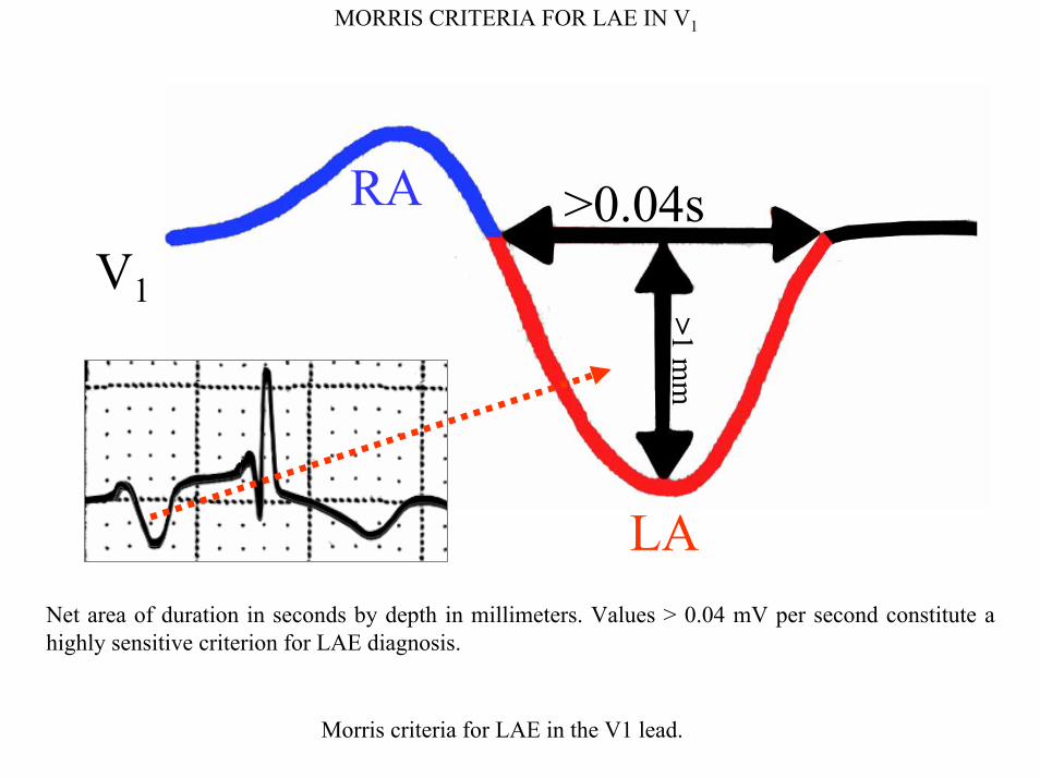
MORRIS CRITERIA FOR LAE IN V1
V1
RA
LA
>0.04s
>1 mm
Net area of duration in seconds by depth in millimeters. Values > 0.04 mV per second constitute a highly sensitive criterion for LAE diagnosis.
Morris criteria for LAE in the V1 lead.

IMPORTANT DILATATION OF THE RIGHT ATRIUM: INDIRECT SIGN OF RAEMORPHOLOGY OF QRS OF qR TYPE IN V1 AND V3R (SODI´S SIGNS)
The volumetric increase of the RA gets it The volumetric increase of the RA gets it Explanation of Sodi sign as indirect sign of RAE with aneurysmal right atrium.
RAqR
closer to the exploring electrode of Vcloser to the exploring electrode of V11, , registering negatively initially in this lead, registering negatively initially in this lead, because the electrode records the epicardial because the electrode records the epicardial morphology of the RA.morphology of the RA.In large megaatria (aneurysmatic atrium) of rheumatic patients, Sodi-Pallares described as indirect sign of RAE, appearance of deep Q waves in complexes in left precordial leads, which results in QR or QS complexes in these leads.(1;2)Note: Kasser I, et al,(3) showed that LAE signs correlate better with left atrial volume than with intraatrial pressure, and frequently they reverse after successful valvulotomy.
1. Sodi-Pallares D, Bisteni A, Herrmann GR. Some views on the significance of qR and QR type complexes in right precordial leads in the absence of myocardial infarction. Am Heart J. 1952 May;43:716-734.
2. Sodi-Pallares D, Ponce de León J, Bisteni A, Medrano GA. Polyparametric electrocardiography concerning new information obtained from clinical electrocardiogram. Prog Cardiovasc Dis. 1970 Jul;13(1):97-117.
3. Kasser I, Kennedy JW. The relationship of increased left atrial volume and pressure to abnormal P waves on the electrocardiogram. Circulation. 1969 Mar;39: 339-343
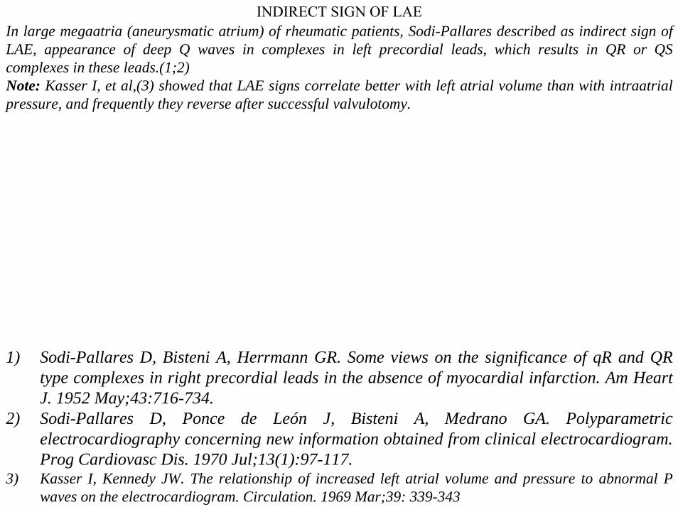
In large megaatria (aneurysmatic atrium) of rheumatic patients, Sodi-Pallares described as indirect sign of LAE, appearance of deep Q waves in complexes in left precordial leads, which results in QR or QS complexes in these leads.(1;2)Note: Kasser I, et al,(3) showed that LAE signs correlate better with left atrial volume than with intraatrialpressure, and frequently they reverse after successful valvulotomy.
INDIRECT SIGN OF LAE
1) Sodi-Pallares D, Bisteni A, Herrmann GR. Some views on the significance of qR and QR type complexes in right precordial leads in the absence of myocardial infarction. Am Heart J. 1952 May;43:716-734.
2) Sodi-Pallares D, Ponce de León J, Bisteni A, Medrano GA. Polyparametricelectrocardiography concerning new information obtained from clinical electrocardiogram. Prog Cardiovasc Dis. 1970 Jul;13(1):97-117.
3) Kasser I, Kennedy JW. The relationship of increased left atrial volume and pressure to abnormal P waves on the electrocardiogram. Circulation. 1969 Mar;39: 339-343

PR INTERVAL IN MITRAL STENOSIS (MS)
PR INTERVAL: Macruz criterion for LAE may be present. It is related to P wave duration (numerator) divided by PR segment duration (denominator). P duration / PRs duration – Normal value 1 to 1.7. In the presence of increase in P wave duration (numerator), the index presents values > 1.7.(1)Pileggiet al (2;3) described four phases in MS:
Phase 1: Macruz index > 1.7;
Phase 2: of hypertrophy of left atrium, characterized by: short PRs, P wave voltage greater than 2.5 mV in inferior leads and presence of notches in P waves of intermediary leads, from V2 to V4;
Phase 3: LA dilatation, characterized by P waves with increased duration (≥120 ms), bimodal with both modules separated by ≥30 ms (distance greater than 40 ms, is called pre-fibrillation P), final component of P wave is slow and negative in V1 and short PR segment;
Phase 4: characterized by sinus rhythm loss: atrial fibrillation.
1) Macruz R, Perloff JK, Case RB. A method for the electrocardiographic recognition of atrialenlargement. Circulation. 1958 May;17(5):882-9.
2) Pileggi F, et al. Cardiología. Chávez Rivera, I.; Capítulo 7, pg 337-338. 19933) Wierzchowska D, Czaplicki S. Usefulness of the Macruz index in the diagnosis of mitral stenosis.
Kardiol Pol. 1965;11:43-5.
PR interval in MS (Macruz index). Stages of MS according to Pileggi.

QRS COMPLEX IN MS
QRS complex: QRS complex modifications may reveal different RVE criteria, less sensitive than LAE signs; however, with high specificity.SAQRS: SAQRS shift to the right from +100º (≈ 30% of cases) constitutes an important sign of RVE diagnosis in MS. Some authors believe that SAQRS shift to the right in the frontal plane in MS could be the most sensitive criterion to detect RVE in ECG, as well as the best indicator of severity in the entity. In MS, SAQRS in the FP located between 0º and +60º in 88% of cases indicates presence of valvular area, estimated by echocardiogram > 1.3 cm2 (normal 4 to 6 cm2). When SAQRS is located to the right from +60º, it indicates valvular area < 1.3 cm2.MS with pressure in pulmonary artery equal or almost systemic, shows SAQRS located around +150º2.Pulmonary vascular resistance > 400 dynes/sec/cm-5 presents SAQRS in the frontal plane located to the right from +77º and pulmonary vascular resistance > 650 dynes presents SAQRS located to the right from +113º.(3)According to Taymor et al,(4) the criterion of SÂQRS shift to the right in the FP has been excessively valued for electrocardiographic diagnosis of RVE in MS. On the contrary, frequent posterior shift of forces in the horizontal plane, has been minimized. The latter is a frequent and characteristic fact in MS with RVE, which is translated in ECG by deep S waves in V2 and QRS loop in VCG of the HP by C-type pattern of RVE: counterclockwise rotation, posterior and rightward shift, and terminal portion located in the right posterior quadrant (> 20% of the total area is located in this quadrant).
1) Hugenholtz, P.G. Hoffman, I.(ed) Vectorcardiography. Amsterdam: North Holland PublishingCompany, 1966.
2) Taymor RC, et al. Circulation 1964;30:865-871.3) Cueto J, et al. Circulation 1966;33:588-598.4) Taymor RC, et al. Circulation 1964;30:865-871.

QRS COMPLEX IN MS
Pulmonary vascular resistance > 400 dynes/sec/cm-5 presents SAQRS in the frontal plane located to the right from +77º and pulmonary vascular resistance > 650 dynes presents SAQRS located to the right from +113º.(1) According to Taymor et al, the criterion of SAQRS shift to the right in the FP has been excessively valued for electrocardiographic diagnosis of RVE in MS. On the contrary, frequent posterior shift of forces in the horizontal plane, has been minimized. The latter is a frequent and characteristic fact in MS with RVE, which is translated in ECG by deep S waves in V2 and QRS loop in VCG of the HP by C-type pattern of RVE: counterclockwise rotation, posterior and rightward shift, and terminal portion located in the right posterior quadrant (> 20% of the total area is located in this quadrant).Other RVE/RVH criteria that are possibly observed in severe MS are:R wave in V1 >7 mm. There is no good correlation between R wave voltage and degree of RV wall thickness;R/S ratio in V1, V3R and V4R >;(3)Ventricular activation time (VAT), or time of increased intrinsic deflection appearance in V1 (>40 ms);R wave in V1 + S wave in V5 and/or V6 ≥ 10.5 mm (RV Sokolow-Lyon index);S wave depth in V1 <2 mm (0.2 mV);Decrease of R wave voltage and concomitant S wave increase in V5 and V6: R/S ratio in such leads ≤ 1. S wave depth in V5-V6: 7 mm (0.7 mv) or more;r wave voltage in V5 and V6:≤ 5 mm (0.5 mv);
1) Cueto J, et al. Circulation 1966;33:588-598.2) Taymor RC, et al. Circulation 1964;30:865-871.3) Fraser HR, et al. Br Heart J. 1955;17:459-483.

THE THREE HEMODYNAMIC ELECTROCARDIOGRAPHIC GROUPS OF MS
Cooksey JD, Dunn M and Massie E, correlated RVE/RVH electrocardiographic manifestations in MS, classifying them in three hemodynamic groups with increasing severity, taking into account RV systolic pressure and pulmonary vascular resistance:
1) GROUP 1 (IN MILD): RV systolic pressure < 70 mmHg and pulmonary vascular resistance < 400 dynes.
2) GROUP 2 (IN MODERATE): RV systolic pressure between 70 and 100 mmHg and pulmonary vascular resistance between 400 and 650 dynes.
3) GROUP 3 (IN SEVERE): RV systolic pressure > 110 mmHg and pulmonary vascular resistance > 650 dynes.
The three electrocardiographic hemodynamic groups in MS (mild, moderate, and severe).

ELECTROVECTOCARDIOGRAPHIC FEATURES OF HEMODYNAMIC GROUP 1 IN MSGROUP 11. Normal ECG or with LAE criteria. 2. In ≈ 20% of cases there is some RVE sign present: triphasic QRS complexes, rSr’ or R/S in V1, SAQRS
located to the right from +70º and frequent absence of normal increase of R/S ratio in intermediary precordial leads V3 and V4.
3. This group corresponds to group 1 of Pileggi, in which right intraventricular pressure is between 30 and 70 mmHg. ECG shows SAQRS located between +60º and +100º, so QRS in I< II. In precordial leads, shift of transition area is observed to the right.
4. In vagotonic patients, QRS complexes may be found in V5 and V6, falsely suggesting LVE, i.e. R wave with great voltage, discrete increase in intrinsic deflection, and positive and apiculate T waves.
5. QRS loop of VCG in the HP that remains with normal counterclockwise rotation.
GROUP 2
1. RV systolic pressure between 70 and 100 mmHg and pulmonary vascular resistance between 400 and 650 dynes. It corresponds to group 2 of Pileggi, with right intraventricular pressure between 70 and 110 mmHg.
2. RVE criteria are present in ≈ 50% of cases; 3. SAQRS located to the right from +130º in the frontal plane;4. R/S ratio in V1, V3R and V4R >1;5. Frequent triphasic QRS complexes (rsR’) in V1, V3R and V4R;6. Left precordial leads (V5 and V6) with R/S or r/S complexes;7. In V1 and V2 ventricular repolarization (ST/T) opposite to QRS complex with negative T waves (T loop
in the HP heading backward and to the left);8. QRS loop in the horizontal plane with abnormal rotation: in eight or clockwise.

GROUP 3:
1. MS with RV systolic pressure > 110 mmHg and pulmonary vascular resistance > 650 dynes.2. MS with RV systolic pressure > 110 mmHg and pulmonary vascular resistance > 650 dynes.3. In 100% of cases, there is RVE criteria;4. Possible presence of biatrial enlargement (BAE);5. QRS complexes of small voltage in V1, in contrast to QRS complexes of normal voltage in V2 (indirect
sign of RAE);6. SAQRS around +150º in the FP (always located to the right from +110º);7. Possible presence of QRS complexes with initial negativity in V1 or V1 and V2 (Sodi sign: qR pattern);8. Monophasic QRS complexes, pure R waves or Rs in V1.
VENTRICULAR REPOLARIZATION IN MITRAL STENOSIS
ST SEGMENTFrequent unspecific ventricular repolarization alterations, secondary to RVE, digitalis effect, electrolytic imbalance by use of diuretics and/or myocardial alterations by rheumatic attack. T WAVES: They present negative polarity from V1 to V3 or up to V5. Hamby et al1 describe backward and leftward shift of SAT in horizontal plane in the presence of severe RVE, originating T waves with negative polarity in right precordial leads (V1-V2) and even in intermediary leads (V3-V4). Biphasic T waves may be possibly observed from V1 to V3 with the first portion being negative, and the final one, positive2.
1. Hamby TI, et al. Dis. Chest. 1966;55:105.2. McCaugham D, et al. Am. J. Cardiol 1967;20:666.

SEVERE MITRAL STENOSIS HEMODYNAMIC GROUP 3
P wave with T-P fusion (“P mitrale”), SAQRS +130º, pure R wave in lead V1 (2 mm). R wave of aVR is prominent. Conclusion: LAE + RVE
Electrocardiographic tracing of patient carrier of mitral stenosis hemodynamic group 3.

DIFFERENTIAL CHARACTERISTICS OF THE THREE HEMODYNAMIC GROUPS IN MS
MILD MS MODERATE MS SEVERE MS
Final diastolic RV pressure: Normal. 5 to 15 mmHg. > 15 mmHg.
Medium RA pressure: Normal. 3 to 12 mmHg. > 12 mmHg.
Medium LA pressure and medium pulmonary capillary pressure.
12 to 20 mmHg. > 25 mmHg. 30 mmHg.
Right intraventricular pressure and in PA:
30 to 70 mmHg. 70 -110 mmHg. > 110 mmHg.
Pulmonary vascular resistance: < 400 dynes. 400 to 650 dynes. > 650 dynes.
Rhythm: Sinus. Sinus. AF in 30 to 50% of cases.
Very frequent AF.
P-wave Normal or mild LAE. SAP + 45º. P-wave voltag > 2.5 mV in II and notches in intermediary precordial leads from V2 to V4. Final slow negative component in V1.
LAE: SAP around + 30ºor less, bimodal P wave duration = or > than 120 ms associated to voltage > 2.5 mm in DII: BAE.
BAE or AF.

MILD MS MODERATE MS SEVERE MS
P wave duration/ PR segment duration (PRs)
ratio:
>1.7. Frequently, the only sign of LAE.
1 to 1.7 or AF
SAQRS in the FP: Between +60º and +100º
To the right from +130º. In average +150º.
Morphology in V1-V2 : rS, possible rsr’. Some criterion of RVE only in 20%.
R>S or rsR’. qR, Rs, monophasic R wave or up to rsR’(*)
Morphology in V5 and V6:
qRs or Rs. R/S or r/S r/S or R/S. R/S ratio = or < 1 frequent.
Ventricular repolarization
ST/T:
Normal. Opposite to QRS. Negative T wave in V1 and V2.
Extreme opposition to QRS: negative T wave from V1 to V3 or even
V5.
P loop: It may display LAE signs early.
Important signs of LAE: final portions backward and leftward in the HP or BAE.
BAE or absent: FA.

MILD MS MODERATE MS SEVERE MS
Rotation of QRS loop in the HP:
CCW rotation and with > 20% of the area of the QRS loop located in the right posterior quadrant: C-type RVE/RVH.
Variable: eight-shaped, clockwise or counterclockwise. C-type RVE pattern predominates.
Frequently CW A-type RVE. QRS loop located predominantly in right anterior quadrant with no final delay (differential diagnosis with CRBBB in case of rsR’ pattern).
Degree of reduction of slant of E-F ramp, of mitral valve anterior fascicle in 1-D echocardiogram:
Minimal: 25 to 35 mm/sec (normal >
35)
Moderate: 15 to 20 mm/sec.
Important: < 15 mm/sec.
Degree of decrease of amplitude of maximal excursion of anterior cuspid opening of mitral valve:
15 to 18 mm. 10 to 15 mm. < 10 mm.
Valvular area estimated by 2-D echocardiogram and by Doppler:
> 1.5 cm2. 1 to 1.5 cm2. < 1 cm2.
Degree of reduction of slant of E-F ramp, of mitral valve anterior fascicle in 1-D echocardiogram:
Minimal: 25 to 35 mm/sec (normal >
35)
Moderate: 15 to 20 mm/sec.
Important: < 15 mm/sec.

MITRAL STENOSIS (MS) WITH PULMONARY HYPERTENSION HEMODYNAMIC GROUP 3
Female, 35 years old. Slow final negative component, P wave in V1-V2: LAE. Triphasic QRS complexes (rsR’) in V1 and V2 and negative T waves from V1 to V5: RVE/RVH
Electrocardiographic tracing of patient carrier of mitral stenosis hemodynamic group 3.

VCG IN MITRAL STENOSISCHARACTERISTICS OF VECTOCARDIOGRAPHIC P LOOP
Lee Y1, et al shown by hemodynamic study, a great VCG sensitivity in comparison to ECG, for the diagnosis of LAE and RVE/RVH in MS.
Vectorcardiographic signs of this entity include:1) LAE criteria;2) RVE pattern, predominantly type C (75%); however, types A (10%) and B (2%) may be found.
VECTOCARDIOGRAPHIC CRITERIA OF LAE IN MS.They are more sensitive for MS diagnosis in comparison to electrocardiographic criteria, since they are present in ≈90% of MS, even in mild forms, in absence of AF. P loop of LAE is the rule in MS.In the next outlines, we show normal P loop and in LAE, in the frontal, horizontal and right sagittal planes (FP, HP and RSP).
1. Lee YC, Scherlis L, Singleton RT. Mitral Stenosis. Hemodynamic, Electrocardiographic, And Vectorcardiographic Studies. Am Heart J. 1965 Apr;69:559-666.
VCG in mitral stenosis. Characteristics of vectocardiographic P loop.

NORMAL P LOOP AND P LOOP IN LAE IN THE FRONTAL PLANE
E0
E0
S Â P + 55 0
X
Y
X
Y
E0
S A P + 55 0
SAP +30º
X
Y
X
Y
LA
RALA
RA
E
D
EE
D
E
LARA LARA
In LAE by MS, SAP in the FP is between +45º and -30º, being in average around +30º (isodiphasic P wave in III).
SAP+60º
NORMAL P LOOP P LOOP IN LEFT ATRIAL ENLARGEMENT
Outline of P loop characteristics in the frontal plane in LAE by mitral stenosis (comparison to normal P loop).

NORMAL P LOOP AND P LOOP IN LAE IN THE HORIZONTAL PLANEIn the Horizontal Plane, wide angle between the initial portions (RA) and final portions (LA) of P loop, are observed in 25% of patients. According to Goochs AS1 et al, this could be the only sign of LAE.
ROTAÇÃOANTI-HORÁRIA
ROTAÇÃOANTI-HORÁRIA
AD
AE
Z Z
XX
V1
V6
V1
V2
V6
0E 0
E
Z Z
XX
V1
V6
V1
V2
V6
Z Z
XX
V1
V6
V1
V2
V6
0E 0
E
Z Z
XX
V1
V6
V1
V2
V6
EERA
LAV2V2V
NORMAL P LOOP Wide angle between the initial portion (RA)
and final portions (LA) of P loop
Outline of P loop characteristics in the horizontal plane in LAE of mitral stenosis (comparison with normal P loop). 1) Goochs AS, et al. Am. Heart. J. 1966;71:727.
CounterclockwiseRotation or
Eight-shaped
Maximalvector of P >0.05 mV
Maximal vector of P and located to the lef
P LOOP IN LAE
t≥0.10 mV in adults and≥ 0.14 mV in < 15 years of age
Bow tie”Morphology
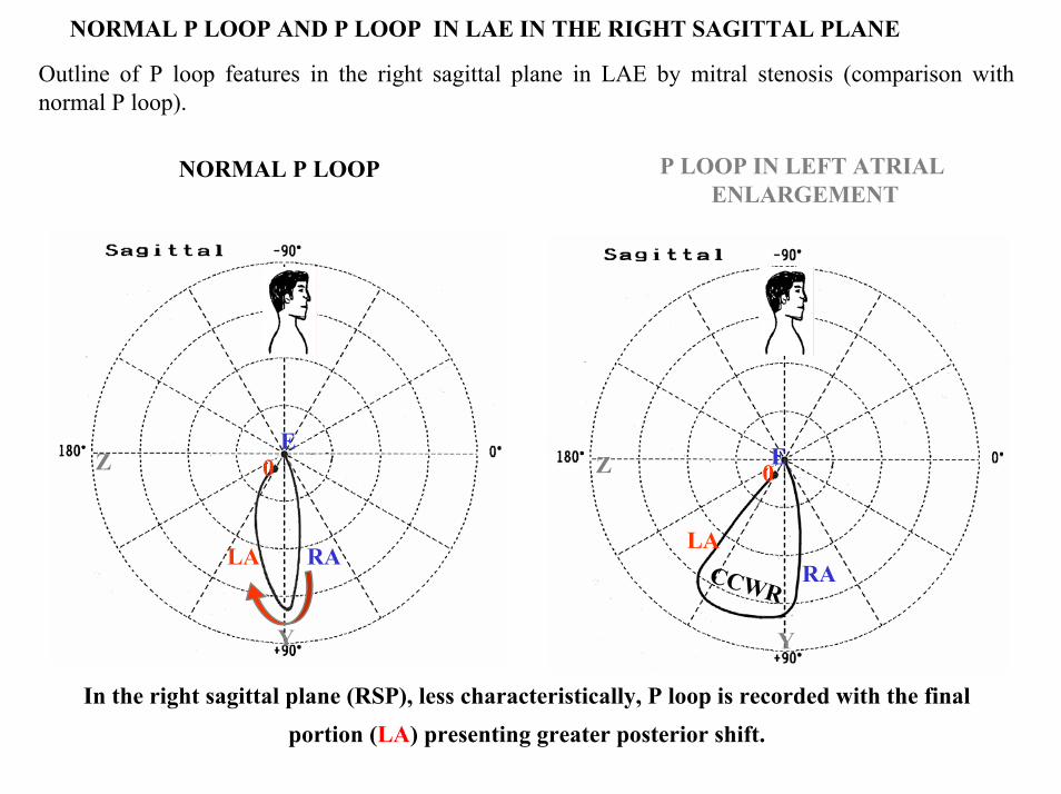
NORMAL P LOOP AND P LOOP IN LAE IN THE RIGHT SAGITTAL PLANE
Outline of P loop features in the right sagittal plane in LAE by mitral stenosis (comparison with normal P loop).
P LOOP IN LEFT ATRIAL ENLARGEMENT
NORMAL P LOOP
In the right sagittal plane (RSP), less characteristically, P loop is recorded with the final portion (LA) presenting greater posterior shift.
0E
RALA
0E
RALA
Z
Y
Z
Y
CCWR

VCG IN MITRAL STENOSIS
The vectorcardiographic record of RVE/RVH in the QRS loop displays a greater sensitivity than ECG, since this is the pattern found most frequently, and known as C1-type RVE/RVH.
The VCG of MS shows:
P loop with mid-final portions heading backward, below and leftward, and with increased magnitude: LAE;Vectors from initial 10 to 20 ms of QRS loop in the HP, more frequently heading forward and to the left
(normally heading forward and to the right, in 80% of cases);QRS loop in the HP maintains normal counterclockwise rotation in 75% of cases, ≈ in 15% the rotation is
eight shaped, and in 10% rotation is inverted (clockwise: A-type RVE);Posterior and rightward shift of QRS loop;20% or > of the total area of QRS loop in the HP, located in the right posterior quadrant with afferent limb
of inscription without end delay;Afferent limb of QRS loop in the FP, located in the right superior quadrant (C1-type RVE) different from
C2-type RVE by emphysema, which presents the afferent limb located below and to the right, in the right inferior quadrant in the FP;
T loop frequently heading backward, leftward and upward: negative T in V1-V2 and in inferior leads DII, DIII and aVF.
Summary of characteristics of the three vectocardiographic loops in MS.

C1-TYPE RVE C2-TYPE RVE
Typical entity that originates vectocardiographic pattern:
MS (75% of cases). Only in 25% of cases of COPD.
Chronic cor pulmonale of emphysema. COPD.
Location of final portion of QRS loop in the HP
Right posterior quadrant: 20% or > of QRS loop area located in this quadrant. More anterior
afferent limb.
Posterior (marked shift), and rightward, and more posterior
afferent limb.
QRS complexes in precordial leads Frequent polyphasic pattern: rsR’ in V1.
rS or QS type from V1 to V4, resembling anterior electrically
inactive area.
Initial vectors in the HP Forward and to the right or left. Forward and to the right or left.
Location of QRS loop in the HP Less posterior shift. Only terminal appendage backward
and rightward.
Marked posterior shift.
Location of final portion of QRS loop in the FP (afferent limb)
In right superior quadrant. In the right inferior quadrant.
Location of QRS loop in RSP In the postero-inferior quadrant with terminal portion of afferent
limb in postero-superior quadrant.
Completely in posteroinferiorquadrant.
Main differences between C1-type (characteristic of MS) and C2-type RVE/RVH (characteristic of COPD).

COMPARISON OF QRS LOOP OF VCG IN THE HP IN MITRAL STENOSIS (C1) AND COPD (C2)
C1-TYPE RVE/RVH C2-TYPE RVE/RVH
V6
V1
V4
V5
V2 V3
X
Z
V6
V1
V4
V5
V2 V3
X
Z
TT
rSR’ QS rS rS
rS
rS
RS
rS rSRS
RS
RS
Comparison of QRS loops in C1-type (characteristics of MS) and C2-type RVE (characteristic of COPD) in the HP.
>20% of QRS looparea located in the
right posterior quadrant
Typical of chronic Cor Pulmonale by emphysema.Characteristic of MS (75% of cases). Only in 25% of cases of COPD
T

X
Y
X
Y
X
Y
NORMAL
C1 RVE/RVH C2 RVE/RVH
COMPARISON OF QRS LOOPS IN
VCG IN THE FP, NORMAL AND
IN RVE BY MITRAL STENOSIS
(C1) AND BY COPD (C2)
TT
QRS loop predominantly
located in the left inferior quadrant
Clockwise Rotation
Afferent limb belowand rightward
Clockwise Rotation
Characteristic of QRS loop in mitral stenosisAfferent
limbRighward
and upward
QRS loop predominantly Located in the right Inferior
quadrant
Comparison of normal QRS-T loops and C1-type (characteristic of mitral stenosis) and C2-type RVE (characteristic of COPD) in the frontal plane.

Z
Y
Z
Y
Z
Y
NORMAL
C1 RVE C2 RVE
COMPARISON OF QRS LOOPS
OF VCG IN RSP: NORMAL AND
IN RVE/RVH BY MS (C1) AND
BY COPD (C2)
QRS LOOP PREDOMINANTLY LOCATED IN THE POSTERO-INFERIOR
QUADRANT.POSTERIOR T LOOP.
T
T
T
AFFERENT LIMB THAT ENDS IN
POSTERO-SUPERIOR QUADRANT
AFFERENT LIMB IN POSTERO-INFERIOR
QUADRANT
CWR
CWR
Comparison of normal QRS-T loop and in C1-type (characteristic of MS) and C2-type RVE/RVH (characteristic of COPD) in the right sagittal plane.
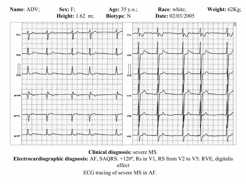
Name: ADV; Sex: F; Age: 35 y.o.; Race: white; Weight: 62Kg; Height: 1.62 m; Biotype: N Date: 02/03/2005
Clinical diagnosis: severe MS.Electrocardiographic diagnosis: AF, SAQRS: +120º, Rs in V1, RS from V2 to V5: RVE, digitalis
effectECG tracing of severe MS in AF.

Name: SDV Sex: F Age: 47y.o. Race: MulattoWeight: 57Kg Height: 1.58 m Biotype: Normal Date: 04/05/2003
Clinical diagnosis: Severe MS.ECG diagnosis: rhythm: coarse fibrillation-flutter that suggests LAE; HR: 60-80 bpm; RVE: SAQRS+100º; R=S in V5, prominent R wave with great voltage from V1 to V3: prominent anterior forces. ST/T opposite to QRS. Negative T wave in V1 and V2. SAT upward and backward: negative T wave in V2, V3 and in inferior leads.
ECG/VCG sequence of severe MS in AF.

ECG/VCG CORRELATION FRONTAL PLANE
aVR aVL
I
IIIII
X
Y
T
aVF
QS
rs
SÂQRS +100º
T-loop opposite to QRS loop
ECG/VCG sequence of severe MS in AF.

ECG/VCG CORRELATION HORIZONTAL PLANE
V6
V1
V4
V5
V2
V3RS
T
RS
CCWR
Type A RVH/RVE
CCWR: QRS loop withClockwise rotation and
Located in anterior quadrantsPAF: Prominent QRS Anterior Forces
ECG/VCG sequence of severe mitral stenosis in AF.

ECG/VCG CORRELATION LEFT SAGITTAL PLANE
Y
Z
T
aVF
Opposite T-loop located in posterosuperior quadrant
CWR
QRS-loop located inInferoanterior quadrant:Prominent anterior forces
Negative T waves in inferior leads
ECG/VCG sequence of severe MS in AF.
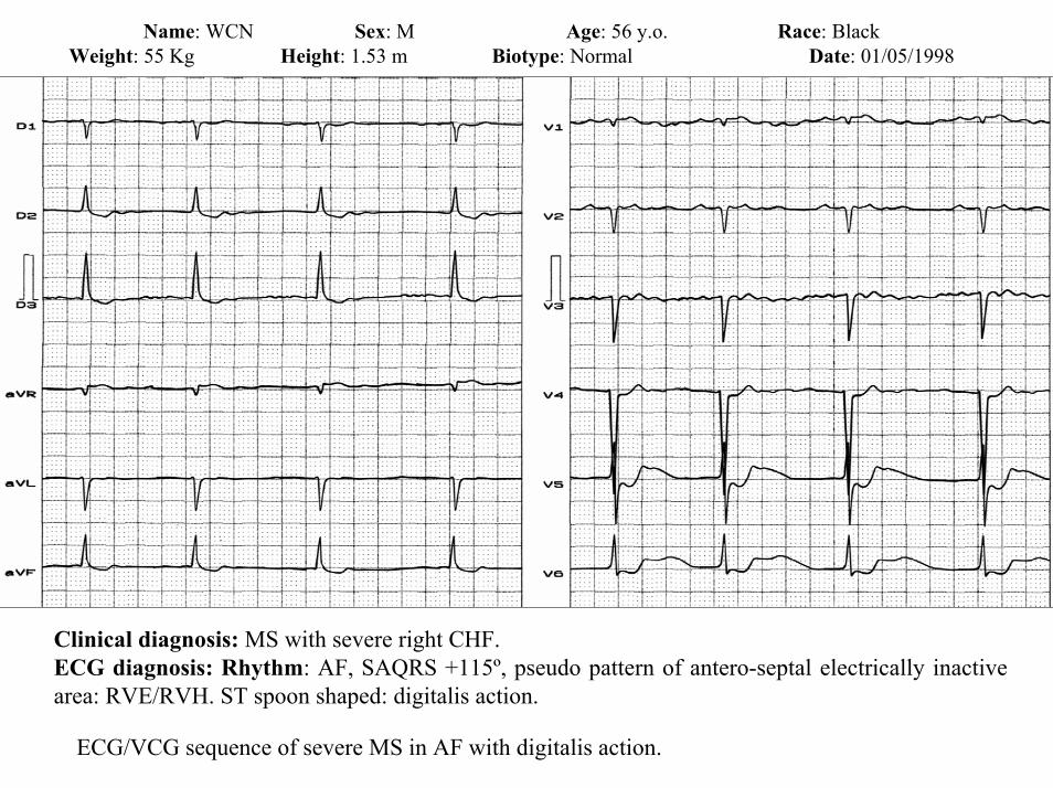
Name: WCN Sex: M Age: 56 y.o. Race: BlackWeight: 55 Kg Height: 1.53 m Biotype: Normal Date: 01/05/1998
Clinical diagnosis: MS with severe right CHF.ECG diagnosis: Rhythm: AF, SAQRS +115º, pseudo pattern of antero-septal electrically inactive area: RVE/RVH. ST spoon shaped: digitalis action.
ECG/VCG sequence of severe MS in AF with digitalis action.
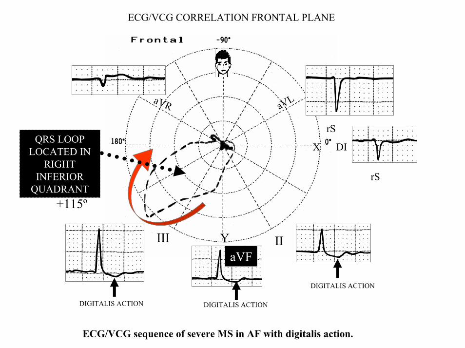
ECG/VCG CORRELATION FRONTAL PLANE
aVR aVL
DI
IIIII
X
Y
SAQRS +115º
aVF
rSQRS LOOP
LOCATED IN RIGHT
INFERIOR QUADRANT
rS
DIGITALIS ACTION
DIGITALIS ACTION DIGITALIS ACTION
ECG/VCG sequence of severe MS in AF with digitalis action.

ECG/VCG CORRELATION HORIZONTAL PLANE
V6
V1
V4
V5
V2
V3
QRS LOOP COMPLETELY LOCATED IN THE RIGHT POSTERIOR QUADRANT:
C-TYPE RVE
RS
LEFTWARD SHIFT FROM TRANSITION AREA
ST/T VECTOR OPPOSITE TO QRS
P LOOP IS NOT OBSERVED: PAF
QS and rS PATTERN IN V1-V4: RVE/RVHECG/VCG sequence of severe MS in AF with digitalis action.

ECG/VCG CORRELATION LEFT SAGITTAL PLANE
Y
Z
QRS LOOP COMPLETELY
LOCATED IN THE POSTEROINFERIOR
QUADRANT: C2-TYPE RVE
aVF
ECG/VCG sequence of severe MS in AF with digitalis action.

Exercise stress testing
Exercise stress testing is indicated in situations where the degree of disability is in question.Stress echocardiography provides information about changes in the transmitral gradient and the degree of limitation of exercise and may guide decisions about valvotomy.
Cardiac catheterization
Cardiac catheterization is indicated when a discrepancy is noted between Doppler-derived hemodynamicsand the clinical status in a symptomatic patient.
Perform percutaneous mitral balloon valvotomy in properly selected patients.
Cardiac catheterization measures absolute left-sided and right-sided pressure when pulmonary artery pressure elevation is out of proportion to mean gradient and valve area.
Coronary angiography may be performed in selected patients.

Prehospital Care
Prehospital care is appropriate for acute pulmonary edema or arrhythmia secondary to MS.Oxygen administration is always indicated for symptomatic patients.In patients with significant acute dyspnea, appropriately trained personnel may administer agents that promote afterload reduction such as nitrates or ACE inhibitors.Clinically significant arrhythmias such as AF with rapid ventricular response should be corrected according to local protocols. Medications appropriate for use by prehospital personnel vary according to local protocol but may include diltiazem, amiodarone, esmolol, or metoprolol.Grossly unstable patients with atrial fibrillation with rapid ventricular response should receive synchronized direct current (DC) cardioversion
Emergency Department CareThe goal is to control symptoms, to prevent or retard disease progression, and to treat complications.
Treatment of congestive heart failureMedications to consider include nitroglycerin, ACE inhibitors, and diuretics.Patients with severe MS should maintain an upright posture and avoid strenuous physical activity.Sodium intake should be restricted, and maintenance doses of oral diuretics should be continued.The data on ββ-blockers are conflicting; β-blockade may be useful for patients with exertional symptoms if the symptoms occur primarily at high heart rates. Prevent or retard disease. Primary and/or secondary prophylaxis against streptococci/endocarditis should be administered.Penicillin is indicated whenever streptococcal infection is suspected in a patient with known rheumatic disease.

Management of atrial fibrillation
Much of the dyspnea related to MS is rate related. Control of atrial fibrillation with rapid ventricular response may be considered with any of the following agents: MetoprololEsmololDiltiazemDigoxinIf the patient is unstable and immediate cardioversion is indicated, then heparin should be administered before, during, and after cardioversion. Otherwise, electrical or chemical cardioversion should be performed after 3 weeks of warfarin anticoagulation.
Transesophageal echocardiography prior to elective cardioversion should be considered.
Anticoagulation is necessary in many patients who are unable to maintain normal sinus rhythm. Anticoagulation may also be beneficial for patients with normal sinus rhythm with a prior embolic event or a left atrial dimension greater than 55 mm Hg noted by echocardiography.
A cardiologist and/or cardiothoracic surgeon should be consulted in the following situations: Known or suspected cases of MS with hemodynamic instability, arrhythmia, or embolizationCases involving a new onset or progression of symptomsThe goal of medical therapy is to control the rapid ventricular rate and to prevent thrombus formation and embolization. Conversion of AF to sinus rhythm may also decrease symptoms.

Antiarrhythmics
These agents alter the electrophysiologic mechanisms that are responsible for arrhythmia.
Amiodarone Class III antiarrhythmic. Has antiarrhythmic effects that overlap all 4 Vaughn-Williams antiarrhythmic classes. May inhibit AV conduction and sinus node function. Prolongs action potential and refractory period in myocardium and inhibits adrenergic stimulation. Only agent proven to reduce incidence and risk of cardiac sudden death, with or without obstruction to LV outflow. Very efficacious in converting AF and flutter to sinus rhythm and in suppressing recurrence of these arrhythmias.Has low risk of proarrhythmia effects, and any proarrhythmic reactions generally are delayed. Used in patients with structural heart disease. Most clinicians are comfortable with inpatient or outpatient loading with 400 mg PO tid for 1 wk because of low proarrhythmic effect, followed by weekly reductions with goal of lowest dose with desired therapeutic benefit (usual maintenance dose for AF 200 mg/d). During loading, patients must be monitored for bradyarrhythmias. Prior to administration, control the ventricular rate and CHF (if present) with digoxin or calcium channel blockers.
Oral efficacy may take weeks. With exception of disorders of prolonged repolarization (eg, LQTS), may be DOC for life-threatening ventricular arrhythmias refractory to β-blockade and initial therapy with other agents.

Esmolol
Ultra–short-acting that selectively blocks beta1-receptors with little or no effect on β2-receptor types. Particularly useful in patients with elevated arterial pressure, especially if surgery is planned. Shown to reduce episodes of chest pain and clinical cardiac events compared with placebo. Can be discontinued abruptly if necessary.May be used with class I antiarrhythmics if digoxin therapy does not abort atrial arrhythmia. Administer in patients needing prompt slowing of ventricular rate in response to atrial flutter or fibrillation and who are most likely to become hemodynamically unstable if left without treatment or in those waiting for the start of the therapeutic effects of digoxin (average, 10 h). Useful in patients at risk for experiencing complications from β-blockade; particularly those with reactive airway disease, mild-moderate LV dysfunction, and/or peripheral vascular disease. Short half-life of 8 min allows for titration to desired effect and quick discontinuation if needed.
Digoxin
Cardiac glycoside that has direct inotropic effects in addition to indirect effects on the cardiovascular system. Effects on myocardium involve a direct action on the cardiac muscle that increases myocardial systolic contractions as well as indirect actions that result in increased carotid sinus nerve activity and enhanced sympathetic withdrawal for any given increase in mean arterial pressure.
Metoprolol
Selective β1-adrenergic receptor blocker that decreases the automaticity of contractions. During IV administration, carefully monitor blood pressure, heart rate, and ECG.

Calcium Channel Blockers
In specialized conducting and automatic cells in the heart, calcium is involved in the generation of the action potential. The calcium channel blockers inhibit movement of calcium ions across the cell membrane, depressing both impulse formation (automaticity) and conduction velocity. During the depolarization, inhibits the calcium ion from entering the slow channels or the voltage-sensitive areas of the vascular smooth muscle and myocardium.
HeparinAugments activity of antithrombin III and prevents conversion of fibrinogen to fibrin. Does not actively
lyse but is able to inhibit further thrombogenesis. Prevents reaccumulation of clot after spontaneous fibrinolysis. Most data are related to use of unfractionated heparin. Low molecular weight heparin probably is as effective but awaits the results from clinical studies.
Warfarin (Coumadin)
Inhibits vitamin K–dependent clotting factors II, VII, IX, and X and anticoagulant proteins C and S. Anticoagulation effect occurs 24 h after drug administration, but peak effect may happen 72-96 h later. Antidotes are vitamin K and FFP.

Percutaneous balloon valvotomy is, in general, the initial procedure of choice for symptomatic patients with moderate-to-severe MS. It can double the mean valve area with a 50-60% decrease in the transmitralpressure gradient, producing a prominent and sustained symptomatic improvement.The 2006 ACC/AHA Practice Guidelines recommend percutaneous mitral balloon valvotomy for symptomatic patients (New York Heart Association [NYHA] Functional Class [FC] II, III, or IV) with moderate or severe MS and favorable valve morphology in the absence of left atrial thrombus or significant mitral regurgitation.In patients with indications for intervention, percutaneous valvotomy has proven superior to closed commissurotomy in some long-term studies. The overall event-free (no death, repeat valvotomy, or valve replacement) survival rate is 80-90% in patients with favorable valve morphology. More than 90% of patients free of events remain in NYHA FC I or II.Surgical commissurotomy is required when the conditions for percutaneous valvotomy are not met. In the United States, open commissurotomy is considered preferable to close commissurotomy.The 2006 ACC/AHA Practice Guidelines gave a class I recommendation for surgical intervention in patients with NYHA FC III-IV symptoms, moderate or severe mitral stenosis, and valve morphology favorable for repair or replacement if one of the following is noted.Percutaneous mitral balloon valvotomy is not available.

A left atrial thrombus is present despite anticoagulation.A nonpliable or calcified valve is present, with the decision to proceed with either repair or replacement made at the time of the operation. Patients with moderate or severe mitral regurgitation should undergo valve replacement.Medications cannot correct mitral stenosis, but therapy can reduce the incidence and severity of symptoms and complications.Drugs that increase diastolic filling time and decrease the heart rate are typically used. Beta-blockers are frequently used in this situation. Calcium channel blockers, such as diltiazem, and digoxin may also be used. β-blockers are preferred over digoxin because they control exercise-induced increases in heart rate.Rhythm control is of questionable clinical significance. Amiodarone may be used to maintain sinus rhythm, but its use may cause complications. Anticoagulation is used in patients with atrial thrombi, in patients with AF, or in patients with a prior thromboembolic event. Some European guidelines for the treatment of group A Streptococcus (GAS) pharyngitis are remarkably different than those in the United States. (1) Indeed, some European guidelines consider acute pharyngitis a self-limiting disease and do not recommend testing for GAS or prescription of antibiotics for GAS pharyngitis.
1. Wilson W, Taubert KA, Gewitz M, et al. Prevention of infective endocarditis: guidelines from the American Heart Association: a guideline from the American Heart Association Rheumatic Fever, Endocarditis, and Kawasaki Disease Committee, Council on Cardiovascular Disease in the Young, and the Council on Clinical Cardiology, Council on Cardiovascular Surgery and Anesthesia, and the Quality of Care and Outcomes Research Interdisciplinary Working Group. Circulation. Oct 9 2007;116:1736-1754.

Primary prophylaxis depends on early diagnosis of GAS pharyngitis. Treatment started within 7-9 days after onset of illness may prevent rheumatic fever. Treatment with penicillin V is recommended.
If β-lactam allergy is present, azithromycin or clarithromycin is now recommended. To date, no strains of streptococci that produce rheumatic fever have been penicillin resistant.
Secondary prophylaxis may delay the progression of MS. Secondary prophylaxis may be individually tailored.
The duration of prophylaxis depends on the presence of carditis and persistent valvular disease.
Rheumatic fever with carditis and residual heart disease requires prophylaxis for 10 years or until age 40 years, whichever is longer.
Rheumatic fever with carditis but no residual heart disease requires prophylaxis for 10 years or until age 21 years, whichever is longer.
Rheumatic fever without carditis requires prophylaxis for 5 years or until age 21 years, whichever is longer.
Endocarditis prophylaxis for routine dental and respiratory procedures is no longer recommended for patients with mitral stenosis unless they have had implantation of an artificial valve. (1)
1. Chiang CW, Lo SK, Ko YS, et al. Predictors of systemic embolism in patients with mitral stenosis. A prospective study. Ann Intern Med. Jun 1 1998;128:885-889.
Complications
Complications of mitral stenosis may include all of the following:1. Thromboembolism2. Atrial fibrillation3. Bacterial endocarditis4. Pulmonary hypertension5. Pulmonary edema6. Complications of balloon valvulotomy (eg, stroke, cardiac perforation, development of mitral
regurgitation)7. Complications of mitral valve replacement (eg, perivalvular leak, thromboembolism, infective
endocarditis, mechanical dysfunction, bleeding due to anticoagulants)
Prognosis
Depending on severity of symptoms of MS, the 10-year survival rate is as follows:1. 85% for no symptom (class I)2. 34-42% for mild symptoms (early class II)3. 40% for moderate-severe symptoms (late class II, class III)4. 0% for class IV (Of class IV patients, survival is 42% at 1 year and 10% at 5 years.)5. The operative mortality rate is 1-2% for mitral commissurotomy and 2-5% for mitral valve
replacement.